Part 6 — CHAPTER XLVIII. THE SMALL INTESTINES. (continued 2)
Reading edition
Resection of the bowel may imply in one case a removal of but two or three inches of its length, while the other extreme is not reached until several feet of bowel have been removed. I have been able to successfully remove eight feet and nine inches of intestine, the lower part including the cecum and a portion of the ascending colon, and there are now on record nearly twenty cases where over 200 Cm. of bowel have been resected, nearly all of them recovering. Success in this procedure depends partly upon the condition necessitating the operation, as well as the general condition of the patient, but in no small measure hangs upon the perfection of the operator’s technique.
Fig. 575
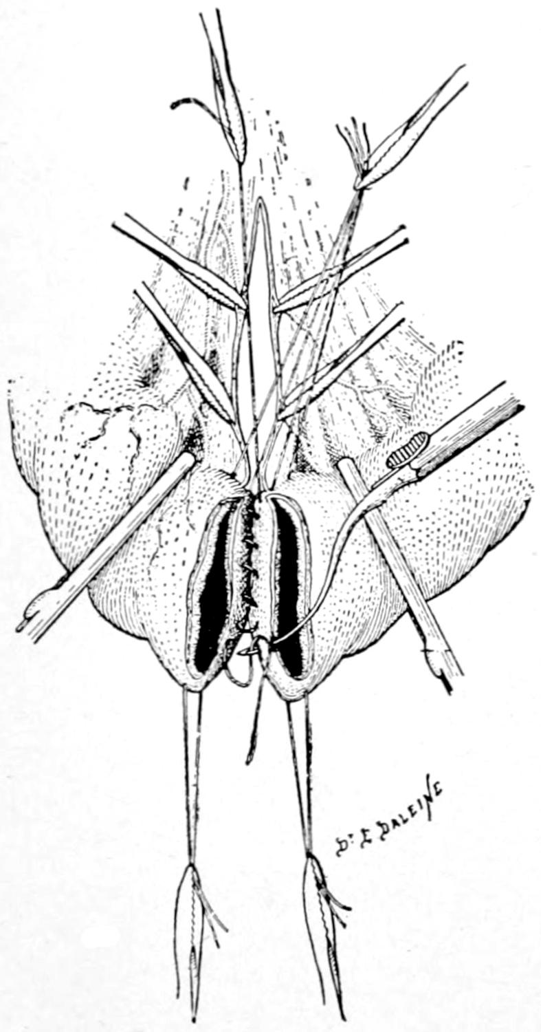
End-to-end or circular anastomosis by enterorrhaphy. First row of distal sutures in serosa. (Type of needle differs from that used in this country). (Lejars.)
Fig. 576
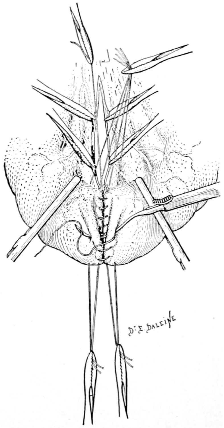
Completion of last row of sutures, begun as shown in Fig. 575. (Lejars.)
Whatever be the condition which requires such resection it should be made sufficiently extensive to completely include and permit the total removal of the diseased or injured portion. The abdominal incision should be large enough to permit the delivery upon the surface of the body of all that portion to be removed. Unless this be done the difficulties are greatly enhanced. Save where there is some distinct indication for opening elsewhere, this incision is made in the middle line. The compromised bowel having been sought and thus delivered and one having decided exactly where to divide it, clamps are so placed both above and below each line of division as to prevent leakage. Underneath the bowel to be thus divided gauze is placed in such a way as to receive the small amount of discharge which will escape from the portion between the clamps. The exposed bowel surfaces should then be thoroughly cleaned, the contaminated gauze removed, fresh pieces substituted for it, and the other division of bowel made in the same way. While in some cases it may be well to tie off the mesenteric border and secure all its vessels before dividing the bowel, this may at other times be delayed until after the division. At all events it is the next step. Whether the mesentery shall be simply separated along the intestinal border and tied off in small portions, one after another, or whether a triangular resection of a portion of the mesentery itself should be made, securing the larger vessels nearer to its root, will depend on the nature of the case and upon whether the mesentery itself be involved in the disease. In dealing with cancer it is often necessary to remove, at the same time, every enlarged lymphatic. It may be inferred that no incision or tear, no matter how short, can be made in these tissues without danger of subsequent hemorrhage unless the parts be secured against it. A series of ligatures and sutures is therefore called for here which may consume no small proportion of the entire time of the operation. (See Figs. 575 and 576.)
All that portion of bowel which has been condemned having been removed and a careful toilet of the parts having been made the surgeon next proceeds to restore the bowel lumen. A V-shaped defect in the mesentery should be united with sutures. The line of former mesenteric border left after removal of bowel should be not only carefully protected with ligatures, but the whole margin should be overcast and so folded in or drawn together in tucks as to make it easy to bring the bowel ends together without undue stress.
Fig. 577
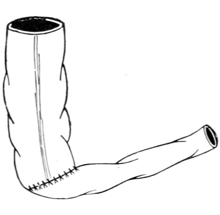
Fig. 578
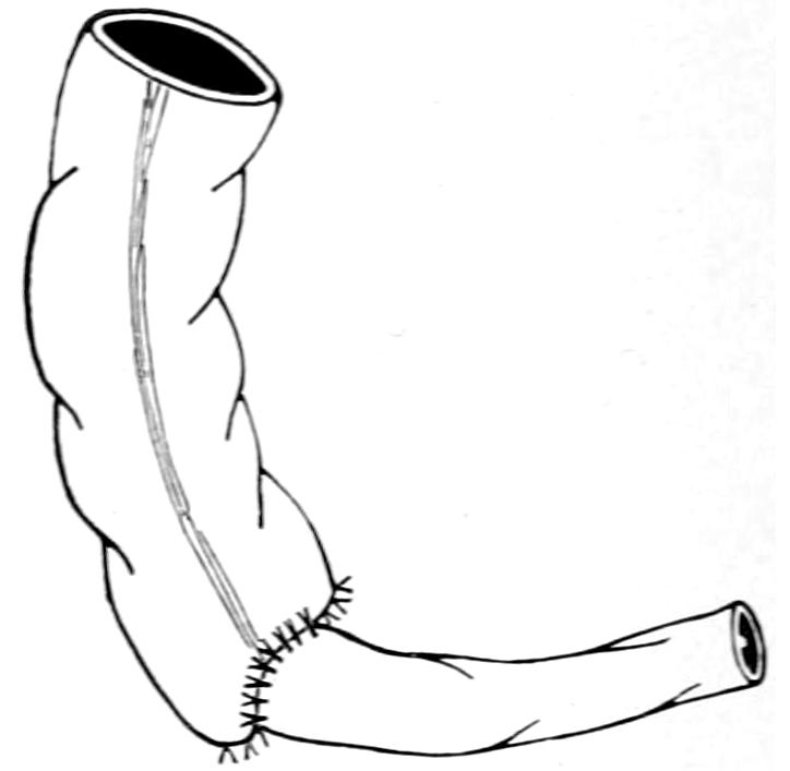
Circular anastomosis of portions of the bowel having different lumina. (Bergmann.)
The sutures by which the divided bowel is restored should begin at the mesenteric border, and every care should be taken to make the joint at this point absolutely water-tight. Suture methods have been described. To unite bowel ends of the same diameter it is an easy matter to suture together first the mucosa and then the outer layer, so long as the intestine is on the outside of the body and equally accessible on all sides (Fig. 578). The surgeon is sometimes compelled to do this work within the body cavity, as in resection of the rectum for cancer. It may be advisable to first place a row of sutures between the serosa and muscularis on the further side of the margins to be united, then to close the mucosa completely around, and then to finish the outer layer of sutures. So long as differences of size are not conspicuous, end-to-end approximation can be made almost anywhere. When, however, it is necessary to attach small bowel to large, the size of the larger opening should be reduced to fit the smaller, or one or both ends may be closed, turning in the stump, as already described, and then making lateral or end-to-side anastomosis. Any such anastomotic opening should be so placed, and bowel so directed, that there shall be no interference in the direction of the natural bowel stream, failure to observe this precaution producing not only added immediate danger but more or less permanent obstruction (Figs. 579 and 580).
Fig. 579
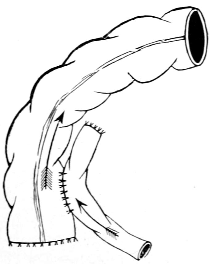
Isoperistaltic lateral apposition.
Fig. 580
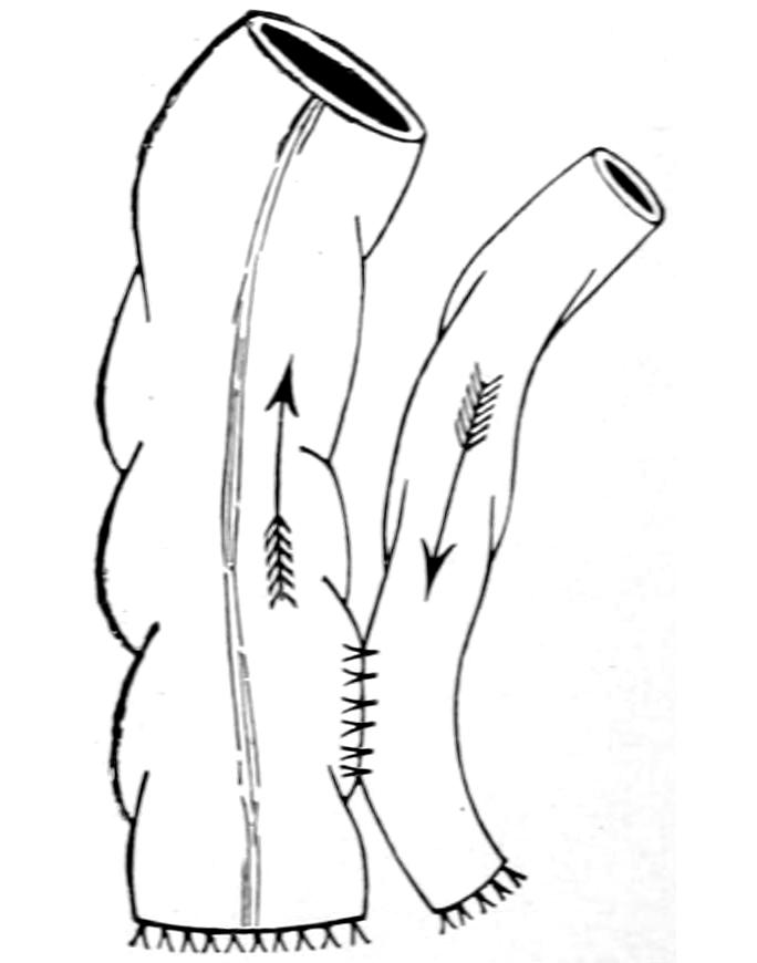
Antiperistaltic lateral apposition (bad).
All that has been said above with regard to the Murphy button and its use in anastomotic operations holds equally good here with regard to its usefulness after resection.
Numerous devices, either instruments for the purpose of holding the bowel together while it is sutured, or of affording substitutes for the Murphy button, have been planned by operators all over the world. There are few of them, however, which give any better results than the simple methods above described, to which I prefer to limit description here because of their very simplicity.
Intestinal suture or any other method of completing the resection having been finished, a careful toilet of all exposed parts should be made, by which bowel may be dropped back into the abdominal cavity and the latter closed without drainage.
The subsequent management of these cases will consist in two or three days’ starvation, in order that peristalsis may be reduced to a minimum, the patient being meanwhile fed by the rectum. Then will come a time when both fluid food, and cathartics a little later, should be gently and discriminately administered. Any satisfactory suture method will rarely give way after forty-eight hours. Buttons, on the contrary, may break loose after many days or even weeks, and this fact affords another argument against their use.
Fig. 581
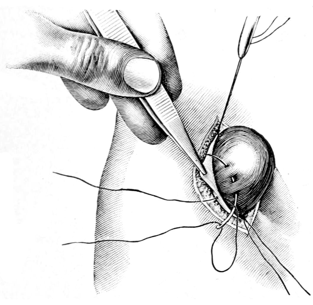
Enterostomy; preliminary fixation of a loop of bowel to the peritoneum. (Lejars.)
Fig. 582
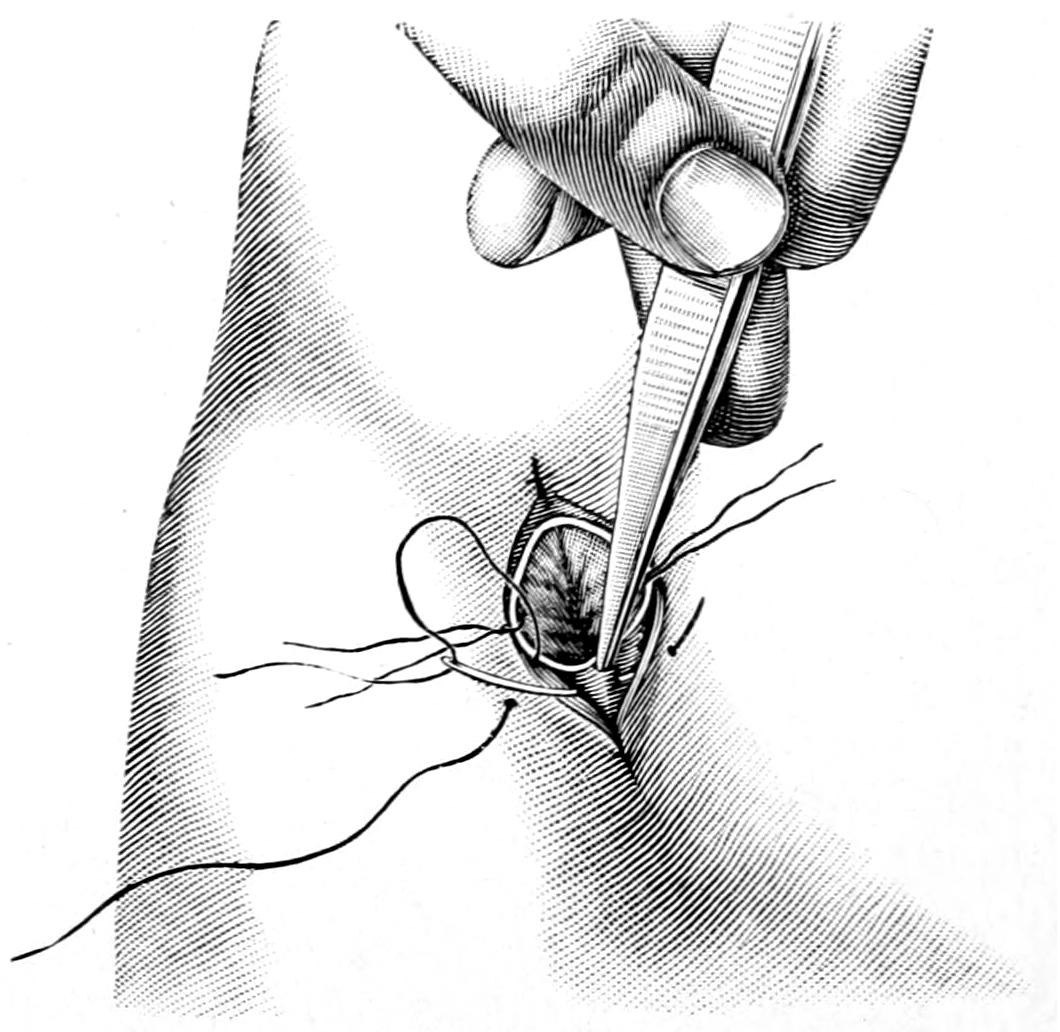
Enterostomy; fixation of margins of opened gut to skin. (Lejars.)
Enterostomy.
—Enterostomy for establishment of fecal fistula, or artificial anus, is performed for relief purposes and sometimes as an emergency measure. It consists in attaching some portion of the bowel, naturally that above the constriction or disease which compels the operation, to the parietal peritoneum through a small wound in the abdominal wall. When the large intestine is opened for this purpose the operation is usually referred to as a colostomy, and this preferably is done in the left iliac region. When enterostomy of the smaller bowel is preferable it may be done at any point on the abdominal surface. Thus if through a median incision a condition be found necessitating it the bowel should be attached at the lower end of the abdominal opening, for here drainage will be better and contamination less likely. When enterostomy is done for acute obstruction, it is preferable to place the opening in one iliac fossa or the other.
Enterostomy consists essentially of the following steps: opening through the abdomen, recognition of the parietal peritoneum, which is seized with forceps on either side, opened and secured with these forceps, after which the first tensely distended loop of bowel which presents is taken, and, with a series of fine sutures in a round needle, the serous surface of the gut is attached to the margins of the parietal peritoneum (Figs. 581 and 582). In the more desperate cases a portion of the bowel may be brought out through the wound and fixed there in such a way that it cannot recede. If the emergency is great the bowel may be immediately punctured, the patient so placed and so protected that fecal contents shall escape away from the body rather than over it. If one can take a little time he may wait a few hours for the adhesion which is sure to take place between the peritoneal surfaces and the consequent shutting off of the abdominal cavity from the outer wound. Thus after twelve hours the surface of bowel exposed through the wound may be punctured either with a knife, scissors, or the actual cautery, and this may be done without causing pain to the patient. Escape of bowel contents will instantly ensue after puncture. After permitting all to escape that will, abundant protection should be provided for the reception of the discharges, which will continue at reduced rate. The best way to do this is to pass into the bowel in the proper direction a rubber tube, as large as it can accommodate, or a glass tube, bent at an angle, which shall connect with a flexible tube, and thus conduct away all discharge.
Another method of performing the operation is to bring out the loop of bowel, open and empty it, then to introduce a glass or rubber tube, around which is snugly fastened the bowel margin. The intestine is then stitched in place and the tube so arranged as to conduct away all discharge.
Just how much may be expected of such a relief opening will depend upon the case. These operations, especially for cancer of the rectum or the lower bowel, may prolong life for two or three years. An emergency opening into the small bowel for relief of acute obstruction may need to be kept open for but a few days, after which the tube may be removed and the fecal fistula be allowed gradually to contract. According to the case an intestinal resection may be made or the opening may be closed by one of the plastic methods.
Appendicostomy.
—Appendicostomy is the more complete form of carrying out a suggestion first made by Hale White, of opening the colon on the right side in cases of intractable colitis. Gibson suggested to accomplish this by a method similar to Kader’s for gastrostomy, making a valvular colostomy through which the colon might be irrigated, without escape of feces. In 1902, Weir, intending to do this operation, found the appendix rising so invitingly into the wound that the inspiration occurred to him, and was promptly acted upon, to utilize it for the purpose.
In performing the operation the smallest possible incision should be made through which the appendix may be delivered, its mesenteric artery is tied, and its mesentery stripped down to its origin. At the latter the cecum is fastened to the parietal peritoneum by a suture on either side, avoiding the appendicular artery itself. The balance of the wound is then closed as usual, the appendix being fastened to the lower angle by suture, the protruding part then wrapped with gutta-percha tissue and included in the dressing. At the end of two days the external portion may be divided about 1 to 4 inches from the skin, after which a catheter is passed along its lumen and the stump tied around it. This serves the double purpose of preventing leakage and severing the appendix flush with the skin. The catheter is introduced from 2 to 4 inches, and its external portion left open to allow escape of gas, or doubled and fastened to prevent leakage, as circumstances may require. Irrigation may be begun on the third or fourth day.
When the appendix is used for the purpose of forming an artificial anus it will be probably in instances where there is more of the emergency element present, and it may be sufficient then to simply utilize it for the purpose of anchoring the cecum to the abdominal wall, or with the purpose of dilating it after the expiration of a few hours. In other words, the method may be modified to meet the indication.
It is scarcely necessary to devote space to any other operative procedures upon the small intestine. Consequently it will simply be mentioned here that the upper part of the jejunum can be used for artificial feeding and jejunostomy made to take the place of gastrostomy under those rare circumstances which may demand it.
Upon the large intestine colopexy may be practised, attaching it to the anterior abdominal wall or to the border of the liver or the gastrohepatic omentum. Andrews’ suggestion to attach the colon to the lower border of the liver, after certain operations upon the biliary passages, will be described in connection with the latter. In cases of extreme dilatation, with loss of muscular tone, etc., involving especially the colon, an enteroplication may be practised corresponding to gastroplication, and having the same purpose, with a technique practically identical with the other. Thus when the sigmoid flexure is so dilated as to largely fill the abdominal cavity, with an enormous S-shape, much can be done by thus reducing its dimensions, the only objection being the fear that the causes which produced the condition will conspire to reproduce it even after enteroplication.