Part 6 — CHAPTER XXXVI. INJURIES AND SURGICAL DISEASES OF THE HEAD.
Reading edition
THE SCALP.
ERYSIPELAS AND CELLULITIS.
Erysipelas and cellulitis of the scalp are the result of the same infections and conditions as when encountered in other regions, but are peculiarly prone to occur here because of the liability to infection from the hair with the material concealed in and upon the surface. They frequently lead to suppuration, in which case abscesses form that may extend inside the cranium, or into the frontal or other sinuses. These are common about the orbit and in the upper eyelid, and unless speedily incised may lead to gangrene. Multiple abscesses are also common. Disturbances of sight and hearing as sequels of these infections occasionally occur. The principal danger from these purulent collections pertains to intracranial infection or general sepsis, usually of pyemic type.
Fig. 366
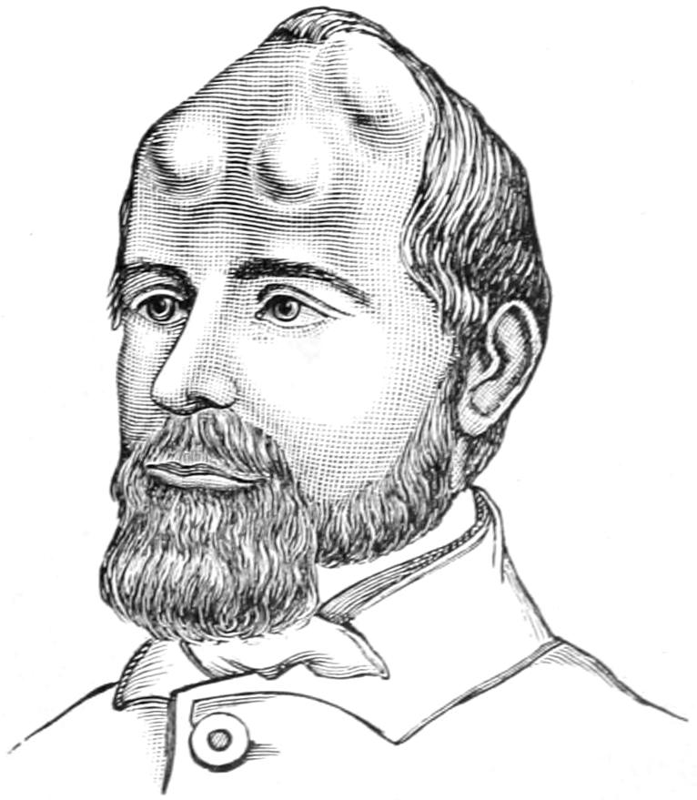
Pneumatocele of cranium. (Warren’s Surg. Obs., 1867.)
GASEOUS TUMORS OF THE SCALP.
The most common of these tumors is ordinary emphysema, which may result from injury to the upper and lower air passages. Thus fractures of the nasal bones or of the base of the skull may permit of distention of the subcutaneous cellular tissue by forcible inspiration of air. Emphysema of the scalp may be a valuable diagnostic feature in certain instances, as after fractures of the upper bones of the face. When connected with a wound it should be enlarged in order to permit the escape of contained air. Otherwise these puffy swellings disappear spontaneously by absorption of air into the veins. In cases of malignant or gangrenous emphysema early and numerous incisions are necessary, after which antiseptic solutions, etc., should be used.
Pneumatocele.
—A pneumatocele is a chronic gaseous tumor, being a cavity distended with air which has escaped from the cells of the underlying bone, bounded on the outside by the scalp and beneath by the cranium. They are found about the mastoid or the frontal regions. Not more than three dozen cases are on record. In consistency these tumors are elastic, while the escape of air upon pressure is sometimes heard on auscultation. Their explanation is usually a defect of the inner wall of the mastoid cells, through which air may be forced from the pharynx through the middle ear by violent effort, or similar defect in the ethmoidal cells by which air is forced anteriorly. Bony defects which might permit this condition are seen in a small percentage of craniums.
Treatment.
—The best results in the way of treatment have been achieved by puncture, with the injection of weak iodine solution (Fig. 366).
TUMORS OF THE SCALP.
Tumors of the scalp may be divided into the congenital and the acquired, as well as into the benign and malignant.
Of the congenital tumors the dermoids are of most interest. Originally the dura and the skin were in contact, and the cranial bones develop later between them. This explains the occurrence of dermoids either beneath or outside of the bone or their simultaneous appearance and possible connection. Many of the so-called atheromatous cysts or wens are of dermoid origin. Those which are extracranial need only antiseptic incision or excision. It will often be sufficient to split such a cyst with a bistoury, after which each half of the sac can be detached from the bed in which it has lain. Should intracranial connection be discovered the bone chisel and sharp spoon will be necessarily called into employment. Some of these dermoids perforate into the orbit, and may have to be followed into that location.
Most varieties of tumors, benign or malignant, may be met with in this region. Subcutaneous collections of fat are not so common, nor are fibromas. Various bony growths may be met, while in certain cases the signs of brain pressure are to be explained only by their extension within the cranium.
Malignant tumors are common about the scalp and the cranium; they assume, however, no conventional appearance, and are seen in any shape or form, those of the scalp alone occurring either as carcinoma or epithelioma from its epithelial elements, or as sarcoma from its mesoblastic elements. Tumors primary in the periosteum or bone are necessarily of sarcomatous nature, while those of the type which perforate to the surface may be either sarcoma or possibly endothelioma. The general character of these growths has been referred to previously. In regard to their extirpation (for there is no other treatment than this) operations of varying degrees of severity may be required. (See Cysts and Tumors and Tumors of Bone.)
Fig. 367
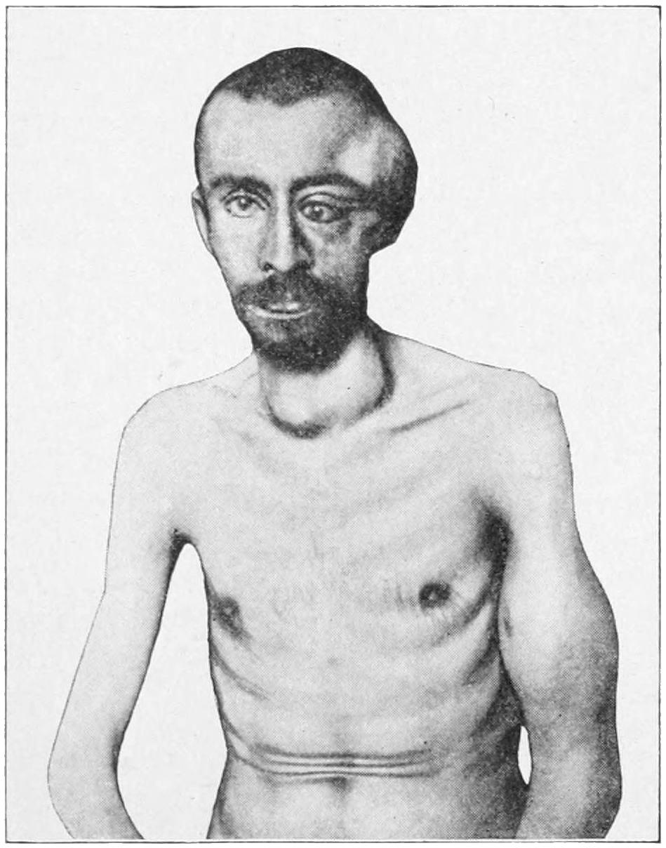
Osteosarcoma of the temporal region. Metastatic tumor in the arm and thyroid. (Parker.)
The superficial epithelioma should be attacked before it has become adherent, in which case everything should be removed down to the underlying periosteum, after which a plastic operation will permit the repair of the defect, so that primary union of the whole surface may be secured. Any malignant growth which is adherent to the underlying cranial bone calls not only for removal of its own substance, but for that of the bone to which it is attached. To fail in this is to invite recurrence. This may necessitate more or less extensive osteoplastic resections of the bone, but the condition permits of no middle course. Extensive resections of bone have been made with success, and need not be abstained from unless there be good reason to fear involvement of the dura or cortex. In this case the advantages and dangers should be carefully weighed before proceeding to operation. During operations on the bone great care should be taken, especially in certain regions, to avoid injury to the intracranial sinuses, although it has been learned that these may be ligated and intervening portions removed. But the wounding of the sinus by the point of an instrument or spicule of bone may lead to a hazardous and annoying complication, and is to be prevented when possible. A small wound in a sinus may be plugged with gauze, which may remain for two or three days. There is always a possibility of air embolism (see pp. 38 and 363) when the sinuses are opened, as their walls do not easily collapse. Hemorrhage from the soft parts may be almost entirely controlled by the use of an elastic tourniquet stretched around the skull. Oozing veins in the diploë or in the bone may often be secured by pressing the tables of the skull together with bone forceps, while at other times an antiseptic wax can be forced into the interstices of the bone and hemorrhage thus checked. In certain cases where it seems impracticable to slide flaps and cover defects the desired end may be obtained by skin grafts, after Thiersch’s method.
A rare and specialized form of blood tumor, seen only on or within the cranium, is the so-called hernial dilatation of the superior longitudinal sinus. It may present through openings in the bone; sometimes pressure upon it will cause vertigo and perhaps greater prominence of adjoining veins, even of the jugulars.
NON INFLAMMATORY DISEASES AND CONGENITAL CONDITIONS OF THE SKULL.
Incomplete Formation of Bone (Aplasia Cranii).
—Incomplete formation of bone is occasionally met with. The bone is a secondary formation in the skull, the dura and skin being originally in contact; consequently this condition can be easily explained as a failure to develop bone where it is normally produced. These defects are most common in the frontal and temporal regions. The bone may fail also to develop to ordinary thickness, and may be found as thin as paper or ossifying only in certain directions. Supernumerary bones may also develop, apparently to take the place of those previously lacking. Aplasia may also be a unilateral defect and contribute toward the formation of meningocele. Atrophy or anostosis—i. e., complete disappearance of cranial bones—is occasionally observed. It may be an interstitial or an eccentric process, and may happen at any point or at several spots. Up to a certain extent it is the rule in the skulls of the aged, when the bones become reduced to the thinness of paper or may in certain places completely disappear. Senile atrophy, in other words, is a normal process, and is to be expected after the sixtieth year of life, its possibility being not forgotten when operations are undertaken upon the skulls of those advanced in years. Eccentric atrophy may also occur from pressure of soft or hard tumors, among them the so-called Pacchionian bodies. It is also stated that increasing hydrocephalus may produce an internal and eccentric anostosis.
Craniotabes, or Cranial Rickets.
—It is particularly in the skull that the manifestations of rickets are most common, the bone becoming unduly thick and the general shape being changed. Usually there is a flattened vertex with delayed ossification, with an abnormally firm union along the suture lines. In spite of these changes, the bone often becomes affected by pressure to such an extent that a rachitic or hydrocephalic child, confined to bed and moving little or not at all, will develop a skull showing the effect of such pressure. Many rachitic skulls show areas of atrophic thinning, dispersed irregularly, while the inner surface may show the markings of the convolutions impressed upon it by the softness of the bone (Fig. 368). (See Rachitis.)
Fig. 368
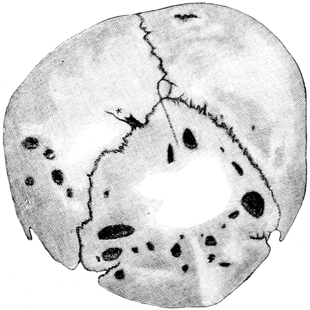
Craniotabes (rachitis). (Bruns.)
SURGICAL AFFECTIONS OF THE CRANIAL BONES.
The acute affections of bones have been considered in Chapter XXXII. Acute periostitis is, in the main, due either to syphilis or to an infection following injury. In the latter case it proceeds from the margin of the wound, and may spread to a considerable distance. It is in some instances secondary to deeper infection extending from the middle ear, and then is found posteriorly to the ear and externally to the mastoid cells. Congenital openings or defects of the sutures about the mastoid seem to have much to do with the travelling of infectious lesions in these localities.
Fig. 369
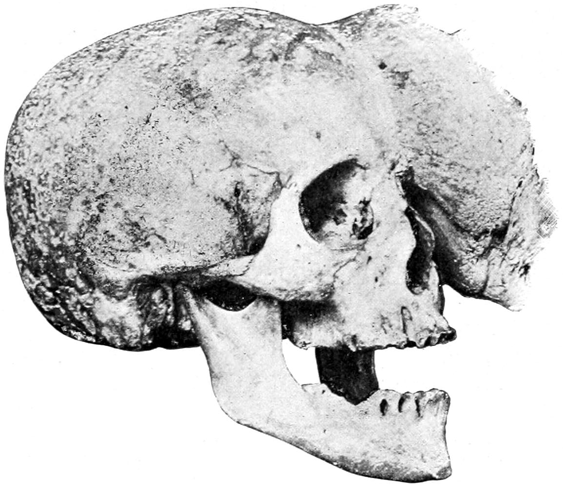
Osteoma of skull. (Mudd.)
Fig. 370
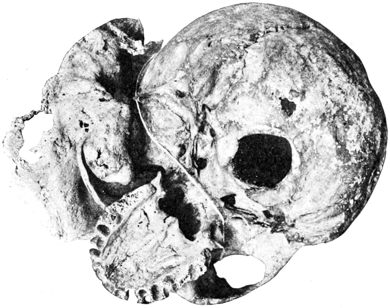
Same as Fig. 369, seen from below.
Acromegaly and Leontiasis
have been considered on pages 437 and 438.
Acute Osteomyelitis.
—Acute osteomyelitis is due to essentially the same causes as those just discussed. In this case it is especially in the diploë that the principal ravages occur. Unless promptly recognized and relieved by surgical measures this is likely to lead to sepsis of the pyemic type and at a relatively early period, the venous arrangement of the diploë favoring such type of disease.
Necrosis of the Skull.
—Necrosis of the skull is ordinarily the result, directly or indirectly, of injury, in which case it is usually of the acute form, a fragment, which has been too much separated from its surroundings to live, giving evidence of early and easily recognizable death. This necrosis is mainly confined to the external table. Necrosis of slow origin is due either to tuberculosis or syphilis, perhaps more often to the latter. Under a cold abscess of the scalp or subperiosteal abscess will often be found a small area of dead external table which needs complete removal. Necrosis has also been observed to follow severe burns of the scalp. It is usually combined with caries of adjoining bone. The caries produced by syphilis is illustrated in Fig. 371.
Fig. 371
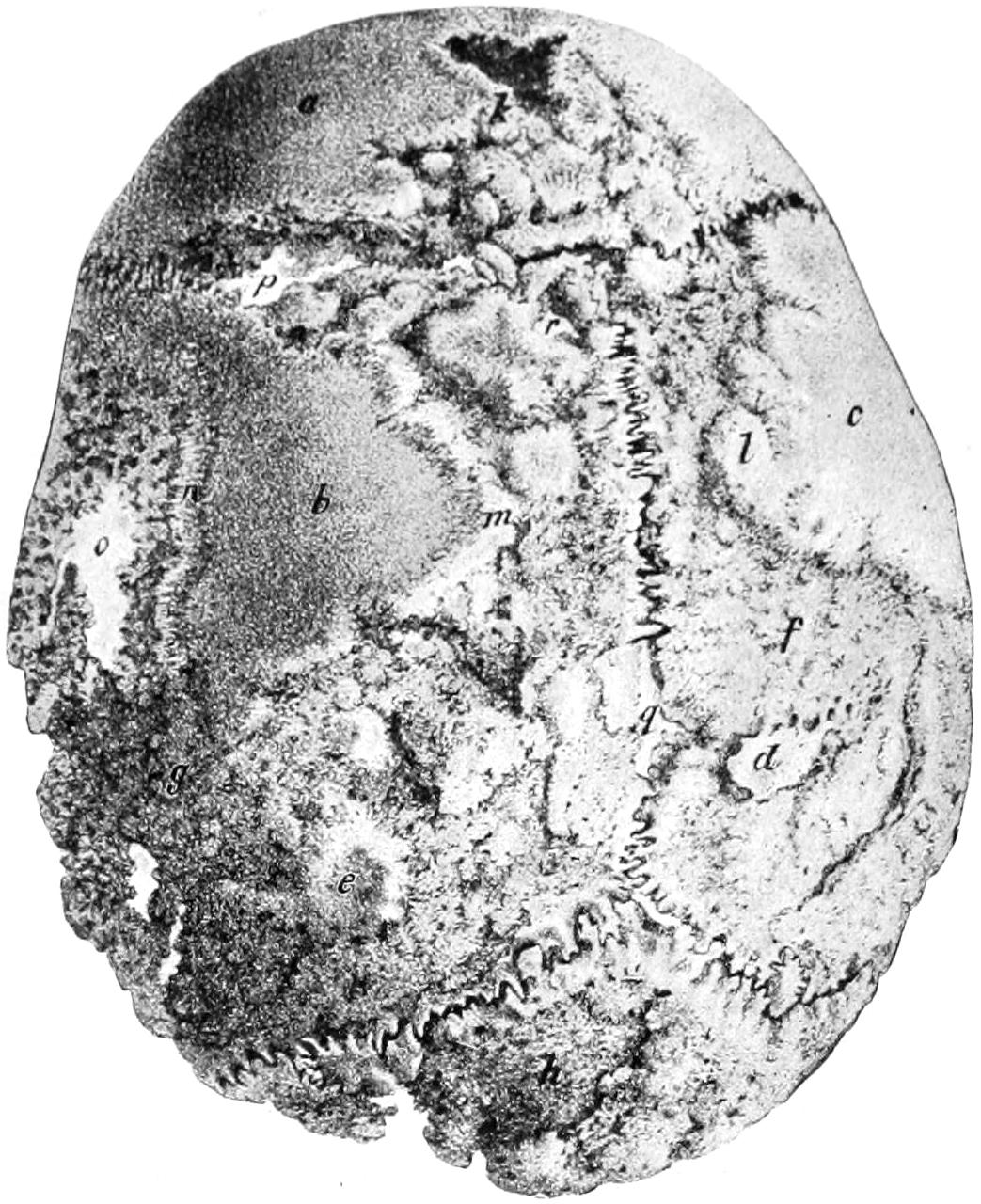
Syphilitic caries of cranium. (Bruns.)
INJURIES TO THE HEAD PREVIOUS TO AND DURING BIRTH.
In utero the head is surrounded by amniotic fluid and is well guarded against injury. Nevertheless as the result of penetrating wounds or of falls on the part of the mother real injuries do occasionally occur. Most of the cases of skull fracture reported as occurring before birth have occurred during delivery. Multiple fractures of the skull of either character have been observed.
During the process of parturition there nearly always appears a tumor of the scalp in the newborn, known as the caput succedaneum, at the point where pressure upon the head has been least. It usually disappears quickly after birth. It is due to a collection of blood, partly an extravasation, as the result of compression or injury. It is composed also of edematous soft tissues of the surface. If incised, blood-stained serum is poured out. When this fails to rapidly resorb during the first days of the infant’s existence, and especially if it fluctuate, it may be incised under antiseptic precautions and blood clot be turned out. In rare cases it suppurates, by which is produced an acute abscess, which should be promptly evacuated.
A collection of fluid blood between the periosteum and the bone is known as the cephalhematoma neonatorum, such a lesion occurring on an average once in two hundred cases. It is generally found over the fissures, and appears to be produced by the sliding of the bones. This collection also usually promptly disappears. In case of failure it may be aspirated or incised. Before resorting to any operative procedure it would be well to make a careful distinction between a possible meningocele or encephalocele, as a congenital defect, and cephalhematoma as an accident of delivery.
Fig. 372
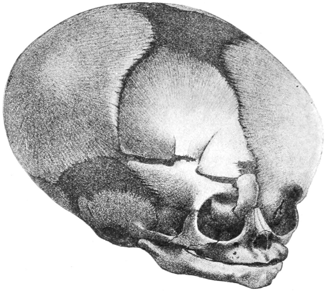
Fracture of right frontal bone in a newborn infant; fracture extending into orbit. (Bruns.)
A depression in the skull of a newborn child which does not quickly right itself or yield to expanding influences from within should not be allowed to go uncorrected, as serious lesions ordinarily of paralytic type may result therefrom. In these days of aseptic surgery there is no reason why such operation as may be necessary to elevate a fragment or an entire bone should not be performed, with the usual precautions.
IMPORTANT POINTS IN THE SURGICAL ANATOMY OF THE SKULL.
The young and the aged have no distinction of tables of the skull, but the diploë which separates the two tables is an affair of middle age, develops slowly, and disappears after the same fashion—sometimes to such an extent as to leave the skull of almost paper-like thinness. In all operations, then, upon the young and the old the surgeon should proceed with extreme caution, as if expecting to find the skull quite thin. The lower limit of the squamous bone proper is the so-called mastosquamosal suture, and operations confined to the squamous plate alone are safe from injuring the sigmoid sinus on its inner side. The ridge at the posterior root of the zygoma indicates, by its lower border, the level of the mastoid antrum. A few lines above this is the level of the base of the brain. The mastoid is present at birth and appears externally by the second year. Its antrum is present also at birth, though its air cells do not develop until after puberty, their location being previously occupied by cancellous tissue. Most of these cells open into the antrum, a few directly into the tympanum. They are not always separated from the sigmoid sinus by bone. The partition between them is perforated by minute veins, forming an easy communication between the sinus and the antrum. Air escaping from the mastoid cells into the overlying tissue may cause emphysema from a basal fracture. In all operations upon the mastoid antrum the operator should keep to its outer side, and the higher and the more closely to the posterior zygomatic ridge he makes the first opening the more sure is he to escape injuring the facial nerve. The groove for the sigmoid sinus extends to the jugular foramen from a point on the outside corresponding to the asterion. The lateral sinus may be indicated externally by a line from the superior border of the mastoid to the inion—i. e., from the asterion to the inion.
The frontal sinuses are usually separated by a septum, which is often incomplete or wanting. They are variable in size and outline, and do not develop until after the seventh year. The infundibulum, by which they empty into the nasal cavity, is often so small that when the lining membrane is involved it becomes closed, and retention, with its accompanying symptoms—pain, tenderness, swelling, etc.—may ensue. Ulceration and erosion, however, may cause perforation internally through the supra-orbital plates, so that pus may penetrate through the inner half of the orbit.
Aside from its direct communication the superior longitudinal sinus connects with the basal sinuses through the middle cerebral and the Sylvian veins, while communications with the middle meningeal veins are abundant. Where the frontal and diploëtic veins enter the longitudinal sinus there frequently are dilatations in which marasmic thromboses often originate. This sinus is also connected with the veins of the nasal septum, so that a septic phlebitis may be propagated from the nose. So much of the lateral sinus as is contained in the sigmoid groove is known as the sigmoid sinus, which connects directly with the exterior through the mastoid and the posterior condyloid veins. In sinus thrombosis this mastoid vein is likewise affected. One or more condyloid veins accompany the hypoglossal nerve through the anterior condyloid foramen, and may also serve for the propagation of infection or exit of pus.
While septic particles may be carried from any part of the lateral or sigmoid sinuses—usually through the internal jugular—they may also be carried by way of the other veins above mentioned or the occipital sinus, all of which empty directly into the subclavian without passing through the internal jugular. These sinuses are all rigid tubes, always open, while the veins are thin and flexible, their caliber constantly varying with inspiration and expiration. The sinuses contain no valves, and these are very rare in the cerebral veins.
So far as the lymphatics are concerned there is free and easy communication between the internal and external plexuses and nodes. Into the superficial nodes, along the external jugular, outside of the deep fascia, empty all the external lymphatics of the head. Intracranial infection shows itself in swelling of the deep cervicals beneath the deep fascia. Lymphatics are abundant in the dura, and pathogenic organisms, once housed within the dura, find it easily open to invasion. The potential interval between the dura and the arachnoid is termed the subdural space, when considerable effusion may occur without marked symptoms, owing to its easy diffusion, while blood here poured out may travel even to the lowest parts of the spine and cause death by pressure upon remote points.
The arachnoid bridges over the convolutions and does not extend into the sulci. It is not vascular; at certain points it is adherent to the pia, at others it does not touch it. The subarachnoid space is formed in the latter way, and within it most of the cerebrospinal fluid is contained. This space is unevenly distributed over the brain surface, most prominently beneath the posterior two-thirds of the brain, where there is a wide interval between the arachnoid and the pia, extending forward around the medulla and pons and as far forward as the optic nerves. This space connects with the ventricles by the foramen of Magendie, as well as with the sheaths of the cranial nerves. Where these nerves escape from the brain or cord they are covered by all three membranes, the layers being most distinct along the optic nerves. Fluid injected into the subdural space may pass along the spinal nerves as far as the limbs. It is essential to realize this in order to appreciate how extensive is the surface exposed in leptomeningitis.
Internal hydrocephalus is often the result of closure of the foramen of Magendie. The cerebrospinal fluid is rapidly reproduced after traumatic escape. External hydrocephalus or accumulation in the subarachnoid space, is a condition frequently due to tuberculous infection.
The pia is the vascular coat of the brain, supplied with an extensive network of fine nerve fibers derived from the sympathetic and the cranial nerves, having intimate relations with the brain, to such an extent that leptomeningitis and encephalitis are almost inseparable. The nerve supply to the cerebral membranes explains the severe pain of meningitis.
INJURIES TO THE SOFT PARTS OF THE CRANIUM.
In direct connection with what has been stated above it is well to emphasize that the venous communications between the exterior and interior of the cranium are numerous, and that the frequency of these anastomoses explains the ease with which extracranial infections are propagated within; in other words, these explain the frequency of septic mischief in the brain after external injuries.
Penetrating and Incised Wounds.
—Penetrating and incised wounds are frequent about the head, their prognosis per se, as well as their proper treatment, varying but little from that of such wounds in other parts, so long as the skull proper and its contents escape injury. Hemorrhage from scalp wounds may be profuse and even fatal. The most dangerous hemorrhages occur from the temporal vessels. Penetrating wounds are short, and the periosteum and underlying bone are usually also injured. Such small articles as blades of penknives, particles of dirt, etc., will often be found when the parts are carefully inspected, a measure never to be neglected. Contusions of the scalp and skull are spoken of as subcutaneous, subaponeurotic, or subperiosteal, and are most frequent in the frontal and lateral regions. Ecchymoses following them may be extensive and discoloration may spread over a large area. In traumatic hematomas resulting from various injuries incision should be an early resort should blood clot fail to resorb.
INJURIES TO THE CRANIAL BONES.
All conceivable degrees of injury to the bones, from a trifling division of the periosteum down to most extensive denudation or mangling of the external table or the entire thickness of the bones, may be encountered. These lesions may be spread over a large area or may be the result of penetrating wounds. In other words, we may have linear, penetrating, or large surface wounds, with such injury to the scalp as perhaps to amount to a total loss of covering for the same. All of these, moreover, may be complicated by fractures of the bone at the point of injury, with or without brain lesions, or by other and more remote lesions.
In regard to most of these, it may be said that non-penetrating injuries, when promptly and properly attended to, have, in most cases, a favorable prognosis. Every penetrating wound of the cranium is a condition justifying grave prognosis, on account of the great danger of infection incurred. Other features of these wounds, with more in regard to prognosis and treatment, will be given under the head of Compound Fractures of the Skull, etc.
It is necessary, however, to say in this place that penetrating wounds of the cranium are often received in a way which does not permit actual diagnosis, as, for instance, when received through the nose or the orbit. Every wound whose history and appearance indicate that penetration may have occurred should be subjected to the most rigid scrutiny and care. Points of fencing foils, umbrella tips, etc., have been forced into the brain cavity through the orbit and elsewhere in ways which left little external evidence of the severity of the injury.
FRACTURES OF THE SKULL.
Following the anatomists, and for general convenience, these are divided into fractures of the vertex, of the lateral region, and of the base, the former being the most frequent as the vertex is the most exposed. A fracture in a given region may be confined to that locality or may radiate widely or extend nearly around the cranium. Of all the fractures of the bony skeleton those of the skull constitute about 2 per cent.
Fractures of the Vertex of the Skull.
Fractures of the vertex are, in most instances, due to actual violence, the force being often expended at the point of application or producing radiating fractures. Those which are limited to the neighborhood of the injury are referred to as direct fractures, in distinction to which we have indirect or radiating, often producing remarkable results. Fractures may vary between the simplest crack or fissure, accompanied by but trifling brain symptoms and never recognized, to the most extensive comminution and destruction of cranial bones which can be imagined.
Splintered or Comminuted Fractures.
—Splintered or comminuted fractures refer to the formation of numerous bony fragments, which are often more or less loosened, sometimes completely so, occasionally dovetailed together, and often driven in or depressed. Such fractures are direct. It is possible to have comminution without depression; the latter makes it the more grave condition.
Fractures with absolute loss of substance may be made by gunshot injuries or by any extensive splintering or by a penetrating body. It is possible to have fracture of one table without that of the other, this being often true of the external table. In isolated fractures of the inner table there is often dislodgement of small fragments which may injure the dura and possibly produce later epileptic or irritative disturbance. When the external table is chipped off the diploë is exposed, and this with its wonderfully fine venous communications opens up a wide area to infection and subsequent pyemia.
Gunshot Fractures.
—Gunshot fractures are always depressed and almost invariably comminuted. The bullet of the modern army rifle possesses a great initial velocity, and the cranium struck by it will probably be disrupted into fragments, causing instant death. The majority of gunshot fractures of the skull seen in ordinary civil practice are due to revolver or pistol bullets from weapons of the prevailing type. In these instances there will usually be penetration, perhaps with perforation of the skull, and the formation thus of one or of two compound fractures, the wound of entrance being always comminuted and depressed, while fragments of bone may be scattered along the course of the bullet, which may also carry infectious material from without, such as hair, particles of hat, and the like (Figs. 373 and 374). (See also Figs. 52, 53 and 54.)
Fig. 373
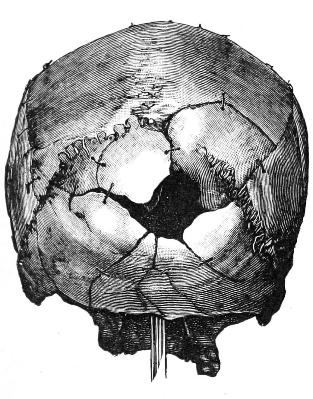
Fig. 374
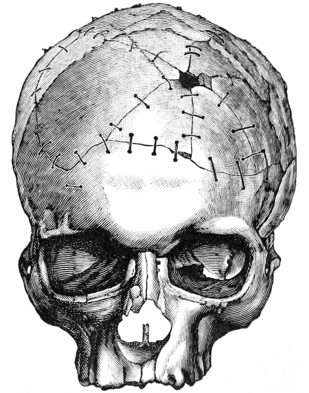
Gunshot fracture of skull. (Helferich.)
Whatever may be the wisdom of operating in other cases where there is room for doubt as to the proper course there rarely is uncertainty as to the proper treatment of gunshot wounds of the skull, which should be invariably subjected to operation.
It will thus be seen that fractures of the skull may be simple or compound, or complicated with other injuries, or depressed, without any reference to whether they are simple fissures or more extensive injuries. On the other hand, depressed and comminuted fractures may occur without being compound in a surgical sense, and with each one of these injuries there may be accompanying disturbance of the brain of any degree of severity, from the mildest concussion or shock up to rapidly fatal compression. Any imaginable complication of these head injuries is not beyond the bounds of possibility.
The essential features in explaining the mechanism of fractures of the vertex are the area involved and the violence of the impact. The skull is often surprisingly elastic, even in the oldest individuals, and fractures occur ordinarily when the natural limits of elasticity have been exceeded and bone cohesion overcome. Children particularly suffer from depression without fracture, which formerly was never operated upon, but which is now regarded as requiring operation. On the other hand, certain skulls are abnormally fragile (see Fragility of the Bones, Chapter XXXII), and among the insane may be found so porous and yielding as to be easily pressed out of shape. In injuries of slight extent it is sufficient that the skull be regarded as composed of an elastic substance, while for injuries produced by greater violence the skull is to be considered rather as a globe or arch possessed of high resistance and elasticity, whose shape will probably yield more or less before a fracture results. Much may be learned from such experiments as those of Félizet, who filled skulls with paraffin and dropped them from varying heights, and then divided the bone, to note in numerous instances that, although the bone had not been fractured, it had yielded at the point of impact to a degree producing a marked depression in the paraffin beneath. After various injuries, especially to the top of the head, the shape of the skull may be altered and its diameters affected. Many fractures, then, are the result of a bursting force, which may be shown by the fact that hair has been found included within apparently closed fissures, and even on the dura. Moreover, particles of bullets have been found within the skull without any visible opening through which they could have entered, showing that the bone has yielded under impact for a fraction of a second. In certain injuries to the head, as when a man is struck to the ground, there is injury at two points nearly opposite.
Fractures of the skull, especially of the vertex, possess surgical interest mainly as they are accompanied by more or less evidence of intracranial complications. So long as there is no evidence of hemorrhage or laceration within they are ordinarily regarded as a feature of the external wound with which they are usually found, and unless there be comminution, depression, or some other good reason for operating they are covered over as the wound is closed and are left to the natural process of repair by formation of minute callus or by the ossification of granulation tissue.
It is unfair to contrast the results of the surgery of today with those of the pre-antiseptic era. Rules then enforced are now abrogated. One respect in which we violate precedent is in our disregard of the periosteum or pericranium. This is sacrificed without hesitation when found to be infected or torn or lacerated beyond repair. A flap of scalp will adhere as readily to denuded bone as to periosteum, and skin grafts can be applied and will adhere to this same bone—if not upon the first day, a little later when granulations have appeared. In the various plastic operations necessitated about the head we may also transplant flaps upon otherwise uncovered bone without the slightest hesitation. Fractures should be treated mainly in accordance with intracranial complications, or through what can be seen either through the wound or through an opening intentionally made under antiseptic precautions for purposes of exploration. It is conceded to be better policy to remove fragments of bone whose vitality is uncertain and to sacrifice tissue injured or lacerated to such an extent that sloughing would probably follow or be so exposed as to have become infected.
Diagnosis of Fractures of the Vertex.
—In the absence of an open wound, and unless incision be made, diagnosis of fractures of the vertex is necessarily conjectural. In the presence of a wound diagnosis is usually easy. In case of a small puncture it will be better to enlarge it sufficiently to permit the introduction at least of the finger. With the finger and the eye we seek to detect differences in level, depressions, fissures, etc. Mistakes arise from the formation of an exudate or a clot, by which a depression of the soft parts may be regarded as depression of the bone. Error occasionally arises from the existence of previous atrophy of the bone or any congenital defects in ossification of the skull; also in the skulls of syphilitic patients where disappearance of a gumma is often followed by absorption of the underlying bone. In case of doubt exploratory incisions should be made under aseptic precautions. These should not be made, however, unless the attendant is ready—i. e., has the facilities immediately at hand—for carrying out any further operative procedure that may be necessary, as elevation of fragments, removal of foreign bodies, etc. Error also may arise from mistaking for fracture a deceptive circular effusion of blood which frequently occurs beneath the scalp after injury. Areas of bloody infiltration often have abrupt margins which are calculated to easily deceive. In children, more especially, we often have a circumscribed bloody tumor which may contain cerebrospinal fluid rather than pure blood. In some of these cases after exploration there will be found material resembling brain matter, which, however, is not always such, although real brain substance may escape, caused by rupture of the overlying membranes. Should it be noted that the fluid used for irrigating and cleansing such a wound begins to pulsate, it will imply connection with the cranial cavity, and, obviously, fracture. A suture should not be mistaken for a line of fracture. This mistake is more easy when Wormian bones are present. Blood may be wiped away from a suture line, but not from that indicating fracture. It is not often possible to diagnosticate an isolated fracture of the inner table. It happened, however, once to Stromeyer to notice that so soon as an injured patient assumed the horizontal position he began to vomit, and that nausea subsided when he was placed in the upright position. On autopsy it was found that there had occurred a depressed splintering of the inner table with perforation of the dura—less irritation was produced in the upright position than when the patient was lying down, which accounted for his vomiting when in the horizontal posture. When a comminution has been produced it is always of prognostic value if an unbroken dura be found. Prolapse of brain substance is a serious complication. Escape of cerebrospinal fluid is relatively rare. Rising temperature after these injuries is always a sign of danger.
Treatment.
—Treatment comprises attention to the local injury and the suitable dealing with the condition of the brain within when injured. The treatment of simple fractures is expectant. In the absence of indication for operation it should be simple, and should consist of physiological rest, aseptic dressings, ice applications to the head, the administration of such laxatives, diuretics, antacids, etc., as may be necessary to favor free excretion and to guard against autointoxication. Whenever there is reason to suspect a depression, exploratory incision should be made. Actual depression, whether the fracture be compound or not, requires operation. This course is justified by the numerous instances in which later consequences have been noted, such as traumatic epilepsy, insanity, etc.
Compound injuries should always be operated upon in some manner, which includes the removal of loosened splinters, the elevation of depressed bone, the removal of foreign matter, the checking of hemorrhage, the excision of bruised and lacerated tissue, and the proper closure of the wound, with or without drainage.
In serious and lacerated cases it is inadvisable to close the wound with the view of attempting primary union. It should be packed with gauze and temporarily closed with secondary sutures. These measures should be seconded by physiological rest (quietude of the head, which may even be enforced by the posterior plaster-of-Paris splint to the head and neck), attention to the primæ viæ, the avoidance of transportation, the prevention of auto-intoxication, etc. The surgeon should use discrimination as to the amount of bone to be removed, the wisdom of opening the dura when not lacerated, of examination of the brain with the exploring needle, the matter of drainage, and the time during which it shall remain. With reference to all these matters exact rules cannot be given. When drainage is made in recent cases it is usually sufficient to drain the scalp wound. Only in cases where there is probability of meningeal infection is it advisable to attempt to drain the dural cavity. This is better accomplished with gauze, catgut, or folded rubber tissue than with drainage tubes.
Skull fractures where the injury is limited to a small area are treated according to a bolder method than was in vogue a number of years ago. There should be careful and judicious operating in every case where distinct depression can be made out, as well as in every case where indications point to injury of parts within the bone. The statistics of trephining in the pre-antiseptic era are valueless as arguments in this consideration. If done according to aseptic precautions, and if good surgical judgment be used in every respect, the operation is per se almost devoid of mortality and should not be regarded as a last resort, but rather in such cases as a first one. I have seen so many instances of later untoward consequences resulting from delay, which corroborate the experience of others, that I would not be misunderstood in this matter. My advice might perhaps be summed up in the following words: Where there are no brain symptoms and no skull symptoms, in fractures of the vertex, let the case alone; when either of these are present, especially the former, it will always be advisable to operate.
Fractures of the Base of the Skull.
In the majority of these fractures the violence is applied at some more or less distant point, and, by transmission through the arch-like structure of the skull, expends itself in fissuring or comminuting the base. The most frequent location of the indirect injury is upon the convexity. The mechanism of these fractures has been a problem for many centuries, but has been cleared up mainly within the past three decades. Félizet has shown, for instance, how the handle of a hammer may be forced into its head by striking it in either one of two different ways, and has compared the mechanism of basal fractures to this fact. The secret of these fractures probably resides in the elasticity of the skull, which varies within wide limits in different individuals, and which breaks, as do the ribs and the pelvis, at points more or less distant from that at which the injury occurred. Were the skull everywhere equally thick and elastic, there would be much less variation in these fractures, but lacerations frequently extend between the most resistant parts; and when violence is applied upon the forehead we find that the resulting fissure extends between the crista and the wings of the sphenoid, upon the same side, in its course toward the base; that when the lateral region of the skull is injured the fissure extends between the sphenoidal wings and the occipital bone; and that when the occipital region receives the first injury the fracture lies between the pyramid and the occipital crests. The analogy between fractures of the skull and cracks made in nutshells (cocoanuts, etc.) when struck with a hammer is too self-evident to be disregarded. Many years since the French introduced the term fracture by contre-coup (counter-stroke)—a practical admission of the occurrence of fracture at a point more or less opposite to that struck.
Fig. 375
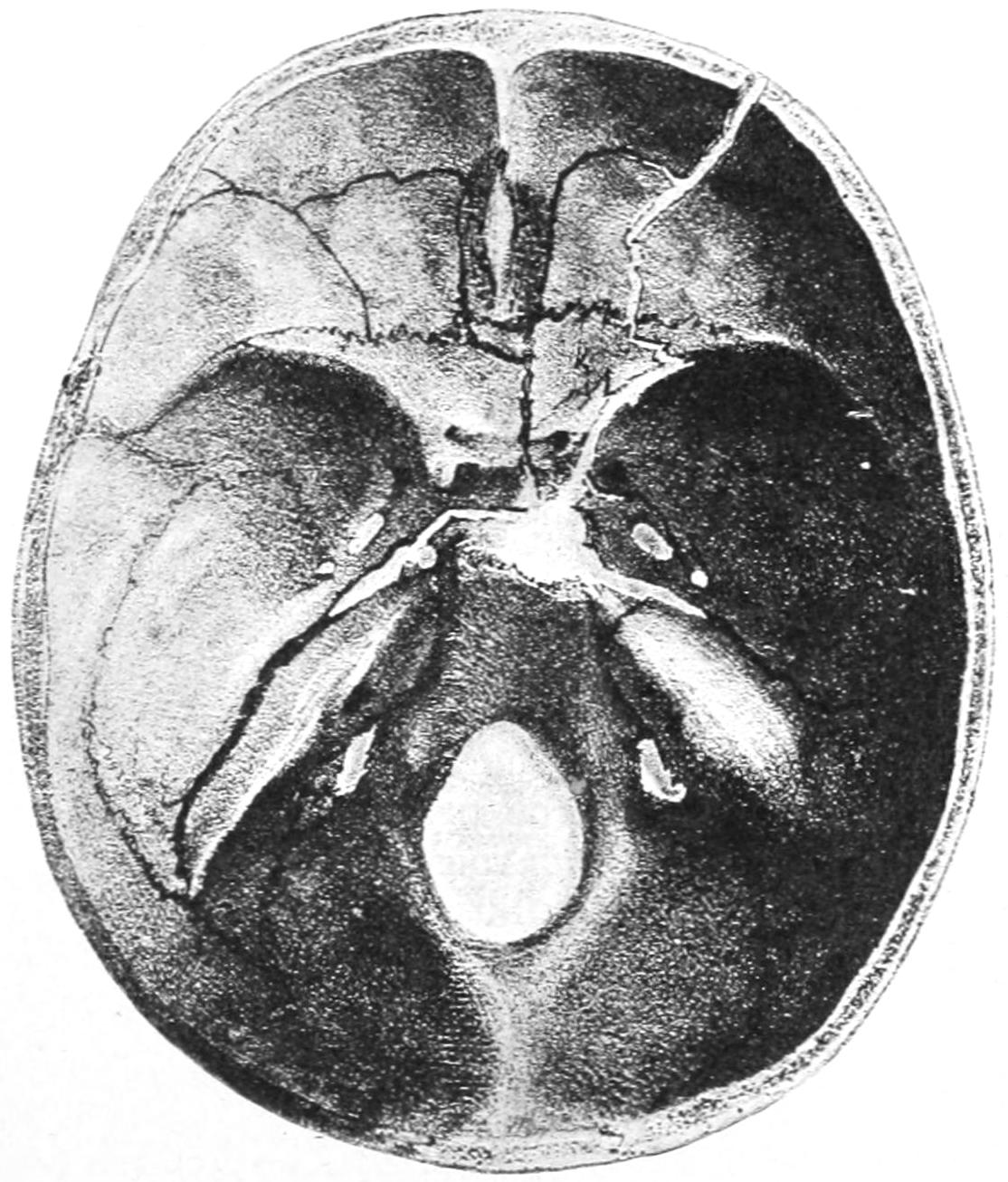
Fracture of base of skull. (Bruns.)
Fig. 376
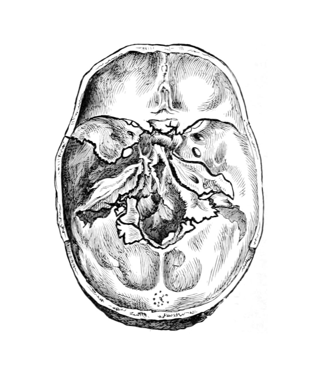
Fracture of base by fall on vertex. Both condyles broken off and driven in. Vertex was fissured.
There is, however, no certainty about these fractures. Extensive fissures of the vertex are almost always extended to the base of the skull, while the reverse is seldom true. There are doubtless also many cases in which a bursting force compromises the bone rather than mere radiation of unexpended violence; but so long as skulls conform to no fixed mathematical figures nor proportions, and are composed of bones varying in shape, density, and strength, it will be impossible to formulate any laws which are sufficiently comprehensive to be satisfactory. Fractures in the posterior fossa occur most often through violence applied posteriorly and from below. There is a ring form of basal fracture produced mainly by the impact of the vertebral column, as when an individual falls upon his head the weight of the body forcing the cranial base in upon the brain.
PLATE XLII
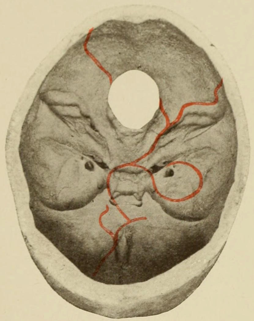
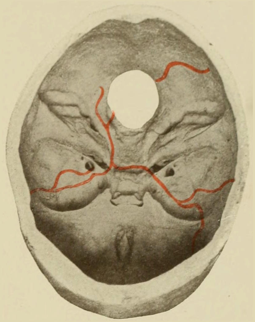
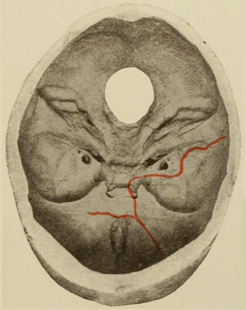
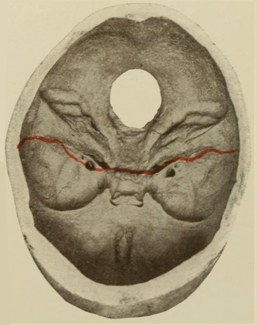
Fractures of the Base of the Skull. Illustrative lines of fissure or fracture are printed in red.
Fractures of the anterior fossa may involve the roof of the orbit; even facial bones may participate in the injury. These considerations are not without importance, for if a patient presents symptoms of injury of the petrous bone, and if these be accompanied by injury to the lateral region of the skull, we are in a position to make a diagnosis of fracture of the middle fossa. (See Plate XLII, and Figs. 375 and 376.)
By all means the majority of basal fractures are mere fissures which open and close instantly upon their production—close so quickly, in fact, as scarcely even to include blood between the broken bony surfaces.
Prognosis.
—The majority of basal fractures are fatal, either because of injuries to the brain, or of hemorrhage or violence along the nerve trunks, or from infection extending along the newly opened paths. Other things being equal, the longer the fissure the greater the danger, particularly so when it takes its origin in the vertex, and because of greater ease of infection. Air infection may occur in any basal fracture by fissures extending into the various air-containing cavities—nose, ears, sinuses, etc. They are then practically compound, though invisibly so. The general prognosis will depend, first, upon the injury to the cranial contents; second, upon the possibility of infection. Statistics are absolutely unreliable, although always possessing interest. Numerous museum specimens show the perfection with which bony repair may occur and the admirable way in which compensation is afforded for defects. Suppuration after basal fractures is mainly that due to purulent basal meningitis, in which case the brain symptoms dominate in the clinical picture, while the appearance of a single drop of pus in the ear or upon the surface is of the greatest significance. The conversion of a serous outflow (e. g., from the ear) into purulent fluid is also pathognomonic. Various paralyses, principally of the cranial nerves, may follow this injury and prove temporary or permanent. Diagnosis is often made by a study of these special nerve lesions.
Diagnosis.
—The most significant diagnostic features are:
1. Spread of blood from the point of fracture until it appears as an ecchymosis at certain points beneath the skin: This will occur early in some cases and late in others. It may appear beneath the skin or beneath the conjunctiva or other mucous membranes, even in the pharynx. Occurring about the mastoid, it implies fracture of the middle or posterior fossa; about the eyelids, of the anterior fossa. Beneath the bulbar conjunctiva it means extravasation along the optic sheath, probably from within the dura. In fractures of the posterior fossa it will come to the surface of the neck, but only after two or three days. The ecchymoses about the lids or orbits occurring after two or three days mean more than those occurring within these days, for the latter may be caused by external bruising. The globe of the eye may be pushed forward by blood accumulating within the orbit. Exophthalmos thus produced is therefore most significant, though not common.
2. Escape of serous fluid, blood, or brain substance from the cavities of the skull: Hemorrhages from this cause occur most often from the ear, the petrous bone being tunnelled with various canals through which blood may thus escape. The surgeon should, however, assure himself in every instance that the blood is escaping from the ear and not from some trifling wound of the external soft parts, the soft walls of the meatus, or the tympanum. Profuse hemorrhage can probably only come from a basal fracture. Escape of serous fluid is usually noted as a sequel to hemorrhage, although it may begin almost immediately after an injury. Rarely more than twenty-four hours elapse before it begins to flow. The quantity of fluid discharged is sometimes considerable. It may occur in frequent drops or during expulsive efforts, like coughing, or may ooze in such a way as to be insensibly collected by the absorbent dressings. In average cases the amount in twenty-four hours is from 100 Cc. to 200 Cc.; 800 Cc. have been noted in occasional instances, and in a very few still more. Occasionally violent expiration will increase the flow.
In some cases the fluid may escape through the Eustachian tube into the pharynx, whence it may escape by the nostrils or be swallowed.
The escape of brain substance is rarely noted, but obviously implies such serious injury as to make the prognosis of the worst.
3. Disturbance of function along particular cranial nerves, paralysis of which is often produced by fractures of the base, especially those involving the foramen of exit of the nerve involved: The nerve may be lacerated or injured in such case by the fragment of bone.
In addition to these distinctive features there will be in the majority of instances brain symptoms, either of contusion or compression, varying in severity within all possible limits, but adding their weight to the value of the testimony.
Other and unusual signs of basal fracture may occur, such as communication between the cavities of the petrous bone and the mastoid cells, leading to the formation of pneumatocele (see page 545), or emphysema of the overlying soft parts, observed mostly about the orbits, when the nasal cavity is involved.
Treatment.
—The treatment of basal fractures is mainly symptomatic. The first effort should be to make antiseptic all those parts of the skull involved, which means to shave the scalp; to thoroughly cleanse and irrigate the external ear and the auditory meatus, using a head mirror and ear speculum for this purpose; to tampon the meatus with antiseptic cotton; to provide a copious absorbent dressing for such fluid as may escape and to change this frequently; to cleanse the nasal cavity as well as the conjunctival sac, for all of which the peroxide of hydrogen is serviceable. All of this should be done promptly, while at the same time studying the patient for evidence of brain injury or of involvement of special nerves. By the time these measures are thoroughly performed a decision as to the necessity for immediate operation should have been reached. Evidence of brain compression wanting, and in the absence of external or compound injury the patient may be left at rest, with cold applications to the head and active purgation. In many of these instances benefit follows the application of a number of leeches to the mastoid region and to the occiput. Operation is necessary later only when brain symptoms supervene, these consisting of evidences of compression, either from blood or from pus, as compression from other causes should have been acting at the time of the first examination, and should have been recognized at that time. When direct fractures are evident the possibility of the entrance of foreign bodies should be also remembered. Thus penetrating fractures of the base have occurred through the orbit as the result of accident or assault, and such weapons or implements as foils, ramrods, drumsticks, canes, umbrella points, etc., have been known not only to penetrate into the brain, but perhaps to leave some portion of their substance—e. g., a foil tip or an umbrella tip—within the cranium after their withdrawal.
Separation of sutures, known also as diastasis of the same, is the occasional result of injury instead of, or complicated with, fissures or other fractures. It is the result of violence, and is virtually a specific form of fracture, from which it differs in no essential particular. Diastasis can only take place along lines of previous suture, but it is possible that Wormian bones may be thus loosened. Sutures thus separated ordinarily heal by fibrous repair rather than osseous union. Diagnosis is possible only as they are exposed to view, although displacement in the middle line or along known suture lines may be regarded as diastasis. The treatment differs in no respect from that of other fractures.
Injuries to the frontal sinuses occasionally complicate fractures of the skull. These sinuses vary in different individuals, are rarely truly symmetrical, and are not found in the young. They connect with the nose in such a way that emphysema of the frontal region is quite possible, while air may be blown beneath the periosteum or may communicate with the interior of the cranium. In wounds of the frontal region the sinuses are occasionally opened—a fact of importance, for infection of the Schneiderian membrane may occur and endanger life, mainly because of the retention of infectious products within its cavities. Moreover, by such wounds the ethmoid may also be injured. Pus which escapes from these sinuses and from the ethmoidal cells is usually thin and bad-smelling. Long continuation of suppuration after such injuries probably means necrosis and formation of sequestra.
INJURIES TO THE BRAIN AND ITS ADNEXA.
By better acquaintance with certain portions of the brain whose function is now generally recognized and described, as well as with the more exact knowledge regarding the entire encephalon, the outcome of many recent studies, the teaching of the past in regard to the nature of various brain lesions has been essentially modified. Especially is this true in regard to the distinction formerly emphasized as between concussion and compression. In discussing brain injuries we should, first of all, distinguish between traumatic disturbances of the entire endocranium and localized injuries to the brain or particular vessels and nerves entering into its composition. In regard to the first, it is possible that the entire blood or lymphatic circulation within the cranium may be affected in such a way as to influence its nutrition and function, by which means activity and function are mildly or seriously perverted. The immediate effect of severe injury to any part of the body is reflex vasomotor spasm, which constitutes the essential feature of the condition known everywhere as shock. It is this condition, with its marked local expressions, which was formerly known as concussion of the brain. When studied upon its merits it is found to be indistinguishable from shock produced by injuries to other parts. The condition for so many years taught and recognized as concussion is but shock following injury to the head. This makes no further demands upon the question of pathology than those prompted by any traumatic disturbance.
Through the mechanism of the cerebrospinal fluid rapid alterations of pressure and of the volume of the brain are produced. There is an easy path between the inelastic cranial cavity and the exceedingly elastic and accommodating spinal canal, which latter serves as a reservoir for the fluid which may be pressed out of the cranium when brain pressure is increased. While the subdural and subarachnoid spaces are each of them absolutely closed sacs and do not communicate one with the other, there is ample accommodation within each to permit a constant equilibrium of pressure under ordinary circumstances, as between the spinal canal and the cranial cavity. The brain expands in volume with every systole of the heart, while with every diastole it contracts. Its size is, moreover, modified by the motions of respiration. Under these extremely accommodating conditions it is scarcely credible that external injuries which leave no internal evidences of violence should do anything more than disturb the equilibrium of fluid distribution.
“CONCUSSION” OF THE BRAIN.
We inherit this term concussion from the earlier masters of our art, by whom, however, it was used in a much broader sense than of late. Its modern significance was given to it by Boirel, who made it apply to a group of cerebral symptoms the result of injuries not accompanied by fracture or perceptible laceration of vessels, symptoms varying in intensity and duration.
Our present position is practically this: The possibility of pure concussion of the brain—i. e., disturbance of brain function without gross mechanical lesions—is admitted, but its general frequency is denied. When present it should either pass away quickly, the condition being equivalent to that called “stunning,” or, if it assume distinct form, its signs and symptoms are indistinguishable from those of shock, consisting essentially of rapid and feeble pulse, quick and shallow respiration, pallor of the skin, copious perspiration, complete or partial unconsciousness, muscle incoördination, with lack of sphincter control, occasional vomiting, the pupils usually reacting in light.
Treatment.
—The treatment for this condition is essentially that for shock, and whatever may be called for in the way of attention to injuries about the head—e. g., sewing up a scalp wound, etc. (See Chapter XVIII, on Blood Pressure.)
CONTUSION OF THE BRAIN.
The condition of shock (cerebral concussion), when of pure type, passes away with reasonable promptness, especially when aided by surgical treatment. Anything which persists in the way of muscle paralysis, disturbance of function of nerves of special sense, or other sign of importance, indicates something more than mere vibratory disturbance: it implies mechanical lesion which could be perceived by the eye were the parts exposed, and constitutes the condition known as contusion. This implies the existence of trifling exudates, or hemorrhages, which lead not only to absorption but even cicatrization. Contusion pure and simple differs from ordinary laceration as a contusion elsewhere may differ from a wound. It cannot be separated, however, from conditions in which there are minute separations of continuity and actual lacerations. It may be divided into three postmortem forms—general hyperemia, with or without edema; punctate or miliary hemorrhages; and thrombosis of minute vessels, which may occur separately or together. Moreover, there may exist similar lesions in the meninges, constituting meningeal contusion. Ordinarily minute vessels of the pia are ruptured and blood is effused in small and thin patches over various parts of the brain. The so-called compression apoplexies of certain authors are inseparable from the conditions above described. Such minute blood clots are only to be distinguished upon very careful sectioning of the brain, and are found most often in the region of the medulla and along the floor of the fourth ventricle. They are probably caused by the forcing into the fourth from the lateral ventricles of the fluid contained in the latter.
Symptoms.
—When the ordinary symptoms of shock, which follow all severe injuries to the head, especially when the deep lesions are not too severe, fail to disappear in a short time under proper treatment, and when new and irregular symptoms are superadded to those of shock alone, it is reasonable to suppose that the intracranial condition is one of contusion rather than of shock. When mental agitation changes into delirium, when the rapid, feeble pulse becomes stronger and slower, the respiration deeper, the limbs move in incoördinate ways, the speech disturbed from muscle incoördination, the patient selects wrong words, or when the mental condition becomes more serious and stupor or coma take place of the delirium, while external irritants have less and less effect, and when the pupils gradually enlarge while failing to respond to light, it may be said that the condition of contusion is making itself apparent. If along with muscle uncertainty there is also muscle spasm or rigidity, with fixation of the fingers in the athetoid position, the evidence to this effect is increasing. If with all this the thermometer fails to show that an active inflammatory condition—i. e., meningitis—is prevailing the diagnosis may be regarded as certain. Error may possibly arise when there are evidences of alcoholism. Coma following head injury ought not to be ascribed to the alcoholic condition except by the strictest process of exclusion. Temperature alone will be of the greatest service in this direction, since in alcoholism it is usually subnormal. In apoplexy and non-traumatic hemorrhages it is also usually subnormal at the commencement of the attack, rising to normal, and remaining there if the patient recover, but continuing to rise in cases where the prognosis is bad.
Treatment.
—The treatment of brain contusion should be managed largely in response to special symptoms. Physiological rest, attention to scalp wounds, fractures, etc., shaving of the scalp, application of ice to the head, with such stimulation to the heart as may be necessary in extreme cases by subcutaneous administration of adrenalin, atropine, etc., by local fomentations over the epigastrium, or by immersion in a hot bath when surroundings permit it—these in a general way constitute most of the methods of treatment in contusion. When only symptoms of diffuse and minute lacerations can be recognized the use of the trephine is impracticable except when indicated by some external marking—i. e., compound fracture or the like. When localizing symptoms are present the trephine is, of course, indicated. When the skull injury is recognized as a basal fracture, venesection or the application of leeches behind the ears will be most serviceable. In every such case there is the greatest necessity for regulating the excretions and preventing auto-intoxication. For this purpose diuretics and laxatives should be used, often in conjunction with intestinal antiseptics. The catheter should be employed whenever indicated by the condition of the bladder, which should be carefully watched. As the days go by, and patients lie more or less helpless and inert, the greatest care should be exercised for the prevention of bed-sores. When mental inertness, muscle rigidity, etc., fail to disappear, potassium iodide should be used internally.
BRAIN PRESSURE OR COMPRESSION.
That the cranial contents—brain, blood, lymph, and cerebrospinal fluid—completely fill the cranial cavity has been already amply shown, as well as that there is no room for anything in the shape of a foreign body without seriously affecting the equilibrium between the brain and the contents of the spinal canal. When, however, any foreign substance exerts pressure upon the brain the results are invariably the same, be this substance what it may, and compression signs are always the same, no matter what the compressing cause. Reduction in capacity of the cranial cavity (i. e., compression) may be produced—
PLATE XLIII
FIG. 1.
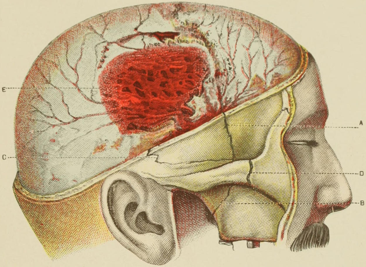
FIG. 2.
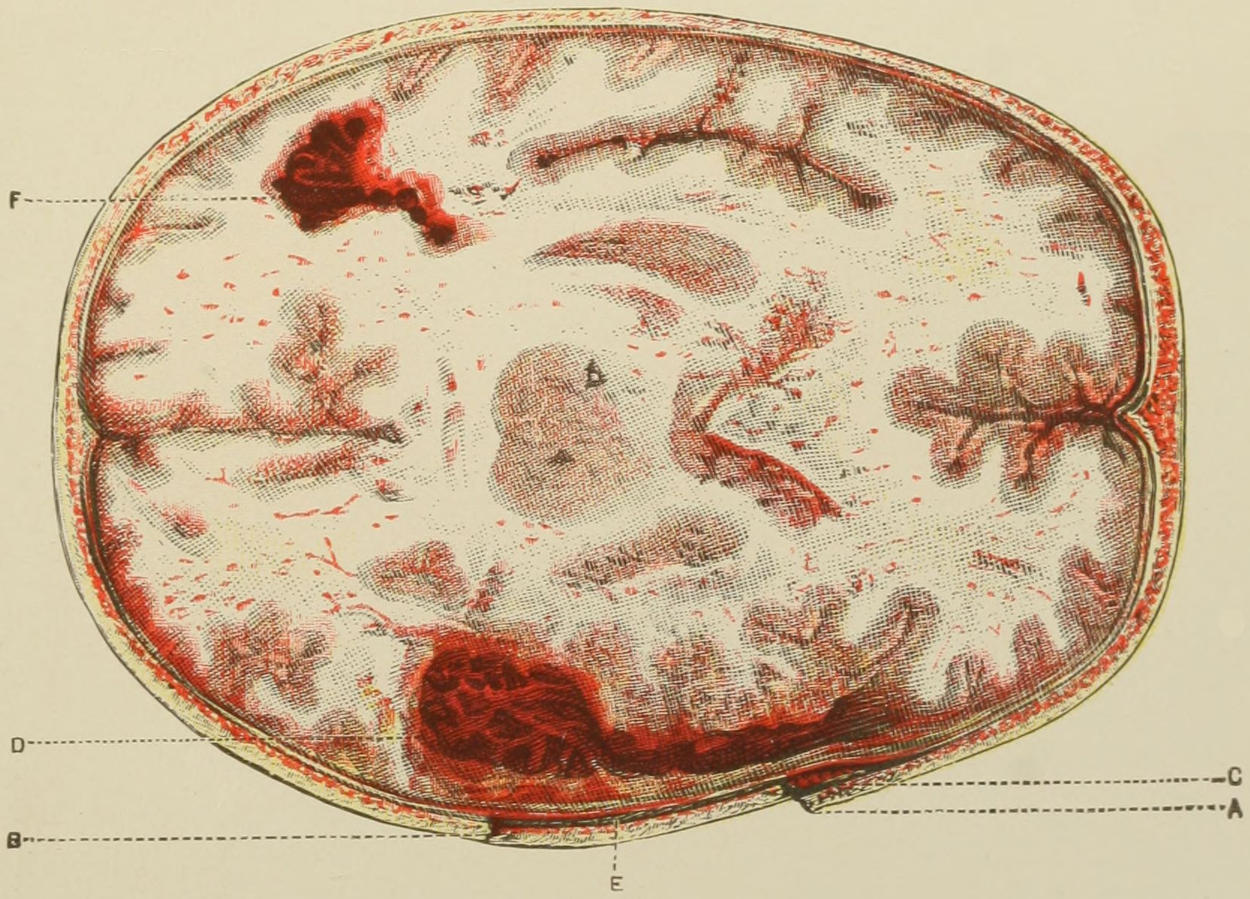
Fig. 1. Compound Fracture of Cranium, with Depression; Fracture of Bones of Face; Extradural Clot from Rupture of Middle Meningeal Artery.
Fig. 2. Horizontal Section of same, showing Depressed Fracture of Bone. (Anger.)
C, extradural clot; D, laceration of brain substance, with extensive intracerebral clot; F, same condition produced by contrecoup. Punctate hemorrhages and minute lacerations at numerous points, characteristic of contusion of the brain.
1. By reducing the dimensions of its enclosing walls (e. g., depressed fractures or by direct pressure);
2. By increase in the quantity of cerebrospinal fluid or of the volume of the brain, which latter may be produced by edema, by serous exudate, or by actual hypertrophy;
3. By foreign bodies, which may enter the skull from without;
4. By pathological conditions—collections of blood or pus, tumors, etc., which may be produced either from the brain substance, its containing bone or membranes, or its vessels.
In every one of these conditions the size and tension of the brain are affected. The cerebrospinal fluid is mainly involved in acute not in chronic conditions. A slow reduction of the diameters of the skull produces such slow alterations of pressure as to cause a minimum of disturbance. So far as compression from traumatic influences is concerned we distinguish mainly between compression—
1. By extravasation of blood (see Plate XLIII);
2. By fractures of the skull with depression, or by foreign bodies penetrating from without;
3. By products of acute infectious inflammation due to septic infection from without.
The result common to all of these is increase of intracranial tension, and its consequence is a less rapid flow of blood and an altered blood supply to the brain and its membranes.
Experiment has established that in compression of the brain cerebrospinal fluid is forced by pressure into the spinal canal, whose membranes are more elastic, and which thus help to accommodate it; it has been also established that compression of the brain by one-sixth of its volume, by any material, is fatal, and that much less is at least serious. That fractures with depression produce sometimes serious, at other times trifling, symptoms is due to the varying accommodation of the spinal canal. Both experiment and observation seem to confirm the view that consciousness pertains to the cortex as a whole, and that unconsciousness is an inhibitory or paralytic condition which is produced in compression.
Temperature is a matter of great importance in studying compression and foretelling its consequences. Elevation of temperature is an early, continuous, and constant symptom in these cases. If temperature be subnormal and subsequently rise, prognosis is bad. Variations of temperature are more reliable guides than conditions of consciousness. As Phelps has remarked, in no condition except sunstroke is temperature so uniformly high as in cases of serious encephalic lesions.
Symptoms.
—As indicated above, the symptoms and signs of compression are practically identical, no matter what the compressing cause. When this cause acts instantly there is no time afforded for differentiation, but when it occurs slowly we note the following symptoms, and about in the order here presented: Irritability or restlessness; visceral disturbances; pain; intense cephalalgia; congestion of the face; narrow pupils; augmented pulse, often seen in the carotids. If compression occur more rapidly, torpor quickly succeeds erethism, after which patients vomit, have convulsions or at least convulsive motions, speech is disturbed, and stupor comes on, from which they neither awake nor can be awakened until the compression is relieved. All of these indications refer to involvement of the cortex, which is generally regarded as the seat of consciousness as well as of projection and imagination. During the night, of the senses produced by pressure upon the cortex only the automatic basal apparatus and that of the spinal cord continue in more or less disturbed operation. Of all the general functions consciousness vanishes first and returns among the last. When intracranial pressure has reached a certain point, epileptiform convulsions result, varying in intensity, affecting all the limbs, and terminating perhaps with rigidity. These form an expression of high pressure. Similar convulsions occur in various head wounds, explanation for which is the result of pressure, which, though not extensive, may produce alteration in the circulation, with its disastrous consequences. The later and constant evidences of compression, and those which in aggravated cases supervene at once, are reduction of pulse rate, due to the action of the pneumogastric, which suffers first an irritation and later a paralysis. The pulse becomes not only slackened but full; the respiration rate is correspondingly reduced, so that breathing during coma is deep, slow, and often stertorous. This feature of stertor is an expression of paralysis of the palatal and pharyngeal muscles, which flap, as it were, in the air current. Vomiting, which may occur before brain tension has risen high, does not occur in the most serious cases. Coma is absolute.
Along with these signs the most important other indications are the paralyses, which may consist of monoplegia, hemiplegia, or paralysis of individual muscle groups, according as pressure is made upon a limited area or upon an entire hemisphere. By the division of the cranial cavity by the falx and the tentorium it is divided into chambers, in any one of which pressure may be more manifest than in the others. Nevertheless a serious compressing cause will affect the tension of the cerebrospinal fluid and produce general expression of pressure. The pupils often vary, and responsiveness to light is occasionally noted. Nystagmus and ocular rotation may be occasionally seen. Choking of the optic disk is also a frequent phenomenon, to be recognized only by ophthalmoscopic examination. This is due to pressure in the subdural and subarachnoid prolongations along the optic nerve. In milder cases of chronic compression disturbances of vision are of very great clinical importance. These pertain especially to diagnosis of hydrocephalus and of brain tumors. When they occur immediately after injury and remain, they depend upon laceration or other severe injury of the optic nerve. Those which quickly disappear depend mainly upon pressure of blood, which is reabsorbed, while those which are later in their appearance depend upon later intracranial complications. A unilateral lesion of the optic nerve depends most often upon injuries to it within the optic canal. When the lesion is bilateral the cause lies deep. General paralysis may be of the type of hemiplegia, single or double—i. e., by “double” I mean paralysis of the entire voluntary musculature of the body, which necessarily implies serious and often fatal hemorrhage.
Prognosis.
—This depends in large degree upon the nature of the compressing cause and of the possibility of its removal. While the nature of the same may ordinarily be determined, how much can be accomplished by way of removal may often not be foretold before the operation at which this should be attempted. In every acute case it is desirable to make this attempt early, for high pressure, which may be borne for a short time, is fatal if continued. Compression to any serious degree is usually fatal. So soon as paralysis of circulatory and respiratory centres is apparent the beginning of the end is at hand. Another reason for hastening operation is that acute softening of brain tissue comes on promptly, as well as general cerebral edema, which has destroyed many a patient during the second to the fourth day after injury.
Treatment.
—The treatment of compression is summed up in one phrase—i. e., to remove the cause when possible. The only cases in which this rule may be safely disregarded are those where the attempt to remove the cause means more danger than to leave it unremoved. This is not true, however, in the ordinary cases of bone depression, meningeal hemorrhage, etc. Before operation, however, or as a substitute for it in cases of minor severity, it may be well to assist venous outflow by venesection, by which blood pressure is reduced. In these cases this may be done from the temporal veins or external jugulars, with the patient in the semi-upright position. Drastic purgatives may also be employed in order to utilize intestinal outpour as a stimulation to resorption of cerebrospinal fluid. The physiological action of cold (ice-bags) may also be secured for the purpose of contracting the cerebral arteries. But all these measures are only to be resorted to when there is uncertainty as to the wisdom of operating, since when operation is indicated it should be done at once, and should take precedence of everything else. This operation means ordinarily the procedure to which the now general term trephining has been, by common consent applied, and comprises any measure by which the skull is opened at a suitable place and the dura or the underlying cortex exposed to such extent as to permit removal of the compressing cause. Whether the opening be made with trephine (annular saw) or with the straight or revolving saw, with bone chisel, with bone forceps, or with anything else, is a matter of choice on the part of the operator. So, too, removal of the compressing cause should include the elevation of depressed bone, the removal of dislodged particles as well as of all foreign bodies, the cleaning out of blood clot, the checking of hemorrhage, and the closure of the wound, with or without drainage or counteropening at some other part of the skull, as may seem desirable in special cases. This entire procedure comes now under the name of trephining, and should in most instances be painstakingly followed.
The operative maneuvers will be discussed in another portion of this chapter.
INJURIES OF INTRACRANIAL VESSELS AND SINUSES.
Intracranial hemorrhages may occur—
(a) From internal sources through the broken bone or between it and the dura (extradural);
(b) Beneath the dura, between or into the membranes (subdural);
(c) Into the brain substance proper or the ventricles (subcortical or intraventricular).
The vessels whose injuries are most often under consideration are the meningeal arteries, the sinuses, the small vessels of the membranes, and the internal carotid. The arteries, like the sinus walls, may be ruptured either by substances forced in from without or by sheer laceration. The longitudinal sinus is most liable to injury from without. When this sinus is exposed, it may be dealt with either by suture if the wound be small, or by ligation, or by tamponing with prepared gauze. Hemorrhage from this source is ordinarily not difficult to check. Fatal air embolism has resulted through an opened sinus not properly plugged. The other sinuses are more rarely injured, as by gunshot wound, fracture of the base, etc. The sinuses have also been injured by compression of the skull during parturition. Bleeding from a sinus is usually indistinguishable from that from a meningeal artery, except that the former occurs more slowly.
Injuries to the Middle Meningeal Artery.
—Injuries to the middle meningeal artery naturally occur in the immediate neighborhood of this vessel, which is not infrequently ruptured by contre-coup. The artery runs sometimes in a groove of the bone, sometimes in the dura, and sometimes entirely in the bone. The more it lies within the bone the more likely it is to be ruptured when this part of the skull is fissured. Basal fractures often follow the groove for this artery. The anterior branch is more often injured than the posterior. Extravasations from this source are more common than from all others combined, the amount of blood varying within wide limits. 240 Gm. of blood clot have been known to collect and the dura to be separated down to the base of the skull. I have repeatedly taken away a small teacupful of blood clot in such cases (Fig. 377 and Plate XLIII).
Fig. 377
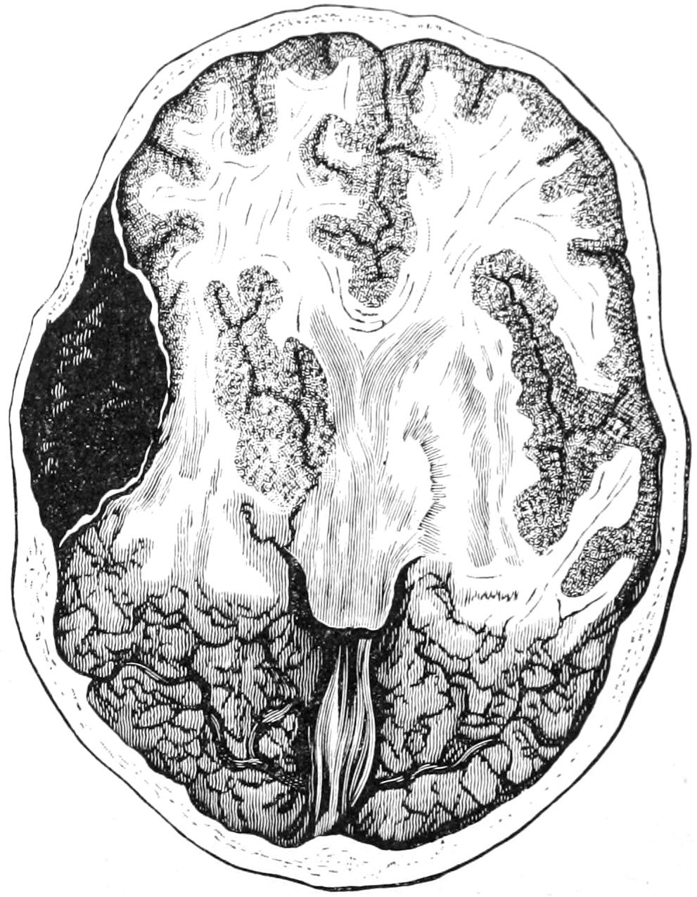
Compression following hemorrhage from the middle meningeal artery. (Helferich.)
Symptoms.
—The symptoms of this hemorrhage are those of compression, while extravasation may be rapid and quickly fatal, delayed for some time, or may take place in two stages, the first but slight and producing no coma. New clots are always dark and disk-shaped, thick in the middle, with a definite margin. As the clots become older they become more adherent and difficult to remove. The symptoms of meningeal hemorrhage consist of an interval of consciousness or lucidity after injury, followed by epileptic or spastic symptoms, alterations in the pupils and pulse, unconsciousness passing into coma, and stertorous respiration. There may or may not be external evidence of head injury. The character of the paralysis (hemiplegia) may indicate that the clot is really upon the side opposite to that of the skull which shows evidence of injury. In this case arterial laceration is the result of contre-coup. According to the rapidity of the symptoms is the extent of the primary lesion. Meningeal hemorrhages involve immediately the motor area, which makes diagnosis all the easier.
Injuries to the Carotid.
—Injuries to the carotid within the cranium are exceedingly rare. Still, it has been injured in basal fractures and penetrating wounds.
Arteriovenous Aneurysm.
—Development of arteriovenous aneurysms after basal injuries is occasionally noted. They will occasionally give rise to pulsating exophthalmos. Pulsating tumors within the orbit which push the eye forward not infrequently occur after serious head injury. Of 77 cases collected by Rivington, 41 had a traumatic origin.
Subdural Hemorrhages.
—Subdural hemorrhages are not infrequent in the skulls of the newborn, and constitute the so-called apoplexia neonatorum. They may occasion convulsions and paralyses of irregular type, while if the extravasations become infected multiple abscess may result.
In adults subdural hemorrhages are most commonly connected with brain lesions which have been already spoken of as contusions. They may be the starting points for pachymeningitis. Their most common results are disturbances of consciousness and mentality. Paralytic dementia follows in some of these cases. Extensive subdural hemorrhage may give a clinical picture corresponding to extradural. Disseminated minute ecchymoses constitute minute focal lesions, which are, however, usually so distributed as to confuse and prevent accurate diagnosis. Apoplexy or intraventricular hemorrhages, especially from the lenticulostriate artery (Charcot’s “artery of hemorrhage”), have until very recently never been regarded as warranting surgical interference. Of late, however, especially in the ingravescent or progressive forms, ligature of the common carotid has been of some service, though in order to render this effective ligation should be done early.
Traumatic Intraventricular Hemorrhage.
—Traumatic intraventricular hemorrhage occurs in much the same way as meningeal, by contre-coup. Individuality of symptoms is lost in the general comatose condition of the patient, but when operation is performed, as it is usually best to perform it, if no extradural clot be found and if brain tension be evidently increased, the dura should be opened; after which, if no subdural clot be seen, the ventricles should be tapped with an exploring instrument. In this case, if blood be removed by aspiration, a knife should be passed directly into the ventricle, after which blood, if present, will promptly escape. Dennis was the first to diagnosticate the presence of intraventricular clot and to deliberately incise into it, and I have myself repeatedly imitated this procedure, both with and without success.
In every case in which superficial or cortical hemorrhage can be recognized—or even suspected—or intraventricular hemorrhage as well, one should insist upon exploration. This means trephining, with perhaps aspiration of the ventricular contents. Tapping of the ventricle is described under Treatment for Hydrocephalus, while trephining is described at the end of this chapter.
LACERATIONS AND INJURIES TO THE BRAIN SUBSTANCE.
These have been mentioned under contusion of the brain. They may be divided into those which occur with or without fracture of the cranial bones. The term contusion was first suggested by Dupuytren. The condition comprises all degrees of injury, from the most minute local disturbances to lesions involving the entire hemisphere. The milder forms show a sprinkling of punctate hemorrhages, numerous in the centre of the injured area, the surrounding tissue taking on a more or less diffuse tint, which fades out toward the periphery, discoloration being due to the imbibition of the coloring matter of the blood. In more extensive injuries clots as large as peas, or larger, are embedded at various points, each surrounded by its area of discoloration. When foreign bodies have been driven into the brain the tissue is also discolored, while various foreign materials may be met. In instances of great violence there may occur absolute rupture of brain tissue extending from cortex to ventricle.
Prognosis.
—Prognosis depends in large degree upon escape from or occurrence of infection. In infected cases the principal dangers are from blood pressure and from later edema or acute softening as well as from meningitis. Brain lacerations may heal by cicatricial repair, but usually with some perversion of function.
The possibility of cystic degeneration of large or small clots is one of great importance. (See Cysts of New Formation in Chapter XXVI, page 264.) A blood clot within the cranium which fails to resorb is essentially a hematoma, in whose interior softening and conversion into a cyst may easily occur. These cysts make room for themselves at the expense of surrounding brain tissue, and when located in the motor area give rise to localizing symptoms as well as to epileptic convulsions. They may be often diagnosticated with certainty after an accurate history of the case and a study of the phenomena which it presents. As they grow older their walls become firmer, and it is often possible to dissect them out.
That foreign bodies may be encapsulated and remain without producing disturbance is now well known. This is particularly true of bullets. As a rule, however, though encapsulated, they produce symptoms like headache, vertigo, etc. (See Plate XLIII.)
Symptoms.
—The general features of brain lacerations are those of contusion. So long as the disturbances are minute, even if multiple, or the foreign body small, compression symptoms are not produced, or at least in very incomplete degree. Minute diagnosis is not easily obtained. The most essential thing is to decide upon the question of operative interference. In the absence of distinctly localizing symptoms or other external markings it is not usually performed. Upon the other hand a lesion which can be localized is probably due to extravasation sufficiently large to be easily reached by opening the skull; and, unless there be other and sufficient reason to the contrary, this should be done (Fig. 378).
In many instances, however, contractures or paralyses of muscle groups occur later, and are followed by spastic conditions which may be permanent. More can be done in these cases by massage, by internal medication, perhaps with external counterirritation, than by distinctly surgical procedures. Tendoplastic or neuroplastic measures for their relief may also be considered. Both albuminuria and glycosuria are known to be the result of injuries herein described, as well as bulbar paralysis and disturbances of special senses. More immediate dangers after these head injuries are those of bronchopneumonia or hemorrhagic or edematous infiltration of the lower lobes of the lungs—conditions often spoken of as hypostatic pneumonia, much resembling those produced experimentally in bilateral division of the pneumogastrics. Some of them are produced by paralysis of the glottis, the result of which is incomplete closure, with aspiration of fluids and solids from the mouth, whose decomposition sets up an infection within the lungs, and is often referred to as aspiration pneumonia. Some form of pulmonary disturbance follows in perhaps one-third of the cases of the injuries above alluded to, and should be anticipated and prevented.
Fig. 378
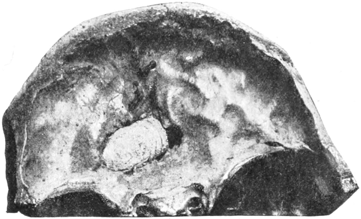
Bullet embedded in anterior fossa. (U. S. Army Med. Museum.)
GUNSHOT WOUNDS OF THE HEAD.
These have already been extensively considered in a previous chapter, so that but little more need be said of them here. Such wounds in the scalp are likely to be followed by sloughing. So far as gunshot fractures of the skull are concerned, there is frequently a marked discrepancy between the wounds of the inner and outer tables, that last perforated by the bullet being almost splintered. Penetrating wounds of the cranium by Mauser and similar bullets are not necessarily fatal. Many men were shot through the head during the Cuban and South African wars and yet did not die as a result of the wound. (See Chapter XXII.)
Treatment.
—So far as treatment is concerned, gunshot injuries of the skull necessitate trephining or exploration, for checking of hemorrhage, disinfection of the bullet track when possible, often for a counterdrainage opening with through drainage either by tube or gauze. The bullet, if it can be found, should be removed. In searching for it the old porcelain-tipped probe of Nélaton has almost completely given way to Fluhrer’s aluminum probe, which is larger and longer and when rightly directed will by slight weight usually glide gently along a bullet track, thus leading often to the missile, and at the same time indicating by its direction where the counteropening should be made. Two other methods of detecting bullets are now in vogue. Girdner, some years ago, invented a telephone probe, by which, so soon as the instrument touches the missile, a telephone circuit is completed and the operator with a telephone receiver applied over his own ear hears the tell-tale “click” indicating the fact. This has been further improved by the substitution of a bell or “buzzer,” which tells its own tale when the probe touches the bullet.
A still more ingenious application of electricity for the purpose is that afforded by Röntgen’s discovery, and during the American and English campaigns of the past few years skiagrams of skulls showing bullets in various locations have become quite common. (See Plate XIII., p. 229.)
PROLAPSUS AND HERNIA CEREBRI.
Escape of brain matter beyond its normal level is not uncommon in connection with compound fractures or their sequels. It may be primary, escaping with the blood at the time of the accident, or secondary, occurring during the ensuing days. Any lesion of this kind in which the brain appears or can be handled is entitled to the term prolapsus, in contradistinction to hernia, which implies that, though escaping from the proper cavity, it is nevertheless covered by other textures—e. g., the dura or scalp.
The protrusion may vary in size from a small tumor to one the size of a fist. It is always the result of uncontrolled intracranial tension, and may be produced by hemorrhage, by serous imbibition, or as the result of brain abscess. When immediate it is of the first variety; when later, of the second or third. When abscess is present it usually delays protrusion, which is produced by degrees. Prolapse occurs through large openings, such as those made by gunshot wounds, the trephine, etc. Prolapse proper implies laceration of the dura. It pertains obviously to the convexity of the skull, occurring, however, in exceedingly rare cases into the orbit (Fig. 379).
Fig. 379
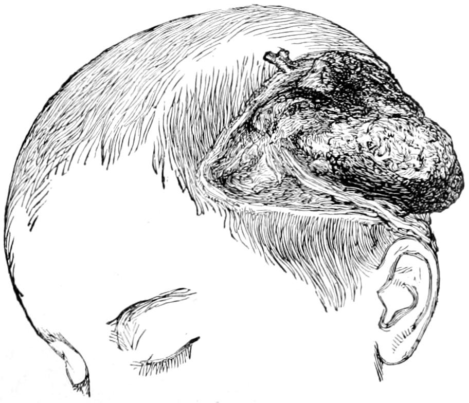
Prolapsus cerebri. (Bryant.)
Prognosis.
—The prognosis is generally unfavorable. There is always risk of edema or infection, either of which may prove fatal.
Infiltration, gangrene, suppuration, or repair by granulation may so disfigure and disguise the real brain substance as to lead to error of diagnosis. It by no means follows that every tumor presenting through an opening in the skull is of this character. When gangrene and spontaneous separation occur, spontaneous recovery may follow, the stump being covered by granulations and finally roofed over by connective tissue.
Treatment.
—Treatment in the primary cases should include the most rigid asepsis with removal of all foreign particles. Localized pressure does some good, especially in those cases where it can be tolerated. Signs of abscess should always be watched for, and deep exploration is often justified or indicated. While excision or cauterization are often heralded as successful, they are by no means without their dangers. Nevertheless in selected and suitable cases excision may be freely practised. Cases that admit of it should wear a protective shield properly molded to the part. Skin transplantation, or even osteoplastic repair of the defect, may give good results in favorable cases.
SEPTIC INFECTIONS WITHIN THE CRANIUM.
Under the general term septic infection are included:
A. Abscess;
B. Thrombosis;
C. Sinus phlebitis;
D. Meningitis;
E. Encephalitis.
These are different manifestations of infection, the clinical picture differing according to the tissues and localities involved. For the production of these infectious conditions no special bacteria other than those already catalogued in Chapter III are comprehended. Their method of activity is there discussed at sufficient length, and we need here only consider the various paths of infection. These may lie along the bloodvessels, the lymphvessels, nerve sheaths, and prolongations of the membranous sacs which extend from the cranial cavity proper.
The most common of all the paths of infection is afforded by the middle ear, especially when involved in a chronic suppurative lesion, which is by no means necessarily connected with a patulous tympanic membrane, and which may consequently be undiscovered, though in more or less constant activity.
A. Abscess of the Brain.
—This may be traumatic or non-traumatic. The former variety is most often due to the direct result of injury, infection displaying its consequences promptly or sometimes not until long periods have elapsed. The ordinary form occurs within the first two weeks, usually as an acute cortical abscess beneath a more or less compromised membrane, surrounded by a zone of red softening, and this by another of brain edema. The chronic traumatic abscesses are less often cortical, but are deeper. They are marked by prolonged suppuration of the external wound, but may occur through some mechanism not understood. Only the chronic abscesses show encapsulation, the capsule partaking of the character of the pyophylactic membrane, elsewhere described. (See Chapter VIII.) It may cover a long period—to my personal knowledge at least nine years, while others have mentioned twenty and more. The non-traumatic abscesses are in the main due to middle-ear disease. When the roof of the tympanum breaks down it is the middle fossa of the skull which is infected; when the posterior wall, naturally the posterior fossa. The most common result of perforation of the tympanic roof is involvement of the mastoid antrum or the sigmoid groove and sinus. In the former case we have temporosphenoidal abscess; in the latter, cerebellar, if any. Previous to actual perforation there is thinning of bone with thrombosis along the minute veins connected with the sinuses. When the dura is exposed by the carious process, granulation tissue often protects it against further inroads, while masses of the same projecting into the tympanum have been mistaken for prolapse. If the sigmoid groove be the site of the first disturbance, extradural abscess may form between the sinus and the remaining bone, the granulating process then involving the whole bony groove. Its later consequence is sinus phlebitis, sinus thrombosis, or intradural infection. If there be adhesion between the dura and the cortex we have actual brain ulceration without formation of a true abscess; but if once the perivascular sheaths have carried infection to the substance of the brain there is a rapid purulent disintegration of the same, and formation of a true subpial or deep abscess, which latter is in effect a purulent encephalitis. Macewen has shown how important it is not merely to evacuate such abscesses, but to eradicate the path of infection from the point of origin, which is rarely easy.
Extradural pus may escape into the mastoid cells by erosion of their inner walls. Such pus may escape suddenly, and serious symptoms thus be mitigated. Even abscess of the bone may thus empty itself by the process of adhesion and pointing toward the surface. Pus from the mastoid cells may perforate the temporomaxillary joint or escape along the digastric groove and form deep cervical abscesses.
When the arachnoidal tissue is involved, both subdural and subarachnoidal spaces participate in the infection, and the brain floats upon a pus-bed rather than a water-bed. Leptomeningitis under these circumstances becomes quickly diffused and fatal. Serous fluid may accumulate so quickly as to produce death by mere obstruction to the cerebral bloodvessels, while distention of the ventricles and an acute infectious internal hydrocephalus is possible. Leptomeningitis may be propagated wherever anatomical paths may carry it, even to the cauda equina and along the spinal nerve sheaths.
The pus within cerebral abscesses is often discolored, sometimes offensive. A greenish color is usually imparted by the Bacillus pyocyaneus, while the offensive odor comes mostly from the Bacillus coli. Around such an abscess is a zone of inflamed cerebral tissue. If within this zone a pyophylactic membrane is produced by condensation the abscess may become encapsulated and life be prolonged. When a capsule fails to form, the process being too acute or rapid, death is the speedy termination of such a case. These abscesses are generally single, but may be multiple. There is also a metastatic expression of abscess formation, seen in typical cases of pyemia, where numerous miliary abscesses are found within the brain. Pressure symptoms are less likely from abscess than from a tumor of the same bulk, while there is much greater liability to edema and sudden infection. Gradually extending paralysis implies pathological activity around the abscess. Large collections of pus are often met in the least vital parts of the brain, as in the frontal or temporosphenoidal lobes.
Symptoms.
—Aside from causal indications (e. g., injury to the head, middle-ear disease, recent operations upon the air-containing cavities, etc.) the first symptoms may be slight. They consist usually of headache, often ascribed to cold or trifling injury, becoming exaggerated, rarely definitely located, radiating widely. In time it is spoken of as “excruciating,” and may be continuous or intermittent. Vomiting is not infrequent, rarely accompanied by nausea. Chills come on early in the history of the case, varying in intensity, duration, and frequency. The more frequent, the more likely is it that the abscess results from some general infection. Temperature is seldom much elevated; it is often subnormal. When exalted it is in proportion to the degree of meningeal involvement. If pressure symptoms become marked we get the usual slow pulse due to increased tension. After evacuation of pus pressure symptoms may subside, but temperature rise. Such discharge from the middle ear as may have been previously noted usually diminishes. A history of cessation of discharge and of increased pain and fever occurring at irregular intervals is very characteristic.
These patients seldom come under the surgeon’s notice until the condition is serious. If they are still conscious, pain is the dominating complaint. This may be aggravated by percussion over the affected region. Rigidity of the sternomastoid on the affected side is a sign of lesion of the sigmoid sinus. Pain elicited by deep pressure in the posterior cervical triangle is also significant. There is mental hebetude, with progressive failure of mental and physical power, as the stupor increases, or coma becomes marked.
Abscess may be often distinguished from infectious thrombosis, as in the latter respirations are quickened and vomiting occurs when the patient is in the upright position.
Vomiting accompanied by cephalalgia is always indicative of intracranial mischief. If it be a special feature throughout the case it may indicate cerebellar lesion. Convulsions are also frequent, but rarely distinctive. They are the result in most cases of secondary irritation of motor areas. Paralysis is the consequence of destructive rather than of irritative lesions.
The ear should be examined, and the use of a probe may give much information.
Brain abscess connected with middle-ear disease will usually be found in the temporosphenoidal lobe, but occasionally occurs beneath the tentorium, in the cerebellum. Many of these cases are connected with self-evident indications of purulent otitis media and mastoid disease, and operation for the latter has often to be combined with the recognition of and suitable treatment for brain abscess. The surgical treatment of mastoid disease will be discussed in separate paragraphs and under a separate heading. Whenever there is any reason to suspect the existence of pus within the cranium the operator should expose the dura by opening above the mastoid; or his operation may already have taken him as far as the sigmoid sinus, in which case, with the dental engine or with other bone-cutting instruments, he may much enlarge the field of operation and thus make access both to the sinus and to the brain itself. An extradural collection of pus may be found within the sinus or above it. Drops of pus may escape as the operator cleans away or even presses apart the granulations. He has often to decide upon further exploration, either to open the sinus expecting to find it filled with disintegrated blood clot and products of decomposition, or to open the dura proper, expose the cortex, and perhaps explore here with the aspirating needle for pus located more deeply. In those cases where evidences of brain abscess are more pronounced, and those of mastoiditis less so, the lateral region of the skull may be exposed and the cranium opened with a trephine before working downward and exposing the mastoid region. In not a few instances both operations are combined and the area of bone to be cut away is relatively large. Thus complete tympanic eventration, with removal of much of the mastoid, may be combined with trephining and opening of a brain abscess, or opening of the sinus, in which latter there may be found such a condition as to make it advisable to ligate the common jugular low in the neck, and irrigate from the sinus to the location of the ligature, where the vein is laid open, or even to pass a small swab upon the end of a flexible probe. Nothing can more predispose to typical pyemia than a breaking-down clot within a sinus or vein involved in thrombophlebitis.
Temporosphenoidal abscess will often be indicated by the escape of pus through the dura, above the roof of the tympanum. Although such an abscess might be evacuated by enlarging the tympanic approach to it, it would ordinarily be much better to open the skull above the ear, and thus make free access and provision for drainage. In any part of such an operation when the dura has once been exposed its appearance should be carefully noted. The coarse of the pial vessels can usually be traced through it. Therefore when it is sufficiently opaque to prevent any appreciation of conditions beneath, or sufficiently distended, it may be opened.
When cerebellar abscess is suspected the trephine should be applied about midway between the tip of the mastoid and the external occipital protuberance (inion), i. e., one inch beneath Reed’s base-line and one and a half inches back of the mastoid. The instrument should here be used with care, as the occipital bone is of irregular and variable thinness. In a brain abscess which can be freely opened gauze packing will be found serviceable, even though its use necessitates the employment of secondary sutures or perhaps leaving the wound open in order to permit of its removal.
Localizing symptoms are only occasional in connection with cerebral abscess, because the majority of these lesions are located without the motor area. Pupillary alterations are indefinite. As an abscess enlarges the size of the pupil may increase. Infective thrombosis rarely affects the pupils, save that when located in the cavernous sinus it may produce ptosis. In temporosphenoidal abscess pain is usually localized in or near the ear upon the same side. As the motor area becomes involved there is a gradual development of localizing phenomena, referred to the opposite side. Facial paralysis is common in advanced destructive lesions in the mastoid and tympanum. When produced by cortical lesion it is rarely so pronounced as when by direct paralysis of the nerve. In frontal abscess there are few localizing phenomena. Abscess in the parietal region is most commonly of traumatic origin, and is to be suspected in accordance with external surface markings. Occipital abscess is exceedingly rare, and cerebellar abscess furnishes few localizing symptoms. Its most prominent clinical features are retraction of the head and neck; slow, feeble pulse and respiration; subnormal temperature; violent yawning; rigidity of the masseters; slow speech; optic neuritis; vertigo and vomiting. If accompanied by thrombosis there is pain upon pressure in the upper part of the neck. In all of these cases when abscess is near the surface there is more or less leptomeningitis, which becomes diffuse at once when the abscess bursts. If meningitis be present we have high temperature without marked remissions, rapid pulse, and general irritability, rapidity of pulse indicating predominance of leptomeningitis over encephalitis, since the more marked the latter the slower the pulse. As distinguished from sinus thrombosis we have in the latter high temperature with marked remission, rapid and weak pulse, frequent chills, profuse sweats, and often symptoms of pulmonary infarct or diarrhea, with cervical and submastoid tenderness and involvement along the jugular vein upon the affected side. If all three conditions be associated the symptoms of thrombosis usually prevail, although there may be retraction of the head due to basilar meningitis. As between tumor and abscess we have in the former absence of explanation of infection, slow progress of symptoms, more definite localizing phenomena, progressive involvement of nerves, pronounced optic neuritis, absence of chill, and alternating periods of mitigation of symptoms. Temperature and pulse afford little help, save that subnormal temperature points rather to abscess.
Prognosis.
—From every direction come statements that the tendency of cerebral abscess is invariably toward fatality. No matter what the cause, unless relief be promptly afforded, death is the sure result. Of the acute cases those not promptly operated usually die within a few weeks. The more chronic or prolonged cases rarely come under surgical treatment; most of those which do are the result of disease in or about the middle ear. Were it possible to early diagnosticate formation of these abscesses prognosis would be much more favorable. When seen before necessarily fatal complications have arisen, in instances where the position can be reasonably well determined, surgical attack is likely to give good results. After proper evacuation even complete mental and bodily recovery is possible. Anchoring of the brain by adhesions may leave a train of disquieting symptoms, which, however, are not so bad as fatality. Abscesses may remain for a long time encysted, and yet be a fruitful source of danger. Multiple abscesses may complicate both the diagnosis and the treatment and produce a condition beyond help.
The operative treatment of these cases will be discussed by itself.
B. Sinus Thrombosis.
—The sinuses are predisposed to thrombosis by virtue of their size, inflexibility, shape, and the fact that they are not emptied during respiration, all of which tend to retard blood flow. If to these be added defect in the blood supply, then everything predisposes toward marasmic thrombosis. This occurs much less frequently than the infective form, is mostly confined to the longitudinal sinus, is noted mainly at the two extremes of life, and is often seen in cases of death following exhausting diarrhea in children. In the marasmic form the clots are dense, firm, stratified, and non-adherent; they rarely occupy the whole caliber. In old cases the clots may be tunnelled sufficiently to permit reëstablishment of circulation. Their principal evil consequences are edema of the frontal lobes and serosanguineous effusion into the ventricles or orbits—in the latter case producing exophthalmos. Sometimes epistaxis is produced. Strabismus, tremor, muscle rigidity, or contractures are more often seen conjoined, especially in children, with convulsions, sometimes unilateral, and choked disk.