Part 1 — CHAPTER II (continued 2)
Reading edition
a. The male, distinguished by a greater development of the fore-limbs, which are armed with temporary excrescences, clasps the female in the axillary region with the fore-limbs: Triton waltli.
b. The male, distinguished by a greater development of the hind-limbs and a prehensile tail, clasps the female in the lumbar and caudal regions: The Euproctus-group of newts: Triton asper, T. rusconii, and T. montanus.
The act of fecundation of most of the other kinds of Urodela, notably Cryptobranchus, Amphiuma, Proteus, has not yet been observed.
Embracing of the two sexes is the universal rule with the Anura, the male creeping on to the back of the female and clasping her firmly with the arms and hands either in the inguinal region, higher up, or under the armpits. See the numerous statements in the systematic part. This often extremely forcible, pressing embrace seems to be necessary, although the females can deposit the eggs without the help of the male, but in such cases the expulsion takes place at irregular intervals instead of at one time. When the eggs appear at last, and this happens in many species many hours, or even some days, after the beginning of the embrace, the male voids the contents of its seminal vesicles over them. Fertilisation is consequently external, with the possible exception of Pipa, q.v. p. 152.
Deposition of the eggs and nursing habits.–The majority of the Amphibia are oviparous, but some Apoda and Urodela are viviparous. It is unnecessary to call the latter condition ovo-viviparous, since this is really a distinction without a difference.
Viviparous forms:–amongst Urodela; Salamandra maculosa, the young burst the egg-membrane in the act of being born, and are provided with long gills; S. atra, the young undergo their whole development and metamorphosis within the uterus (see p. 119); Spelerpes fuscus, the young are likewise born in the perfect condition: amongst Apoda; Typhlonectes compressicauda and Dermophis thomensis.
The oviparous Apoda, at least Ichthyophis and Hypogeophis, and a few of the Urodela, as Desmognathus and Amphiuma, take care of their eggs by coiling themselves around them in a hole underground.
Nursing habits are very common amongst the Anura. Boulenger[28] has summarised the various conditions concerning the deposition and care that is taken of the eggs, in the following list, in which more recent discoveries have been interpolated.
I. The ovum is small, and the larva leaves it in a comparatively early embryonic condition.
A. The eggs are laid in the water:–
a. Without further care or preparations: probably the majority of Anura; all European forms, except Alytes.
b. The eggs are laid in a specially walled-in part of the pond: Hyla faber.
B. The eggs are deposited out of the water:–
a. In holes, or under grass, near the banks of pools. The larvae are liberated and washed into the water by the next heavy rain: Leptodactylus ocellatus, L. mystacinus, Paludicola gracilis, Pseudophryne australis and P. bibroni.
b. On leaves above the water, the larvae dropping down when leaving the egg: Chiromantis rufescens, Phyllomedusa iheringi, Ph. hypochondrialis.
II. The yolk is very large and the young undergoes the whole or part of the metamorphosis within the egg; at any rate the larva does not assume an independent existence until after the loss of the gills.
A. The eggs are deposited in damp situations, or on leaves. The young escape as:–
a. Tadpoles: Arthroleptis seychellensis, Rhacophorus schlegeli, Rh. maculatus.
b. Perfect, air-breathing frogs: Rana opisthodon, Hylodes martinicensis, Hyla nebulosa.
B. The eggs are carried by a parent.
a. By the male:–
α. Round the legs; the young leaves the egg in the tadpole stage: Alytes.
β. In the enlarged vocal sacs; the young leave in the perfect state: Rhinoderma.
b. By the female:–
α. Attached to the belly: Rhacophorus reticulatus.
β. Attached to the back; the young complete their metamorphosis within the egg: Pipa.
γ. In a dorsal pouch which the young leave as tadpoles: Nototrema marsupiatum;–or in the perfect state: Nototrema testudineum, N. cornutum, N. oviferum, N. fissipes, and Hyla goeldii.
The development and metamorphosis of many species have been described in the systematic part. The following is a short general account of some of the more important features. Metamorphosis in the Apoda and Urodela is restricted chiefly to the reduction of the gills, the closing of the clefts, and the loss of the gill-chamber and the finny margins of the tail; but the change from the tadpole to the final Anurous animal implies an almost entire reorganisation.
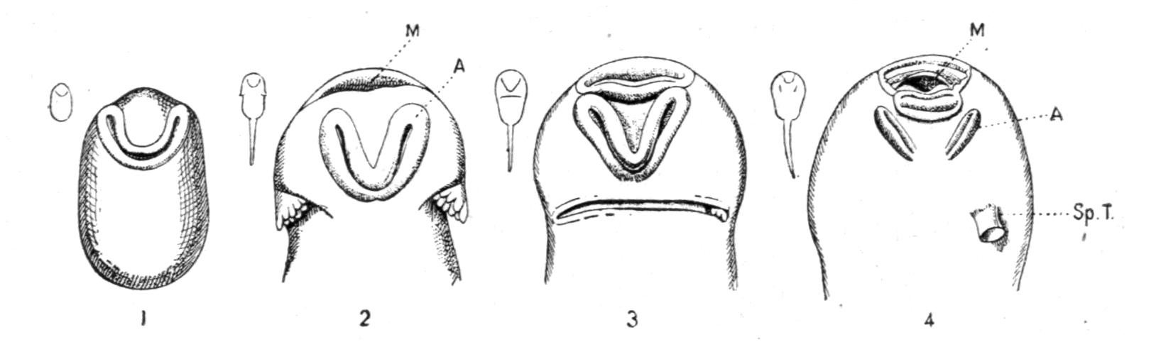
Fig. 9.–Four stages of the development of the adhesive apparatus (A) of Bufo vulgaris; M, Mouth; Sp.T. spiracular tube. In 3 the gills are almost completely hidden by the united right and left opercular folds. The small outlined figures indicate the shape and natural size of the tadpoles. (After Thiele.)
In the earliest condition the embryo consists of a large head and body, while the tail is still absent. Behind the beginnings of the future mouth appears a transverse crescentic fold, with the convexity looking backwards, which develops into the paired or unpaired adhesive apparatus. This consists of large complex glands, developed in the Malpighian layer, originally covered by the cuticula, which soon disappears, whereupon the sticky secretion enables the larva to attach itself to the gelatinous mantle of the egg, later on to weeds or other objects in the water. The name of suckers, often applied to this apparatus, conveys a wrong idea, there being neither muscles nor any suctorial function. The shape of this organ undergoes many changes during the early life of the individual, and differs much in the various genera, affording thereby diagnostic characters.[29] At first a crescent, it divides into a right and a left oval or disc, which either remain asunder and behind the mouth (Rana, Bufo), or they move forwards to the corners of the mouth (Hyla) or further back, and unite again more or less completely, as in Discoglossus and Bombinator. It is mostly of short duration, and disappears by the time that the larva, by the proper development of the gills and the tail and the functional mouth, changes into the tadpole. But in a few species these discs transform themselves into an elaborate ventral disc. Such an organ persists throughout the greater part of the tadpole-stage in certain Oriental species of Rana, all of which, when adult, possess fully webbed toes and strongly dilated discs on the fingers and toes, e.g. Rana whiteheadi, R. natatrix, and R. cavitympanum of Borneo, R. jerboa of Java (this larva was originally described and figured as that of Rhacophorus reinwardti), and R. afghana of the Himalayan system. These tadpoles, at least those of R. jerboa, are further remarkable for having the "spiracular" opening very far back on the left side, nearer to the base of the tail than to the snout, so as to be well out of the way when the creature has attached itself by the adhesive disc.
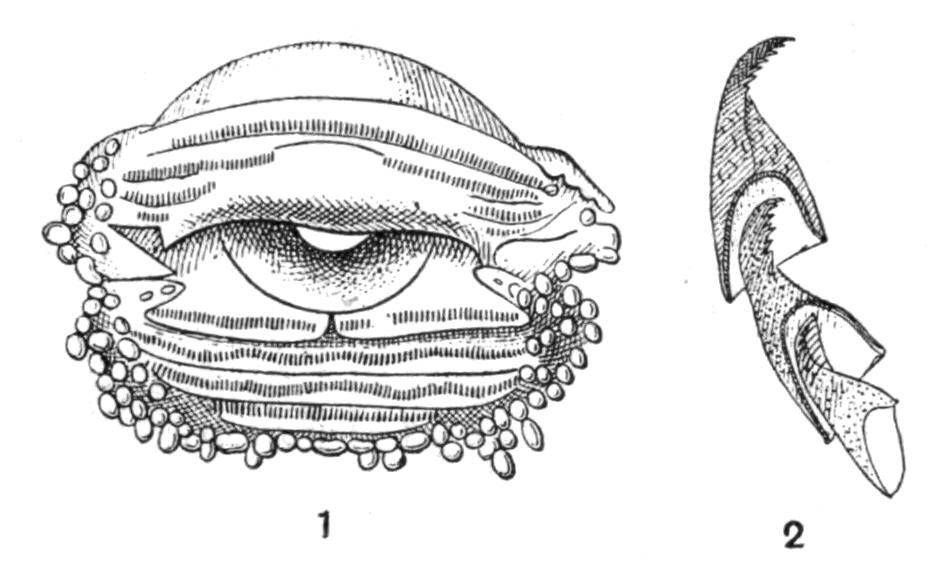
Fig. 10.–1, Front view of the mouth of a tadpole of Rana temporaria, showing the transverse rows of tiny horny teeth; 2, three successive horny teeth, much magnified. (After Gutzeit.)
The mouth of the tadpoles of Anura is furnished with horny armaments, substitutes for teeth. Their development and that of the mouth in general has been well described by Gutzeit.[30] In the young larvae of Rana temporaria, one or two days after hatching, a shallow groove appears above the conspicuous pair of adhesive organs. The groove becomes rhombic in outline, and when the mouth has been formed in its centre, the jaws appear in the median corners of the rhombus. The epidermis then rises like a circular wall around the jaws, and divides into an upper and lower lip; furrows appear on them, and between these various papillae and comb-like transverse plates of teeth. The papillae are possibly tactile organs, but although nerves enter them, nerve-endings of a sensory nature have not yet been discovered. On the fourth day the jaws become black, by the tenth day horny teeth have appeared upon all the plates of the mouth-armature, and on the seventeenth day the mouth-apparatus has reached the configuration typical of the tadpole, which is now about 14 mm. long. The number of horny teeth in R. temporaria amounts to about 640. These teeth are not cuticular products, but cornified cells; they are very small, and consist each of one horny cell, which is shaped like a nightcap, the apex of which is curved back and serrated. The little teeth are shed continuously, the renewal taking place by successive cells growing into the bases of the older series. The shape and size differ much in the various genera and species. The comb-like plates, composed of those teeth which surround the lips, seem to be used chiefly for the fixing or hooking of the food, while those which compose the horny beak proper, the armature of the jaws, are used like the radulae of snails. These beaks are likewise composed of a great number of individual teeth, closely packed together in several rows, but the teeth themselves are simple and not serrated.
In Hyla arborea there are in all about 560 teeth. The development of the mouth does not begin before the eleventh day; the horny teeth break through, and the jaws get black edges, on the eighteenth. In Pelobates fuscus the number of horny teeth is increased to about 1100. In Borborocoetes taeniatus the horny teeth form series of five bells, which fit into each other like the joints of a rattlesnake's tail.
One of the most extraordinary kinds of tadpoles is that of Megalophrys montana.[31] Mr. Annandale (Skeat Expedition) found it at Bukit Besar, Malay Peninsula, from 2000 to 3000 feet above the level of the sea. The tadpoles (Fig. 11) were found in the beginning of the month of May 1899 in sandy streams and in pools of rain-water; they floated in a vertical position, the peculiar membranous funnel-shaped expansion of the lips acting as surface-floats. The inside of the funnel is beset with radiating series of little horny teeth, and the whole apparatus is possibly used for scraping the under-surface of the leaves of water-plants in search of food. Total length of the tadpoles 1 inch.[32]
The gills, the formation of the operculum, and the modifications of the branchial arterial arches have been described fully on p. 43; those of the hyo-branchial skeleton on p. 31. Fusion of the opercular fold with the skin of the neck, across the branchial region, causes the head to become confluent with the trunk (cf. Fig. 9, 3, p. 57). The body becomes oval, more or less globular, and the alimentary canal is greatly elongated and stowed away in the shape of a neat, very regular spiral, shining through the ventral wall of the body; the anus opens at the end of a somewhat protruding tube, either in the median line, just in front of the ventral fin (Discoglossidae, Pelobates, Bufo), or it assumes an asymmetrical position by turning to the right side (Hyla, Rana).
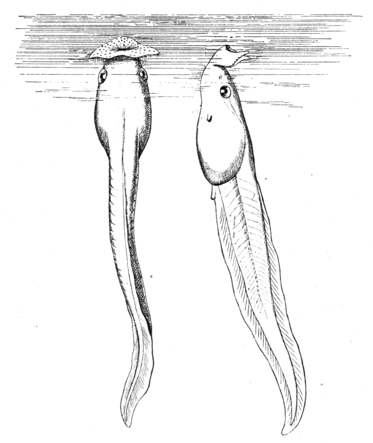
Fig. 11.–Tadpoles of Megalophrys montana from Bukit Besar, Malay Peninsula. × 3.
Although both pairs of limbs begin to bud simultaneously, or the fore-limbs even earlier, the hind-limbs are hurried on, and appear first, long before the fore-limbs. The latter lie ready beneath the skin of the gill-chamber, and the right always breaks through the skin, while the left does the same in the Mediogyrinidae, while in the Laevogyrinidae it is generally pushed through the left-sided spiracular opening, immediately behind the outer gills. According to Barfurth the right limb appears, in about 80 per cent. of Rana esculenta, from two to eight hours before the left.
Meanwhile the lungs are being developed, and the tadpole occasionally rises to the surface to breathe air. The gills, which, as has been explained elsewhere, are less ancestral than they are larval organs, degenerate, and all the organs are modified for the coming terrestrial life. The fins of the tail are absorbed, the horny armature of the mouth and lips is shed in pieces and makes room for the true teeth, the eyes receive lids, and the whole cranium, especially the apparatus of the jaws, undergoes the final modifications–widening and lengthening of the mouth, arresting of the mento-Meckelian cartilages, elongation of the Meckelian cartilages or lower jaw proper, shifting backwards of the suspensorium, and lengthening of its orbital process to form the pterygo-palatine bridge.
The tadpole ceases to feed, the whole intestinal canal is voided of its contents, and by "histolysis" is thoroughly rebuilt, becoming wider and shrinking to about one-sixth of its original length,–undoing thereby the spiral–preparatory for the coarser food, which consists of insects, worms, and other strictly animal, living matter. Hitherto the tadpoles have lived on "mud," confervae, Diatoms, rotting vegetable and animal matter. The anal tube collapses, becomes ultimately absorbed, and a new vent is formed at and below the root of the tail.
Barfurth[33] has made interesting observations and experiments with regard to the absorption of the tail and other organs which disappear during the metamorphosis. This is retarded by low temperature; it is accelerated by rest and freedom from mechanical disturbances, as, for instance, concussion of the water. Hunger shortens or hurries on the last stages of metamorphosis, the absorption of the tail taking place in four instead of five days. Amputation of the tail has no retarding influence; it is followed at once by regeneration, although the tadpole may be on the verge of reducing the tail. Whilst hungering the whole organism draws upon its available store of material, naturally first upon those parts which sooner or later are to become superfluous. This applies eminently to the tail, which represents a considerable amount of "edible" matter, and also to that portion of the skin which still covers the fore-limbs. The elements of the cutis are resorbed, thereby thinning the skin; and consequently the limbs break through earlier in fasting than in well-fed specimens. Nature herself seems to apply hunger as an accelerator. Mlle. von Chauvin found that the larvae of Urodela normally fast during the transformation, and according to Barfurth the larvae of Rana temporaria eat less after their hind-limbs are fully developed. This is, however, also preparatory for the reorganisation of the gut, which has to be more or less empty during the shortening process.
The loss of the tail is not due to a sudden dropping off of this organ–a crude but by no means uncommon belief–but is brought about by a very gradual process of resorbtion. When the fore-limbs begin to break through the skin, the tip of the tail shrinks and becomes black, owing to an increase, or rather concentration, of the pigment cells. The reduction proceeds from the tip forwards until on about the fifth day there remains only a short, conical, black stump. From the beginning of this process of reduction the tail is scarcely used for locomotion, the tadpole rowing with its legs, or it crawls and hops about, although the tail may still be 20 mm. long. The cells of the epidermis atrophy, shrink, and peel off, while those of the cutis, blood-vessels, nerves, muscles, and chorda dorsalis become disintegrated, often undergoing fatty degeneration. The leucocytes eat up the débris and other dissolved tissue, and carry it away through the lymphatic vessels, to be used as new building material in the rest of the animal.
Barfurth asks very properly, Why do these tissues degenerate and die? Because the vasomotor nerve-fibres cease to regulate the circulation. And why does this trophic influence of the central nervous system stop? Because the function of the tail becomes superfluous through the appearance of the fore-limbs. The tail is doomed, and degenerates like any other organ without a function. The whole process is, of course, a recapitulation of ancestral, phylogenetic evolution.