Chapter IX · THE ORDER UNGULATA (continued 3)
Reading edition
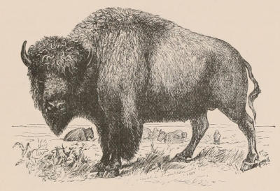
Fig. 148.—The American Bison (Bos americanus). After Hornaday.
The American Bison formerly ranged over about one-third of the North American continent. Thus, to quote from Mr. Hornaday,[251] “starting almost at tide-water on the Atlantic coast, it extended across the Alleghany mountain system to the prairies along the Mississippi, and southward to the delta of that great system. Although the great plain country of the West was the natural home of the species, where it flourished most abundantly, it also wandered south across Texas to the burning plains of North-Eastern Mexico, westward across the Rocky Mountains into New Mexico, Utah, and Idaho, and northward across a vast treeless waste to the bleak and inhospitable shores of the Great Slave Lake itself.” In consequence of the settlement of the country by Europeans the area inhabited by the Bison was gradually contracted, till about 1840 one mighty herd occupied the centre of its former range. The completion of the Union Pacific Railway in 1869 divided this great herd into a southern and a northern division, the former comprising a number of individuals estimated at nearly four millions, while the latter contained about a million and a half. Before 1880 the southern herd had, however, practically ceased to exist; while the same fate overtook the northern one in 1883. In 1889 some twenty stragglers in Texas represented the last of the southern herd; while there were a few others in Colorado, Wyoming, Montana, and Dakota. A herd of some two hundred wild individuals, derived from the northern herd, is preserved by the United States Government in the Yellowstone National Park; and it is believed that some five hundred of the race known as Wood-Bison exist in British territory; but with these exceptions this magnificent species is exterminated. The multitudes in which the American Bison formerly existed are almost incredible; the prairies being absolutely black with them as far as the eye could reach, and the numbers in the herds being, as we have said, reckoned by millions. Mr. Hornaday even considers that the whole of the game in South Africa was never equal to the number of Bison on an equal area of the American prairies.
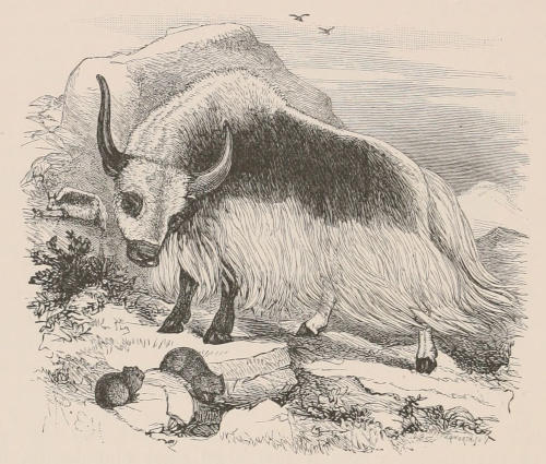
Fig. 149.—The Yak (Bos grunniens), domestic variety.
An extinct Bison from the Pleistocene of Texas, known as Bos latifrons, was probably the ancestor of the recent American species.
The Yak (Bos grunniens) appears to be allied both to the Bisons and the true Oxen, being distinguished from the former by the different position occupied by the long hair, which forms a fringe investing the shoulders, flanks, and thighs, and grows over the whole of the tail. In the skull the orbits are less tubular, the forehead flatter, and the premaxillæ less widely separated from the nasals. There is no distinct dewlap. Wild Yaks inhabit the higher regions of Chinese Tibet and the region of the Karakoram, as well as the more outlying parts of Ladak, such as the Changchemo valley. Owing, however, to incessant pursuit those now found within the territories of the Maharaja of Kashmir are stragglers from Chinese Tibet. The height of the Yak is somewhat lower than that of the larger domestic cattle. The colour of the wild race is black, tending to brown on the flanks; but many of the tame breeds which have been crossed with ordinary cattle have more or less white (Fig. 149), and it is the white tails of these half-breeds that are so esteemed in India as “chowries.” Yaks are exceedingly intolerant of heat, and the wild ones always live at very great elevations. Tame Yaks are extensively used as beasts of burden in Tibet, where they are extremely valuable in crossing the high and desolate wastes of that region; they have, however, the great drawback that they refuse to eat corn, so that in districts where there is no grass it is frequently necessary to make forced marches with wearied beasts in order to prevent them (and thus the whole party) perishing from starvation.
The skull of an extinct species from the Pliocene of Northern India, described as Bos sivalensis, appears to indicate a species allied to the Yak.
With the Bibovine group we come to the consideration of three Oriental species which connect the preceding forms with the typical Oxen. The three species are the Gaur (B. gaurus) the Gayal (B. frontalis, Fig. 150) of India, and the Banteng (B. sondaicus) of Burma, Java, Bali, and Lambok. In this group, as in the true Oxen, there are thirteen pairs of ribs, against fourteen in the Bisons. All the three species are characterised by the great height of the spines of the anterior dorsal vertebræ, causing a prominent ridge down the back. The horns, which are of a greenish colour in the Gaur, are somewhat flattened, and after running outwards are directed upwards instead of backwards; they occupy the vertex of the skull. The frontals are more or less concave, the premaxillæ do not join the nasals, and the occipital aspect of the skull is characterised by the deep incisions made by the temporal fossæ. The lower part of the legs is white (Fig. 150), and the hoofs are comparatively small and pointed. The Gaur (B. gaurus) is the largest of the three species, and inhabits all the large forests of India from near Cape Comorin to the foot of the Himalaya; it is commonly known to sportsmen as the Indian Bison. It stands fully 6 feet in height at the withers, which are much elevated; and since the whole back is arched the line from the nose to the root of the tail forms an almost continuous curve. The most characteristic feature of the animal is, however, the large and convex intercornual frontal crest, which curves forward, and thus gives a concave profile to this part of the skull. As a rule the Gaur prefers hilly regions, although it is sometimes met with on the flat. It is very shy and readily frightened; and it has never been domesticated. The Gayal, or Mithan, of which a figure is given in woodcut 150, is at once distinguished from the Gaur by the straight line between the horns (which are black in colour), owing to the absence of the intercornual crest of the latter. The horns are also shorter, more rounded, and less curved. In the Indian Museum, Calcutta, there are, however, skulls which are to a great extent intermediate between those of typical Gaurs and those of typical Gayals, but these may belong to hybrids. The Gayal occurs in Assam, Chittagong, and adjacent districts, but it appears that these animals exist in a semi-domesticated condition, no wild race being known to Europeans, although it is probable that such may exist in the unexplored Mishmi Hills.
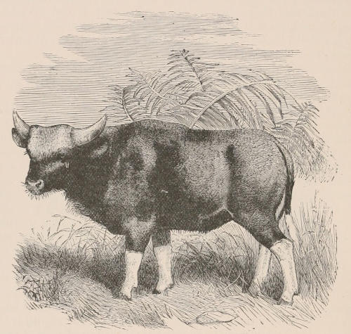
Fig. 150.—The Gayal (Bos frontalis). From Sclater, List of Animals in Zoological Society’s Gardens, 1883.
The Banteng (B. sondaicus) is a smaller and lighter built animal than either of the preceding, with a longer and sharper head, and more rounded and slender horns. The dorsal ridge is, moreover, but slightly developed; while the bright dun colour of the body of the female readily distinguishes it from the darker hue of the Gaur and Gayal.
A fossil skull from the Pleistocene deposits of the Narbada valley, India, described as Bos palæogaurus, is believed to indicate a species nearly allied to the Gaur, if indeed it be specifically distinct.
The true Oxen, or Taurine group, are now represented solely by Bos taurus and Bos indicus. Both of these species are now known only by domesticated races, unless the herds of the former preserved at Chillingham and some other British parks are the survivors of an original wild race. The dorsal ridge of the Bibovine group is here wanting; the horns are rounded, with their extremities directed backwards, and are placed at the extreme vertex of the skull; while the long frontal region is nearly flat; the temporal fossæ scarcely intrude upon the occipital aspect of the skull; and the premaxillæ reach the nasals. The hoofs are large and rounded. It is known that wild Oxen were abundant in the forests of Europe at the time of Julius Cæsar, by whom they were described as the Urus, equal to the German Aurochs; and the large skulls found in turbary and Pleistocene deposits, and described under the name of Bos primigenius, can only be regarded as having belonged to the large original race of B. taurus, of which it has been thought the Chillingham cattle are smaller descendants.[252] The subfossil skulls described as B. longifrons and B. frontosus must also be looked upon as referable to smaller races of the same species. That the domestic cattle of Europe are descendants from the various races of the same original species there can be no doubt, but in the case of the humped cattle of India (B. indicus) it is quite probable that their origin may be, at least in part, different. The extinct Bos namadicus, of the Pleistocene deposits of India, was a species with the general characters of the Taurine group, but with an inclination to a flattening of the horns, and with an approximation to a Bibovine type of occiput, as well as with the separation of the premaxillæ from the nasals.
The earliest representatives of this group occur in the Pliocene of the Siwalik Hills in Northern India. One of these species (B. planifrons) appears to be allied to B. namadicus; but the other (B. acutifrons) was a gigantic species characterised by the sharp median angulation of the frontal region, and the pyriform section of the enormous horn-cores.
The extinct B. elatus, from the Upper Pliocene of France and Italy, is the representative of a generalised type, which may be known as the Leptobovine group. The males had rounded horn-cores widely separated at their bases, and placed low down on the forehead. The females (which have been described as Leptobos) were often or always hornless. The limbs were unusually slender. This group also occurs in the Pliocene of the Siwalik Hills.
Suborder Perissodactyla.
This is a perfectly well-defined group of Ungulate mammals, represented in the actual fauna of the world by only three distinct types or families—the Tapirs, the Rhinoceroses, and the Horses—poor in genera and species, and (except in the case of the two domesticated species of Equus, which have been largely multiplied and diffused by man’s agency) not generally numerous in individuals, though widely scattered over the earth’s surface. Palæontological records, however, show very clearly that these are but the surviving remnants of a very extensive and much-varied assemblage of animals, which flourished upon the earth through the Tertiary geological period, and which, if it could be reconstructed in its entirety, would not only show members filling up structurally the intervals between the existing apparently isolated forms, but would also show several marked lines of specialisation which have become extinct without leaving any direct successors.
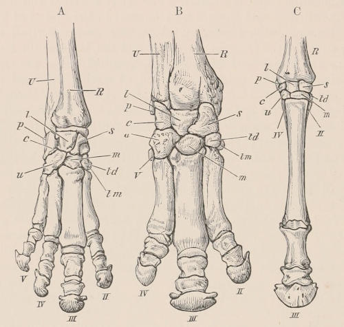
Fig. 151.—Bones of right fore foot of existing Perissodactyles. A, Tapir (Tapirus indicus), × ⅕; B, Rhinoceros (Rhinoceros sumatrensis), × ⅙; C, Horse (Equus caballus), × ⅛. U, ulna; R, radius; c, cuneiform; l, lunar; s, scaphoid; u, unciform; m, magnum; td, trapezoid; tm, trapezium.—From Flower, Osteology of Mammalia.
The following are the principal characters distinguishing them from the Artiodactyla. Premolar and molar teeth in continuous series, with massive, quadrate, transversely ridged or complex crowns,—the posterior premolars often resembling the true molars in size and structure. Crown of the last lower molar commonly bilobed, and if a third lobe is present in this tooth it is wanting in the last lower milk-molar. Dorso-lumbar vertebræ never fewer than twenty-two, usually twenty-three in the existing species. Nasal bones expanded posteriorly. An alisphenoid canal. Femur with a third trochanter.[253] The middle or third digit on both fore and hind feet larger than any of the others, and symmetrical in itself, the free border of the ungual phalanx being evenly rounded (see Fig. 151). This may be the only functional toe, or the second and fourth may be subequally developed on each side of it. In the Tapirs and many extinct forms, the fifth toe also remains on the fore limb, but its presence does not interfere with the symmetrical arrangement of the remainder of the foot around the median line of the third or middle digit. Traces of a hallux have only been found in some extremely ancient and primitive forms. The astragalus has a pulley-like surface above for articulation with the tibia, but its distal surface is flattened and unites to a much greater extent with the navicular than with the cuboid, which bone is of comparatively less importance than in the Artiodactyla. The calcaneum does not articulate with the lower or distal extremity of the fibula. The stomach is always simple, the cæcum is large and capacious, the placenta diffused, and the mammæ are inguinal. The gall-bladder is invariably absent.
As regards the dentition, the whole of the premolar series may be preceded by milk-teeth; and it has been demonstrated in Rhinoceros that when there is no displacement of the first cheek-tooth that tooth is a persistent milk-molar; the same condition apparently holding good in Palæotherium. This feature indicates considerable dental specialisation, the milk-molars, according to the theory generally accepted by the leading English zoologists, being the acquired, and the premolars the original series. Another peculiar feature of the dentition of the Perissodactyla, very rarely met with among the Artiodactyla, is that the premolars tend to resemble the true molars; this feature occurring in all the existing genera, although not found in the earlier generalised types. The cheek-teeth of all the members of the suborder are primarily constructed on some modification of what is known as the lophodont plan. Thus the upper molars (Fig. 155, p. 375) have an outer antero-posterior wall from which proceed two transverse ridges, formed by the coalescence of the primitive inner and outer columns, towards the inner aspect of the crown; while in the lower molars there may be either two simple transverse ridges, or these ridges may be curved into crescents, coming into contact with one another at their extremities. Those forms having brachydont teeth show this plan of structure in its simplest modification; but in cases, as in the Horse, where the teeth assume an extremely hypsodont form, the original plan is so obscured by infoldings of the enamel that it can only be traced with difficulty.
At the present day the Perissodactyla are sharply differentiated into Horses, Tapirs, and Rhinoceroses, but the knowledge already gained of the extinct representatives of the suborder shows such a close alliance between these groups that it is exceedingly difficult to make any satisfactory classification of the whole. This is of course exactly what might have been expected; and the same would doubtless be the case with all other groups if we knew as much of their past history as we do of that of the Perissodactyles.
The detailed account of the anatomy of the Horse given in the sequel will afford much information as to the general structure of the members of the suborder.
Family Tapiridæ.
Both upper and lower cheek-teeth brachydont and simply bilophodont; hinder premolars as complex as the molars; last lower molar without third lobe; first upper cheek-tooth with a milk-predecessor.[254] Outer columns of upper molars conical. Four digits in the manus, and three in the pes.
Tapirus.[255]—Dentition i ³⁄₃, c ¹⁄₁, p ⁴⁄₃, m ³⁄₃; total 42. Of the upper incisors, the first and second are nearly equal, with short, broad crowns; the third is large and conical, considerably larger than the canine, which is separated from it by an interval. Lower incisors diminishing in size from the first to the third; the canine, which is in contact with the third incisor, large and conical, working against (and behind) the canine-like third upper incisor. In both jaws there is a diastema between the canines and the commencement of the teeth of the cheek-series, which are all in contact. First upper premolar with a triangular crown, narrow in front owing to the absence of the anterior inner cusp. The other upper premolars and molars all formed on the same plan and of nearly the same size, with four roots and quadrate crowns, rather wider transversely than from before backwards, each having four cusps, connected by a pair of transverse ridges, anterior and posterior. The first lower premolar compressed in front; the others composed of a simple pair of transverse crests, with a small anterior and posterior circular ridge.
Skull elevated and compressed. Orbit and temporal fossa widely continuous, there being no true postorbital process from the frontal bone. Anterior narial apertures very large, and extending high on the face between the orbits; nasal bones short, elevated, triangular, and pointed in front. Vertebræ: C 7, D 18, L 5, S 6, C about 12. Limbs short and stout. Forefeet with four toes, having distinct hoofs: the first is absent, the third the longest, the second and fourth nearly equal, the fifth the shortest and scarcely reaching the ground in the ordinary standing position. Hind feet with the typical Perissodactyle arrangement of three toes,—the middle one being the largest, the two others nearly equal. Nose and upper lip elongated into a flexible, mobile snout or short proboscis, near the end of which the nostrils are situated. Eyes rather small. Ears of moderate size, ovate, erect. Tail very short. Skin thick and but scantily covered with hair.
The existing species of Tapir may be grouped into two sections, the distinctive characters of which are only recognisable in the skeleton. (A) With a great anterior prolongation of the ossification of the nasal septum (mesethmoid), extending in the adult far beyond the nasal bones, and supported and embraced at the base by ascending plates from the maxillæ (genus Elasmognathus, Gill). Two species, both from Central America, Tapirus bairdi and T. dowi. The former is found in Mexico, Honduras, Nicaragua, Costa Rica, and Panama; the latter in Guatemala, Nicaragua, and Costa Rica. (B) With ossification of the septum not extending farther forward than the nasal bones (Tapirus proper). Three species, T. indicus, the largest of the genus, from the Malay Peninsula (as far north as Tavoy and Mergui), Sumatra, and Borneo, distinguished by its peculiar coloration, the head, neck, fore and hind limbs, being glossy black, and the intermediate part of the body white; T. americanus (T. terrestris, Linn.), the common Tapir of the forests and lowlands of Brazil and Paraguay (Fig. 152); and T. roulini, the Pinchaque Tapir of the high regions of the Andes. All the American species are of a nearly uniform dark brown or blackish colour when adult; but it is a curious circumstance that when young (and in this the Malay species conforms with the others) they are conspicuously marked with spots and longitudinal stripes of white or fawn colour on a darker ground.
The habits of all the kinds of Tapirs appear to be very similar. They are solitary, nocturnal, shy, and inoffensive, chiefly frequenting the depths of shady forests and the neighbourhood of water, to which they frequently resort for the purpose of bathing, and in which they often take refuge when pursued. They feed on various vegetable substances, as shoots of trees and bushes, buds, and leaves. They are hunted by the natives of the lands in which they live for the sake of their hides and flesh.
The singular fact of the existence of so closely allied animals as the Malayan and the American Tapirs in such distant regions of the earth, and in no intervening places, is accounted for by what is known of the geological history of the race; for the Tapirs must once have had a very wide distribution. There is no proof of their having lived in the Eocene epoch, but in deposits of Miocene and Pliocene date remains undistinguishable generically from the modern Tapirs, and described as T. priscus, T. arvernensis, etc., have been found in France, Germany, and in the Red Crag of Suffolk. Tapirs appear, however, to have become extinct in Europe before the Pleistocene period, since none of their bones or teeth have been found in any of the caverns or alluvial deposits in which those of Elephants, Rhinoceroses, and Hippopotamuses occur in abundance; but in other regions their distribution at this age was far wider than at present, as they are known to have extended eastward to China (T. sinensis, Owen) and westwards over the greater part of the southern United States of America, from South Carolina to California. Lund also distinguished two species or varieties from the caves of Brazil, one of which appears identical with T. americanus. Thus we have no difficulty in tracing the common origin in the Miocene Tapirs of Europe of the now widely separated American and Asiatic species. It is, moreover, interesting to observe how very slight an amount of variation has taken place in forms isolated during such an enormous period of time.
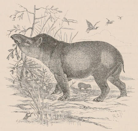
Fig. 152.—The American Tapir (Tapirus americanus).
The anatomy of the soft parts of the Tapirs[256] conforms to the general Perissodactyle type, as exemplified in the Rhinoceros and the Horse, although on the whole (as might have been expected) presenting a closer resemblance to the former. T. americanus differs from T. indicus by the absence, or at any rate the less development, of the intestinal valvulæ conniventes, the presence of a moderator band in the heart, the shape of the glans penis, and the more elongated cæcum, which is sacculated by four distinct longitudinal fibrous bands. The convolutions of the hemispheres of the brain of the Tapirs are simpler than in other Perissodactyles, thus tending to confirm the inferences which may be drawn from the skeleton and teeth as to the comparatively low or generalised organisation of these animals.
Palæotapirus.—This name has been applied to an imperfectly known form from the Upper Eocene Phosphorites of Central France, which is regarded by Dr. Filhol as referable to this family.
Family Lophiodontidæ.
Molars brachydont and bilophodont, those of the lower jaw with either straight or imperfectly crescentoid ridges; premolars smaller and usually simpler than the molars; last lower molar generally with a third lobe. Outer columns of upper molars conical or flattened. Digits usually as in the preceding family.
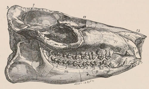
Fig. 153.—Right side of skull of Hyracotherium leporinum, from the London Clay. ½ natural size. (After Owen.) 3, Occiput; 7, sagittal crest; 11, frontals; 15, nasals; 21, maxilla; 22, premaxilla; d, mandibular condyle; a, aperture of facial nerve; p 1-4, premolars; m 1-3, molars.
This family includes a number of more or less imperfectly known forms, all of which are extinct and apparently confined to the Eocene period, and ranging from the size of a Rabbit to that of a Rhinoceros. Although some of these appear to have died out without giving rise to more specialised forms, it is probable that this family contained the ancestral types from which most or all of the modern Perissodactyles have been derived. Only very brief mention can be made here of some of the leading genera. Lophiodon, of the Middle and Upper Eocene of Europe, with the dental formula, i ³⁄₃, c ¹⁄₁, p ³⁄₃, m ³⁄₃, includes the largest representatives of the family, and is generally regarded as a stock which has died out without giving rise to later forms. The ridges of the lower molars are straight, and the last of these teeth has a third lobe; while the second transverse ridge of the last upper premolar is usually incomplete; the outer columns of the upper molars are flattened, as in the next genus. Hyrachyus, of the Upper Eocene of the United States, and probably also occurring in the French Eocenes, is an allied genus, with four premolars and no third lobe to the last lower molar; the fourth upper premolar having the two ridges uniting internally to form a crescent. This genus has been regarded as the ancestor of the Rhinocerotic Hyracodon. The genus Hyracotherium was established in 1839 by Owen for a small animal no larger than a Hare, the skull of which was found in the London Clay at Herne Bay. A more nearly perfect specimen, apparently of the same species, was afterwards (in 1857) described under the name of Pliolophus vulpiceps, of which the skull is figured in the accompanying woodcut. Other forms referable to the same genus have been obtained from the Wasatch Eocene of the United States, and were described by Professor Marsh under the name of Eohippus. There were four premolars, the fourth being unlike the molars, and in the upper jaw having only one inner cusp. The upper molars are of the general type of those of Lophiodon, but have conical outer columns, and the anterior transverse ridge imperfect, while the ridges of the lower molars are crescentoid. Systemodon differs from Hyracotherium by the absence of a diastema between the first and second premolars; it occurs in the Wasatch Lower Eocene of the United States. In Pachynolophus (Lophiotherium, Orotherium, or Orohippus), which is common to the Middle and Upper Eocene of Europe and the Bridger Eocene of North America, the outer columns of the upper molars are flattened, and in some cases, at least, the last premolar resembles the molars, that of the upper jaw having two inner cusps.[257] This genus, indeed, so closely connects Hyracotherium with the genera Epihippus and Anchilophus as to show that the distinction between the Lophiodontidæ and Palæotheriidæ is really an arbitrary one. Epihippus, of the Upper Eocene of the United States, has both the third and fourth upper premolars as complex in the molars, and is distinguished from Anchilophus by the lower cusps and more imperfect transverse ridges of these teeth. The so-called Orohippus agilis belongs to this genus. Isectolophus is another American Eocene genus which may be provisionally placed in this family; it is regarded by Professors Scott and Osborn as connecting Systemodon with the Tapiridæ; the fourth and probably the third upper premolar approximating in structure to the molars; the upper molars have conical outer columns. Helaletes is another closely allied form, with similar premolars, but with the outer columns of the upper molars flattened.
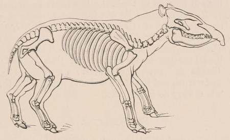
Fig. 154.—Restoration of Palæotherium (Upper Eocene). After Cuvier.
Family Palæotheriidæ.
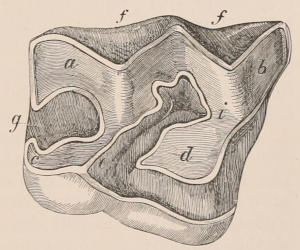
Fig. 155.—A half-worn right upper molar of Palæotherium magnum. (After Owen.) f, f, External surfaces of outer columns; a, postero-external column (metacone); b, antero-external column (paracone); c, postero-internal column (hypocone); d, antero-internal column (protocone); i, anterior intermediate column (protoconule); e, median valley; g, posterior valley.
Molars (Fig. 155) brachydont, with the valleys between the ridges never filled with cement; upper premolars either simpler than or as complex as the molars; lower molars with crescentoid ridges, and the last of the series with or without a third lobe. Outer columns of upper molars flattened. Orbit (at least usually) confluent with temporal fossa. Three digits on each foot. This family includes extinct genera ranging from the Middle and Upper Eocene to the Miocene, and passes so gradually into the following one that the maintenance of the two can only be supported on the ground of convenience. The typical genus, Palæotherium, was made known to science in the early part of the present century by Cuvier, who restored the skeleton (Fig. 154) with a short neck like that of the Tapirs, although it has been subsequently found that the neck was considerably longer. This genus (which may be taken to include Paloplotherium) ranges from the Middle to the Upper Eocene of Europe, and usually has the full typical dentition, although the first premolar may disappear. The last lower molar has a third lobe; and in the typical forms the last premolar is as complex as the molars, the diastema is short, and the canines are not large. In other forms, however, the hinder ridge of the fourth upper premolar may be aborted. The first upper cheek-tooth is generally a well-developed tooth, which may have a deciduous predecessor. Anchilophus, of the Upper Eocene of Europe, and Anchitherium, of the Miocene of Europe and North America, connect the preceding forms with the Equidæ. In the latter genus there is the full number of teeth, the last lower molar has almost completely lost the third lobe of Anchilophus, and the surfaces of the two outer lobes of the upper molars (Figs. 157, 158) lack the median vertical ridges of that genus. In the American species of Anchitherium (which have been described as Mesohippus and Miohippus) the lateral digits are larger than in the European Middle Miocene Anchitherium aurelianense; a mere splint represents the fifth metacarpal, and the meso- and ento-cuneiform of the tarsus do not unite as they do in the latter.
Family Equidæ.
Molars hypsodont, with the outer columns of the upper ones flattened, the valleys completely filled with cement, and the enamel thrown into folds and plications; upper premolars as complex as molars, which they slightly exceed in size; ridges of lower molars crescentoid, and complicated by enamel-foldings; no distinct third lobe to last lower molar; summits of incisors with a central infolding of enamel. Orbit completely surrounded by bone. Digits three or one, but in the former case the median one is alone of functional importance; ulna and fibula incomplete; meso- and ento-cuneiform of tarsus united.
Such are the leading characters which serve to distinguish the existing Horses and their nearest fossil allies from the Palæotheriidæ. The Horse, as being the best known of the Perissodactyle Ungulates, is selected for a somewhat detailed description; but before proceeding to this it will be advisable to take a brief survey of the relations of the Equidæ to the extinct forms already noticed, and also of the modifications of the family at present existing.
The earliest form which can be certainly included in this line of descent is the American Lower Eocene genus Phenacodus (noticed below under the head of the suborder Condylarthra), in which there were five complete digits to the feet. From this form there is but a step to Systemodon and Hyracotherium, in which the functional digits of the manus were reduced to four, as in Pachynolophus (Fig. 156, a), although one species retained a rudiment of the metacarpal of the pollex.
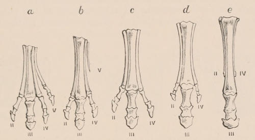
Fig. 156.—Successive stages of modification of the feet of extinct forms of Horse-like animals (chiefly from Marsh), showing gradual reduction of the outer and enlargement of the middle toe (III). a, Pachynolophus (Eocene); b, Anchitherium (Early Miocene); c, Anchitherium (Late Miocene); d, Hipparion (Pliocene); e, Equus (Pleistocene).
The transition from these animals of the Eocene period to the Horses of modern times has been accompanied by a gradual increase in size. The diminutive Hyracotherium of the Lower, and Pachynolophus of the Middle and Upper Eocene were succeeded in the Miocene period by the forms to which the name of Anchitherium has been given, of the size of sheep; these again in Pliocene times by Hipparion and Protohippus, as large as the modern donkeys; and it is mainly in the Pleistocene period that Equidæ occur which approach in size the existing Horse. Important structural modifications have also taken place, with corresponding changes in the mode of life of the animal. Thus the neck has become elongated, the skull altered in form, the teeth greatly modified, and the limbs have undergone remarkable changes. The last two require to be described more in detail.
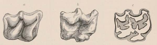
Fig. 157.—a, Grinding surface of unworn molar tooth of Anchitherium; b, corresponding surface of unworn molar of young Horse; c, the same tooth after it has been some time in use. The uncoloured portions are the dentine or ivory, the shaded parts the cement filling the cavities and surrounding the exterior. The black line separating these two structures is the enamel or hardest constituent of the tooth.
The teeth in the Eocene forms had, as mentioned above, the characteristic number of forty-four. This number has been retained throughout the series, at least theoretically; but one tooth on either side of each jaw, the anterior premolar, which in all the Eocene and Miocene species was well developed, persisting through the lifetime of the animal, is in all modern Horses rudimentary, functionless, and generally lost at an early period of life, evidently passing through a stage which must soon lead to its complete disappearance. The canines have also greatly diminished in size, and are rarely present in the female sex, so that practically a very large number of adult Horses of the present day have eight teeth less than the number possessed by their predecessors. The diastema or interval between the incisor and premolar teeth (of essential importance in the domesticated Horse to his master, as without it there would be no room for inserting the special instrument of subjugation to his commands, the bit) already existed in the earliest known forms, but has gradually increased in length. The incisors have undergone in comparatively recent times that curious change producing the structure more fully described hereafter, which distinguishes the Horse’s incisors from those of all other known animals, with the exception of the extinct Macrauchenia. Lastly, the molars have undergone a remarkable series of modifications, much resembling in principle those that have taken place in several other groups of herbivorous animals. Distinctions in form which existed between the premolars, at least in the anterior part of the series, and the true molars have gradually disappeared, the teeth becoming all very uniform in the shape and structure of their grinding surface. The crowns of all these teeth in the early forms were very short (see Fig. 158, a); there was a distinct constriction, or neck, between the crown and roots; and when the tooth was developing, as soon as the neck once rose fairly above the alveolar margin, the tooth remained permanently in this position. The term “brachydont” expresses this condition of teeth, the mode of growth of which may be illustrated by those of man. The free surface had two nearly transverse curved ridges, with valleys between (Fig. 157, a); but the valleys were shallow and had no deposit of cement filling them, the whole exposed surface of the unworn tooth being formed of enamel. When the ridges became worn down the dentine of the interior was exposed, forming islands surrounded by enamel. With the progress of time the crowns of the teeth gradually became longer, the valleys deeper, and the ridges not only more elevated but more curved and complex in arrangement. To give support to these high ridges and save them from breaking in use, the valleys or cavities between them became filled up to the top with cement, and as the crown wore down an admirable grinding surface consisting of patches and islands of the two softer substances, dentine and cement, separated by variously reduplicated and contorted lines of intensely hard enamel, resulted (Fig. 157, c). The crown continued lengthening until in the modern Horses it has assumed the form called “hypsodont” (Fig. 158, b). Instead of contracting into a neck, and forming roots, its sides continue parallel for a considerable depth in the socket, and as the surface wears away, the whole tooth slowly pushes up, and maintains the grinding edge constantly at the same level above the alveolus, much as in the perpetually growing Rodent’s teeth. But in existing Horses there is still a limit to the growth of the molar. After a length is attained which in normal conditions supplies sufficient grinding surface for the lifetime of the animal, a neck and roots are formed, and the tooth is reduced to the condition of that of the brachydont ancestor. It is perfectly clear that this lengthening of the crown adds greatly to the power of the teeth as organs of mastication, and enables the animals in which it has taken place to find their sustenance among the comparatively dry and harsh herbage of the open plains, instead of being limited to the more succulent vegetable productions of the marshes and forests in which their predecessors probably dwelt.
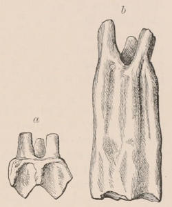
Fig. 158.—a, Outer view of second upper molar teeth of Anchitherium (brachydont form); b, corresponding tooth of Horse (hypsodont form).
The modifications of the limbs which took place pari passu with those of the teeth must have been associated with increased speed, especially over firm and unyielding ground. Short, stout legs, and broad feet, with numerous toes, spreading apart from each other when the weight of the creature is borne on them, are sufficiently well adapted for plodding deliberately over marshy and yielding surfaces, and the Tapirs and the Rhinoceroses, which in the structure of the limbs have altered but little from the primitive Eocene forms, still haunt the borders of streams and lakes and the shady depths of the forests, as was probably the habit of their ancient representatives, while the Horses are all inhabitants of the open plains, for life in which their whole organisation is in the most eminent degree adapted. The length and mobility of the neck, position of the eye and ear, and great development of the organ of smell, give them ample means of becoming aware of the approach of enemies, while the length of their limbs, the angles the different segments form with each other, and especially the combination of firmness, stability, and lightness in the reduction of all the toes to a single one, upon which the whole weight of the body and all the muscular power are concentrated, give them speed and endurance surpassing that of almost any other animal. When surprised, however, they are by no means helpless, both fore and hind feet becoming at need powerful weapons of defence.
If we were not so habituated to the sight of the Horse as hardly ever to consider its structure, we should greatly marvel at being told of a mammal so strangely constructed that it had but a single toe on each extremity, on the end of the nail of which it walked or galloped. Such a conformation is without a parallel in the vertebrate series, and is one of the most remarkable instances of specialisation, or deviation from the usual type, in accordance with particular conditions of life. It is clear, both from the structure of the foot itself, and also by an examination of the intermediate forms, that this toe corresponds to the middle or third digit of the complete typical or pentadactyle foot; and there is very strong evidence to show that by a gradual concentration of all the power of the limb upon this toe, and the concurrent dwindling away and final disappearance of all the others, the present condition of the Horse’s foot has been produced.
Protohippus.[258]—In this Lower Pliocene North American genus (also described as Merychippus) the cheek-teeth resemble those of the generalised species of Equus, but have shorter crowns; while the milk-molars approximate to the permanent molars of Anchitherium. Each foot has three digits.
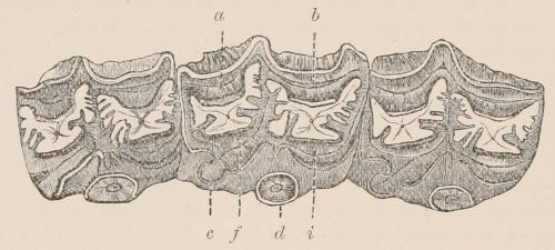
Fig. 159.—Three right upper cheek-teeth of Hipparion. a, Antero-external column; b, postero-external column; c, postero-internal column, or posterior pillar; d, antero-internal column, or anterior pillar; f, posterior intermediate column; i, anterior intermediate column. (From the Palæontologia Indica.)
Hipparion.[259]—Upper cheek-teeth (Fig. 159), with the antero-internal column, or anterior pillar as it may be conveniently termed in this family, detached throughout the greater part of its height from the adjacent column. Either a single or three digits in each foot. First upper premolar large and persistent. This genus was very widely distributed in the Pliocene, occurring in Europe, Asia, and North America. In the typical European forms, and also in those of North America, there were three digits in the feet (Fig. 156, d); but in the Indian H. antilopinum (separated by Cope as Hippodactylus) the lateral digits seem to have disappeared. There is some doubt whether or no Hipparion should occupy a place in the direct ancestry of the Horse, and Professor Cope suggests that while in America the intermediate place between Anchitherium and Equus was held by Protohippus, in Europe the same position was occupied by Hipparion—a view which involves the dual origin of the Horses of the New and Old Worlds.
Equus.[260]—Upper cheek-teeth with the anterior pillar (except in a very early stage of wear) joined by a narrow neck to the adjacent column (Fig. 157, c). Each foot with a single complete digit, but with remnants of the proximal portions of the second and fourth metapodials (Fig. 156, e); some extinct forms having claw-like rudiments of the terminal phalangeals of the lateral digits. First upper premolar very small or altogether absent in existing species, but in some fossil species larger and persistent; first lower premolar only occasionally developed in some fossil forms. Ears long. Tail long, with long hairs either at the end or throughout. A callosity on the inner side of the fore limb above the carpus.
Fossil Species.—In the Pleistocene Horses of South America described as Hippidium, as well as in the closely allied ones from North America for which the name Pliohippus has been proposed, the upper molars are shorter and more curved than in the existing species, while their anterior pillar is not longer antero-posteriorly than in Hipparion; the lateral claw-like hoofs persisting. Some of the European Pliocene species (like E. stenonis) agree with these species in the form of the grinding surface of the anterior pillar of the upper molars. In one of the species from the Lower Pliocene of India (E. sivalensis)—which was a contemporary of Hipparion—and in all the existing species, the grinding surface of the pillar in question is greatly elongated in the antero-posterior direction, as in Fig. 157, c.
Fossil remains of Horses are found abundantly in deposits of the most recent geological age in almost every part in America, from Eschscholtz Bay in the north to Patagonia in the south. In that continent, however, they became quite extinct, and no Horses, either wild or domesticated, existed there at the time of the Spanish conquest, which is the more remarkable as, when introduced from Europe, the Horses that ran wild proved by their rapid multiplication in the plains of South America and Texas that the climate, food, and other circumstances were highly favourable for their existence. The former great abundance of Equidæ in America, their complete extinction, and their perfect acclimatisation when reintroduced by man, form curious but as yet unsolved problems in geographical distribution.
Existing Species.—The existing species of the genus are the following:—
The Horse, Equus caballus, is distinguished from the others by the long hairs of the tail being more abundant and growing quite from the base as well as the end and sides, and also by possessing a small bare callosity on the inner side of the hind leg, just below the “hock” or heel joint, in addition to the one on the inner side of the fore limb above the carpus, common to all the genus. The mane is also longer and more flowing, and the ears are shorter, the limbs longer, the hoofs broader, and the head smaller.
Though the existing Horses are not usually marked in any definite manner, or only irregularly dappled, or spotted with light surrounded by a darker ring, many examples are met with showing a dark median dorsal streak like that found in all the other members of the genus, and even with dark stripes on the shoulders and legs indicating “the probability of the descent of all the existing races from a single dun-coloured, more or less striped, primitive stock, to which our horses still occasionally revert.”[261]
In Europe wild Horses were extremely abundant in the Neolithic or polished-stone period. Judging from the quantity of their remains found associated with those of the men of that time, the chase of these animals must have been among man’s chief occupations, and they must have furnished him with one of his most important food supplies. The characters of the bones preserved, and certain rude but graphic representations carved on bones or reindeers’ antlers, enable us to know that these Horses were rather small in size, and heavy in build, with large heads and rough shaggy manes and tails, much like, in fact, the present wild horses of the steppes of the south of Russia. They were domesticated by the inhabitants of Europe before the dawn of history, but it is doubtful whether the majority of the animals now existing on the Continent are derived directly from them, as it is more probable that they are descendants from Horses imported through Greece and Italy from Asia, derived from a still earlier domestication, followed by gradual improvement through long-continued attention bestowed on their breeding and training. Horses are now diffused by the agency of man throughout almost the whole of the inhabited parts of the globe, and the great modifications they have undergone in consequence of domestication and selective breeding are well exemplified by comparing such extreme forms as the Shetland pony, dwarfed by uncongenial climate, the thoroughbred racer, and the London dray-horse. In Australia, as in America, horses imported by the European settlers have escaped into the unreclaimed lands, and multiplied to a prodigious extent, roaming in vast herds over the plains where no hoofed animal ever trod before.
A wild Horse from Central Asia, named E. prezevalskii,[262] is described as having callosities on both limbs and broad hoofs like E. caballus; but the long hairs of the tail do not begin until about half way down its length. It also differs from E. caballus in having a short erect mane and no forelock; neither is there any dorsal stripe. The ears are of moderate size; the whole body is of a whitish-gray, paler beneath, and reddish on the head and upper parts of the limbs. If rightly described this form would appear to be intermediate between the true Horses and the Asses.
The second species is the domestic Ass (E. asinus), and the wild Asses of Africa (E. asinus, var. africanus and var. somalicus[263]). The domestic Ass, which is now nearly as widely diffused and useful to man as the Horse, was known in Egypt long before the latter, and is doubtless of African origin. The ears are long, the mane erect, the tail without long hairs at the base, and there are no callosities on the hind limbs. There is a dark dorsal stripe, and another across the shoulders; while the limbs are frequently banded. Of the wild forms the Nubian race (var. africanus) has distinct dorsal and shoulder stripes, but the rings on the limbs are often very indistinct; while in the Somali race the dorsal stripe is indistinct, and the shoulder stripe wanting, but the rings on the limbs are very boldly marked. Teeth and bones from a Pleistocene cavern deposit in Madras have been referred to E. asinus.
The Asiatic wild Asses, which roam in small herds in the open plains of Syria, of many parts of Persia, of the north-west of India, and the highlands of Tartary and Tibet, from the shores of the Caspian to the frontiers of China, differ from the last in being of a more rufous or isabelline colour, instead of pure gray, in wanting the dark streak across the shoulder, and having smaller ears. They have all a dark-coloured median dorsal stripe. Though it is considered probable by many zoologists that they form but a single species[264] (E. hemionus), they present such marked variations in size and form that they have commonly been divided into three—the Syrian Wild Ass (E. hemippus), the Onager (E. onager) from Persia, Baluchistan, the Punjab, Sind, and the desert of Kach, and the Kiang or Dzeggetai (E. hemionus) of the high table-lands of Tibet, where it is usually met with at an elevation of 15,000 feet and upwards above the sea-level. The last is considerably larger than either of the others, and differs from them in external appearance, having more the aspect of the horse. They are all remarkably swift, having been known to outstrip the fleetest Horse in speed.
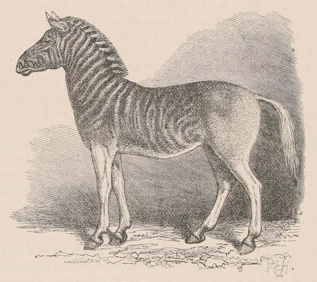
Fig. 160.—The Quagga (Equus quagga).
Lastly, there are four striped species, all inhabitants of Africa. These constitute the genus Hippotigris of Hamilton-Smith, but they are not separable except by their coloration from the true Asses, and one of them, the Quagga (E. quagga), may be considered as intermediate. This animal was formerly met with in vast herds on the great plains of South Africa, between the Cape Colony and the Vaal River, but now, in common with most of the larger wild animals of that region, is becoming extremely scarce, owing to the encroachments of European civilisation, if, indeed, it is not already extinct. In length of ears and character of tail it more resembles the Horse than it does the Ass, although it agrees with the latter in wanting the callosity on the inner side of the hind leg, just below the hock, characteristic of the Horse. The colour of the head, neck, and upper parts of the body is reddish-brown, irregularly banded and marked with dark brown stripes, stronger on the head and neck and gradually becoming fainter until lost behind the shoulder. There is a broad dark median dorsal stripe. The under surface of the body, the legs, and tail are nearly white, without stripes. The crest is very high, surmounted by a standing mane, banded alternately brown and white. Though never really domesticated, Quaggas have occasionally been trained to harness. The accompanying figure is reduced from a painting made from one of a pair which were driven in Hyde Park in the early part of the present century. The name is an imitation of the shrill barking neigh of the animal—“ouag-ga, ouag-ga,” the last syllable very much prolonged. It must be remembered, however, in reading books of African travel that the same word is very commonly applied by hunters to Burchell’s Zebra.
Of the Zebras proper, the one which was first known to Europeans, and was formerly considered the most common, is the True Zebra (E. zebra), sometimes called the Mountain Zebra. It inhabits the mountainous regions of the Cape Colony; but now, owing to the advances of civilised man into its somewhat restricted range, it has become very scarce, and is even, like the Quagga, threatened with extermination at no distant date. The second species, Burchell’s Zebra (E. burchelli), still roams in large herds over the plains to the north of the Orange River, but in yearly diminishing numbers. Both species are subject to considerable individual variations in marking, but the following are the principal characters by which they can be distinguished.
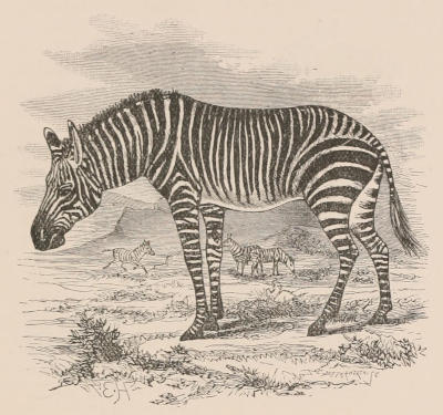
Fig. 161.—True or Mountain Zebra (Equus zebra).
E. zebra (Fig. 161) is the smaller of the two (about 4 feet high at the shoulders), and has longer ears, a tail more scantily clothed with hair, and a shorter mane. The general ground colour is white, and the stripes are black; the lower part of the face is bright brown. With the exception of the abdomen and the inside of the thighs, the whole of the surface is covered with stripes, the legs having narrow transverse bars reaching quite to the hoofs, and the base of the tail being also barred. The outsides of the ears have a white tip and a broad black mark occupying the greater part of the surface, but are white at the base. Perhaps the most constant and obvious distinction between this species and the next is the arrangement of the stripes on the hinder part of the back, where there are a number of short transverse bands passing from the median longitudinal dorsal stripe towards, and sometimes joining with, the uppermost of the broad stripes which run obliquely across the haunch from the flanks towards the root of the tail. There is often a median longitudinal stripe under the chest.
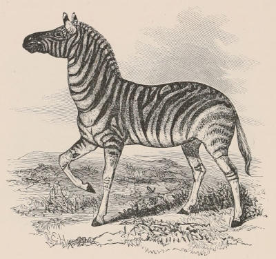
Fig. 162.—Burchell’s Zebra (Equus burchelli).
E. burchelli (Fig. 162) is a rather larger and more robust animal, with smaller ears, a longer mane, and fuller tail. The general ground colour of the body is pale yellowish-brown, the limbs nearly white, the stripes dark brown or black. In the typical form they do not extend on to the limbs or the tail; but there is a great variation in this respect, even in animals of the same herd, some being striped quite down to the hoofs (this form has been named E. chapmani). There is a strongly marked median longitudinal ventral black stripe, to which the lower ends of the transverse side stripes are usually united, but the dorsal stripe (also strongly marked) is completely isolated in its posterior half, and the uppermost of the broad haunch stripes runs nearly parallel to it. A much larger proportion of the ears is white than in the other species. In the middle of the wide intervals between the broad black stripes of the flanks and haunches fainter stripes are generally seen.
E. grevyi.—Under this name a Zebra has been described which was sent in 1882 to Paris from the Galla country, lying to the south of Abyssinia, the most northern locality in which Zebras have previously been met with. In many of its characters it resembles E. zebra, but the stripes are much finer and more numerous than in the typical examples of that species, and it has a strong, black, and isolated dorsal stripe. Even allowing for the great variations that are met with in the markings of animals of this group, the aberrant characters of this individual are quite sufficient to separate it specifically from the true Zebra of South Africa. Other similar specimens have been recently brought from the Somali country.
The flesh of the Zebras is relished by the natives as food, and their hides are very valuable for leather. Although the many attempts that have been made to break in and train these animals for riding or driving have sometimes been rewarded with partial success, they have never been domesticated in the true sense of the word.
There are thus at least seven modifications of the Horse type at present existing, sufficiently distinct to be reckoned as species by all zoologists, and easily recognised by their external characters. They are, however, all so closely allied that each will, at least in a state of domestication or captivity, breed with perfect freedom with any of the others. Cases of cross breeds are recorded between the Horse and the Quagga, the Horse and Burchell’s Zebra, the Horse and the Hemionus or Asiatic wild Ass, the common Ass and the Zebra, the common Ass and Burchell’s Zebra, the common Ass and the Hemionus, the Hemionus and the Zebra, and the Hemionus and Burchell’s Zebra. The two species which are perhaps the farthest removed in general structure, the Horse and the Ass, produce, as is well known, hybrids or Mules, which in some qualities useful to man excel both their progenitors, and in some countries, and for certain kinds of work, are in greater requisition than either. Although occasional instances have been recorded of female Mules breeding with the males of one or other of the pure species, it is doubtful if any case has occurred of their breeding inter se, although the opportunities of doing so must have been great, as Mules have been reared in immense numbers for at least several thousands of years. We may therefore consider it settled that the different species of the group are now in that degree of physiological differentiation which enables them to produce offspring with each other, but does not permit of the progeny continuing the race, at all events unless reinforced by the aid of one of the pure forms.
The several members of the group show mental differences quite as striking as those exhibited by their external form, and more than perhaps might be expected from the similarity of their cerebral organisation. The patience of the Ass, the high spirit of the Horse, the obstinacy of the Mule, have long been proverbial. It is very remarkable that, out of so many species, two only should have shown any aptitude for domestication, and that these two should have been from time immemorial the universal and most useful companions and servants of man, while all the others remain in their native freedom to this day. It is, however, still a question whether this really arises from a different mental constitution causing a natural capacity for entering into relations with man, or whether it may not be owing to their having been brought gradually into this condition by long-continued and persevering efforts when the need of their services was keenly felt. It is quite possible that one reason why most of the attempts to add new species to the list of our domestic animals in modern times have ended in failure is that it does not answer to do so in cases in which existing species supply all the principal purposes to which the new ones might be put. It can hardly be expected that Zebras and Quaggas fresh from their native mountains and plains can be brought into competition as beasts of burden and draught with Horses and Asses, whose naturally useful qualities have been augmented by the training of thousands of generations of progenitors.
Not unfrequently instances occur of domestic Horses being produced with a small additional toe with complete hoof, usually on the inside of the principal toe, and, though far more rarely, three or more toes may be present. These malformations are often cited as instances of reversion to the condition of some of the earlier forms of equine animals previously mentioned. Such explanations, however plausible they appear at first sight, are nevertheless very doubtful. All the feet of polydactyle horses which we have examined bear little resemblance to those of Hipparion or Anchitherium, but look rather as if due to that tendency to reduplication of parts which occurs so frequently as a teratological condition, especially among domestic animals, and, whatever its origin, certainly cannot in many instances, as the cases of entire limbs superadded, or of six digits in man, be attributed to reversion.
Anatomy.—The anatomical structure of the Horse has been described in great detail in several works devoted to the subject, which will be mentioned in the bibliography, though these have generally been written from the point of view of the veterinarian rather than of the comparative anatomist. The limits of the present work will only admit of the most salient points being indicated, particularly those in which the Horse differs from the other Ungulata. Unless otherwise specified, it must be understood that all that is stated here, although mostly derived from observation upon the Horse, applies equally well to the other existing members of the group.
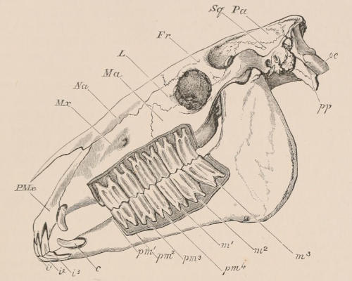
Fig. 163.—Side view of skull of Horse, with the bone removed so as to expose the whole of the teeth. PMx, Premaxilla; Mx, maxilla; Na, nasal; Ma, malar or jugal; L, lachrymal; Fr, frontal; Sq, squamosal; Pa, parietal; oc, occipital condyle; pp, paroccipital process; i¹, i², and i³³, the three incisors; c, the canine; pm¹, the situation of the rudimentary first premolar, which has been lost in the lower, but is present in the upper jaw; pm², pm³, and pm⁴, the three fully developed premolars; m¹, m², and m³, the three true molars.
Skeleton.—The skull (Fig. 163) as a whole is greatly elongated, chiefly in consequence of the immense size of the face as compared with the hinder or true cranial portion. The basal line of the cranium from the lower border of the foramen magnum to the incisor border of the palate is very nearly straight. The orbit, of nearly circular form, though small in proportion to the size of the whole skull, is distinctly marked, being completely surrounded by a strong ring of bone with prominent edges. Behind it, and freely communicating with it beneath the osseous bridge (the postorbital process of the frontal) forming the boundary between them, is the small temporal fossa occupying the whole of the side of the cranium proper, and in front is the great flattened expanse of the “cheek,” formed chiefly by the maxilla, giving support to the long row of cheek-teeth, and having a prominent ridge running forward from below the orbit for the attachment of the masseter muscle. The lachrymal occupies a considerable space on the flat surface of the cheek in front of the orbit, and below it the jugal or malar does the same. The latter sends a horizontal or slightly ascending process backwards below the orbit to join the under surface of the zygomatic process of the squamosal, which is remarkably large, and, instead of ending as usual behind the orbit, runs forwards to join the greatly developed postorbital process of the frontal, and even forms part of the posterior and inferior boundary of the orbit, an arrangement not met with in other mammals. The closure of the orbit behind distinguishes the skull of the Horse from that of the Rhinoceros and Tapir, and also from all of the Perissodactyles of the Eocene period. In front of the cerebral cavity, the great tubular nasal cavities are provided with well-developed turbinal bones, and are roofed over by very large nasals, broad behind, and ending in front in a narrow decurved point. The opening of the anterior nares is prolonged backwards on each side of the face between the nasals and the elongated slender premaxillæ. The latter expand in front, and are curved downwards to form the semicircular alveolar border supporting the large incisor teeth. The palate is narrow in the interval between the incisor and cheek-teeth, in which are situated the large anterior palatine foramina. Between the cheek-teeth it is broader, and it ends posteriorly in a rounded excavated border opposite the hinder edge of the penultimate molar. It is mainly formed by the maxillæ, as the palatines are very narrow. The pterygoids are delicate slender slips of bone attached to the hinder border of the palatines, and supported externally by, and generally ankylosed to, the rough pterygoid plates of the alisphenoid, with no pterygoid fossa between. They slope very obliquely forwards, and end in curved, compressed, hamular processes. There is a distinct alisphenoid canal for the passage of the internal maxillary or main branch of the external carotid artery. The base of the cranium is long and narrow; the alisphenoid is very obliquely perforated by the foramen rotundum, but the foramen ovale is confluent with the large foramen lacerum medium behind. The glenoid surface for the articulation of the mandible is greatly extended transversely, concave from side to side, convex from before backwards in front, and hollow behind, and is bounded posteriorly at its inner part by a prominent post-glenoid process. The squamosal enters considerably into the formation of the temporal fossa, and, besides sending the zygomatic process forwards, it sends down behind the meatus auditorius a post-tympanic process which aids to hold in place the otherwise loose tympano-periotic bone. Behind this the exoccipital gives off a very long paroccipital process. The periotic and tympanic are ankylosed together, but not with the squamosal. The former has a wide but shallow floccular fossa on its inner side, and sends backwards a considerable “pars mastoidea,” which appears on the outer surface of the skull between the post-tympanic process of the squamosal and the exoccipital. The tympanic forms a tubular meatus auditorius externus directed outwards and slightly backwards. It is not dilated into a distinct bulla, but ends in front in a pointed styliform process; and completely embraces the truncated cylindrical tympanohyal, which is of great size, in correspondence with the large development of the whole anterior arch of the hyoid. This consists mainly of a long and compressed stylohyal, expanded at the upper end, where it sends off a triangular posterior process. The basihyal is remarkable for the long, median, pointed, compressed “glossohyal” process, which it sends forward from its anterior border into the base of the tongue. A similar but less developed process is found in the Rhinoceros. The mandible is largely developed, especially the region of the angle, which is expanded and flattened, giving great surface for the attachment of the masseter muscle. The condyle is greatly elevated above the alveolar border; its articular surface is very wide transversely, and narrow and convex from before backwards. The coronoid process is slender, straight, and inclined backwards. The horizontal ramus, long, straight, and compressed, gradually narrows towards the symphysis, where it expands laterally to form with the ankylosed opposite ramus the wide, semicircular, shallow alveolar border for the incisor teeth.
The vertebral column consists of seven cervical, eighteen dorsal, six lumbar, five sacral, and fifteen to eighteen caudal vertebræ. There may be nineteen rib-bearing vertebræ, in which case five only will be reckoned as belonging to the lumbar series. The odontoid process of the atlas is wide, flat, and hollowed above, as in the Ruminants. The bodies of the cervical vertebræ are elongated, strongly keeled, and markedly opisthocœlous, or concave behind and convex in front. Their neural laminæ are very broad, the spines almost obsolete, except in the seventh, and the transverse processes not largely developed. In the trunk vertebræ the opisthocœlous character of the centrum gradually diminishes. The spinous processes of the anterior thoracic region are high and compressed. To these is attached the powerful elastic ligament, ligamentum nuchæ, or “paxwax,” which passing forwards in the middle line of the neck above the neural arches of the cervical vertebræ, to which it is also connected, is attached to the occiput and supports the weight of the head. The transverse processes of the lumbar vertebræ are long, flattened, and project horizontally outwards or slightly forwards from the arch. The metapophyses are moderately developed, and there are no anapophyses. The caudal vertebræ, except those quite at the base, are slender and cylindrical, without processes and without chevron-bones beneath. The ribs are eighteen or nineteen in number on each side, flattened, and united to the sternum by short, stout, tolerably well ossified sternal ribs. The sternum consists of six pieces; the anterior or presternum being extremely compressed, and projecting forwards like the prow of a boat. The segments which follow gradually widen, and the hinder part of the sternum is broad and flat.
As in all other Ungulates, there are no clavicles. The scapula is long and slender; the suprascapular border is rounded, and slowly and imperfectly ossified. The spine is very slightly developed; rather above the middle its edge is thickened and somewhat turned backwards, but it gradually subsides at the lower extremity without forming any acromial process. The coracoid process is a prominent rounded nodule. The humerus is stout and rather short, and has a double bicipital groove. The ulna is quite rudimentary, being only represented by little more than the olecranon. The shaft gradually tapers below, and is firmly ankylosed to the radius. The latter bone is of nearly equal width throughout. The three bones of the first row of the carpus (the scaphoid, lunar, and cuneiform) are subequal in size. The second row consists of a very broad and flat magnum, supporting the great third metacarpal, having to its radial side the trapezoid, and to its ulnar side the unciform, which are both small, and articulate distally with the rudimentary second and fourth metacarpals. The pisiform is large and prominent, flattened, and curved; articulating partly with the cuneiform and partly with the lower end of the radius. The large metacarpal is called in veterinary anatomy “cannon-bone”; the small lateral metacarpals, which gradually taper towards their lower extremities, and lie in close contact with the large one, are called “splint-bones.” The single digit consists of a moderate-sized proximal (os suffraginis, or large pastern), a very short middle (os coronæ, or small pastern), and a wide, semilunar, ungual phalanx (os pedis, or coffin-bone). There is a pair of large nodular sesamoids behind the metacarpo-phalangeal articulation, and a single large transversely extended sesamoid behind the joint between the second and third phalanx, called the “navicular bone.”[265]
The carpal joint, corresponding to the wrist of man, is commonly called the “knee” of the Horse, the joint between the metacarpal and the first phalanx the “fetlock,” that between the first and second phalanges the “pastern,” and that between the second and third phalanges the “coffin-joint.”
In the hind limb the femur is marked, as in other Perissodactyles, by the presence of a “third trochanter,” a flattened process, curving forwards, arising from the outer side of the bone, about one-third of the distance from the upper end. The fibula is reduced to a mere styliform rudiment of the upper end; its lower part being absent or completely fused with the tibia. The calcaneum has a long and compressed calcaneal process. The astragalus has a large flat articular surface in front for the navicular, and a very small one for the cuboid. The navicular and the external cuneiform bones are very broad and flat. The cuboid is small, and the internal and middle cuneiform bones are small and united together. The metapodials and phalanges resemble very closely those of the fore limb, but the principal metatarsal is more laterally compressed at its upper end than is the corresponding metacarpal. The joint between the femur and tibia, corresponding to the knee of man, is called the “stifle joint”; while that between the tibia and tarsus, corresponding to the ankle of man, is termed the “hock.” The bones and joints of the foot have the same names as in the fore limb. The Horse is eminently “digitigrade,” standing on the extremity of the single digit of each foot, which is kept habitually in a position approaching to vertical.
The muscles[266] of the limbs are modified from those of the ordinary mammalian type in accordance with the reduced condition of the bones and the simple requirements of flexion and extension of the joints, no such actions as pronation and supination, or opposition of digits, being possible or needed. The muscles, therefore, which perform these functions in other mammals are absent or rudimentary.
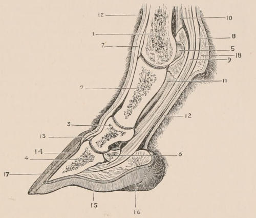
Fig. 164.—Section of foot of Horse. 1, Metacarpal bone; 2, first phalanx (os suffraginis); 3, second phalanx (os coronæ); 4, third or ungual phalanx (os pedis, or coffin-bone); 5, one of the upper sesamoid bones; 6, lower sesamoid or “navicular” bone; 7, tendon of anterior extensor of the phalanges; 8, tendon of superficial flexor (fl. perforatus); 9, tendon of deep flexor (fl. perforans); 10, suspensory ligament of fetlock; 11, inferior or short sesamoid ligament; 12, derma or skin of the foot, covered with hair, and continued into 13, the coronary cushion, 14, the podophyllous or laminar membrane, and 15, the keratogenous membrane of the sole; 16, plantar cushion; 17, hoof; 18, fatty cushion of fetlock.
Below the carpal and tarsal joints the fore and hind limbs correspond almost exactly in structure as well as function. On the anterior or extensor surface of the limb a powerful tendon (7 in Fig. 164), that of the anterior extensor of the phalanges (corresponding to the extensor communis digitorum of the arm and extensor longus digitorum of the foot of man) passes down over the metacarpal bone and phalanges, to be inserted mainly into the upper edge of the anterior surface of the last phalanx or pedal bone. There is also a much smaller second extensor on the outer side of this in each limb, the lateral extensor of the phalanges. In the fore leg the tendon of this muscle (which corresponds with the extensor minimi digiti of man) receives a slip from that of the principal extensor, and is inserted into the first phalanx. In the hind leg (where it is the homologue apparently of the peroneus brevis of man) the tendon becomes blended with that of the large extensor.
A very strong ligamentous band behind the metapodium, arising from near the upper extremity of its posterior surface, divides into two at its lower end, and each division, being first connected with one of the paired upper sesamoid bones, passes by the side of the first phalanx to join the extensor tendon of the phalanges. This is called in veterinary anatomy the “suspensory ligament of the sesamoids,” or of the “fetlock” (10 in Fig. 164); but its attachments and relations, as well as the occasional presence of muscular fibres in its substance, show that it is the homologue of the short flexor muscle of other mammals, curiously modified both in structure and function to suit the requirements of the Horse’s foot. Behind or superficial to this are placed the two strong tendons of the long flexor muscles, the most superficial, or flexor perforatus (8), dividing to allow the other to pass through, and then inserted into the middle phalanx. The flexor perforans (9) is as usual inserted into the terminal phalanx. In the fore leg these muscles correspond with those similarly named in man. In the hind leg, the perforated tendon is a continuation of that of the plantaris, passing pulley-wise over the tuberosity of the calcaneum. The perforating tendon is derived from the muscle corresponding with the long flexor of man, and the smaller tendon of the oblique flexor (tibialis posticus of man) is united with it.
The hoof of the Horse corresponds to the nail or claw of other mammals, but is so constructed as to form a complete and very solid case to the expanded termination of the toe, giving a firm basis of support formed of a nonsensitive substance, which is continually renewed by the addition of material from within as its surface wears away by friction against the ground. The terminal phalanx of the toe is greatly enlarged and modified in form to support this hoof, and the size of the internal framework of the foot is further increased by a pair of lateral fibro-cartilaginous masses attached on each side to the hinder edges of the bone, and by a fibro-cellular and adipose plantar cushion in the median part. These structures are all enclosed in the keratogenous membrane or “subcorneous integument,” a continuation of the ordinary derma of the limb, but extremely vascular, and having its superficial extent greatly increased by being developed into papillæ or laminæ. From this the horny material which constitutes the hoof is exuded. A thickened ring encircling the upper part, called coronary cushion (13), and the sole (15), are covered with numerous thickly set papillæ or villi, and take the greatest share in the formation of the hoof; the intermediate part constituting the front and side of the foot (14), corresponding with the wall of the hoof, is covered with parallel, fine longitudinal laminæ, fitting into corresponding depressions in the inner side of the horny hoof.
The horny hoof is divided into a wall or crust consisting of the front and sides, the flattened or concave sole, and the “frog,” a triangular median prominence, notched posteriorly, with the apex turned forwards, situated in the hinder part of the sole. It is formed of pavement epithelial cells, mainly grouped in a concentric manner around the vascular papillæ of the keratogenous membrane, so that a section near the base of the hoof, cut transversely to the long axis of these papillæ, shows a number of small circular or oval orifices, with cells arranged concentrically round them. The nearer the surface of the hoof, or farther removed from the seat of growth, the more indistinct the structure becomes.
Small round or oval plates of horny epidermis called “chestnuts,” growing like the hoof from enlarged papillæ of the skin, are found on the inner face of the fore limb, above the carpal joint, in all species of Equidæ, and in the Horse (E. caballus) alone similar formations occur near the upper extremity of the inner face of the metatarsus. Their use is unknown.
Behind the joint between the metapodium and the first phalanx is a prominence formed by the fatty cushion of the fetlock (18 in Fig. 164). On the middle of this is a small bare patch covered with thickened epidermis, the ergot or spur, generally concealed beneath the long hair which grows around it. This is the functionless vestige of the large callous pad found in this situation in the Tapir, and in fact in all mammals in which this part reaches the ground in walking.
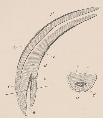
Fig. 165.—Longitudinal and transverse section of upper incisor of Horse. p, Pulp cavity; d, dentine or ivory; e, enamel; c, outer layer of cement; c′, inner layer of cement, lining a, the pit or cavity of the crown of the tooth.
Dentition.—The dentition of the Horse, when all the teeth are in place, is, as stated before, expressed by the formula i ³⁄₃, c ¹⁄₁, p ⁴⁄₃, m ³⁄₃ = 42. The incisors of each jaw are placed in close contact, forming a semicircle. The crowns are broad, somewhat awl-shaped, and of nearly equal size. They have all the great peculiarity, not found in the teeth of any other living mammal, of an involution of the external surface of the tooth (see Fig. 165) forming a deep fossa or pit, the bottom of which becomes partially filled up with cement. As the tooth wears, the surface, besides the external enamel layer as in an ordinary simple tooth, shows in addition a second inner ring of the same hard substance surrounding the pit, thus of course adding greatly to the efficiency of the tooth as an organ for biting tough, fibrous substances. This pit, generally filled in the living animal with particles of food, is conspicuous from its dark colour, and constitutes the “mark” by which the age of the horse is judged, as in consequence of its extending only to a certain depth, it becomes obliterated as the crown wears away, when the tooth assumes the character of an ordinary incisor, consisting only of a core of dentine surrounded by the external enamel layer. It is not quite so deep in the lower as in the upper teeth. The canines are either quite rudimentary or entirely absent in the female. In the male they are compressed, pointed, and smaller than the incisors, from which they are separated by a slight interval. The teeth of the cheek series are all in contact with each other, but separated from the canines by a considerable toothless space. The anterior premolars are quite rudimentary, often, especially in the lower jaw, not developed at all, and generally fall by the time the animal attains maturity, so that there are but six functional grinding teeth—three that have predecessors in the milk-dentition, and hence are considered as premolars, and three true molars, but otherwise, except the first and last of the series, not distinguishable in form or structure. These teeth in both upper and lower jaws are extremely long-crowned or hypsodont (Fig. 158), successive portions being pushed out as the surface wears away;—a process which continues until the animal becomes advanced in age. The enamelled surface is infolded in a complex manner (a modification of that found in other Perissodactyles, see Figs. 155, 167), the folds extending quite to the base of the crown, and the interstices being filled and the surface covered with a considerable mass of cement, which binds together and strengthens the whole tooth. As the teeth wear, the folded enamel, being harder than the other constituents—the dentine and cement—forms projecting ridges on the surface arranged in a definite pattern, which give it great efficiency as a grinding instrument (see Fig. 157, b and c). The free surfaces of the upper teeth are quadrate, except the first and last, which are nearly triangular. The lower teeth are much narrower than the upper.
The milk dentition consists of i ³⁄₃, c ⁰⁄₀, m ³⁄₃ = 24,—the canines and first or rudimentary premolars having apparently no predecessors. In form and structure they much resemble the permanent teeth, having the same characteristic enamel-foldings. Their eruption commences a few days after birth, and is complete before the end of the first year, the upper teeth usually appearing somewhat earlier than those of the lower jaw. The first teeth to appear are the first and second milk-molars (about five days), then the central incisor (from seven to ten days); this is followed by the second incisor (at one month), then by the third molar, and finally by the third incisor. Of the permanent teeth the first true molar appears a little after the end of the first year, followed by the second molar before the end of the second year. At about two and a half years the first premolar replaces its predecessor. Between two and a half and three years the first incisor appears. At three years the second and third premolars and the third true molar have appeared; at from three and a half to four years the second incisor; at four to four and a half years the canine; and, finally, at five years the third incisor, completing the permanent dentition. Up to this period the age of the horse is clearly shown by the state of the dentition, and for some time longer indications can be obtained from the wear of the incisor teeth, though this depends to a certain extent upon the hardness of the food or other accidental circumstances. As a general rule, the depression caused by the infolding of the surface of the incisor (the “mark”), is obliterated in the first or central incisor at six years, in the second at seven years, and in the third at eight years. In the upper teeth, as the depressions are deeper, this obliteration does not take place until about two years later. After this period no certain indications can be obtained of the age of the horse from the teeth.
Digestive Organs.—The lips are flexible and prehensile. The membrane that lines them and the cheeks is quite smooth. The palate is long and narrow; its mucous surface has seventeen pairs of not very sharply defined oblique ridges, extending as far back as the last molar tooth, beyond which the velum palati extends for about 3 inches, having a soft corrugated surface, and ending posteriorly in an arched border without uvula. This embraces the base of the epiglottis, and shuts off all communication between the cavity of the mouth and the nasal passages, respiration being, under ordinary circumstances, carried on exclusively through the nostrils. Between the mucous membrane and the bone of the hard palate is a dense vascular and nervous plexus. The membrane lining the fauces is soft and corrugated. An elongated raised glandular mass, 3 inches long and 1 inch from above downwards, extending backwards from the root of the tongue along the side of the fauces, with openings on the surface leading into crypts with glandular walls, represents the tonsil. The tongue, corresponding to the general form of the mouth, is long and narrow. It consists of a compressed intermolar portion with a flat upper surface, broad behind and becoming narrower in front; and of a depressed anterior part rather shorter than the former, which is narrow behind but widens towards the evenly rounded apex. The dorsal surface generally is very soft and smooth. There are two large circumvallate papillæ near the base, rather irregular in form, about a quarter of an inch in diameter and half an inch apart. The conical papillæ are very small and close set, though longer and more filamentous on the intermolar portion. There are no fungiform papillæ on the dorsum, but a few not very conspicuous ones scattered along the sides of the organ.
Of the salivary glands the parotid is by far the largest; elongated in the vertical direction, and narrower in the middle than at either upper or lower extremity. Its upper extremity embraces the lower surface of the cartilaginous ear-conch; its lower end reaches the level of the inferior margin of the mandible, along the posterior margin of which it is placed. Its duct leaves the inferior anterior angle, at first descends a little, and runs forward under cover of the rounded inferior border of the mandibular ramus, then curves up along the anterior margin of the masseter muscle, becoming superficial, pierces the buccinator, and enters the mouth by a simple aperture opposite the middle of the crown of the third premolar tooth. It is not quite so thick as a goose-quill when distended, and nearly a foot in length.
The submaxillary gland is of very similar texture to the last, but much smaller; it is placed deeper, and lies with its main axis horizontal. It is elongated and slender, and flattened from within outwards. Its posterior end rests against the anterior surface of the transverse process of the atlas, from which it extends forwards and downwards, slightly curved, to beneath the ramus of the jaw. The duct which runs along its upper and internal border passes forwards in the usual course, lying in the inner side of the sublingual gland, to open on the outer surface of a distinct papilla, situated on the floor of the mouth, half an inch from the middle line, and midway between the lower incisor teeth and the attachment of the frænum linguæ. The sublingual is represented by a mass of glands lying just beneath the mucous membrane of the floor of the mouth on the side of the tongue, causing a distinct ridge, extending from the frænum backwards, and the numerous ducts opening separately along the summit of the ridge. The buccal glands are arranged in two rows parallel with the molar teeth. The upper ones are the largest, and are continuous anteriorly with the labial glands, the ducts of which open on the mucous membrane of the upper lip.
The stomach of the Horse is simple in its external form, with a largely developed right cul de sac, and is a good deal curved on itself, so that the cardiac and pyloric orifices are brought near together. The antrum pyloricum is small and not very distinctly marked off. The interior is divided by the character of the lining membrane into two very distinct portions, right and left. Over the latter the dense white smooth epithelial lining of the œsophagus is continued, terminating abruptly by a raised crenellated border. Over the right part (rather the larger portion) the mucous membrane has a grayish-red colour and a velvety appearance, and contains very numerous peptic glands, which are wanting in the cardiac portion. The œsophageal orifice is very small, and is guarded by a strong crescentic or rather horse-shoe-like band of muscular fibres, which is supposed to be the cause of the difficulty of vomiting in the Horse. The small intestine is of great length (80 to 90 feet), its mucous membrane being covered with numerous fine villi. The cæcum is of conical form, about 2 feet long and nearly a foot in diameter; its walls are sacculated, especially near the base, having four longitudinal fibrous bands; and its capacity is about twice that of the stomach. It lies with its base near the lower part of the abdomen, and its apex directed towards the thorax. The colon is about one-third the length of the small intestine, and very capacious in the greater part of its course. As usual, it may be divided into an ascending, transverse, and descending portion; but the middle or transverse portion is folded into a great loop, which descends as low as the pubis; so that the colon forms altogether four folds, generally parallel to the long axis of the body. The descending colon is much narrower than the rest, and not sacculated, and being considerably longer than the distance it has to traverse, is thrown into numerous folds.
The liver (Fig. 166) is tolerably symmetrical in its general arrangement, being divided nearly equally into segments by a well-marked umbilical fissure. Each segment is again divided by lateral fissures, which do not extend quite to the posterior border of the organ; of the central lobes thus cut off, the right is rather the larger, and has two fissures in its free border subdividing it into lobules. The extent of these varies, however, in different individuals, being not usually so marked as in the figure, which is from a fœtal specimen. The two lateral lobes are subtriangular in form. The Spigelian lobe is represented by a flat surface between the portal fissure and the posterior border, not distinctly marked off from the left lateral by a fissure of the ductus venosus, as this vessel is buried deep in the hepatic substance, but the caudate lobe is distinct and tongue-shaped, its free apex reaching nearly to the border of the right lateral lobe. In most works on the anatomy of the Horse this has been confounded with the Spigelian lobe of man. There is no gall-bladder (as in all other Perissodactyles), and the biliary duct enters the duodenum about 6 inches from the pylorus. The pancreas has two lobes or branches—a long one passing to the left and reaching the spleen, and a shorter right lobe. The principal duct enters the duodenum with the bile-duct, and there is often a second small duct which opens separately near to this.
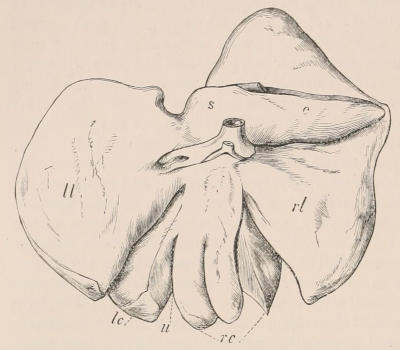
Fig. 166.—Under surface of the liver of the Horse. u, Umbilical fissure; ll, left lateral lobe; lc, left central lobe; rc, right central lobe; rl, right lateral lobe; s, Spigelian lobe; c, caudate lobe.
Circulatory and Respiratory Organs.—The heart has the form of a rather elongated and pointed cone. There is one anterior vena cava, formed by the union of the two jugular and two axillary veins. The aorta gives off a large branch (the anterior aorta) very near its origin, from which arise—first, the left axillary, and afterwards the right axillary and the two carotid arteries.
Under ordinary circumstances the Horse breathes entirely by the nasal passages, the communication between the larynx and the mouth being closed by the velum palati. The nostrils are placed laterally, near the termination of the muzzle, and are large and very dilatable, being bordered by cartilages upon which several muscles act. Immediately within the opening of the nostril, the respiratory canal sends off on its upper and outer side a diverticulum or blind pouch (called “false nostril”) of a conical form, and curved, 2 to 3 inches in depth, lying in the notch formed between the nasal and premaxillary bones. It is lined by mucous membrane continuous with that of the nasal passage, but its use is not apparent. It is longer in the Ass than in the Horse. A similar structure is found in the Rhinoceros, and in a much more developed condition in the Tapir. Here may be mentioned the guttural pouches, large air sacs, diverticula from the Eustachian tubes, and lying behind the upper part of the pharynx. These are likewise found in other Perissodactyles, but their use is also still not clearly understood. The larynx has the lateral sacculi well developed, though entirely concealed within the alæ of the thyroid cartilage. The trachea divides into two bronchi, one for each lung.
Nervous System.—The brain differs little, except in details of arrangement of convolutions, from that of other Ungulates. The cerebral hemispheres are rather elongated and subcylindrical, the olfactory lobes are large and project freely in front of the hemispheres, and the greater part of the cerebellum is uncovered. The eye is provided with a nictitating membrane or third eyelid, at the base of which the ducts of the Harderian gland open.
Reproductive System.—The testes are situated in a distinct sessile or slightly pedunculated scrotum, into which they descend from the sixth to the tenth month after birth. The accessory generative glands are the two vesiculæ seminales, with the median third vesicle, or uterus masculinus, lying between them, the single bilobed prostate, and a pair of globular Cowper’s glands. The penis is large, cylindrical, with a truncated, expanded, flattened termination. When in a state of repose it is retracted by a muscle arising from the sacrum, within the prepuce, a cutaneous fold attached below the symphysis pubis.
The uterus is bicornuate. The vagina is often partially divided by a membraneous septum or hymen. The mammæ (as in other members of the suborder), are two, inguinally placed. The surface of the chorion is covered evenly with minute villi, constituting a diffuse non-deciduate placenta. The period of gestation is eleven months.
Bibliography.—M. S. Arloing, “Organisation du pied chez le cheval,” Ann. Sci. Nat. 1867, viii. pp. 55-81; H. Burmeister, Los caballos fosiles de la Pampa Argentina, Buenos Ayres, 1875; Chanveau and Arloing, Traité d’anatomie comparée des animaux domestiques, Paris, 1871, and English edition by G. Fleming, 1873; E. Cuyer and E. Alix, Le Cheval, 1886; A. Ecker, “Das Europäische Wildpferd und dessen Beziehungen zum domesticirten Pferd,” Globus, Bd. xxxiv. Brunswick, 1878; Forsyth-Major, “Beiträge zur Geschichte der fossilen Pferde besonders Italiens,” Abh. Schw. Pal. Ges. iv. pp. 1-16, pt. iv.; George, “Études zool. sur les Hémiones et quelques autres espèces chevalines,” Ann. Sci. Nat. 1869, xii. p. 5; E. F. Gurlt, Anatomische Abbildungen der Haussäugethiere, 1824, and Hand. der vergleich. Anat. der Haussäugethiere, 2 vols. 1822; Huet, “Croisement des diverses espèces du genre cheval,” Nouv. Archives du Muséum, 2d sér. tom. ii. p. 46, 1879; Leisering, Atlas der Anatomie des Pferdes, Leipsic, 1861; J. M’Fadyean, The Anatomy of the Horse, 1884; O. C. Marsh, “Notice of New Equine Mammals from the Tertiary Formation,” Am. Journ. of Science and Arts, vol. vii. March 1874; Id. “Fossil Horses in America,” Amer. Naturalist, vol. viii. May 1874; Id. “Polydactyle Horses,” Am. Journ. of Science and Arts, vol. xvii. June 1879; Franz Müller, Lehrbuch der Anatomie des Pferdes, Vienna, 1853; R. Owen, “Equine Remains in Cavern of Bruniquel,” Phil. Trans. vol. clix. (1870), p. 535; W. Percivall, The Anatomy of the Horse, 1832; G. Stubbs, Anatomy of the Horse, 1766. F. H. Huth’s Bibliographical Record of Hippology (1887) contains a list of nearly four thousand works on Horses and Equitation, published in the various languages of the civilised world.
Family Rhinocerotidæ.
Although the existing members of this family are readily distinguished from the other living representatives of the suborder by the simple crescentoid form assumed by the ridges of the lower cheek-teeth, yet it is exceedingly difficult to give a definition by which they can be distinguished from the Lophiodontidæ, from some members of which they are, indeed, probably derived. The outer columns of the upper molars (Fig. 167) are, however, so excessively flattened as to produce a continuous thick and nearly straight outer wall, which is often produced in advance of the anterior transverse ridge; both transverse ridges being but little curved, and intimately connected with the outer wall. The upper premolars are in most cases nearly or quite as complex as the molars, and the ridges of the lower cheek-teeth are crescentoid. The last lower molar has no third lobe. The height of the crowns of the cheek-teeth is variable. The skull is large, with the orbit confluent with the temporal fossa. There are either three or four digits in the manus, and three in the pes. One or more dermal horns are attached to the fronto-nasal region of the skull of existing forms, but these were wanting in some of the fossil species.
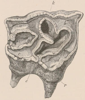
Fig. 167.—A partially worn second right upper molar of Rhinoceros antiquitatis. Letters as in Fig. 155 (p. 375), except k, which indicates a prolongation of the median valley. (After Owen.)
Rhinoceros.[267]—Incisors variable, reduced in number, often quite rudimentary, and early deciduous. Upper canines absent. Molar series, consisting of the full number of four premolars and three molars above and below, all in contact and closely resembling each other, except the first, which is much smaller than the rest and often deciduous; and the last, in which the hinder lobe is partly aborted, so that the contour of the crown is triangular. Head large, skull elongated, elevated posteriorly into a transverse occipital crest. No postorbital processes. Nasal bones large and stout, co-ossified, and standing out freely above the premaxillæ, from which they are separated by a deep and wide fissure; the latter small, generally not meeting in the middle line in front, often quite rudimentary. Tympanics small, not forming a bulla. Brain cavity very small for the size of the skull. Vertebræ: C 7, D 19-20, L 3, S 4, C about 22. Limbs stout, and of moderate length. Three completely developed toes, with distinct broad rounded hoofs on each foot (Fig. 151, p. 368), some fossil forms having a fourth in the manus. Eyes small. Ears of moderate size, oval, erect, prominent, placed near the occiput. Skin very thick, in many species thrown into massive folds. Hairy covering scanty. When one horn is present it is situated over the conjoined nasal bones; when two, the hinder one is over the frontals. These horns, which are of a more or less conical form and usually recurved, often grow to a great length (three or even four feet), and are composed of a solid mass of hardened epidermic cells growing from a cluster of long dermal papillæ. The cells formed on each papilla constitute a distinct horny fibre, like a thick hair, and the whole are cemented together by an intermediate mass of cells which grow up from the interspaces between the papillæ. It results from this that the horn has the appearance of a mass of agglutinated hairs, which, in the newly growing part at the base, readily fray out on destruction of the softer intermediate substance; but the fibres differ from true hairs in growing from a free papilla of the derm, and not within a follicular involution of the same.