Chapter II · GENERAL ANATOMICAL CHARACTERS
Reading edition
I. TEGUMENTARY STRUCTURES
Hair.—The external surface of the greater number of members of the class is thickly clothed with a peculiarly modified form of epidermis, commonly called hair. This consists of hard, elongated, slender, cylindrical or tapering, filiform, unbranched masses of epidermic material, growing from a short papilla sunk at the bottom of a follicle in the derm or true skin. Such hairs upon different parts of the same animal, or upon different animals, assume various forms, and are of various sizes and degrees of rigidity,—as seen in the delicate soft velvety fur of the Mole, the stiff bristles of the Pig, and the spines of the Hedgehog and Porcupine, all modifications of the same structures. Each hair is composed usually of a cellular pithy internal portion, containing much air, and a denser or more horny cortical part. In some animals, as Deer, the substance of the hair is almost entirely composed of the medullary or cellular substance, and it is consequently very easily broken; in others the horny part prevails almost exclusively, as in the bristles of the Wild Boar. In the Three-toed Sloth (Bradypus) the hairs have a central horny axis and a pithy exterior. Though generally nearly smooth, or but slightly scaly, the surface of some hairs is strongly imbricated, notably so in some Bats; while in the Two-toed Sloth (Cholœpus) the hairs are longitudinally grooved or fluted. Though usually more or less cylindrical or circular in section, hairs are often elliptical or flattened, as in the curly-haired races of men, the terminal portion of the hair of Moles and Shrews, and conspicuously in the spines of the Rodents Xerus and Platacanthomys. Hair having a property of mutual cohesion or “felting,” which depends upon a roughened scaly surface and a tendency to curl, as in domestic Sheep (in which animal this property has been especially cultivated by selective breeding), is called “wool.”
In a large number of mammals hairs of one kind only are scattered pretty evenly over the surface; but in many there are two kinds, one longer, stiffer, and alone appearing on the surface, and the other shorter, finer, and softer, constituting the under fur, analogous to the down of birds. This under fur, or pashm as it is called by the natives of Kashmir, is especially abundant in the mammals inhabiting the cold plateau of Tibet and the adjacent regions. In many cases hairs of a different character from those of the general surface grow in special regions, forming ridges or tufts on the median dorsal or ventral surface or elsewhere. The tail is very often completed in this way by variously disposed elongated hairs. The margins of the eyelids are almost always furnished with a special row of stiffish hairs, called cilia or eyelashes; and in most mammals specially modified hairs, constituting the vibrissæ or whiskers, and endowed, through the abundant nerve supply of their basal papillæ, with special tactile powers, grow from the lips and cheeks. In some mammals the hairy covering is partial and limited to particular regions; in others, as the Hippopotamus and the Sirenia, though scattered over the whole surface, it is extremely short and scanty; but in none is it reduced to so great an extent as in the Cetacea, in which it is limited to a few small bristles confined to the neighbourhood of the lips and nostrils, and often only present in the young or even fœtal condition.
Some kinds of hairs, as those of the mane and tail of the Horse, appear to persist throughout the lifetime of the animal; but more generally, as in the case of the body hair of the same animal, they are shed and renewed periodically, generally annually. Many mammals have a longer hairy coat in winter, which is shed as summer comes on; and some few, which inhabit countries covered in winter with snow, as the Arctic Fox, Variable Hare, and Ermine, undergo a complete change of colour in the two seasons, being white in winter, and gray or brown in summer. The several species of Cape Mole (Chrysochloris), the Desmans or Water Moles (Myogale), and Potamogale velox, are remarkable as being the only mammals whose hair reflects those iridescent tints so common in the feathers of tropical birds.
The principal and most obvious purpose of the hairy covering is to protect the skin against external influences, especially cold and damp. Its function in the hairless Cetacea is supplied by the specially modified and thickened layer of adipose tissue beneath the skin, called “blubber.”
Colour.—From the consideration of hair we are easily led to that of colour. As a general rule, bright and primary colours are absent in the class; but among the Baboons we find brilliant patches of scarlet or blue on some of the bare portions of the body, and one of the South American Monkeys (Brachyurus) has its whole face of a bright crimson. The most general colours are various shades of gray, brown, and tawny, with a frequent tendency to whiteness of the ventral surface of the body; but among the Squirrels, and more especially those provided with a parachute for flying, we find brilliant russets, passing into orange and red. Dark brown or black is also not very uncommon, as in the Bears and the Sable Antelope of South Africa. Entirely white mammals are rare, and mostly characteristic of the polar regions, or of countries having a long and snowy winter. An entirely white Bat (Diclidurus albus) occurs, however, in South America. In the large majority of mammals that exhibit a varied coloration, the upper and most exposed parts of the surface present the richest and darkest colours, the under parts being pale or often quite white. The Ratels, Gluttons, Ælurus, Hamsters, and some others are exceptions to this rule. A large number of mammals having a ground colour of gray, tawny, or dun are marked by stripes or spots, which are generally of a darker hue than the ground colour, as in many Carnivora, but more rarely are lighter, as in the Fallow and Axis Deer and several species of Antelope. These stripes very generally run transversely to the axis of the body, as in the Tasmanian Thylacine, the Tiger, and the Zebra; but they may be longitudinal, as in several of the Civet family. There has been considerable discussion as to whether the striped or the spotted is the more primitive type of coloration; but no very conclusive arguments have been brought forward in favour of either view. It is, however, manifest that in several groups of mammals there is a tendency to lose the spots, and more rarely the stripes, and to assume a uniform colour. Thus the young of nearly all the species of Deer are spotted, whereas the adults of only the Fallow and Axis Deer are so marked. The same is true of most of the Pigs; and the young of the Malayan and American Tapirs are marked by light-coloured stripes and spots on a dark ground. In like manner the young of the Lion and the Puma exhibit distinct spots which disappear with advancing age. In most of our domestic horses of various shades of bay and brown we may detect “dappling” on the under hair when the outer coat has been removed, which is not apparent on the surface of the latter. Many varieties of the Ass and the Horse also exhibit a tendency to the presence of stripes on the legs, which would seem to indicate a descent from a striped Zebra-like type.
A peculiar feature, which is, however, common to many other groups of animals, is the tendency to what is known as melanism, or the production of black or dark individuals or races of particular species, due to an excess of pigment in the skin and hair. Thus we may have black Leopards and Jaguars, black Wolves, and black Rabbits.
The opposite to melanism, and of more frequent occurrence, is albinism—a condition in which the pigment or colouring matter usually present in the tissues constituting the external coverings of the body, and which gives them their characteristic hue, is absent. When it occurs the hair is of an opaque white, the claws, hoofs, etc., of a pale horn-colour, and the skin and eyes pink, in consequence of the colour of the blood which circulates through them being no longer concealed by the stronger hues of the pigments. An animal in this condition is called an albino. In complete albinism there is a total absence of pigment throughout the system. This condition occurs occasionally as an individual peculiarity among wild animals of many kinds; but it has never been perpetuated among them in distinct races or species. The disadvantage of absence of pigment in the eye, causing a certain amount of intolerance of light, is probably sufficient to account for this. Several races of true albinos, as White Ferrets, Rabbits, Rats, and Mice, have, however, been established under the protection of man, and in them this abnormal condition is propagated from generation to generation.
Partial albinism—a condition in which the absence of pigment is limited to portions of the surface, or, at all events, does not extend to the eyes—is much more common as an individual variation both in domestic and in wild animals. It is possible that the artificial conditions incident to domestication increase the tendency to its occurrence; but, whether this be so or not, it certainly becomes perpetuated more frequently among domesticated than among wild animals. This may be accounted for partly by its proving of no disadvantage to them, and partly by the frequent selection by man of animals of such colour in preference to others. The result is that there is no completely domestic animal of which white races do not exist. On the other hand, to most wild animals even partial albinism seems to be a disadvantage in the struggle for existence, since, except in the case of species inhabiting lands continually covered with snow, it renders them more conspicuous objects both to their enemies and their prey, and hence it is rarely perpetuated. In northern regions, however, a large proportion of species are regularly and normally of a white colour, either, as the Polar Bear, all the year through, or, as the Ermine or Stoat, Arctic Fox, and Alpine Hare, during the winter season. The coloration in these cases is obviously protective, as it is also to a great extent in many other instances throughout the class.
Among conspicuously coloured mammals, it has been observed that the vertical black and tawny stripes of the Tiger harmonise so well with the brown and green grasses of its native jungle as to render the animal almost invisible when lying among them; while the dappled hide of the Giraffe is said to agree equally well with the chequered splashes of light and shade in the clumps of tall mimosas among which it feeds. The uniformly tawny hue of the Lion accords well with the prevailing tint of its native desert; and any one who has seen an Elephant or Buffalo in the deep shades of an Indian forest will realise how perfectly adapted is their dull, slaty colour to concealment in such a spot. The dun colour of the Wild Ass of India is equally well suited to the sandy deserts of Kutch; it is also stated that the brilliant stripes of the Zebras of Africa are arranged in such proportion as exactly to match the pale tint which arid ground possesses when seen by moonlight.[1] The most remarkable instance of protective coloration is, however, to be found in the Sloths of South America, in which the coarse gray hairs so closely resemble a mass of lichenous growth that it is almost impossible to distinguish these animals when at rest from the gnarled and lichen-clad boughs from which they suspend themselves. This resemblance is increased by the fact that the hairs actually develop a growth of lichens upon themselves. That the sombre coloration of these animals has been produced to harmonise with their present surroundings seems to be evident by the circumstance that when the long hair is plucked off the under fur is seen to present a bold alternation of black and yellow stripes, which may probably be regarded as the original primitive coloration of this group.
Scales, etc.—True scales, or flat imbricated plates of horny material, covering the greater part of the body, so frequently occurring in reptiles, are found only in one family of mammals, the Manidæ or Pangolins; but these are also associated with hairs growing from the intervals between the scales, or on the parts of the skin not covered by them. Similarly, imbricated epidermic productions form the covering of the under surface of the tail of the flying Rodents of the genus Anomalurus; and flat scutes, with the edges in apposition, and not overlaid, clothe both surfaces of the tail of the Beaver, Rats, and others of the same order, and also of some Insectivores and Marsupials. The Armadillos alone have an ossified exoskeleton, composed of plates of true bony tissue, developed in the derm or corium, and covered with scutes of horny epidermis. Other epidermic appendages are the horns of Ruminants and Rhinoceroses,—the former being elongated, tapering, hollow caps of hardened epidermis of fibrillated structure, fitting on and growing from conical projections of the frontal bone, and always arranged in pairs, while the latter are of similar structure, but solid and without any internal bony support, and (in all existing species) situated in the median line. Callosities, or bare patches covered with hardened and thickened epidermis, are found covering the pads under the soles of the feet and undersurfaces of the toes of nearly all mammals, upon the ischial tuberosities of many Apes, the sternum of Camels, on the inner side of the limbs of the Equidæ, the grasping under surface of the tail of the prehensile-tailed Monkeys, etc. The greater part of the skin of both species of one-horned Asiatic Rhinoceros is immensely thickened and stiffened by increase of the tissue both of the derm and epiderm, constituting the well-known jointed “armour-plated” hide of those animals.
Nails, Claws, and Hoofs.—With very few exceptions, the terminal extremities of the digits of both limbs are more or less protected or armed by epidermic plates or sheaths, constituting the various forms of nails, claws, or hoofs. These are wanting in the Cetacea alone. A perforated spur, with a special secreting gland in connection with it, is found attached to the hind leg of the males of the three genera of Monotremata, Ornithorhynchus, Proechidna, and Echidna.
Odour-secreting Glands.—Besides the universally distributed sebaceous glands connected with the pilose system, most mammals have special glands situated in modified portions of the integument, often involuted to form a shallow recess or a deep sac with a narrow opening, situated in various parts of the surface of the body, and secreting odorous substances, by the aid of which individuals appear to recognise one another, and probably affording the principal means by which wild animals are able to become aware of the presence of other members of the species, even at great distances. Although the commencement of the modifications of portions of the external covering for the formation of special secretions may be at present difficult to understand, the principle of natural selection will readily explain how such organs become fixed and gradually increase in development in any species, especially as there would probably be a corresponding modification and increased sensibility of the olfactory organs. Such individuals as by the intensity and peculiarity of their scent had greater power of attracting the opposite sex would certainly be those most likely to leave descendants to inherit and in their turn propagate the modification.
To this group of structures belong the suborbital gland or “crumen” of Antelopes and Deer, the frontal gland of the Muntjac and of Bats of the genus Hipposiderus, the submental gland of the Chevrotains and of Taphozous and some other Bats, the post-auditory follicle of the Chamois, the temporal gland of the Elephant, the lateral glands of the Musk-Shrew, the dorsal gland of the Peccary, the inguinal glands of Antelopes, the preputial glands of the Musk-Deer and Beaver (already alluded to in connection with the use made of their powerfully odorous secretion in medicine and perfumery) and also of the Swine and Hare, the anal glands of Carnivora, the perineal gland of the Civet (also of commercial value), the caudal glands of the Fox and Goat, the gland on the humeral membrane of Bats of the genus Saccopteryx, the post-digital gland of the Rhinoceros, the interdigital glands of the Sheep and many Ruminants, and numerous others. In some of these cases the glands are peculiar to, or more largely developed in, the male; in others they are found equally developed in both sexes.
II. DENTAL SYSTEM
The dental system of mammals may be considered rather more in detail than space permits for some other portions of their structure, not only on account of the important part it plays in the economy of the animals of this class, but also for its interest to zoologists as an aid in the classification and identification of species. Owing to the imperishable nature of their tissues, teeth are preserved for an indefinite time, and in the case of extinct species frequently offer the only indications available from which to derive an idea of the characters, affinities, and habits of the animals to which they once belonged. Hence even their smallest modifications have received great attention from comparative anatomists, and they have formed the subject of many special monographs.[2]
Teeth are present in nearly all mammals, and are applied to various purposes. They are, however, mainly subservient to the function of alimentation, being used either in procuring food, by seizing and killing living prey or gathering and biting off portions of vegetable material, and more indirectly in tearing or cutting through the hard protective coverings of food substances, as the husks and shells of nuts, or in pounding, crushing, or otherwise mechanically dividing the solid materials before swallowing, so as to prepare them for digestion in the stomach. Certain teeth are also in many animals most efficient weapons of offence and defence, and for this purpose alone, quite irrespective of subserviency to the digestive process, are they developed in the male sex of many herbivorous animals, in the females of which they are absent or rudimentary.
Teeth belong essentially to the tegumentary or dermal system of organs, and, as is well seen in the lower vertebrates, pass by almost insensible gradations into the hardened spines and scutes formed upon the integument covering the outer surface of the body; but in mammals they are more specialised in structure and limited in locality. In this class they are developed only in the gums or fibro-mucous membrane covering the alveolar borders of the upper and lower jaws, or, in other words, the premaxillary and maxillary bones and the mandible. In the process of development, for the purpose of giving them that support which is needful for the performance of their functions, they almost always become implanted in the bone,—the osseous tissue growing up and moulding itself around the lengthening root of the tooth, so that ultimately they become apparently parts of the skeleton. In no mammal, however, does ankylosis or bony union between the tooth and jaw normally take place, as in many fishes and reptiles,—a vascular layer of connective tissue, the alveolo-dental membrane, always intervening.[3] The presence of two or more roots, frequently met with in the cheek-teeth of mammals, implanted in corresponding distinct sockets of the jaw, is now peculiar to animals of this class.[4]
Structure.—The greater number of mammalian teeth when fully formed are not simple and homogeneous in structure, but are composed of several distinct tissues, which are enumerated below.
The pulp, a soft substance, consisting of a very delicate gelatinous connective tissue, in which numerous cells are imbedded, and abundantly supplied with blood-vessels and nerves, constitutes the central axis of all the basal part of the tooth, and affords the means by which the vitality of the whole is preserved. The nerves which pass into the pulp and endow the tooth with sensibility are branches of the fifth pair of cranial nerves. The pulp occupies a larger relative space, and performs a more important purpose, in the young growing tooth than afterwards, as by the calcification and conversion of its outer layers the principal hard constituent of the tooth, the dentine, is formed. In teeth which have ceased to grow the pulp occupies a comparatively small space, which in the dried tooth is called the pulp-cavity. This communicates with the external surface of the tooth by a small aperture at the apex of the root, through which the branches of the blood-vessels and nerves, by which the tooth receives its nutrition and sensitiveness, pass in to be distributed in the pulp. In growing teeth the pulp-cavity is widely open, while in advanced age it often becomes obliterated, and the pulp itself entirely converted into bone-like material.
The dentine or ivory forms the principal constituent of the greater number of teeth. When developed in its most characteristic form, it is a very hard but elastic substance, white, with a yellowish tinge, and slightly translucent. It consists of an organic matrix, something like, but not identical with, that of bone, richly impregnated with calcareous salts (chiefly calcium phosphate), these constituting in a fresh human tooth 72 per cent of its weight. When subjected to microscopical examination it is seen to be everywhere permeated by nearly parallel branching tubes which run, in a slightly curving or wavy manner, in a general direction from the centre towards the free surface of the tooth. These tubes communicate by open mouths with the pulp-cavity, and usually terminate near the periphery of the dentine by closed ends or loops, though in Marsupials and certain other mammals they penetrate into the enamel. They are occupied in the living tooth by soft gelatinous fibrils connected with the cells of the pulp. A variety of dentine, permeated by canals containing blood-vessels, met with commonly in fishes and in some few mammals, as the Megatherium, is called vaso-dentine. Other modifications of this tissue occasionally met with are called osteodentine and secondary dentine,—the latter being a dentine of irregular structure which often fills up the pulp-cavity of old animals.
The enamel constitutes a thin investing layer, complete or partial, of the outer or exposed and working surface of the dentine of the crown of the teeth of most mammals. This is the hardest tissue met with in the animal body, containing from 95 to 97 per cent of mineral substances (chiefly calcium phosphate and some carbonate, with traces of fluoride). Its ultimate structure consists of prismatic fibres, placed generally with their long axes at right angles to the free surface of the tooth. Enamel is easily distinguished from dentine with the naked eye by its clear, bluish-white, translucent appearance.
The cement or crusta petrosa is always the most externally placed of the hard tissues of which teeth are composed, as will be understood when the mode of development of these organs is considered. It is often only found as a thin layer upon the surface of the root; but sometimes, as in the complex-crowned molar teeth of the Horse and Elephant, it is a structure which plays a very important part, covering and filling in the interstices between the folds of the enamel. In appearance, histological structure, and chemical composition it is closely allied to osseous tissue, containing lacunæ and canaliculi, though only when it is of considerable thickness are Haversian canals present in it.
Development.—The two principal constituents of the teeth, the dentine and the enamel, are developed from the two layers of the mucous membrane of the jaw—the dentine from the deeper or vascular, the enamel from the superficial or epithelial layer. The latter dips down into the substance of the gum, and forms the enamel-organ or germ, the first rudiment of the future tooth, which is constantly present even in those animals in which the enamel is not found as a constituent of the perfectly-formed tooth. Below the mass of epithelial cells thus embedded in the substance of the gum, and remaining connected by a narrow neck of similar structure with the epithelium of the surface, a portion of the vascular areolar tissue becomes gradually separated and defined from that which surrounds it, and assumes a distinct form, which is that of the crown of the future tooth,—a single cone in the case of simple teeth, or with two or more eminences in the complex forms. This is called the dental papilla or dentine germ, and by the gradual conversion of its tissue into dentine the bulk of the future tooth is formed, the uncalcified central portion remaining as the pulp. The conversion of the papilla into hard tissue commences at the outer surface of the apex, and gradually proceeds downwards and inwards, so that the form of the papilla exactly determines the form of the future dentine, and no alteration either in shape or size of this portion of the tooth, when once calcified, can take place by addition to its outer surface. In the meanwhile, calcification of a portion of the cells of the enamel-organ, which adapts itself like a cap round the top of the dentinal papilla, and has assumed a somewhat complex structure, results in the formation of the enamel-coating of the crown of the tooth. While these changes are taking place the tissues immediately surrounding the tooth-germ become condensed and differentiated into a capsule, which appears to grow up from the base of the dental papilla, and encloses both this and the enamel-germ, constituting the follicle or tooth-sac. By the ossification of the inner layer of this follicle the cement is formed. This substance, therefore, unlike the dentine, increases from within outwards, and its growth may accordingly be the cause of considerable modification of form and enlargement, especially of the roots, of certain teeth, as those of Seals and some Cetacea. The delicate homogeneous layer coating the enamel surface of newly-formed teeth, in which cement is not found in the adult state, and known as Nasmyth’s membrane, is considered by Tomes as probably a film of this substance, too thin to exhibit its characteristic structure, though by others it is believed to be derived from the external layer of the enamel-organ. The homology of the teeth with the dermal appendages, hairs, scales, and claws, has already been alluded to, and it will now be seen that in both cases two of the primary embryonic layers are concerned in their development—the mesoblast and epiblast—although in very different proportions respectively. Thus in the hair or nail the part derived from the epiblast forms the principal bulk of the organ, the mesoblast only constituting the papilla or matrix. But in the tooth the epiblastic portion is limited to the enamel, and is always of relatively small bulk and often absent, while the dentine (the principal constituent of the tooth) and the cement are formed from the mesoblast.
When more than one set of teeth occur in mammals, those of the second set are developed in a precisely similar manner to the first, but the enamel-germ, instead of being derived directly from an independent part of the oral epithelium, is formed from a budding out of the neck of the germ of the tooth succeeded. In the case of the true molars, which have no predecessors, the germ of the first has an independent origin, but that of the others is derived from the neck of the germ of the tooth preceding it in the series. The foundations of the permanent teeth are thus laid as it were almost simultaneously with those of their predecessors, although they remain in many cases for years before they are developed into functional activity.
Although the commencement of their formation takes place at an early period of embryonic life, teeth are in nearly all mammals still concealed beneath the gum at the time of birth. The period of eruption, or “cutting” of the teeth as it is called, that is, their piercing through and rising above the surface of the mucous membrane, varies much in different species. In some, as Seals, the whole series of teeth appears almost simultaneously; but more often there are considerable intervals between the appearance of the individual teeth, the front ones usually coming into place first, and those at the back of the mouth at a later period.
Forms of Teeth.—The simplest form of tooth may be exemplified on a large scale by the tusk of the Elephant (Fig. 1, I.) It is a hard mass almost entirely composed of dentine, of a conical shape at first, but during growth becoming more and more cylindrical or uniform in width. The enamel-covering, present on the apex in its earliest condition, soon disappears, but a thin layer of cement covers the circumference of the tooth throughout life. In section it will be seen that the basal portion is hollow, and contains a large conical pulp, as broad at the base as the tooth itself, and deeply imbedded in the bottom of a recess, or socket, in the maxillary bone. This pulp continues to grow during the lifetime of the animal, and at the same time is converted at its surface into dentine. The tooth therefore continually elongates, but the use to which the animal subjects it in its natural state causes the apex to wear away, at a rate generally proportionate to the growth at the base, otherwise it would become of inconvenient length and weight. Such teeth of indefinite growth are said to be “rootless,” or to have “persistent pulps.”
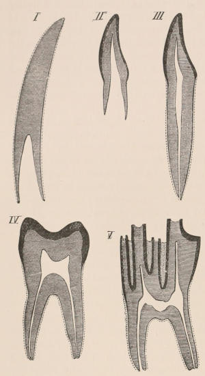
Fig. 1.—Diagrammatic Sections of various forms of Teeth. I. Incisor or tusk of Elephant, with pulp-cavity persistently open at base. II. Human incisor during development, with root imperfectly formed, and pulp-cavity widely open at base. III. Completely formed human incisor, with pulp-cavity contracted to a small aperture at the end of the root. IV. Human molar, with broad crown and two roots. V. Molar of the Ox, with the enamel covering the crown deeply folded, and the depressions filled up with cement. The surface is worn by use; otherwise the enamel coating would be continuous at the top of the ridges. In all the figures the enamel is black, the pulp white, the dentine represented by horizontal lines, and the cement by dots.
One of the corresponding front teeth of man (Fig. 2, II. and III.) may be taken as an example of a very different condition. After its crown is fully formed by calcification of the germ, the pulp, though continuing to elongate, begins to contract in diameter; a neck or slight constriction is formed; and the remainder of the pulp is converted into the root (often, but incorrectly, called “fang”), a tapering conical process imbedded in the alveolar cavity of the bone, and having at its extremity a minute perforation, through which the vessels and nerves required to maintain the vitality of the tooth enter the pulp-cavity, which is very different from the widely open cavity at the base of the growing tooth. When the crown of the tooth is broad and complex in character, instead of having a single root, it may be supported by two or more roots, each of which is implanted in a distinct alveolar recess or socket, and to the apex of which a branch of the common pulp-cavity is continued (Fig. 1, IV.) Such teeth are called “rooted teeth.” When they have once attained their position in the jaw, with the neck a little way above the level of the free margin of the alveolus, and embraced by the gum or tough fibrovascular membrane covering the alveolar border, and having the root fully formed, they can never increase in length or alter their position; if they appear to do so in old age, it being only in consequence of absorption and retrocession of the surrounding alveolar margins. If, as often happens, their surface wears away in mastication, it is never renewed. The open cavity at the base of the imperfectly developed tooth (Fig. 1, II.) causes it to resemble the persistent condition of the rootless tooth. The latter is therefore a more primitive condition, the formation of the root being a completion of the process of tooth development. Functionally it is, however, difficult to say that the one is a higher form than the other, since they both serve important and different purposes in the animal economy.
As is almost always the case in nature, intermediate conditions between these two forms of teeth are met with. Thus some teeth, as the molars of the Horse, and of many Rodents, are for a time rootless, and have growing pulps producing very long crowns with parallel sides, the summits of which may be in use and beginning to wear away while the bases are still growing; but ultimately the pulp contracts, forms a neck and distinct roots, and ceases to grow. The canine tusks of the Musk Deer and of the Walrus have persistent pulps, and are open at their base until the animal is of advanced age, when they close, and the pulp ceases to be renewed. The same sometimes happens in the tusks of very old Boars.
The simplest form of the crown of a tooth is that of a cone; but this may be variously modified. Thus it may be flattened, with its edges sharp and cutting, and pointed at the apex, as in the laterally compressed premolars of most Carnivora; or it may be chisel- or awl-shaped, with a straight truncated edge, as in the human incisors; or it may be broad, with a flat or rounded upper surface. Very often there is a more or less prominent ridge encircling the whole or part of the base of the crown just above the neck, called the cingulum, which serves as a protection to the edge of the gum in masticating, and is most developed in flesh-eating and insectivorous animals, in which the gums are liable to be injured by splinters of bone or other hard fragments of their food. The form of the crown is frequently rendered complex by the development upon its surface of elevations or tubercules called cusps or cones, or by ridges usually transverse, but sometimes variously curved or folded. When the crown is broad and the ridges are greatly developed, as in the molars of the Elephant, Horse, and Ox (Fig. 1, V.), the interspaces between them are filled with cement, which supports them and makes a solid compact mass of the whole tooth. When such a tooth wears away at the surface by friction against the opposed tooth of the other jaw, the different density of the layers of the substances of which it is composed—enamel, dentine, and cement—arranged in characteristic patterns, causes them to wear unequally, the hard enamel ridges projecting beyond the others, and thus giving rise to a grinding surface of great mechanical advantage.
Succession.—The dentition of all mammals consists of a definite set of teeth, almost always of constant and determinate number, form, and situation, and, with few exceptions, persisting in a functional condition throughout the natural term of the animal’s life. In many species these are the only teeth which the animal ever possesses,—the set which is first formed being permanent, or, if accidentally lost, or decaying in extreme old age, not being replaced by others. These animals are called Monophyodont. But in the larger number of mammals, certain of the teeth are preceded by others, which may be only of a very transient, rudimentary, and functionless character (being in the Seals, for example, shed either before or within a few days after birth), or may be considerably developed, and functionally occupy the place of the permanent teeth for a somewhat lengthened period, during the growth and development of the latter and of the jaws. In all cases these teeth disappear (by the absorption of their roots and shedding of the crowns) before the frame of the animal has acquired complete maturity, as evidenced by the coalescence of the epiphyses of the osseous system. As these teeth are, as a general rule, present during the period in which the animal is nourished by the milk of the mother, the name of “milk-teeth” (French dents de lait, German milchzähne) has been commonly accorded to them, although it must be understood that the epoch of their presence is by no means necessarily synchronous with that of lactation. Animals possessing such teeth are called Diphyodont. No mammal is known to have more than two sets of teeth; and the definite and orderly replacement of certain members of the series is a process of quite a different nature from the indefinite succession which takes place in all the teeth continuously throughout the lifetime of the lower vertebrates.
When the milk-teeth are well developed, and continue in place during the greater part of the animal’s growth, as is especially the case with the Ungulata, and, though to a less degree, with the Primates and Carnivora, their use is obvious, since taken all together they form structurally a complete epitome on a small scale of the more numerous and larger permanent set (see Fig. 3), and, consequently, are able to perform the same functions, while time is allowed for the gradual maturation of the latter, and especially while the jaws of the growing animal are acquiring the size and strength sufficient to support the permanent teeth. Those animals, therefore, that have a well-developed and tolerably persistent set of milk-teeth may be considered to be in a higher state of development, as regards their dentition, than those that have the milk-teeth absent or rudimentary.
It is a very general rule that individual teeth of the milk and permanent set have a close relationship to one another, being originally formed, as mentioned above, in exceedingly near proximity, and with, at all events so far as the enamel-germ is concerned, a direct connection. Moreover, since the latter ultimately come to occupy the position in the alveolar border temporarily held by the former, they are spoken of respectively as the predecessors or successors of each other. But it must be understood that milk-teeth may be present which have no successors in the permanent series, and, what is far more general, permanent teeth may have no predecessors in the milk series.
The complete series of permanent teeth of most mammals forms a complex machine, with its several parts adapted for different functions,—the most obvious structural modification for this purpose being an increased complexity of the individual components of the series from the anterior towards the posterior extremity of such series. Since, as has just been said, the complete series of the milk teeth often presents structurally and functionally a similar machine, but composed of fewer individual members, and the anterior of which are as simple, and the posterior as complex as those occupying corresponding positions in the permanent series,—and since the milk-teeth are only developed in relation to the anterior or lateral, never to the most posterior of the permanent series,—it follows that the hinder milk-teeth are usually more complex than the teeth of which they are the predecessors in the permanent series, and represent functionally, not their immediate successors, but those more posterior permanent teeth which have no direct predecessors. This character is clearly seen in those animals in which the various members of the molar series are well differentiated from each other in form, as the Carnivora, and also in Man.
In animals which have two sets of teeth the number of those of the permanent series which are preceded by milk-teeth varies greatly, being sometimes, as in Marsupials and some Rodents, as few as one on each side of each jaw, and sometimes including the larger portion of the series.
Although there are difficulties in some cases in arriving at a satisfactory solution of the question, it is, on the whole, safest to assume that when only one set of teeth is present, this corresponds to the permanent teeth of the Diphyodonts. When this one set is completely developed, and remains in use throughout the animal’s life, there can be no question on this subject. When, on the other hand, the teeth are rudimentary and transient, as in the Whalebone Whales, it is possible to consider them as representing the milk series; but there are weighty reasons in favour of the opposite conclusion.[5]
Arrangement, Homologies, and Notation of Teeth.—The teeth of the two sides of the jaws are always alike in number and character, except in cases of accidental or abnormal variation, and in the one remarkable instance of constant deviation from bilateral symmetry among mammals, the tusks of the Narwhal (Monodon), in which the left is of immense size, and the right rudimentary. In certain mammals, such as the Dolphins and some Armadillos, which have a very large series of similar teeth, not always constant in number in different individuals, there may be differences in the two sides; but, apart from these, in describing the dentition of any mammal, it is quite sufficient to give the number and characters of the teeth of one side only. Since the teeth of the upper and the lower jaws work against each other in masticating, there is a general correspondence or harmony between them, the projections of one series, when the mouth is closed, fitting into corresponding depressions of the other. There is also a general resemblance in the number, characters, and mode of succession of both series, so that, although individual teeth of the upper and lower jaws may not be in any strict sense of the term homologous parts, there is a great convenience in applying the same descriptive terms to the one as are used for the other.
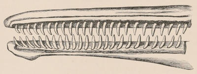
Fig. 2.—Upper and Lower Teeth of one side of the Mouth of a Dolphin (Lagenorhynchus) as an example of the homodont type of dentition. The bone covering the outer side of the roots of the teeth has been removed to show their simple character.
The simplest dentition as a whole is that of many species of Dolphin (Fig. 2), in which the crowns are single-pointed, slightly curved cones, and the roots also single and tapering, and all alike in form from the anterior to the posterior end of the series, though it may be with some slight difference in size, those at the two extremities of the series being rather smaller than the others. Such a dentition is called Homodont, and in the case cited, as the teeth are never changed, it is also Monophyodont. Such teeth are adapted only for catching slippery living prey, as fish.
In a very large number of mammals the teeth of different parts of the series are more or less differentiated in character, and have different functions to perform. The front teeth are simple and one-rooted, and are adapted for cutting and seizing. They are called “incisors.” The back- or cheek-teeth have broader and more complex crowns, tuberculated or ridged, and are supported on two or more roots. They crush or grind the food, and are hence called “molars.” Many animals have, between these two sets, a tooth at each corner of the mouth, longer and more pointed than the others, adapted for tearing or stabbing, or for fixing struggling prey. From the conspicuous development of such teeth in the Carnivora, especially the Dogs, they have received the name of “canines.” A dentition with its component parts so differently formed that these distinctive terms are applicable to them is called Heterodont. In most cases, though by no means invariably, animals with Heterodont dentition are also Diphyodont.
This general arrangement is extremely obvious in a considerable number of mammals; and closer examination shows that, under very great modification in detail, there is a remarkable uniformity of essential characters in the dentition of a large number of members of the class belonging to different orders and not otherwise closely allied; so much so indeed that it has been possible (chiefly through the researches of Sir Richard Owen) to formulate a common plan of dentition from which the others have been derived by the alteration of some and suppression of other members of the series, and occasionally, but very rarely, by addition. The records of palæontology fully confirm this view, as by tracing back many groups now widely separated in dental characters we find a gradual approximation to a common type. In this generalised form of mammalian dentition (which is best exemplified in the genera Anoplotherium and Homalodontotherium) the entire number of teeth present is 44, or 11 above and 11 below on each side. Those of each jaw are placed in continuous series without intervals between them; and, although the anterior teeth are simple and single-rooted, and the posterior teeth complex and with several roots, the transition between the two kinds is gradual.
In dividing and grouping such teeth for the purpose of description and comparison, more definite characters are required than those derived merely from form or function. The first step towards a classification has been made by the observation that the upper jaw is composed of two bones, the premaxilla and the maxilla, and that the suture between these bones separates the three anterior teeth from the others. These three teeth, then, which are implanted by their roots in the premaxilla, form a distinct group, to which the name of “incisor” is applied. This distinction is, however, not so important as it appears at first sight, for, as mentioned when speaking of the development of the teeth, their connection with the bone is only of a secondary nature, and, although it happens conveniently for our purpose that in the great majority of cases the segmentation of the bone coincides with the interspace between the third and fourth tooth of the series, still, when it does not happen to do so, as in the case of the Mole, we must not give too much weight to this fact, if it contravenes other reasons for determining the homologies of the teeth. The eight remaining teeth of the upper jaw offer a natural division, inasmuch as the posterior three never have milk-predecessors; and, although some of the anterior teeth may be in the same case, the particular one preceding these three always has such a predecessor. These three then are grouped apart as the “molars,” or, since some of the teeth in front of them often have a molariform character, “true molars.” Of the five teeth between the incisors and molars the most anterior, or that which is usually situated close behind the premaxillary suture, almost always, as soon as any departure takes place from the simplest and most homogeneous type, assumes a lengthened and pointed form, and is the tooth so developed as to constitute the “canine” or “laniary” tooth of the Carnivora, the tusk of the Boar, etc. It is customary therefore to call this tooth, whatever its size or form, the “canine.” The remaining four are the “premolars” or “false molars.” This system of nomenclature has been objected to as being artificial, and in many cases not descriptive, the distinction between premolars and canine especially being sometimes not obvious; but the terms are now in such general use, and are so practically convenient—especially if, as it is best to do in all such cases, we forget their original signification and treat them as arbitrary signs—that it is not likely they will be superseded by any that have been proposed as substitutes for them.
With regard to the lower teeth the difficulties are greater, owing to the absence of any suture corresponding to that which defines the incisors above; but since the number of the teeth is the same, the corresponding teeth are preceded by milk-teeth, and in the large majority of cases it is the fourth tooth of the series which is modified in the same way as the canine (or fourth tooth) of the upper jaw, it is quite reasonable to adopt the same divisions as with the upper series, and to call the first three, which are implanted in the part of the mandible opposite to the premaxilla, the incisors, the next the canine, the next four the premolars, and the last three the molars. It may be observed that when the mouth is closed, especially when the opposed surfaces of the teeth present an irregular outline, the corresponding upper and lower teeth are not exactly opposite, otherwise the two series could not fit into one another; but as a rule the points of the lower teeth shut into the interspaces in front of the corresponding teeth of the upper jaw. This is seen very distinctly in the canine teeth of the Carnivora, and is a useful guide in determining the homologies of the teeth of the two jaws. Objections have certainly been made to this view, because, in certain rare cases, the tooth which, according to it, would be called the lower canine has the form and function of an incisor (as in Ruminants and Lemurs), and on the other hand (as in Cotylops, an extinct Ungulate from North America) the tooth that would thus be determined as the first premolar has the form of a canine; but it should not be forgotten that, as in all such cases, definitions derived from form and function alone are quite as open to objection as those derived from position and relation to surrounding parts, or still more so.
Dental formulæ.—For the sake of brevity the complete dentition, arranged according to these principles, is often described by the following formula, the numbers above the line representing the teeth of the upper, those below the line those of the lower jaw:—incisors ³⁻³⁄₃₋₃, canines ¹⁻¹⁄₁₋₁, premolars ⁴⁻⁴⁄₄₋₄, molars ³⁻³⁄₃₋₃ = ¹¹⁻¹¹⁄₁₁₋₁₁; total 44. Since, however, initial letters may be substituted for the names of each group, and it is quite unnecessary to give more than the numbers of the teeth on one side of the mouth, the formula may be conveniently abbreviated into—
i ³⁄₃, c ¹⁄₁, p ⁴⁄₄, m ³⁄₃ = ¹¹⁄₁₁; total 44.
The individual teeth of each group are always enumerated from before backwards, and by such a formula as the following—
i 1, i 2, i 3, c, p 1, p 2, p 3, p 4, m 1, m 2, m 3
i 1, i 2, i 3, c, p 1, p 2, p 3, p 4, m 1, m 2, m 3
or more briefly—
i 1,2,3 c 1 p 1,2,3,4 m 1,2,3 .
1,2,3, 1, 1,2,3,4, 1,2,3
A special numerical designation is thus given by which each one can be indicated. In mentioning any single tooth, such a sign as m¹⁄ will mean the first upper molar, ⁄m₁ the first lower molar, and so on. The use of such signs saves much time and space in description.[6]
It was part of the view of the founder of this system of dental notation that, at least throughout the group of mammals whose dentition is derived from this general type, each tooth has its strict homologue in all species, and that in those cases in which fewer than the typical number are present (as in all existing mammals except the genera Sus, Gymnura, Talpa, and Myogale), the teeth that are missing can be accurately defined. According to this view, when the number of incisors falls short of three it is assumed that the absent ones are missing from the outer and posterior end of the series. Thus, when there is but one incisor present, it is i 1; when two, they are i 1 and i 2. Furthermore, when the premolars and the molars are below their typical number, the absent teeth are missing from the fore part of the premolar series, and from the back part of the molar series. If this were invariably so, the labours of those who describe teeth would be greatly simplified; but there are so many exceptions that a close scrutiny into the situation, relations, and development of a tooth is required before its nature can be determined, and in some cases the evidence at our disposal is scarcely sufficient for the purpose. In other instances, however, as among the Polyprotodont Marsupials, we have decisive evidence to show that the missing premolar teeth are not those at the extremity of the series.
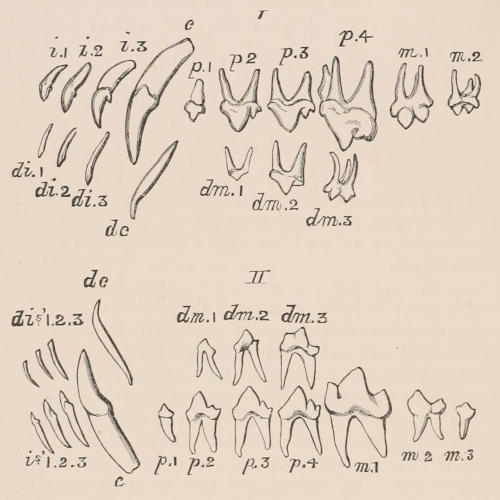
Fig. 3.—Milk and Permanent Dentition of Upper (I.) and Lower (II.) Jaw of the Dog (Canis familiaris), with the symbols by which the different teeth are commonly designated. The third upper molar (m.3) is the only tooth wanting in this animal to complete the typical heterodont mammalian dentition.
The milk-dentition is expressed by a similar formula, d for deciduous or m for milk being commonly prefixed to the letter expressive of the nature of the tooth. Since the three molars, and almost invariably the first premolar of the permanent series, have no predecessors, the typical milk-dentition would be expressed as follows—di ³⁄₃, dc ¹⁄₁, dm ³⁄₃, = ⁷⁄₇, total 28. In a few Ungulates, however, such as the Hyrax and Tapir, and in some instances the Rhinoceros and the extinct Palæotherium, the whole of the four premolars are preceded by milk-teeth; when we have the fullest development of cheek-teeth in the whole of the Eutheria. The teeth which precede the premolars of the permanent series are all called molars in the milk-dentition, although as a general rule, in form and function they represent in a condensed form the whole premolar and molar series of the adult. When there is a marked difference between the premolars and molars of the permanent dentition, the first milk-molar resembles a premolar, while the last has the characters of the posterior true molar.
The dentition of all the members of the orders Primates, Carnivora, Insectivora, Chiroptera, and Ungulata can clearly be derived from the above-described generalised type. The same may be said of the Rodents, and even the Proboscidea, though at least in the existing members of the order with greater modification. It is also apparent in certain extinct Cetacea, as Zeuglodon and Squalodon, but it is difficult to find any traces of it in existing Cetacea, Sirenia, or any of the so-called Edentata. All the Marsupials, different as they are in their general structure and mode of life, and variously modified as is their dentition, present in this system of organs some deep-lying common characters which show their unity of origin. The generalised type to which their dentition can be reduced presents considerable resemblance to that of the placental mammals, yet differing in details. It is markedly heterodont, and susceptible of division into incisors, canines, premolars, and molars upon the same principles. The whole number is, however, not limited to forty-four. The incisors may be as numerous as five on each side above, and they are almost always different in number in the upper and the lower jaw. The premolars and molars are commonly seven, as in the placental mammals, but their arrangement is reversed, as there are four true molars and three premolars.
The larger number of incisive and molar teeth among the Marsupials suggests that their additional teeth have disappeared in the Eutheria,[7] and Mr. O. Thomas has endeavoured to construct a generalised dental formula from which both the Marsupial and Eutherian modifications may have been derived by the suppression of particular teeth. Thus the hypothetical formula i ¹,²,³,⁴,⁵⁄₁,₂,₃,₄,₅, c ¹⁄₁, p ¹,²,³,⁴⁄₁,₂,₃,₄, m ¹,²,³,⁴,⁵⁄₁,₂,₃,₄,₅, by the loss of the fifth lower incisor, and of the second premolars (which we know to be those which disappear in the Marsupials) and the fifth molars, will give i ¹,²,³,⁴,⁵⁄₁,₂,₃,₄,₀, c ¹⁄₁, p ¹,⁰,³,⁴⁄₁,₀,₃,₄, m ¹,²,³,⁴⁄₁,₂,₃,₄; or the formula of the Opossum (Didelphys), usually written i ⁵⁄₄, c ¹⁄₁, p ³⁄₃, m ⁴⁄₄. Again, in the same formula the loss of the fourth and fifth incisors in both jaws, and also of the fourth molars, gives us i ¹,²,³,⁰,⁰⁄₁,₂,₃,₀,₀, c ¹⁄₁, p ¹,²,³,⁴⁄₁,₂,₃,₄, m ¹,²,³⁄₁,₂,₃, or the formula of a typical Eutherian, like the Pig, which we generally write as i ³⁄₃, c ¹⁄₁, p ⁴⁄₄, m ³⁄₃. Such a generalised formula will admit of modification into that of all existing, and a large number of fossil Marsupials, but it is possible that some of the Mesozoic types may have had more than four premolars, although there is no absolutely decisive evidence that such was the case. The presence of seven or eight true molars in some Mesozoic forms merely entails the addition of two or three additional figures to the ideal generalised formula.
The milk-dentition of all known Marsupials, existing or extinct, is (if not entirely absent) limited to a single tooth on either side of each jaw, this being the predecessor of the last permanent premolar. And if the view that the milk-dentition is an additional series grafted upon the original permanent series be correct, it is evident that we have in this single replacement the first stage of this additional development.
In very few mammals are teeth entirely absent. Even in the Whalebone Whales their germs are formed in the same manner and at the same period of life as in other mammals, and even become partially calcified, but they never rise above the gums, and completely disappear before the birth of the animal. In some species of the order Edentata, the true Anteaters and the Pangolins, no traces of teeth have been found at any age. The adult Monotremata are likewise devoid of teeth of the same structure as those of ordinary mammals; but well-developed molars occur in the young Ornithorhynchus, although no traces of teeth have hitherto been detected in Echidna.
Modifications of the Teeth in Relation to their Functions.—The principal functional modifications noticed in the dentition of mammalia may be roughly grouped as piscivorous, carnivorous, insectivorous, omnivorous, and herbivorous, each having, of course, numerous variations and transitional conditions.
The essential characters of a piscivorous dentition are best exemplified in the Dolphins, and also (as modifications of the carnivorous type) in the Seals. This type consists of an elongated, rather narrow mouth, wide gape, with numerous subequal, conical, sharp-pointed, recurved teeth, adapted simply to rapidly seize, but not to divide or masticate, active, slippery, but not powerful prey. All animals which feed on fish as a rule swallow and digest them entire, a process which the structure of prey of this nature, especially the intimate interblending of delicate, sharp-pointed bones with the muscles, renders very advantageous, and for which the above-described type of dentition is best adapted.
The carnivorous type of dentition is shown in its most specialised development among existing mammals in the Felidæ. The function being here to seize and kill struggling animals, often of large size and great muscular power, the canines are immensely developed, trenchant, and piercing, and are situated wide apart, so as to give the firmest hold when fixed in the victim’s body. The jaws are as short as is consistent with the free action of the canines, so that no power may be lost. The incisors are very small, so as not to interfere with the penetrating action of the canines, and the crowns of the molar series are reduced to scissor-like blades, with which to pare off the soft tissues from the large bones, or to divide into small pieces the less dense portions of the bones for the sake of nutriment afforded by the blood and marrow they contain. The gradual modification between this and the two following types will be noticed in their appropriate places.
In the most typical insectivorous animals, as the Hedgehogs and Shrews, the central incisors are elongated, pointed, and project forwards, those of the upper and lower jaw meeting like the blades of a pair of forceps, so as readily to secure small active prey, quick to elude capture, but powerless to resist when once seized. The crowns of the molars are covered with numerous sharp edges and points, which, working against each other, rapidly cut up the hard-cased insects into little pieces fit for swallowing and digestion.
The omnivorous type, especially that adapted for the consumption of soft vegetable substances, such as fruits of various kinds, may be exemplified in the dentition of Man, of most Monkeys, and of the less modified Pigs. The incisors are moderate, subequal, and cutting. If the canines are enlarged, it is usually for other purposes than those connected with food, and only in the male sex. The molars have their crowns broad, flattened, and elevated into rounded tubercles. The name Bunodont, or hillock-toothed, has been proposed for molars of this type, and will frequently be found convenient.
In the most typically herbivorous forms of dentition, as seen in the Horse and Kangaroo, the incisors are well developed, trenchant, and adapted for cutting off the herbage on which the animals feed; the canines are rudimentary or suppressed; the molars are large, with broad crowns, which in the simplest forms have strong transverse ridges, but may become variously complicated in the higher degrees of modification which this type of tooth assumes.
Various forms of teeth of this type will be noticed among the Ungulates and Rodents.
The natural groups of mammals, or those which in our present state of knowledge we have reason to believe are truly related to each other, may each contain examples of more than one of these modifications. Thus the Primates have both omnivorous and insectivorous forms. The Carnivora show piscivorous, carnivorous, insectivorous, and omnivorous modifications of their common type of dentition. The Ungulata and the Rodentia have among them the omnivorous and various modifications, both simple and complex, of the herbivorous type. The Marsupialia exhibit examples of all forms, except the purely piscivorous. Other orders, more restricted in number or in habits, as the Proboscidea and Cetacea, naturally do not show so great a variety in the dental structure of their members.
Taxonomy.—In considering the taxonomic value to be assigned to the modifications of teeth of mammals, two principles, often opposed to each other, which have been at work in producing these modifications, must be held in view:—(1) the type, or ancestral form, as we generally now call it, characteristic of each group, which in most mammals is itself derived from the still more generalised type described above; and (2) variations which have taken place from this type, generally in accordance with special functions which the teeth are called upon to fulfil in particular cases. These variations are sometimes so great as completely to mask the primitive type, and in this way the dentition of many animals of widely different origin has come to present a remarkable superficial resemblance, as in the case of the Wombat (a Marsupial), the Aye-Aye (a Lemur), and the Rodents, or as in the case of the Thylacine and the Dog. In all these examples indications may generally be found of the true nature of the case by examining the earlier conditions of dentition; for the characters of the milk-teeth or the presence of rudimentary or deciduous members of the permanent set will generally indicate the route by which the specialised dentition of the adult has been derived. It is perhaps owing to the importance of the dental armature to the well-being of the animal in procuring its sustenance, and preserving its life from the attacks of enemies, that great changes appear to have taken place so readily, and with such comparative rapidity, in the forms of these organs—changes often accompanied with but little modification in the general structure of the animal. Of this proposition the Aye-Aye (Chiromys) among Lemurs, the Walrus among Seals, and the Narwhal among Dolphins form striking examples; since in all these forms the superficial characters of their dentition would entirely separate them from the animals with which all other evidence (even including the mode of development of their teeth) proves their close affinity.
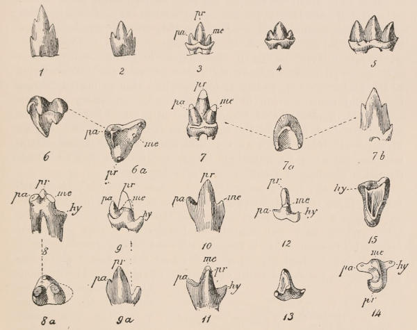
Fig. 4.—Molar teeth of Mesozoic Mammals (enlarged). Triconodont type—1, Dromatherium; 2, Microconodon; 3, Amphilestes; 4, Phascolotherium; 5, Triconodon. Tritubercular type—6, 7, Spalacotherium; 10, Asthenodon. Tubercular sectorial type—8, Amphitherium; 9, Peramus; 11-13, Amblotherium; 14 (?) Amblotherium. pr, Protocone; hy, hypocone; pa, paracone; me, metacone, in the upper teeth; and protoconid, hypoconid, paraconid, and metaconid in the lower. 6 and 15 are upper molars, and the rest lower molars. (After Osborn.)
Trituberculism.—Recent researches, and more especially those of Professors Cope and Osborn, tend to show that almost all of the extremely different forms of tooth-structure found among Mammals may be traced to one common type, in which the crown of each tooth carried three cusps, and hence termed the tritubercular type; these three cusps being arranged in a triangle, with the apex directed inwardly in the upper teeth (Fig. 4, ₆), and outwardly in the lower ones (Fig. 4, ₇). It is further probable that this tritubercular type was itself derived from a type of dentition in which the teeth were in the form of almost a quite simple cone; such a presumably primitive type of dentition—being apparently retained among some existing Edentates, like the Armadillos, while it is possible that we should regard the dentition of the existing Cetacea (Fig. 2) as a reversion to the same primitive type. None of the Mesozoic mammals at present known exhibit this simple conical type of teeth, although we have an approximation to it in the extremely generalised genus Dromatherium. Starting then from this presumed simple cone it appears that the teeth of Dromatherium (Fig. 4, ₁) present the first stage towards trituberculism, the crown of each tooth having one main cone, with minute lateral cusps, and the root being grooved. In the next or true Triconodont stage (Fig. 4, ₃₋₅) the crown has become elongated antero-posteriorly, and consists of one central and two lateral cones or cusps, while the root is divided. From this the transition is easy to the tritubercular type, in which the three cusps, instead of being placed in a line, are arranged in a triangle; the upper teeth (Fig. 4, ₆) having one inner and two outer cusps, while the reverse condition obtains in those of the lower jaw (Fig. 4, ₇). These three cusps of the simple tritubercular tooth are collectively designated as the primitive triangle; in the upper tooth the inner cusp is termed the protocone, the antero-external one the paracone, and the postero-external the metacone; the corresponding cusps of the lower tooth being named protoconid, paraconid, and metaconid—the protoconid being here on the outer side of the crown.
It is thus apparent that in the first, or haplodont type, as well as in the triconodont type, the upper and lower molars are alike; while in the simple tritubercular type they have a similar pattern, but with the arrangement of the cusps reversed. This simple tritubercular type occurs in the Mesozoic genus Spalacotherium (Fig. 4, ₆ and ₇), and apparently in the existing Chrysochloris; but in the majority of tritubercular forms, while this primitive triangle forms the main portion of the crown, other secondary cusps are added, the homologies of which in the upper and lower teeth are somewhat doubtful. At the same time that we have the addition of these secondary cusps we also find trituberculism differentiating into a secodont and a bunodont series, according as to whether the dentition becomes of a cutting or a crushing type.
Thus in the lower molars (Fig. 4, ₈ and ₉) we very frequently find the three cusps of the primitive triangle elevated and connected by cross crests, while there is an additional low posterior heel or talon, which may be termed the hypoconid. This tubercular-sectorial sub-type, as it is termed, is found in the lower molars of many Polyprotodont Marsupials and Insectivores, and it also occurs in the lower carnassial teeth of the true Carnivora. The presence of two cusps (inner and outer) to the talon converts this modification into a quinquetubercular form; while, by the suppression of one of the three primitive cusps, it develops into the quadritubercular type of the bunodont series.
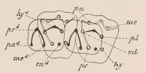
Fig. 5.—Diagram of two upper and two lower left quadritubercular molars in mutual apposition. The cusps and ridges of the upper molars in double lines, and those of the lower in black lines. The lower molars are looked at from below, as if transparent. pr, Protocone; hy, hypocone; pa, paracone; me, metacone; ml, protoconule; pl, metaconule; prd, protoconid; hyd, hypoconid; pad, paraconid; med, metaconid; end, entoconid. (After Osborn.)
In the upper molars the primitive triangle in the secodont series may remain purely tricuspid; but the addition of intermediate cusps, both in the secodont and bunodont series, may give rise to a quinquetubercular type; these intermediate cusps being respectively designated as the protoconule and metaconule (Fig. 5, ml, pl). Finally, in the bunodont series, the addition of a postero-internal cusp (Fig. 5, hy), termed the hypocone, forms the sextubercular molar.
The following table exhibits, in a collective form, the names and relations of all the above-mentioned cusps, and the letters by which they are indicated in the figures:—
Upper Molars.
Antero-internal cusp = protocone = pr.
Postero ” or 6th cusp = hypocone = hy.
Antero-external cusp = paracone = pa.
Postero ” ” = metacone = me.
Anterior intermediate cusp = protoconule = ml.
Posterior ” ” = metaconule = pl.
Lower Molars.
Antero-external cusp = protoconid = prd.
Postero ” ” = hypoconid = hyd.
Antero-internal or 5th cusp = paraconid = pad.
Intermediate (or in quadritubercular molars antero-internal) cusp = metaconid = med.
Postero-internal cusp = entaconid = end.
The common occurrence of trituberculism in the mammals of the earlier geological epochs is, as remarked by Osborn, very significant of the uniformity of molar origin. Thus, among the Mesozoic mammals (with the exception of the group known as Multituberculata, in which the molars are constructed on a different type), trituberculism occurs in the great majority of the genera; while out of 82 species, belonging to five different suborders from the Lowest or Puerco Eocene of the United States, all but four exhibit this feature; and the same holds good for the mammals of the corresponding European horizon. At the present day trituberculism persists in the Lemuroidea, Insectivora, Carnivora, and Marsupialia. In the Carnivora there is a tendency to lose the metaconid, while in the bunodont molars of the Ungulata it is the paraconid that disappears.
III. THE SKELETON.
Definition.—The skeleton is a system of hard parts, forming a framework which supports and protects the softer organs and tissues of the body. It consists of dense fibrous and cartilaginous tissues, portions of which remain through life in this state, but the greater part is transformed during the growth of the animal into bone or osseous tissue. This is characterised by a peculiar histological structure and chemical composition, being formed mainly of a gelatinous basis, strongly impregnated with salts of calcium, chiefly phosphate, and disposed in a definite manner, containing numerous minute nucleated spaces or cavities called lacunæ, connected together by delicate channels or canaliculi, which radiate in all directions from the sides of the lacunæ. Parts composed of bone are, next to the teeth, the most imperishable of all the organs of the body, often retaining their exact form and internal structure for ages after every trace of all other portions of the organisation has completely disappeared, and thus, in the case of extinct animals, affording the only means of attaining a knowledge of their characters and affinities.[8]
In the Armadillos and their extinct allies alone is there an ossified exoskeleton, or bony covering developed in the skin. In all other mammals the skeleton is completely internal. It may be described as consisting of an axial portion belonging to the head and trunk, and an appendicular portion belonging to the limbs. There are also certain bones called splanchnic, being developed within the substance of some of the viscera. Such are the os cordis and os penis found in some mammals.
It is characteristic of all the larger bones of the mammalia that their ossification takes its origin from several distinct centres. One near the middle of the bone, and spreading throughout its greater portion, constitutes the diaphysis, or “shaft,” in the case of the long bones. Others near the extremities, or in projecting parts, form the epiphyses, which remain distinct during growth, but ultimately coalesce with the rest of the bone.
Axial skeleton.—The axial skeleton consists of the skull, the vertebral column (prolonged at the posterior extremity into the tail), the sternum, and the ribs.
Skull.—In the skull of adult mammals, all the bones, except the lower jaw, the auditory ossicles, and the bones of the hyoid arch, are immovably articulated together, their edges being in close contact, and often interlocking by means of fine denticulations projecting from one bone and fitting into corresponding depressions of the other; they are also held together by the investing periosteum, or fibrous membrane, which passes directly from one to the other, and permits no motion, beyond perhaps a slight yielding to external pressure. In old animals there is a great tendency for the different bones to become actually united by the extension of ossification from one to the other, with consequent obliteration of the sutures. The cranium, thus formed of numerous originally independent ossifications, which may retain throughout life more or less of their individuality, or be all fused together, according to the species, the age, or even individual peculiarity, consists of a brain-case, or bony capsule for enclosing and protecting the brain, and a face for the support of the organs of sight, smell, and taste, and of those concerned in seizing and masticating the food. The brain-case articulates directly with the anterior cervical vertebra, by means of a pair of oval eminences, called condyles, placed on each side of the large median foramen which transmits the spinal cord. It consists of a basal axis, continuous serially with the axes or centra of the vertebræ, and of an arch above, roofing over and enclosing the cavity which contains the cephalic portion of the central nervous system (see Fig. 6). The base with its arch is composed of three segments placed one before the other, each of which is comparable to a vertebra with a greatly expanded neural arch. The hinder or occipital segment consists of the basioccipital, exoccipital, and supraoccipital bones; the middle segment of the basisphenoid, alisphenoid, and parietal bones; and the anterior segment of the presphenoid, orbitosphenoid, and frontal bones. The axis is continued forwards into the mesethmoid, or septum of the nose, around which the bones of the face are arranged in a manner so extremely modified for their special purposes that anatomists who have attempted to trace their serial homologies with the more simple portions of the axial skeleton have arrived at very diverse interpretations. The characteristic form and structure of the face of mammals is mainly dependent upon the size and shape of (1) the orbits, a pair of cup-shaped cavities for containing the eyeball and its muscles, which may be directed forwards or laterally, placed near together or wide apart, and may be completely or only partially encircled by bone; (2) the nasal fossæ, or cavities on each side of the median nasal septum, forming the passage for the air to pass between the external and the internal nares, and containing in their upper part the organ of smell; (3) the zygomatic arch, a bridge of bone for the purpose of muscular attachment, which extends from the side of the face to the skull, overarching the temporal fossa; (4) the roof of the mouth, with its alveolar margin for the implantation of the upper teeth. The face is completed by the mandible, or lower jaw, consisting of two lateral rami, articulated by a hinge joint with the squamosal (a cranial bone interposed between the posterior and penultimate segment of the brain-case, where also the bony capsule of the organ of hearing is placed), each being composed of a single solid piece of bone, and the two united together in the middle line in front, at the symphysis,—which union may be permanently ligamentous or become completely ossified. Into the upper border of the mandibular rami the lower teeth are implanted.
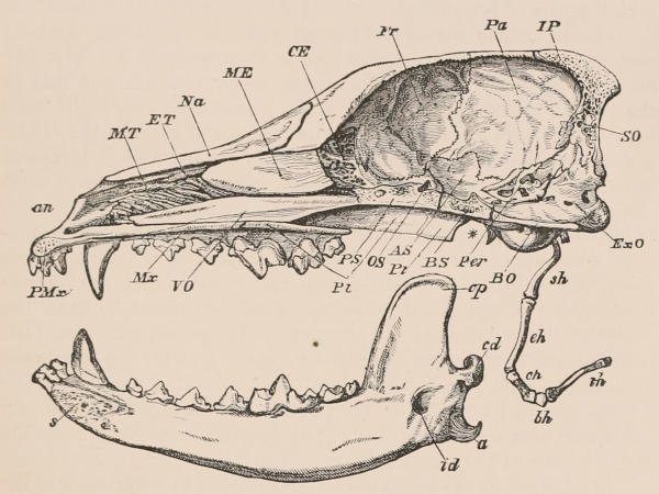
Fig. 6.—Longitudinal and vertical section of the skull of a Dog (Canis familiaris), with mandible and hyoid arch. an, Anterior narial aperture; MT, maxillo-turbinal bone; ET, ethmo-turbinal; Na, nasal; ME, ossified portion of the mesethmoid; CE, cribriform plate of the ethmo-turbinal; Fr, frontal; Pa, parietal; IP, interparietal; SO, supraoccipital; ExO, exoccipital; BO, basioccipital; Per, periotic; BS, basisphenoid; Pt, pterygoid; AS, alisphenoid; OS, orbitosphenoid; PS, presphenoid; PI, palatine; VO, vomer; Mx, maxilla; PMx, premaxilla; sh, stylohyal; eh, epihyal; ch, ceratohyal; bh, basihyal; th, thyrohyal; s, symphysis of mandible; cp, coronoid process; cd, condyle; a, angle; id, inferior dental canal. The mandible is displaced downwards, to show its entire form; the * indicates the part of the cranium to which the condyle is articulated.[9]
In addition to the bones already mentioned as entering into the formation of the cranium, there are many others, the most important of which may be briefly noticed. The anterior extremity of the skull is formed by the premaxillæ (Figs. 6, 7, PMx), which carry the incisors; behind them are the maxillæ, in which all the remaining upper teeth are implanted. Both the premaxillæ and maxillæ meet in a median suture on the palate, where they form a floor to the nasal passage; this floor being continued backwards by the plate-like palatines, at the hinder extremity of which the posterior nares are usually situated. In a few instances, however, as in certain Edentates and Cetaceans, the small pair of bones forming the posterior continuation of the lateral borders of the palatines, and known as the pterygoids (Fig. 6, Pt), likewise meet in the middle line below the nasal passage, and thus cause the aperture of the posterior nares to be situated near the occiput. On the upper, or frontal aspect of the cranium the paired nasals roof over the nasal passage and fill the interval left between the premaxilla and maxilla of either side. Behind the nasals and maxillæ, the anterior part of the brain-case is formed by the large paired frontals (Figs. 6, 7, Fr), behind which are the parietals, which may be of still larger size, and form the greater part of the brain-case. A median interparietal ossification (Fig. 6, IP) may divide the parietals posteriorly, and is itself articulated with the supraoccipital, to the lateral borders of which the parietals are also joined. The squamosal (Fig. 7, Sq) forms the lateral wall of the hinder part of the brain-case, and articulates superiorly with the parietal, and posteriorly with the exoccipital. The glenoid cavity (Fig. 8), for the reception of the articular condyle of the mandible, is formed by the inferior portion of the squamosal, at the point where it gives off the zygomatic process to form the hinder portion of the zygomatic arch. The middle portion of that arch is formed by the jugal, or malar bone (Fig. 7, Ma), which articulates posteriorly with the zygomatic process of the squamosal, and anteriorly with the maxilla. The jugal (as in Fig. 7) may also articulate with a small bone situated on the anterior border of the orbit known as the lachrymal. It is important to observe that the zygomatic or temporal arch is a squamoso-maxillary one, and that an arcade thus composed is found elsewhere only among the extinct Anomodont reptiles, which have already been mentioned as showing signs of mammalian affinity. The relative position occupied by the orbito- and alisphenoid is sufficiently indicated in Fig. 7.
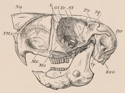
Fig. 7.—Side view of skull of Cape Jumping Hare (Pedetes caffer). × ⅗ PMx, Premaxilla; Mx, maxilla, Ma, jugal or malar; Fr, frontal; L, lachrymal; Pa, parietal; Na, nasal; Sq, squamosal; Ty, tympanic; ExO, exoccipital; AS, alisphenoid; OS, orbitosphenoid; Per, mastoid bulla.
Wedged in between the squamosal and the bones of the occipital and basisphenoidal region are the bones connected with the organ of hearing, known as the periotic and tympanic. The position of the periotic, which encloses the labyrinth or essential organ of hearing, is shown in Fig. 6. The periotic is divided into a very dense antero-internal moiety known as the petrosal, and a postero-external or mastoid portion (Fig. 8), which appears on the outer wall of the brain-case. The tympanic is produced horizontally outwards to form the external auditory meatus or tube of the ear, while the inner and under surface is frequently dilated into a shell-like auditory bulla (Fig. 8). The small bones of the internal ear known as the malleus, incus, and stapes are contained in the membranous tympanic cavity, which is situated in a space left among this group of bones. Further mention of these bones is made below under the head of the sense organs.
In the Carnivora and some other groups the foramina on the base of the skull for the passage of blood-vessels and nerves are of considerable taxonomic importance. The position of the more important of these foramina is indicated in Fig. 8; but for details the reader may refer to the work on the Osteology of the Mammalia already mentioned. Attention may, however, be particularly directed to the so-called alisphenoid canal, the position of which is shown in Fig. 8, since this is a feature of some importance in the classification of the Carnivora. This canal is a short channel running horizontally forward from near the foramen ovale through the alisphenoid, and opening anteriorly with the foramen rotundum; it is traversed by the external carotid artery.
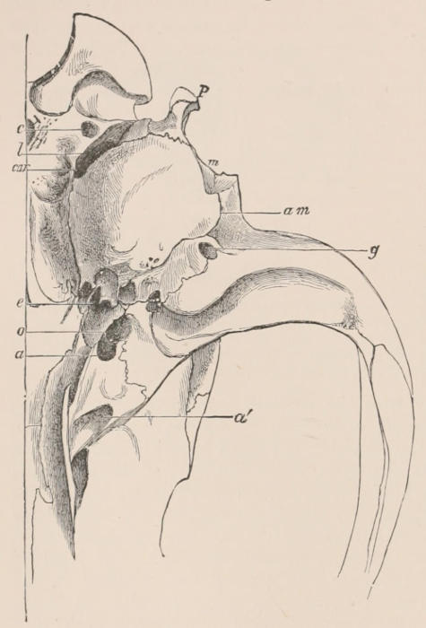
Fig. 8.—The right half of the hinder part of the base of the cranium of the Wolf (Canis lupus). c, Condyloid foramen; l, foramen lacerum posticum; car, carotid canal; e, eustachian canal; o, foramen ovale; a, posterior, and a′, anterior aperture of alisphenoid canal; P, paroccipital process of exoccipital; m, mastoid process of periotic; am, external auditory meatus; g, glenoid foramen, below which is the glenoid cavity for the condyle of the mandible. (Flower, Proc. Zool. Soc., 1869, p. 25.)
Only in those species, as Man and the smaller kinds of the Primates and some other orders, in which the brain holds a large relative proportion to the rest of the body, does the external form of the skull receive much impress from the real shape of the cavity containing the brain. The size and form of the mouth, and the modifications of the jaws for the support of teeth of various shape and number, the ridges and crests on the cranium for the attachment of the muscles necessary to put this apparatus in motion, and outgrowths of bone for the enlargement of the external surface required for the support of sense organs or of weapons, such as horns or antlers (which outgrowths, to prevent undue increase of weight, are filled with cells containing air), cause the principal variations in the general configuration of the skull. These variations are, however, only characteristically developed in perfectly adult animals, and are in many cases more strongly marked in the male than the female sex. Throughout all the later stages of growth up to maturity the size and form of the brain-case remain comparatively stationary, while the accessory parts of the skull rapidly increase and assume their distinctive development characteristic of the species.
The hyoidean apparatus in mammals (Fig. 6) supports the tongue and larynx, and consists of an inferior median portion termed the basihyal, from which two pairs of half arches, or cornua, extend upwards and outwards. The anterior is the more important, being connected with the periotic bone of the cranium. It may be almost entirely ligamentous, but more often has several ossifications, the largest of which is usually the stylohyal. The posterior cornu (thyrohyal) is united at its extremity with the thyroid cartilage of the larynx, which it suspends in position. The median portion, or basihyal, is sometimes, as in the Howling Monkeys, enormously enlarged and hollowed, admitting into its cavity an air-sac connected with the organ of voice.
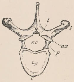
Fig. 9.—Anterior surface of Human thoracic vertebra (fourth). c, Body or centrum; nc, neural canal; p, pedicle, and l, lamina of the arch; t, transverse process; az, anterior zygapophysis.
Vertebral Column.—The vertebral column consists of a series of distinct bones called vertebræ, arranged in close connection with each other along the dorsal side of the neck and trunk, and in the median line.[10] It is generally prolonged posteriorly beyond the trunk, to form the axial support of the appendage called the tail. Anteriorly it is articulated with the occipital region of the skull. The number of distinct bones composing the vertebral column varies greatly among the Mammalia, the main variation being due to the degree of elongation of the tail. Apart from this, in most mammals the number is not far from thirty, though it may fall as low as twenty-six (as in some Bats), or rise as high as forty (Hyrax and Cholœpus). The different vertebræ, with some exceptions, remain through life quite distinct from each other, though closely connected by means of fibrous structures which allow of a certain, but limited, amount of motion between them. The exceptions are the following:—(1) near the posterior part of the trunk, in nearly all mammals which possess completely developed hinder limbs, two or more vertebræ become ankylosed together to form the “sacrum,” or portion of the vertebral column to which the pelvic girdle is attached; (2) in some species of Whales and Armadillos there are constant ossific unions of certain vertebræ of the cervical region.
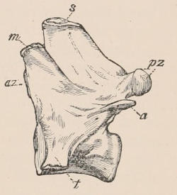
Fig. 10.—Side view of the first lumbar vertebra of a Dog (Canis familiaris). s, Spinous process; az, anterior zygapophysis; pz, posterior zygapophysis; m, metapophysis; a, anapophysis; t, transverse process.
Although the vertebræ of different regions of the column of the same animal or of different animals present great diversities of form, yet there is a certain general resemblance among them, or a common plan on which they are constructed, which is more or less modified by alteration of form or proportions, or by the addition or suppression of parts to fit them to fulfil their special purpose in the economy. An ordinary or typical vertebra consists, in the first place, of a solid piece of bone, termed the body or centrum (Fig. 9, c), of the form of a disk or short cylinder. The bodies of contiguous vertebræ are connected together by a very dense, tough, and elastic material called the “intervertebral substance,” of peculiar and complex arrangement. This substance forms the main, and in some cases the only, union between the vertebræ. Its elasticity provides for the vertebræ always returning to their normal relation to each other and to the column generally, when they have been disturbed therefrom by muscular action. A process (p) arises on each side from the dorsal surface of the body. These processes, meeting in the middle line above, form an arch, surmounting a space or short canal (nc). Since it contains the posterior prolongation of the great cerebro-spinal nervous axis, or spinal cord, this space is called the neural canal, and the arch the neural arch, in contradistinction to another arch on the ventral surface of the body of the vertebræ, called the hæmal arch. The latter is, however, never formed in mammals by any part of the vertebra itself, but by certain distinct bones placed more or less in apposition to it, namely the ribs in the thoracic, and the “chevron bones” in the caudal region. In most cases the arch of one vertebra is articulated with that of the next by distinct surfaces with synovial joints, placed one on each side, called “zygapophyses” (az, pz), but these are often entirely wanting when flexibility is more needed than strength, as in the greater part of the caudal region of long-tailed animals. In addition to the body and the arch, there are certain projecting parts called processes, chiefly serving for the attachment of the numerous muscles which move the vertebral column. Of these two are single and median, viz. the spinous process, neural spine, or neurapophysis (s), arising from the middle of the upper part of the arch, and the hypapophysis from the under surface of the body. The latter, however, is as frequently absent as the former is constant. The other processes are paired and lateral. They are the transverse processes (t), of which there may be two, an upper and a lower, in which case the former is called, in the language of Owen (to whom we are indebted for the terminology of the parts of vertebræ in common use), “diapophysis,” and the latter “parapophysis.” Other processes less constantly present are called respectively “metapophyses” (m) and “anapophyses” (a).
The vertebral column is divided for convenience of description into five regions—the cervical, thoracic or dorsal, lumbar, sacral, and caudal. This division is useful, especially as it is not entirely arbitrary, and in most cases is capable of ready definition; but at the contiguous extremities of the regions the characters of the vertebræ of one are apt to blend into those of the next region, either normally or as peculiarities of individual skeletons.
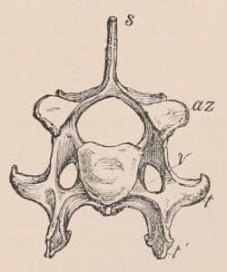
Fig. 11.—Anterior surface of sixth cervical vertebra of Dog. s, Spinous process; az, anterior zygapophysis; v, vertebrarterial canal; t, transverse process; t′, its inferior lamella.
Cervical Vertebræ.—The cervical region constitutes the most anterior portion of the column, or that which joins the cranium. The vertebræ which belong to it are either entirely destitute of movable ribs, or if they have any these are small, and do not join the sternum. As a general rule they have a considerable perforation through the base of the transverse process (the vertebrarterial canal, Fig. 11, v); or, as it is sometimes described, they have two transverse processes, superior and inferior, which meet at their extremities to enclose a canal. This, however, rarely applies to the last vertebra of the region, in which only the upper transverse process is usually developed. The transverse process, moreover, very often sends down near its extremity a more or less compressed plate (inferior lamella), which, being considered serially homologous with the ribs of the thoracic vertebræ (though not developed autogenously), is often called the “costal” or “pleurapophysial” plate. This is usually largest on the sixth, and altogether wanting on the seventh vertebra. The first and second cervical vertebræ, called respectively “atlas” and “axis,” are specially modified for the function of supporting and permitting the free movements of the head. They are not united together by the intervertebral substance, but connected only by ordinary ligaments and synovial joints.
The cervical region in mammals presents the remarkable peculiarity that, whatever the length or flexibility of the neck, the number of vertebræ is the same, viz. seven, with the exception of the Manatee and Hoffman’s Two-toed Sloth (Cholœpus hoffmanni), which both have but six, and the Three-toed Sloth (Bradypus tridactylus), which has nine, though in this case the last two usually support movable ribs, which are not sufficiently developed to reach the sternum.
According to Parker there may occasionally be eight cervicals in the Pangolins (Manis).
Dorsal Vertebræ.—The dorsal (or, as it would be more correctly termed, thoracic) region consists of the vertebræ succeeding those of the neck, which have ribs movably articulated to them. These ribs arch round the thorax—the anterior one, and usually the greater number of those that follow, being attached below to the sternum.
Lumbar Vertebræ.—The lumbar region consists of those vertebræ of the trunk in front of the sacrum which bear no movable ribs. It may happen that, as the ribs decrease in size posteriorly (the last being sometimes more or less rudimentary), the step from the thoracic to the lumbar region may be gradual and rather undetermined in a given species; but most commonly this is not the case, and the distinction is as well defined here as in any other region. As a general rule there is a certain relation between the number of the thoracic and lumbar vertebræ, the whole number being tolerably constant in a given group of animals, and any increase of the one being at the expense of the other. Thus in all known Artiodactyle Ungulata there are 19 dorso-lumbar vertebræ; but these may consist of 12 dorsal and 7 lumbar vertebræ, or 13 dorsal and 6 lumbar, or 14 dorsal and 5 lumbar. The smallest number of dorso-lumbar vertebræ in mammals occurs in some Armadillos, which have but 14. The number found in Man, the higher Apes, and most Bats, viz. 17, is exceptionally low; 19 prevails in the Artiodactyla, nearly all Marsupials, and very many Rodents; 20 or 21 in Carnivora and most Insectivora; and 23 in Perissodactyla. The highest and quite exceptional numbers are in the Two-toed Sloth (Cholœpus) 27, and the Hyrax 30. The prevailing number of rib-bearing vertebræ is 12 or 13, any variation being generally in excess of these numbers.
Sacral Vertebræ.—The sacral region offers more difficulties of definition. Taking the human “os sacrum” as a guide for comparison, it is generally defined as consisting of those vertebræ between the lumbar and caudal regions which are ankylosed together to form a single bone. It happens, however, that the number of such vertebræ varies in different individuals of the same or nearly allied species, especially as age advances, when a certain number of the tail vertebræ generally become incorporated with the true sacrum. Other suggested tests—as those vertebræ which have a distinct additional (pleurapophysial) centre of ossification between the body and the ilium, those to which the ilium is directly articulated, or those in front of the insertion of the ischiosacral ligaments—being equally unsatisfactory or unpractical, the old one of ankylosis, as it is found to prevail in the average condition of adults in each species, is used in the enumeration of the vertebræ in the following pages. The Cetacea, having no iliac bones, have no part of the vertebral column modified into a sacrum.
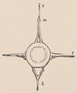
Fig. 12.—Anterior surface of fourth caudal vertebræ of Porpoise (Phocæna communis). s, Spinous process; m, metapophysis; t, transverse process; h, chevron bone.
Caudal Vertebræ.—The caudal vertebræ are those placed behind the sacrum, and terminating the vertebral column. They vary in number greatly—being reduced to 5, 4, or even 3, in a most rudimentary condition, in Man and in some Apes and Bats, and being numerous and powerfully developed, with strong and complex processes, in many mammals, especially among the Edentata, Cetacea, and Marsupialia. The highest known number, 46, is possessed by the African Long-tailed Pangolin. Connected with the under surface of the caudal vertebræ of many mammals which have the tail well developed are certain bones formed more or less like an inverted arch, called chevron bones, or by the French os en V. These are always situated nearly opposite to an intervertebral space, and are generally articulated both to the vertebra in front and the vertebra behind, but sometimes chiefly or entirely either to one or the other.
In some of the Anomodont Reptiles and Labyrinthodont Amphibians these chevrons are attached to the intercentra—or imperfect disks alternating with the true centra—which suggests that they are primarily intercentral elements which have been transferred to the edges of the centra by the disappearance of the intercentra.
Sternum.—The sternum of mammals is a bone, or generally a series of bones, placed longitudinally in the mesial line, on the inferior or ventral aspect of the thorax, and connected on each side with the vertebral column by a series of more or less ossified bars called “ribs.” It is present in all mammals, but varies much in character in the different groups. It usually consists of a series of distinct segments placed one before the other, the anterior being called the presternum or “manubrium sterni” of human anatomy, and the posterior the xiphisternum, or xiphoid or ensiform process, while the intermediate segments, whatever their number, constitute the mesosternum or “body.” In the Whalebone Whales the presternum alone is developed, and but a single pair of ribs is attached to it.