Chapter II · GENERAL ANATOMICAL CHARACTERS (continued 2)
Reading edition
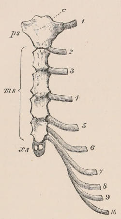
Fig. 13.—Human sternum and sternal ribs. ps, Presternum; ms, mesosternum; xs, xiphisternum; c, point of attachment of clavicle; 1 to 10, the cartilaginous sternal ribs.
Ribs.—The ribs form a series of long, narrow, and more or less flattened bones, extending laterally from the sides of the vertebral column, curving downwards towards the median line of the body below, and mostly joining the sides of the sternum. The posterior ribs, however, do not directly articulate with that bone, but are either attached by their extremities to the edges of each rib in front of them, and thus only indirectly join the sternum, or else they are quite free below, meeting no part of the skeleton. These differences have given rise to the division into “true” and “false” ribs (by no means good expressions), signifying those that join the sternum directly and those that do not; and of the latter, those that are free below, are called “floating” ribs. The portion of each rib nearest the vertebral column and that nearest the sternum differ in their characters, the latter being usually but imperfectly ossified, or remaining permanently cartilaginous. These are called “costal cartilages,” or when ossified “sternal ribs.”
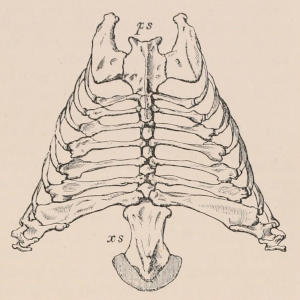
Fig. 14.—Sternum and strongly ossified sternal ribs of Great Armadillo (Priodon gigas). ps, Presternum; xs, xiphisternum.
In the anterior part of the thorax the vertebral extremity of each rib is divided into two parts, “head” or “capitulum,” and “tubercle”; the former is attached to the side of the body of the vertebra, the latter to its transverse process; the former attachment corresponds to the interspace between the vertebræ, the head of the rib commonly articulating partly with the hinder edge of the body of the vertebra antecedent to that which bears its tubercle. Hence the body of the last cervical vertebra usually supports part of the head of the first rib. In the posterior part of the series the capitular and tubercular attachments commonly coalesce, and the rib is attached solely to its corresponding vertebra. The number of pairs of ribs is of course the same as that of the thoracic vertebræ.
The circumstance that in some of the Anomodont reptiles and Labyrinthodonts the capitula of the ribs articulate with the intercentral elements of the vertebral column has suggested, as in the instance of the chevron bones, that the intercentral capitular articulation of the ribs of mammals is a feature directly inherited from those extinct types by the gradual disappearance of the intercentra.
Appendicular Skeleton.—The appendicular portion of the framework consists, when completely developed, of two pairs of limbs, anterior and posterior (Fig. 15).
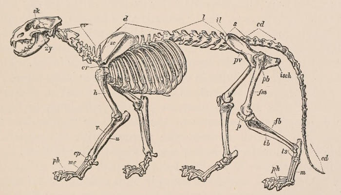
Fig. 15.—Skeleton of Lion (Felis leo). cd, Caudal vertebræ; cp, carpus; cr, coracoid process of scapula, cv, cervical vertebræ; d, dorsal vertebræ; fb, fibula; fm, femur; h, humerus; il, ilium; isch, ischium; l, lumbar vertebræ; m, metatarsus; mc, metacarpus; p, patella; pb, pubis; ph, phalanges; pv, pelvis; r, radius; s, sacral vertebræ; sc, scapula; sk, skull; tb, tibia; ts, tarsus; u, ulna; zy, zygomatic arch.
Anterior Limb.—The anterior limb is present and fully developed in all mammals, being composed of a shoulder girdle and three segments belonging to the limb proper; viz. the upper arm or brachium, the forearm or antebrachium, and the hand or manus.
Shoulder-girdle.—The shoulder or pectoral girdle in the large majority of mammals is in a rudimentary or rather modified condition, compared with that in which it exists in other vertebrates. In the Monotremata (Ornithorhynchus and Echidna) alone is the ventral portion, or coracoid, complete and articulated with the sternum below, as in the Sauropsida; and in this group alone do we find an anterior ventral element, apparently corresponding with the pre-coracoid of the Anomodont reptiles, although generally known as the epi-coracoid. In all other mammals the coracoid, though ossified from a distinct centre, forms only a process, sometimes a scarcely distinct tubercle, projecting from the anterior border of the glenoid cavity of the scapula. The last-named cavity, which in the Monotremes is formed jointly by the scapula and coracoid, receives the head of the humerus, or arm-bone. The scapula is always well developed, and generally broad and flat (whence its vernacular name “blade bone”), with a ridge called the “spine” on its outer surface, which usually ends in a free curved process, the “acromion.” As the scapula affords attachment to many of the muscles which act upon the anterior limb, its form and the development of its processes are greatly modified according to the uses to which the member is put. Thus it is most reduced and simple in character in those animals whose limbs are mere organs of support, as the Ungulates; and most complex when the limbs are also used for grasping, climbing, or digging. The development or absence of the clavicle or “collar-bone,” an accessory bar which connects the sternum with the scapula and steadies the shoulder-joint, has a somewhat similar relation, though its complete absence in the Bears shows that this is not an invariable rule. A complete clavicle is found in Man and all the Primates, in Chiroptera, all Insectivora (except Potamogale), in many Rodents, in most Edentates, and in all Marsupials, except Perameles. More or less rudimentary clavicles (generally suspended freely in the muscles) are found in the Cat, Dog, and most Carnivora, Myrmecophaga, and some Rodents. Clavicles are altogether absent in most of the Ursidæ, all the Pinnipedia, Manis among Edentates, the Cetacea, Sirenia, Ungulates, and some Rodents.
The Monotremes are peculiar in possessing a T-shaped interclavicle like that of many reptiles, lying upon the sternum, and articulating superiorly with the clavicles.
Brachium and Antebrachium.—The proximal segment of the anterior or pectoral limb proper contains a single bone, the humerus, and the second segment two bones, the radius and the ulna, placed side by side, and articulating with the humerus at their proximal, and with the carpus at their distal extremity (Fig. 15). In their primitive and unmodified condition these bones may be considered as placed one on each border of the limb, the radius being preaxial or anterior, and the ulna postaxial or posterior, when the distal or free end of the limb is directed outwards, or away from the trunk. This is their position in the earliest embryonic condition, and is best illustrated among adult mammals in the Cetacea, where the two bones are fixed side by side and parallel to each other. In the greater number of mammals the bones assume a very modified and adaptive position, usually crossing each other in the forearm, the radius in front of the ulna, so that the preaxial bone (radius), though external (in the ordinary position of the limb) at the upper end, is internal at the lower end; and the hand, being mainly fixed to the radius, also has its preaxial border internal. In the large majority of mammals the bones are fixed in this position, but in some few, as in Man, a free movement of crossing and uncrossing—or pronation and supination, as it is termed—is allowed between them, so that they can be placed in their primitive parallel condition, when the hand (which moves with the radius) is said to be supine, or they may be crossed, when the hand is said to be prone.
The humerus frequently has a foramen piercing the inner border of the distal extremity, known as the entepicondylar foramen, which corresponds with a similar one found in the Anomodont reptiles. The hollow in the head of the ulna for the reception of the head of the humerus is known as the greater sigmoid cavity, and that for the head of the radius as the lesser sigmoid cavity (Fig. 16). The term olecranon is applied to that process of the ulna which forms the prominence of the elbow.
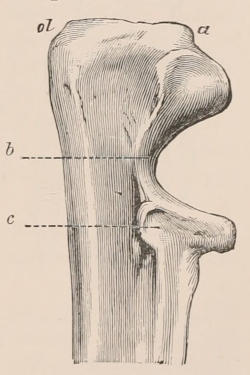
Fig. 16.—Outer aspect of the proximal extremity of the right ulna of a Bear (Ursus). a, Anterior tubercle; ol, olecranon; b, greater sigmoid cavity; c, lesser do.
In most mammals walking on four limbs, in which the hand is permanently prone, the ulna is much reduced in size, and the radius increased, especially at the upper end; so that the articular surface of the latter, instead of being confined to the external side of the trochlea of the humerus, extends all across its anterior surface, and the two bones, instead of being external and internal, are anterior and posterior. In many hoofed or Ungulate mammals, and in Bats, the ulna is reduced to little more than its upper articular extremity, and firmly ankylosed to the radius—stability of these parts being more essential than mobility.
Manus.—The terminal segment of the anterior limb is the hand or manus. Its skeleton consists of three divisions: (1) the “carpus,” a group of small, more or less rounded or angular bones with flattened surfaces applied to one another, and, though articulating by synovial joints, having scarcely any motion between them; (2) the “metacarpus,” a series of elongated bones placed side by side, with their proximal ends articulating by almost immovable joints with the carpus; (3) the “phalanges” or bones of the digits, usually three in number to each, articulating to one another by freely movable hinge-joints, the first being connected in like manner to the distal end of the metacarpal bone to which it corresponds.
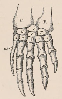
Fig. 17.—Dorsal surface of the right manus of a Water Tortoise (Chelydra serpentina). After Gegenbaur. U, Ulna; R, radius; u, ulnare; i, intermedium; r, radiale; c, centrale; 1-5, the five bones of the distal row of the carpus; m¹-m⁵, the five metacarpals.
Carpus.—To understand thoroughly the arrangement of the bones of the carpus in mammals, it is necessary to study their condition in some of the lower vertebrates. Fig. 17 represents the manus in one of its fullest and at the same time most generalised forms, as seen in one of the Water Tortoises (Chelydra serpentina). The carpus consists of two principal rows of bones. The upper or proximal row contains three bones, to which Gegenbaur has applied the terms radiale (r), intermedium (i), and ulnare (u), the first being on the radial or preaxial side of the limb.[11] The lower or distal row contains five bones, called carpale 1, 2, 3, 4, and 5 respectively, commencing on the radial side. Between these two rows, in the middle of the carpus, is a single bone, the centrale (c). In this very symmetrical carpus it will be observed that the radiale supports on its distal side two bones, carpale 1 and 2; the intermedium is in a line with the centrale and carpale 3, which together form a median axis of the hand, while the ulnare has also two bones articulating with its distal end, viz. carpale 4 and 5. Each of the carpals of the distal row supports a metacarpal.
In the carpus of the Mammalia there are usually two additional bones developed in the tendons of the flexor muscles, one on each side of the carpus, which may be called the radial and ulnar sesamoid bones; the latter, which is the more constant and generally larger, is commonly known as the pisiform bone. The fourth and fifth carpals of the distal row are always united into a single bone, and the centrale is very often absent. As a general rule all the other bones are present and distinct, though it not unfrequently happens that two may have coalesced to form a single bone, or one or more may be altogether suppressed.
The following table shows the principal names in use for the various carpal bones,—those in the second column being the terms generally employed by English anatomists:—
Radiale = Scaphoid = Naviculare.
Intermedium = Lunar = Semilunare, Lunatum.
Ulnare = Cuneiform = Triquetrum, Pyramidale.
Centrale = Central = Intermedium (Cuvier).
Carpale 1 = Trapezium = Multangulum majus.
Carpale 2 = Trapezoid = Multangulum minus.
Carpale 3 = Magnum = Capitatum.
Carpale 4 } = Uneiform = Hamatum, Uncinatum.
Carpale 5 }
The radial and ulnar sesamoids are regarded by Bardeleben[12] as the rudiments of a prepollex and a postminimus digit; the primitive number of digits being thus supposed to have been seven. These bones have been observed in all orders of mammals having five complete digits. Occasionally, as in Pedetes caffer, the so-called prepollex consists of two bones, of which the distal one bears a distinct nail-like horny covering. In Bathyergus maritimus the pisiform, or postminimus, is likewise double; the two elements being regarded by their describer as representing the carpal and metacarpal of the presumed seventh digit.
Similarly in the posterior limb the tibial sesamoid, and a fibular ossification corresponding to the pisiform, are regarded as representing a prehallux and a postminimus.
Metacarpus and Phalanges.—The metacarpal bones, with the digits which they support, are never more than five in number, and are described numerically—first, second, etc., counting from the radial towards the ulnar side. The digits are also sometimes named (1) the pollex, (2) index, (3) medius, (4) annularis, (5) minimus. One or more may be in a rudimentary condition, or altogether suppressed. If one is absent, it is most commonly the first. Excepting the Cetacea, no mammals have more than three phalanges to each digit, but they may occasionally have fewer by suppression or ankylosis. The first or radial digit is an exception to the usual rule, one of its parts being constantly absent, since, while each of the other digits has commonly a metacarpal and three phalanges, it has only three bones altogether; whether the missing one is a metacarpal or one of the phalanges is a subject which has occasioned much discussion, and has not yet been satisfactorily decided. The terminal phalanges of the digits are usually specially modified to support the nail, claw, or hoof, and are called “ungual phalanges.” In walking, some mammals (as the Bears) apply the whole of the lower surface of the carpus, metacarpus, and phalanges to the ground; to these the term “plantigrade” is applied. Many others (as nearly all the existing Ungulata) only rest on the last one or two phalanges of the toes, the first phalanx and the metacarpals being vertical and in a line with the forearm. These are called “digitigrade.” Intermediate conditions exist between these two forms, to which the terms “phalangigrade” (as the Camel) and “subplantigrade” (as in most Carnivora), are applied. When the weight is borne entirely on the distal surface of the ungual phalanx, and the horny structures growing around it, as in the Horse, the mode of progression is called “unguligrade.”
In the Chiroptera the digits are enormously elongated, and support a cutaneous expansion constituting the organ of flight. In the Cetacea the manus is formed into a paddle, being covered by continuous integument, which conceals all trace of division into separate digits, and shows no sign of nails or claws. In the Sloths the manus is long and very narrow, habitually curved, and terminating in two or three pointed curved claws in close apposition with each other, and incapable, in fact, of being divaricated; so that it is reduced to the condition of a hook, by which the animal suspends itself to the boughs of the trees among which it lives. These are only examples of the endless modifications to which the distal extremity of the limb is subjected in adaptation to the various purposes to which it is applied.
Posterior Limb.—The posterior limb is constructed upon a plan very similar to that of the anterior extremity. It consists of a pelvic girdle and three segments belonging to the limb proper, viz. the thigh, the leg, and the foot or pes (Fig. 15).
Pelvic Girdle.—The pelvic girdle is present in some form in all mammals, though in the Cetacea and the Sirenia it is in an exceedingly rudimentary condition. In all mammals except those belonging to the two orders just named, each lateral half of the pelvic girdle consists essentially, like the corresponding part of the anterior limb, of a flattened rod of bone crossing the long axis of the trunk, having an upper or dorsal and a lower or ventral end. The upper end diverges from that of the opposite side, but the lower end approaches, and, in most cases, meets it, forming a symphysis, without the intervention of any bone corresponding to the sternum. The pelvic girdle differs from the shoulder girdle in being firmly articulated to the vertebral column, thus giving greater power to the hinder limb in its function of supporting and propelling the body. Like the shoulder girdle, it bears on its outer side, near the middle, a cup-shaped articular cavity (“acetabulum”), into which the proximal end of the first bone of the limb proper is received. Each lateral half of the girdle is called the “os innominatum,” or innominate bone, and consists originally of three bones which unite at the acetabulum. The “ilium” or upper bone is that which articulates with the sacral vertebræ. Of the two lower bones the anterior or “pubis” unites with its fellow of the other side at the symphysis; the posterior is the “ischium.” These lower elements form two bars of bone, united above and below, but leaving a space between them in the middle, filled only by membrane, and called the “thyroid” or “obturator” foramen. The whole circle of bone formed by the two innominate bones and the sacrum is called the pelvis. In the Monotremata and Marsupialia, a pair of thin, flat, elongated ossifications called epipubic or marsupial bones are attached to the fore part of the pubis, and project forward into the muscular wall of the abdomen.
Thigh and Leg.—The first segment of the limb proper has one bone, the femur, corresponding with the humerus of the anterior limb. The second segment has two bones, the tibia and fibula, corresponding with the radius and ulna. These bones always lie in their primitive unmodified position, parallel to each other, the tibia on the preaxial and the fibula on the postaxial side, and are never either permanently crossed or capable of any considerable amount of rotation, as in the corresponding bones of the fore limb. In the ordinary walking position the tibia is internal, and the fibula external. In many mammals the fibula is in a more or less rudimentary condition, and it often ankyloses with the tibia at one or both extremities. The patella or “knee-cap,” which is found in an ossified condition in all mammals, with the exception of some of the Marsupialia, is a large sesamoid bone developed in the tendon of the extensor muscles of the thigh, where the tendon passes over the front of the knee-joint, to which it serves as a protection. There are frequently smaller ossicles, one or two in number, situated behind the femoral condyles, called “fabellæ.” The processes for the attachment of muscles near the upper end of the femur are termed trochanters; and the third trochanter, found on the hinder aspect of the shaft of this bone in many forms is of considerable taxonomic importance.
Pes.—The terminal segment of the hind limb is the foot or pes. Its skeleton presents in many particulars a close resemblance to that of the manus, being divisible into three parts: (1) a group of short, more or less rounded or square bones, constituting the tarsus; (2) a series of long bones placed side by side, forming the metatarsus; and (3) the phalanges of the digits or toes.
The bones of the tarsus of many of the lower Vertebrata closely resemble both in number and arrangement those of the carpus, as shown in Fig. 17. They have been described in their most generalised condition by Gegenbaur under the names expressed in the first column of the following table. The names in the second column are those by which they are generally known to English anatomists, while in the third column some synonyms occasionally employed are added.
Tibiale (?) } = Astragalus[13] = Talus.
Intermedium }
Fibulare = Calcaneum = Os calcis.
Centrale = Navicular = Scaphoideum.
Tarsale 1 = Internal cuneiform = Entocuneiforme.
Tarsale 2 = Middle cuneiform = Mesacuneiforme.
Tarsale 3 = External cuneiform = Ectocuneiforme.
Tarsale 4 } = Cuboid.
Tarsale 5 }
The bones of the tarsus of mammals present fewer diversities of number and arrangement than those of the carpus. The proximal row (see Fig. 18) always consists of two bones, namely the astragalus (a), which probably represents the coalesced scaphoid and lunar of the hand, and the calcaneum (c). The former is placed more to the dorsal side of the foot than the latter, and almost exclusively furnishes the tarsal part of the tibio-tarsal or ankle-joint. The calcaneum, placed more to the ventral or “plantar” side of the foot, is elongated backwards to form a more or less prominent tuberosity, the “tuber calcis,” to which the tendon of the great extensor muscles of the foot is attached. The navicular bone (n) is interposed between the proximal and distal row on the inner or tibial side of the foot, but on the outer side the bones of the two rows come into contact. The distal row, when complete, consists of four bones, which, beginning on the inner side, are the three cuneiform bones, internal (c¹), middle (c²), and external (c³), articulated to the distal surface of the navicular, and the cuboid (cb), articulated with the calcaneum. Of these the middle cuneiform is usually the smallest in animals in which all five digits are developed; but when the hallux is wanting the internal cuneiform may be rudimentary or altogether absent. The three cuneiform bones support respectively the first, second, and third metatarsals, and the cuboid supports the fourth and fifth; they thus exactly correspond with the four bones of the distal row of the carpus.
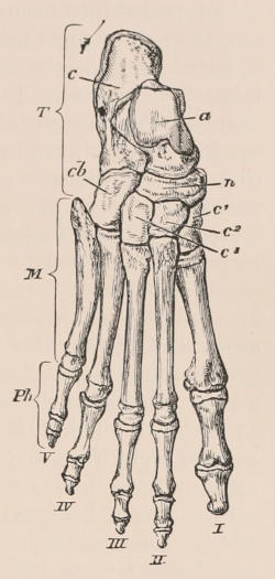
Fig. 18.—Bones of the right Human foot. T, Tarsus; M, metatarsus; Ph, phalanges, c, calcaneum; a, astragalus; cb, cuboid; n, navicular; c¹, internal cuneiform; c², middle cuneiform; c³, external cuneiform. The digits are indicated by Roman numerals, counting from the tibial to the fibular side.
In addition to these constant tarsal bones, there may be supplemental or sesamoid bones: one situated near the middle of the tibial side of the tarsus, largely developed in many Carnivora and Rodentia; another, less frequent, on the fibular side; and a third, often developed in the tendons of the plantar surface of the tarsus, is especially large in Armadillos. There is also usually a pair of sesamoid bones on the plantar aspect of each metatarso-phalangeal articulation. In the young of the carnivorous genus Crytoprocta there may be a second centrale, which usually coalesces with the ectocuneiform.
The metatarsal bones never exceed five in number, and the phalanges follow the same numerical rule as in the manus, never exceeding three in each digit. Moreover, the first digit, counting from the tibial side, or hallux, resembles the pollex of the hand in always having one segment less than the other digits. As the function of the hind foot is more restricted than that of the hand the modifications of its structure are less striking. In the Cetacea and the Sirenia it is entirely wanting, though in some existing members of the first-named order rudiments of the bones of both the first and second segments of the limb have been detected, and a femur is present in the Miocene Sirenian Halitherium.
IV. THE DIGESTIVE SYSTEM.
General Considerations.—The search after the purpose which every modification of structure subserves in the economy is always full of interest, and, if conducted with due caution and sufficient knowledge of all the attendant circumstances, may lead to important generalisations. It must always be borne in mind, however, that adaptation to its special function is not the only cause of the particular form or structure of an organ, but that this form, having in all probability been arrived at by the successive and gradual modification of some other different form from which it is now to a greater or less degree removed, has other factors besides use to be taken into account. In no case is this principle so well seen as in that of the organs of digestion. These may be considered as machines which have to operate upon alimentary substances in very different conditions of mechanical and chemical combination, and to reduce them in every case to the same or precisely similar materials; and we might well imagine that the apparatus required to produce flesh and blood out of coarse fibrous vegetable substances would be different from that which had to produce exactly the same results out of ready-made flesh or blood; and in a very broad sense we find that this is so. Thus, if we take a large number of carnivorous animals, belonging to different fundamental types, and a large number of herbivorous animals, and strike a kind of average of each, we shall find that there is, pervading the first group, a general style, if we may use the expression, of the alimentary organs, different from that of the others. That is to say, there is a specially carnivorous and a specially herbivorous modification of these parts. But, if function were the only element which has guided such modification, it might be inferred that, as one form must be supposed to be best adapted in its relation to a particular kind of diet, that form would be found in all the animals consuming such diet. But this is far from being the case. Thus the Horse and the Ox, for instance—two animals whose food in the natural state is precisely similar—are most different as regards the structure of their alimentary canal, and the processes involved in the preparation of that food. Again, the Seal and the Porpoise, both purely fish-eaters, which seize, swallow, and digest precisely the same kind of prey, in precisely the same manner, have a totally different arrangement of the alimentary canal. If the Seal’s stomach is adapted in the best conceivable manner for the purpose it has to fulfil, why is not the Porpoise’s stomach an exact facsimile of it, and vice versâ? We can only answer that the Seal and Porpoise belong to different natural groups of animals, formed either on different primitive types, or descended from differently constructed ancestors. On this principle only can we account for the fact that, whereas, owing to the comparatively small variety of the different alimentary substances met with in nature, few modifications would appear necessary in the organs of digestion, there is really endless variety in the parts devoted to this purpose.
Mouth.—The digestive apparatus of mammals, as in other vertebrates, consists mainly of a tube with an aperture placed at or near either extremity of the body,—the oral and the anal orifice,—and furnished with muscular walls, the fibres of which are so arranged as by their regular alternate contraction and relaxation to drive onwards the contents of the tube from the first to the second of these apertures. The anterior or commencing portion of this tube and the parts around it are greatly and variously modified in relation to the functions assigned to them of selecting and seizing the food, and preparing it by various mechanical and chemical processes for the true digestion which it has afterwards to undergo before it can be assimilated into the system. For this end the tube is dilated into a chamber or cavity called the mouth, bordered externally by the lips, which are usually muscular and prehensile, and supported by a movable framework carrying the teeth; the structure and modifications of which have been already described. The roof of the mouth is formed by the palate, terminating behind by a muscular, contractile arch, having in Man and some few other species a median projection called the uvula, beneath which the mouth communicates with the pharynx. The anterior part of the palate is composed of mucous membrane tightly stretched over the flat or slightly concave bony lamina separating the mouth from the nasal passages, and is generally raised into a series of transverse ridges, which sometimes, as in Ruminants, attain a considerable development. In the floor of the mouth, between the rami of the mandible, and supported behind by the hyoidean apparatus, lies the tongue; an organ the free surface of which, especially in its posterior part, is devoted to the sense of taste, but which also, by its great mobility (being composed almost entirely of muscular fibres), performs important mechanical functions connected with masticating and procuring food. Its modifications of form in different mammals are very numerous. Between the long, extensile, vermiform tongue of the Anteaters, which is essential to the peculiar mode of feeding of those animals, and the short, sessile, and almost functionless tongue of the Porpoise, every intermediate condition is found. Whatever the form, the upper surface is always covered with numerous fine papillæ, in which the terminal filaments of the gustatory nerve are distributed.
Salivary Glands.—The fluid known as the saliva is secreted by an extensive and complex system of glands discharging into the cavity of the mouth (buccal cavity), the position and relation of some of which are exhibited in the woodcut on the next page (Fig. 19).
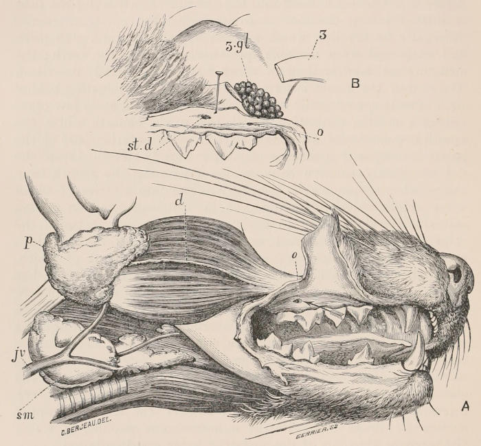
Fig. 19.—Salivary Glands of the Genet. A, Right side of the head dissected; p, parotid gland; d, Steno’s duct; sm, submaxillary gland, traversed by the jugular veins (jv); o, aperture of Steno’s duct. B, Part of the head with the lip drawn up to show (st.d) aperture of Steno’s duct; z.gl, zygomatic gland; o, aperture of do.; z, zygomatic arch (Mivart, Proc. Zool. Soc. 1882, p. 504.)
This apparatus consists of small glands embedded in the mucous membrane or submucous tissue lining the cavity of the mouth, which are of two kinds (the follicular and the racemose), and of others in which the secreting structure is aggregated in distinct masses removed some distance from the cavity; other tissues besides the lining membrane being usually interposed, and pouring their secretion into the cavity by a distinct tube or duct, which traverses the mucous membrane. To the latter alone the name of “salivary glands” is ordinarily appropriated, although the distinction between them and the smaller racemose glands is only one of convenience for descriptive purposes, their structure being more or less nearly identical; and, since the fluids secreted by all become mixed in the month, their functions are, at all events in great part, common. Under the name of salivary glands are commonly included—(1) the “parotid” (p), situated very superficially on the side of the head, below or around the cartilaginous external auditory meatus, and the secretion of which enters the mouth by a duct (often called Steno’s or Stenson’s) which crosses the masseter muscle and opens into the upper and back part of the cheek (Fig. 19); and (2) the “submaxillary” (sm), situated in the neck, near or below the angle of the mandible, and sending a long duct (Wharton’s) forwards to open on the forepart of the floor of the cavity of the mouth, below the apex of the tongue. These are the most largely developed and constant of the salivary glands, being met with in various degrees of development in almost all animals of the class. Next in constancy are (3) “the sublingual,” closely associated with the last-named, at all events in the locality in which the secretion is poured out; and (4) the “zygomatic” (z.gl), found only in some animals in the cheek, just under cover of the anterior part of the zygomatic arch, its duct entering the buccal cavity near that of the parotid.
The most obvious function common to the secretion of these various glands, and to that of the smaller ones placed in the mucous membrane of the lips, the cheeks, the tongue, the palate, and fauces, is the mechanical one of moistening and softening the food, to enable it the more readily to be tasted, masticated, and swallowed, though each kind of gland may contribute in different manner and different degree to perform this function. The saliva is, moreover, of the greatest importance in the first stage or introduction to the digestive process, as it dissolves or makes a watery extract of all soluble substances in the food, and so prepares them to be further acted on by the more potent digestive fluids met with subsequently in their progress through the alimentary canal. In addition to these functions it seems now well established by experiment that saliva serves in Man and many animals to aid directly in the digestive process, particularly by its power of inducing the saccharine transformation of amylaceous substances. As a general rule, in mammals the parotid saliva is more watery in its composition, while that of the submaxillaries, and still more the sublingual, contains more solid elements and is more viscid;—so much so that some anatomists consider the latter, together with the small racemose glands of the cheeks, lips, and tongue, as mucous glands, retaining the name of salivary only for the parotid. These peculiar properties are sometimes illustrated in a remarkable degree, as, for example, the great secretion of excessively viscid saliva which lubricates the tongue of the Anteaters and Armadillos, associated with enormously developed submaxillary glands; while, on the other hand, the parotids are of great size in those animals which habitually masticate dry and fibrous food.
Stomach.—After the preparation which the aliment has undergone in the mouth,—the extent of which varies immensely in different forms, being reduced almost to nothing in such animals as the Seals and Cetaceans, which, to use the familiar expression, “bolt” their food entire, and most fully carried out in the Ruminants, which “chew the cud,”—it is swallowed, and carried along the œsophagus by the action of its muscular coats into the stomach. In the greater number of mammals this organ is a simple saccular dilatation of the alimentary canal, as in Figs. 20, 21, but in others it undergoes remarkable modifications and complexities. The lining of the stomach is thickly beset with tubular glands, which are generally considered to belong to two different forms, recognisable by their structure, and different in their function—the most numerous and important secreting the gastric juice (the active agent in stomachic digestion), and hence called “peptic” glands, while the others are concerned only in the elaboration of mucus. The relative distribution of these glands in different regions of the walls of the stomach varies greatly in different animals, and in many species there are large tracts of the mucous membrane which do not secrete a fluid having the properties of gastric juice, but often constitute more or less distinct cavities devoted to storing and perhaps softening or otherwise preparing the food for digestion. Sometimes there is a great aggregation of glands forming distinct thickened patches of the stomach wall, as in the Beaver and Koala, or even collected in pyriform pouches with a common narrow opening into the cavity, as in the Manatee and the curious African Rodent Lophiomys. The action of the gastric fluid is mainly exerted upon the nitrogenous elements of the food, which it dissolves and modifies so as to render them capable of undergoing absorption, effected partly by the blood-vessels of the stomach, although the greater part, passes through the pylorus, an aperture surrounded by a circular muscular valve, into the intestinal canal. Here it comes in contact with the secretion of a vast number of small glands called the crypts of Lieberkuhn, somewhat similar to those of the stomach; and also of several special glands of a different character, namely, the small racemose, duodenal, or Brunner’s glands, the pancreas, and the liver; the position of the ducts of the two latter organs being indicated in Fig. 20.
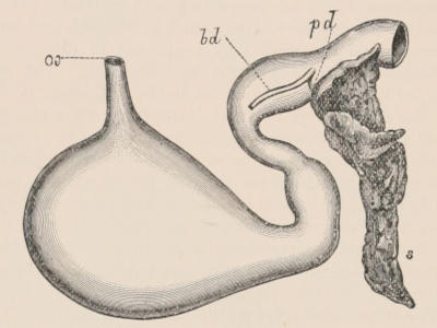
Fig. 20.—Stomach and pancreas of the Genet. Posterior or dorsal surface, œ, Œsophagus; s, pancreas; pd, pancreatic duct; bd, biliary duct from the liver. (From Mivart, Proc. Zool. Soc. 1882, p. 305.)
Intestinal Canal.—The intestinal canal varies greatly in relative length and capacity in different animals, and it also offers manifold peculiarities of form, being sometimes a simple cylindrical tube of nearly uniform calibre throughout, but more often subject to alterations of form and capacity in different portions of its course,—the most characteristic and constant being the division into an upper and narrower, and lower and wider portion, called respectively the small and the large intestine, the former being divided quite arbitrarily and artificially into duodenum, jejunum, and ileum, and the latter into colon and rectum. One of the most striking peculiarities of this part of the alimentary canal is the frequent presence of a diverticulum or blind pouch, the caput cæcum coli, as it was first called, a name generally abbreviated into “cæcum,” situated at the junction of the large and the small intestine, a structure presenting an immense variety of development, from the smallest bulging of a portion of the side wall of the tube to a huge and complex sac, greatly exceeding in capacity the whole of the remainder of the alimentary canal. It is only in herbivorous animals that the cæcum is developed to this great extent, and among these there is a curious complementary relationship between the size and complexity of this organ and that of the stomach. Where the latter is simple the cæcum is generally the largest, and vice versâ. Both the cæcum and colon are often sacculated, a disposition caused by the arrangement of the longitudinal bands of muscular tissue in their walls; but the small intestine is always smooth and simple-walled externally, though its lining membrane often exhibits various contrivances for increasing the absorbing surface without adding to the general bulk of the organ, such as the numerous small villi by which it is everywhere beset, and the more obvious transverse, longitudinal, or reticulating folds projecting into the interior, met with in many animals, of which the “valvulæ conniventes” of Man form well-known examples.
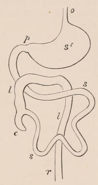
Fig. 21.—Diagrammatic plan of the general arrangement of the alimentary canal in a typical Mammal. o, Œsophagus; st, stomach; p, pylorus; s, s, small intestine; (abbreviated); c, cæcum; l, l, large intestine or colon, ending in r, the rectum.
Besides the crypts of Lieberkuhn found throughout the intestinal canal, and the glands of Brunner confined to the duodenum, there are other structures in the mucous membrane, about the nature of which there is still much uncertainty, called “solitary” and “agminated” glands; the latter being more commonly known by the name of “Peyer’s patches.” These were formerly supposed to be secretory organs, which discharged some kind of fluid into the intestine, but are now more generally considered to belong to that group of structures of somewhat mysterious function of which the lymphatic and lacteal glands are members. The solitary glands are found scattered irregularly throughout the whole intestinal tract; the agminated, on the other hand, are always confined to the small intestine, and are most abundant in its lower part. They are subject to great variation in number and in size, and even in different individuals of the same species, and also differ in character at different periods of life, becoming atrophied in old age.
Liver.—The distinct glands situated outside the walls of the intestinal canal, but which pour their secretion into it, are the pancreas and the liver. The latter is the more important on account of its size, if not on account of the direct action of its secretion in the digestive process. This large gland, so complex in structure and function, is well developed in all mammals, and its secreting tube, the bile-duct, always opens into the duodenum, or that portion of the canal which immediately succeeds the stomach. It is situated on the right side of the abdomen in contact with the diaphragm and the stomach, but varies greatly in relative size, and also in form, in different groups of mammals. In most mammals a gall-bladder, consisting of a pyriform diverticulum from the bile-duct, is present, but in many this appendage is wanting, and it is difficult to find the rationale of its presence or absence in relation to use or any other circumstance in the animal economy.
The descriptions of the livers of various animals to be met with in treatises or memoirs on comparative anatomy are very difficult to understand for want of a uniform system of nomenclature. The difficulty usually met with arises from the circumstance that this organ is divided sometimes, as in Man, Ruminants, and the Cetacea, into two main lobes, which have been always called respectively right and left, and in other cases, as in the lower Monkeys, Carnivora, Insectivora, and several other orders, into a larger number of lobes. Among the latter the primary division usually appears at first sight tripartite, the whole organ consisting of a middle, called “cystic” or “suspensory” lobe, and two lateral lobes, called respectively right and left lobes. This introduces confusion in describing livers by the same terms throughout the whole series of mammals, since the right and left lobes of the Monkey or Dog, for instance, do not correspond with parts designated by the same names in Man and the Sheep. There are, moreover, conditions where neither the bipartite nor the tripartite system of nomenclature will answer, so that we should have considerable difficulty in describing them without some more general system. In order to arrive at such a system it appears desirable to consider the liver in all cases as primarily divided by the umbilical vein (see Fig. 22, u) into two segments, right and left. This corresponds with its development and with the condition characteristic of the organ in the inferior classes of vertebrates. The situation of this division can almost always be recognised in adult animals by the persistence of some traces of the umbilical vein in the form of the round ligament, and by the position of the suspensory ligament.
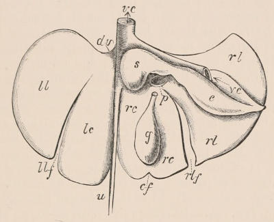
Fig. 22.—Diagrammatic plan of the inferior surface of a multilobed liver of a Mammal. The posterior or attached border is uppermost. u, Umbilical vein of the fœtus, represented by the round ligament in the adult, lying in the umbilical fissure; dv, the ductus venosus; vc, the inferior vena cava; p, the vena portæ entering the transverse fissure; llf, the left lateral fissure; rlf, the right lateral fissure; cf, the cystic fissure; ll, the left lateral lobe; lc, the left central lobe; rc, the right central lobe; rl, the right lateral lobe; s, the Spigelian lobe; c, the caudate lobe; g, the gall-bladder.
When the two main parts into which the liver is thus divided are entire, as in Man, the Ruminants, and Cetacea, they may be spoken of as the right and left lobes; when fissured, as the right and left segments of the liver, reserving the term lobe for the subdivisions. This will involve no ambiguity, for the terms right and left lobe will no longer be used for divisions of the more complex form of liver. In the large majority of mammals each segment is further divided by a fissure, more or less deep, extending from the free towards the attached border, which are called right and left lateral fissures (Fig. 22, rlf and llf). When these are more deeply cut than the umbilical fissure (u), the organ has that tripartite or trefoil-like form just spoken of, but it is easily seen that it is really divided into four regions or lobes, those included between the lateral fissures being the right and left central (rc and lc) separated by the umbilical fissure, and those beyond the lateral fissures on each side being the right and left lateral lobes (rl and ll). The essentially bipartite character of the organ and its uniformity of construction throughout the class are thus not lost sight of, even in the most complex forms. The left segment of the liver is rarely complicated to any further extent, except in some cases by minor or secondary fissures marking off small lobules, generally inconstant and irregular, and never worthy of any special designation. On the other hand, the right segment is usually more complex. The gall-bladder, when present, is always attached to the under surface of the right central lobe, sometimes merely applied to it, in other cases deeply embedded in its substance. In many instances the fossa in which it is sunk is continued to the free margin of the liver as an indent, or even a tolerably deep fissure (cf). The portal fissure (p), through which the portal vein and hepatic artery enter and the bile-duct emerges from the liver, crosses the right central lobe transversely, near the attached border of the liver. The right lateral lobe always has the great vena cava (vc) either grooving its surface or tunnelling through its substance near the inner or left end of its attached border; and a prolongation of this lobe to the left, between the vein and the portal fissure, sometimes forming a mere flat track of hepatic substance, but more often a prominent tongue-shaped process, is the so-called “Spigelian lobe” (s). From the under surface, of the right lateral lobe a portion is generally partially detached by a fissure, and called the “caudate lobe” (c). In Man this lobe is almost obsolete, but in most mammals it is of considerable magnitude, and has very constant and characteristic relations. It is connected by an isthmus at the left (narrowest or attached) end to the Spigelian lobe, behind which isthmus the vena cava is always in relation to it, channelling through or grooving its surface. It generally has a pointed apex, and is deeply hollowed to receive the right kidney, to the upper and inner side of which it is applied.
Considerations derived from the comparatively small and simple condition of the liver of the Ungulata, compared with its large size and complex form in the Carnivora, have led to the perhaps too hasty generalisation that the first type is related to a herbivorous and the latter to a carnivorous diet. The exceptions to such a proposition are very numerous. The fact of the great difference between the liver of the Cetacea and that of the Seals cannot be accounted for by difference of habits of life, though it perhaps may be by difference of origin.[14]
V. CIRCULATORY, ABSORBENT, RESPIRATORY, AND URINARY SYSTEMS.
Blood.—The blood of mammals is always red, and during the life of the animal hot, having a nearly uniform temperature, varying within a few degrees on each side of 100° Fahr. The corpuscles are, as usual in the vertebrates, of two kinds: (1) colourless, spheroidal, nucleated, and exhibiting amœboid movements; while (2) the more numerous, on which depends the characteristic hue of the fluid in which they are suspended, are coloured, non-nucleated, flattened, slightly biconcave discs, with circular outline in all known species except the Camels and Llamas, where they have the elliptical form characteristic of the red corpuscles of nearly all the other vertebrates, though adhering to the mammalian type in the absence of nucleus and relatively small size. As a rule they are smaller as well as more numerous than in other classes, but vary considerably in size in different species, and not always in relation to the magnitude of the animal; a Mouse, for instance, having as large corpuscles as a Horse. Within the limits of any natural group there is, however, very often some such relation, the largest corpuscles being found among the large species and the smallest corpuscles among the small species of the group, but even to this generalisation there are many exceptions. The transverse diameter of the red corpuscles in Man averages ¹⁄₃₂₀₀ of an inch, which is exceptionally large, and only exceeded by the Elephant (¹⁄₂₇₄₅), and by some Cetacea and Edentata. They are also generally large in Apes, Rodents, and the Monotremata, and small in the Artiodactyles, least of all in the Chevrotains (Tragulus), being in T. javanicus and meminna not more than ¹⁄₁₂₃₂₅.[15]
Heart.—The heart of mammals consists of four distinct cavities, two auricles and two ventricles. Usually the ventricular portion is externally of conical form, with a simple apex, but in the Sirenia it is broad and flattened, and a deep notch separates the apical portion of each ventricle. A tendency to this form is seen in the Cetacea and the Seals. It is characteristic of mammals alone among vertebrates that the right auriculo-ventricular valve is tendinous like the left, consisting of flaps held in their place by fibrous ends (chordæ tendiniæ) and arising from projections of the muscular walls of the ventricular cavity (musculi papillares). In the Monotremata a transition between this condition and the simple muscular flap of the Sauropsida is observed. In most of the larger Ungulates a distinct but rather irregular ossification (os cordis) is developed in the central tendinous portion of the base of the heart.
Blood-vessels.—The orifices of the aorta and pulmonary artery are each guarded by three semilunar valves. The aorta is single, and arches over the left bronchial tube. After supplying the tissues of the heart itself with blood by means of the coronary arteries, it gives off large vessels (“carotid”) to the head and (“brachial”) to the anterior extremities. The mode in which these vessels arise from the aorta varies much in different mammals, and the study of their disposition affords some guide to classification. In nearly all cases the right brachial and carotid have a common origin (called the “innominate artery” in anthropotomy). The other two vessels may come off from this, as is the rule in Ungulates, the common trunk constituting the “anterior aorta” of veterinary anatomy; or they may be detached in various degrees, both arising separately from the aorta, as in Man, or the left carotid from the innominate and the left brachial from the aorta, a very common arrangement; or the last two from a common second or left innominate, as in some Bats and Insectivores. The aorta, after giving off the intercostal arteries, passes through the diaphragm into the abdomen, and, after supplying the viscera of that cavity by means of the gastric, hepatic, splenic, mesenteric, renal, and spermatic vessels, gives off in the lumbar region a large branch (iliac) to each of the hinder extremities, which also supplies the pelvic viscera, and is continued onwards in the middle line, greatly diminished in size, along the under surface of the tail as the caudal artery. In certain mammals, arterial plexuses, called retia mirabilia, formed by the breaking up of the vessel into an immense number of small trunks, which may run in a straight course parallel to one another (as in the limbs of Sloths and Slow Lemurs), or form a closely packed network, as in the intracranial plexuses of Ruminants, or a sponge-like mass of convoluted vessels, as in the intercostals of Cetaceans, are peculiarities of the vascular system the meaning of which is not in all cases clearly understood. In the Cetacea they are obviously receptacles for containing a large quantity of oxygenated blood available during the prolonged immersion, with consequent absence of respiration, to which these animals are subject.
The vessels returning the blood to the heart from the head and upper extremities usually unite, as in Man, to form the single vena cava superior or precaval vein, but in some Insectivores, Chiroptera, and Rodents, in the Elephant, and all Marsupials and Monotremes, the two superior caval veins enter the right auricle without uniting, as in birds. In Seals and some other diving mammals there is a large venous sinus or dilatation of the inferior vena cava immediately below the diaphragm. In the Cetacea the purpose of this is supplied by the immense abdominal venous plexuses. As a rule the veins of mammals are furnished with valves, but these are said to be altogether wanting in the Cetacea, and in the superior and inferior cava, subclavian and iliac veins, the veins of the liver (both portal and hepatic), heart, lungs, kidneys, brain, and spinal cord of other mammals. Many of the veins within the cranium are included in spaces formed by the separation of the laminæ of the dura mater, and do not admit of being dilated beyond a certain size; these are termed sinuses. The portal circulation in mammals is limited to the liver, the portal vein being formed by the superior and inferior mesenteric, the splenic, the gastro-epiploic, and the pancreatic veins. The kidney is supplied solely by arterial blood, and its veins empty their contents only into the inferior cava.
Lymphatic Vessels.—The absorbent or lymphatic system of vessels is very fully developed in the Mammalia. Its ramifications extend through all the soft tissues of the body, and convey a colourless fluid called lymph, containing nucleated corpuscles, and also, during the process of digestion, the chyle, a milky fluid taken up by the lymphatics (here called lacteals) of the small intestine, and pour them into the general vascular system, where they mix with the venous blood. The lymphatic vessels of the hinder extremities, as well as those from the intestinal canal, unite in the abdomen to form the “thoracic duct,” the hinder end or commencement of which has a dilatation called the receptaculum chyli. This duct, which is of irregular size and sometimes double, often dividing and uniting again in its course, or even becoming plexiform, passes forwards close to the bodies of the thoracic vertebræ, and empties itself, by an orifice guarded by a valve, into the great left brachio-cephalic vein, having previously received the lymphatics from the thorax and the left side of the head and left anterior extremity. The lymphatics from the right side of the head and right anterior limb usually enter by a small distinct trunk into the corresponding part of the right brachio-cephalic vein. The duct, and also the principal lymphatic vessels, are provided with valves.
Lymphatic glands, rarely met with in the Sauropsida, are usually present in mammals, both in the general and in the lacteal system; the latter being called “mesenteric glands.” They are round or oval masses, situated upon the course of the vessels, which break up in them and assume a plexiform arrangement, and then reunite as they emerge. No structures corresponding to the pulsating “lymphatic hearts” of the lower vertebrates have been met with in mammals.
Ductless Glands.—Associated with the vascular and lymphatic systems are certain bodies (the functions of which are not properly understood), usually, on account of their general appearance, grouped together under the name of “ductless glands.” The largest of these is the “spleen,” which is single, and always placed in mammals in relation to the fundus or left end of the stomach, to which it is attached by a fold of peritoneum. It is dark-coloured and spongy in substance, and has a depression or “hilus” on one side, into which the splenic artery, a branch of the cœliac axis of the abdominal aorta, enters, and from which the vein joining the portal system emerges. The spleen varies much in size and form in different mammals, being relatively very small in the Cetacea. It is sometimes almost spherical, but more often flattened, oval, triangular, or elongated, and occasionally, as in Monotremes and most Marsupials, triradiate. The “suprarenal bodies” or “adrenals” are two in number, each situated either in contact with, or at a short distance in front of the anterior extremity of the kidney. They are abundantly supplied with nerves, and are much larger relatively in early than in adult life. The “thyroid bodies,” of which there are generally two, though in Man and some other species they are connected by an isthmus passing across the middle line, are constant in mammals, though only met with in a rudimentary condition, if at all, in other vertebrates. They are situated in the neck, in contact with the sides of the anterior extremity of the trachea. The “thymus” lies in the anterior part of the thorax, between the sternum and the great vessels at the base of the heart, and differs from the thyroid in being median and single, and having a central cavity. It attains its greatest development during the period in which the animal is nourished by its mother’s milk, and then it diminishes, and generally disappears before full growth is attained.
Nostrils.—Mammals breathe occasionally through the mouth, but usually, and in many cases exclusively, through the nostrils or nares. Those are apertures, always paired (except in the toothed Cetacea, where they unite to form a single external opening), and situated at the fore part of the face, generally at or beneath the end of the muzzle, a median prominence above the mouth. This is sometimes elongated to form a proboscis, to the extremity of which the nostrils are carried, and which attains its maximum of development in the Elephant. In the Cetacea the nostrils are situated at a considerable distance behind the anterior end of the face, upon the highest part of the head, and are called “blowholes,” from the peculiar mode of respiration of those animals. The nostrils are kept open by means of cartilages surrounding their aperture, which many animals have the power of moving so as to cause partial dilatation or contraction. In diving animals, as Seals and Cetacea, they can be completely closed at will so as to prevent the entrance of water when beneath the surface. The passage to which the nostrils lead is in most mammals filled by a more or less complex sieve-like apparatus, formed of the convoluted turbinal bones and cartilages, over which a moist, vascular, ciliated mucous membrane is spread, which intercepts particles of dust, and also aids in warming the inspired air before it reaches the lungs. In the Proboscidea, in which these functions are performed by the walls of the long tubular proboscis, this apparatus is entirely wanting.
Trachea.—The narial passages have the organ of smell situated in their upper part, and communicate posteriorly with the pharynx, and through the glottis with the “trachea” or windpipe, a tube by which the air is conveyed to and from the lungs. The permanent patency of the trachea during the varied movements of the neck is provided for by its walls being stiffened by a series of cartilaginous rings or hoops, which in most mammals are incomplete behind. Having entered the thorax, the trachea bifurcates into the two bronchi, one of which enters, and, dividing dichotomously, ramifies through each lung. In some of the Cetacea and Artiodactyla a third bronchus is given off from the lower part of the trachea, above its bifurcation and enters the right lung.
Larynx.—The upper end of the trachea is modified into the organ of voice or “larynx,” the air passing through which to and from the lungs is made use of to set the edges of the “vocal cords,” or fibrous bands stretched one on each side of the tube, into vibration. The larynx is composed of several cartilages, such as the “thyroid,” the “cricoid,” and the “arytenoid” which are moved upon one another by muscles, and suspended from the hyoidean arch. By alteration of the relative position of these cartilages the cords can be tightened or relaxed, approximated or divaricated, as required to modulate the tone and volume of the voice. A median tongue-shaped fibro-cartilage at the top of the larynx, the “epiglottis,” protects the “glottis,” or aperture by which the larynx communicates with the pharynx, from the entry of particles of food during deglutition. The form of the larynx and development of the vocal cords present many variations in different members of the class, the greatest modification from the ordinary type being met with in the Cetacea, where the arytenoid cartilages and epiglottis are united in a tubular manner, so as to project into the nasal passage, and, being grasped by the muscular posterior margin of the palate, provide a direct channel of communication from the lungs to the external surface. An approach to this condition is met with in the Hippopotamus and some other Ungulates; it is indeed so general as an abnormality, that Howes suggests that an internarial epiglottis may have been a primitive feature common throughout the class. Nearly all mammals have a voice, although sometimes it is only exercised at seasons of sexual excitement. Some Marsupials and Edentates appear to be quite mute. In no mammal is there an inferior larynx, or “syrinx,” as in birds.
Diaphragm.—The thoracic cavity of mammals differs from that of the Sauropsida in being completely separated from the abdomen by a muscular partition, the “diaphragm,” attached to the vertebral column, the ribs, and the sternum. This is much arched, with the convexity towards the thorax, so that when its fibres contract and it is flattened the cavity of the thorax is increased, and when they are relaxed the cavity is diminished.
Lungs.—The lungs are suspended freely in the thorax, one on each side of the heart, being attached only by the root, which consists of the bronchus or air-tube and pulmonary arteries and veins by which the blood is passed backwards and forwards between the heart and the lungs. The remaining part of the surface of each lung is covered by serous membrane, the “pleura”; and whatever the state of distension or contraction of the chest-wall, is accurately in contact with it. Inspiration is effected by the contraction of the diaphragm and by the intercostal and other muscles elevating or bringing forward the ribs, and thus throwing the sternum farther away from the vertebral column. As the surface of the lung must follow the chest-wall, the organ itself is expanded, and air rushes in through the trachea to fill all the minute cells in which the ultimate ramifications of the bronchi terminate. In ordinary expiration very little muscular power is expended, the elasticity of the lungs and surrounding parts being sufficient to cause a state of contraction and thus drive out at least a portion of the air contained in the cells, when the muscular stimulus is withdrawn. The lungs are sometimes simple externally, as in the Sirenia (where they are greatly elongated) and the Cetacea, but are more often divided by deep fissures into one or more lobes. The right lung is usually larger and more subdivided than the left. It often has a small distinct lobe behind, wanting on the left side, and hence called lobulus azygos.
Air-sacs.—Most mammals have in connection with the air passages certain diverticuli or pouches containing air, the use of which is not always easy to divine. The numerous air sinuses situated between the outer and inner tables of the bones of the head, represented in Man by the antrum of Highmore and the frontal and sphenoidal sinuses, and attaining their maximum of development in the Indian Elephant, are obviously for the mechanical purpose of allowing expansion of the osseous surface without increase of weight. They are connected with the nasal passages. The Eustachian tubes pass from the back of the pharynx to the cavity of the tympanum, into which and the mastoid cells they allow air to pass. In the Equidæ there are large post-pharyngeal air-sacs in connection with them. The Dolphins have an exceedingly complicated system of air-sacs in connection with the nasal passages just within the nostrils, and the Tapirs, Rhinoceroses, and Horses have blind sacs in the same situation. In the males of some Seals (Cystophora and Macrorhinus) large pouches, which the animal can inflate with air, and which are not developed in the young animal or the female, arise from the upper part of the nasal passages, and lie immediately under the skin of the face. These appear analogous, although not in the same situation, to the gular pouch of the male Bustard. The larynx frequently has membranous pouches in connection with it, into which air passes. These may be lateral and opening just above the vocal cords, when they constitute the sacculi laryngis, found in a rudimentary state in Man, and attaining an enormous development, so as to reach to the shoulders and axillæ, in some of the Anthropoid Apes; or they may be median, opening in front either above or below the thyroid and cricoid cartilages, as in the Howling and other Monkeys, and also in the Whalebone Whales and Great Anteater.
Urinary Organs.—The kidneys of mammals are more compact and definite in form than in other vertebrates, being usually more or less oval, with an indent on the side turned towards the middle line, from and into which the vessels and ducts pass. They are distinctly divided into a cortical secretory portion, composed mainly of convoluted tubes, and containing the so-called Malpighian bodies; and a medullary excreting portion, formed of straight tubes converging towards a papilla, embraced by the commencement of the ureter or duct of the organ. The kidneys of some mammals, as most Monkeys, Carnivores, Rodents, etc., are simple, with a single papilla into which all the renal tubuli enter. In others, as Man, there are many pyramids of the medullary portion, each with its papilla, opening into a division (calyx) of the upper end of the ureter. Such kidneys, either in the embryonic condition only, or throughout life, are lobulated on the surface. In some cases, as in Bears, Seals, and especially the Cetacea, the lobulation is carried further, the whole organ being composed of a mass of renules, loosely united by connective tissue, and with separate ducts, which soon join to form the common ureter.
Bladder.—In all mammals except the Monotremes the ureters terminate by slit-like valvular openings in the urinary bladder. This receptacle when filled discharges its contents through the single median urethra, which in the male is almost invariably included in the penis, and in the females of some species of Rodents, Insectivores, and Lemurs has a similar relation to the clitoris. In the Monotremes, though the bladder is present, the ureters do not enter into it, but join the urino-genital canal some distance below it, with the orifice of the genital duct intervening.
VI. NERVOUS SYSTEM AND ORGANS OF SENSE.
Brain.—The brain of mammals shows a higher condition of organisation than that of other vertebrates. The cerebral hemispheres have a greater preponderance compared with other parts, especially to the so-called optic lobes, or corpora quadrigemina, which are completely concealed by them. The commissural system of the hemispheres is much more complex, both fornix and corpus callosum being present in some form; and when the latter is rudimentary, as in Marsupials and Monotremes, its deficiency is made up for by the great size of the anterior commissure. The lateral lobes of the cerebellum, wanting in lower vertebrates, are well developed and connected by a transverse commissure, the pons Varolii. The whole brain, owing especially to the size of the cerebral hemispheres, is considerably larger relatively to the bulk of the animal than in other classes, but it must be recollected that the size of its brain depends upon many circumstances besides the degree of intelligence which an animal possesses, although this is certainly one. Man’s brain is many times larger than that of all other known mammals of equal bulk, and even three times as large as that of the most nearly allied Ape. Equal bulk of body is here mentioned, because, in drawing any conclusions from the size of the brain compared with that of the entire animal, it is always necessary to take into consideration the fact that in every natural group of closely allied animals the larger species have much smaller brains relatively to their general size than the smaller species, so that, in making any effective comparison among animals belonging to different groups, species of the same size must be selected. It may be true that the brain of a Mouse is, as compared with the size of its body, larger than that of a Man, but, if it were possible to reduce an animal having the general organisation of a Man to the size of a Mouse, its brain would doubtless be very many times larger; and conversely, as shown by the rapid diminution of the relative size of the brain in all the large members of the Rodent order, a Mouse magnified to the size of a Man would, if the general rule were observed, have a brain exceedingly inferior in volume. Although the brain of the large species of Whales is, as commonly stated, the smallest in proportion to the bulk of the animal of any mammal, this does not invalidate the general proposition that the Cetacea have very large brains compared with terrestrial mammals, like the Ungulata, or even the aquatic Sirenia, as may be proved by placing the brain of a Dolphin by the side of that of a Sheep, a Pig, or a Manatee of equal general weight. It is only because the universally observed difference between the slower ratio of increase of the brain compared with that of the body becomes so enormous in these immense creatures that they are accredited with small brains.
The presence or absence of “sulci” or fissures on the surface of the hemisphere, dividing it into “convolutions” or “gyri,” and thus increasing the superficies of the cortical gray matter, as well as allowing the pia mater with its nutrient blood-vessels to penetrate into the cerebral substance, follow somewhat similar rules. The sulci are related partly to the high or low condition of organisation of the species, but also in a great degree to the size of the cerebral hemispheres. In very small species of all groups, even the Primates, they are absent, and in the largest species of groups so low in the scale as the Marsupials and Edentates they are found. They reach their maximum of development in the Cetacea.
The accompanying woodcut (Fig. 23) shows the principal parts of a mammalian brain, as seen from the superior, lateral, and inner surfaces. The sylvian fissure (sf) is one of the most constant of the sulci found in the hemispheres.
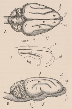
Fig. 23.—Brain of the Genet (Genetta tigrina). A, From above; B, from the right side; C, inner surface of right hemisphere; cc, corpus callosum; c.m.s, calloso-marginal sulcus; c, notch representing central sulcus of other forms; d, depression on superior lateral gyrus of hemisphere; hg, hippocampal gyrus; i, inferior lateral gyrus of hemisphere; m, middle lateral gyrus of do.; s, superior lateral gyrus of do.; os, supraorbital sulcus of do.; sf, sylvian fissure of do.; ol, olfactory lobes. The deeply convoluted part behind the cerebral hemisphere is the cerebellum, below which lies the medulla oblongata, or commencement of the spinal cord. (Mivart, Proc. Zool. Soc. 1882, p. 516.)
The researches of Palæontologists, founded upon studies of casts of the interior of the cranial cavity of extinct forms, have shown that, in many natural groups of mammals, if not in all, the brain has increased in size, and also in complexity of surface foldings, with the advance of time,—indicating in this, as in so many other respects, a gradual progress from a lower to a higher type of development.
Nerves.—The twelve pairs of cranial nerves generally recognised in vertebrates are usually all found in mammals, though the olfactory nerves are excessively rudimentary, if not altogether absent, in the Toothed Whales. The spinal cord, or continuation of the central nervous axis, lies in the canal formed by the neural arches of the vertebræ, and gives off the compound double-rooted nerves of the trunk and the extremities, corresponding in number to the vertebræ, through the interspaces between which they pass out to their destination. The cord is somewhat enlarged at the two points where it gives off the great nerves to the anterior and the posterior extremities, which, from their interlacements soon after their origin, are called respectively the brachial and lumbar plexuses. The ganglionic or sympathetic portion of the nervous system is well developed, and presents few modifications.
Sense of Touch.—The sense of touch is situated in the skin generally, but is most acute in certain regions more or less specialised for the purpose by the presence of tactile papillæ, such as portions of the face, especially the lips and end of the snout, and the extremities of the limbs when these are used for other purposes than mere progression, and the under surface of the end of the tail in some Monkeys. The “vibrissæ” or long stiff bristles situated on the face of many mammals are rendered extremely sensitive to touch by the abundant supply of branches from the fifth nerve to their basal papillæ. In Bats the extended wing membranes, and probably also the large ears and the folds and prominences of skin about the face of some species, are so sensitive as to receive impressions even from the different degrees of resistance of the air, and so enable the animals to avoid coming in contact with obstacles to their nocturnal flight.
Taste and Smell.—The organs of the other special senses are confined to the head. Taste is situated in the papillæ scattered on the dorsal surface of the tongue. The organ of smell is present in all mammals except the Toothed Whales. It consists of a ramification of the olfactory nerves over a plicated, moist, mucous membrane, supported by folded plates of bone, placed on each side of the septum nasi in the roof, or often in a partially distinct upper chamber, of the nasal passage, so arranged that, of the air passing into the lungs in inspiration, some comes in contact with it, causing the perception of any odorous particles with which it may be charged. Many mammals possess intense powers of smelling certain odours which others are quite unable to appreciate, and the influence which this sense exercises over the well-being of many species is very great, especially in indicating the proximity of others of the same kind, and giving warning of the approach of enemies. The development and modification of the sense of smell is probably associated with that of the odorous secretion of the cutaneous glands.
Sight.—The organ of sight is quite rudimentary, and even concealed beneath the integument, in some burrowing Rodents and Insectivores, and is most imperfectly developed in the Platanista, or Freshwater Dolphin of the rivers of India. In all other mammals the eyeball has the structure characteristic of the organ in the higher Vertebrata, consisting of parts through which the rays of light are admitted, regulated, and concentrated upon the sensitive expansion of the optic nerve lining the posterior part of the ball. A portion of the fibrovascular and highly pigmented layer, the choroid, which is interposed between the retina and the outer sclerotic coat, is in many mammals modified into a brilliantly-coloured light-reflecting surface, the tapetum lucidum. There is never a pecten or marsupium like that of the Sauropsida, nor is the sclerotic ever supported by a ring of flattened ossicles, as is so frequently the case in the lower vertebrated classes. The eyeball is moved in various directions by a series of muscles—the four straight, two oblique, and, except in the higher Primates, a posterior retractor muscle called choanoid. The superior oblique muscle passes through a tendinous pulley fastened to the roof of the orbit, which is a feature not found beyond the limits of the mammalian class. The eye is protected by the lids, generally distinctly separated into an upper and a lower movable flap, which, when closed, meet over the front of the eye in a more or less nearly horizontal line: but sometimes, as in the Sirenia, the lids are not distinct, and the aperture is circular, closing to a point. In almost all mammals below the Primates, except the Cetacea, a “nictitating membrane” or third eyelid is placed at the inner corner of the eyeball, and works horizontally across the front of the ball within the true lids. Its action is instantaneous, being apparently for the purpose of cleaning the front of the transparent cornea;—a function unnecessary in animals whose eyes are habitually bathed in water, and which in Man and his nearest allies is performed by winking the true eyelids. Except in Cetacea the surface of the eye is kept moist by the secretion of the lachrymal gland, placed under the upper lid at its outer side, and the lids are lubricated by the Harderian and Meibornian glands, the former being situated at the inner side of the orbit, and especially related to the nictitating membrane, the latter in the lining membrane of the lids.
Hearing.—The organ of hearing is inclosed in a bony capsule (periotic) situated in the side of the head, intercalated between the posterior (occipital) and the penultimate (parietal) segment of the skull. It has, in common with other vertebrates, three semicircular canals and a vestibule, but the cochlea is more fully developed than in the Sauropsida, and, except in the Monotremes, spirally convoluted. The tympanic cavity is often dilated below, forming a smooth rounded prominence on the base of the skull, the auditory bulla (Fig. 8). The three principal ossicles, the “malleus,” “incus,” and “stapes,” are always present, but variable in characters. In the Sirenia, Cetacea, and Seals they are massive in form, being in the first-named order of larger size than in any other mammals. In the Cetacea the malleus is ankylosed to the tympanic; but in other mammals it is connected only with the membrana tympani. The stapes in the lower orders—Edentates, Marsupials, and Monotremes—has a great tendency to assume the columnar form of the corresponding bone in Sauropsida, its two rami entirely or partially coalescing.[16] The tympanic membrane (drum of the ear) forms the outer wall of the cavity. In the fœtal state it is level with the external surface of the skull, and remains so permanently in a few mammals as the American Monkeys; but commonly, by the growth of the squamosal bone, it becomes deeply buried at the bottom of a bony tube (meatus auditorus externus), which is continued to the surface of the skin in a fibrous or fibro-cartilaginous form. In Whales, owing to the thickness of the subcutaneous adipose tissue, this meatus is of great length, and is also extremely narrow. In most aquatic and burrowing animals it opens upon the surface by a simple aperture, but in the large majority of the class there is a projecting fold of skin, strengthened by fibro-cartilages, called the pinna, auricle, or “external ear,” of very variable size and shape, generally movably articulated on the skull, and provided with muscles to vary its position; this pinna helping to collect and direct the vibrations of sound into the meatus.
VII. REPRODUCTIVE ORGANS.
Testes.—In the male the testes retain nearly their primitive or internal position throughout life in the Monotremata, Sirenia, Cetacea, most Edentata, Hyracoidea, Proboscidea, and Seals, but, in other groups they either periodically (as in Rodentia, Insectivora, and Chiroptera) or permanently pass out of the abdominal cavity through the inguinal canal, forming a projection beneath the skin of the perineum, or becoming suspended in a distinct pouch of integument called the scrotum. All the Marsupials have a pedunculated scrotum, the position of which differs from that of other mammals, being in front of, instead of behind, the preputial orifice. As regards the presence, absence, or comparative size and number of the accessory generative glands—prostate, vesicular, and Cowper’s glands, as they are called—there is much variation in different groups of mammals.
Penis.—The penis is almost always completely developed, consisting of two corpora cavernosa attached to the ischial bones, and of a median corpus spongiosum enclosing the urethra, and forming the glans at the distal portion of the organ. In Marsupials, Monotremes, and the Sloths and Anteaters, the corpora cavernosa are not attached directly to the ischia, and in the last-named the penis is otherwise of a very rudimentary character, the corpus spongiosum not being present. In many Marsupials the glans penis is bifurcated. In most Primates, Carnivora, Rodentia, Insectivora, and Chiroptera, but in no other orders, an os penis is present.
Ovaries and Oviduct.—In the female, the ovaries permanently retain their original abdominal position, or only descend a short distance into the pelvis. They are of comparatively smaller size than in other vertebrates, have a definite flattened oval form, and are enclosed in a more or less firm “tunica albiginea.” The oviduct has a trumpet-like, and usually fimbriated abdominal aperture, and is more or less differentiated into three portions:—(1) a contracted upper part, called in Man and the higher mammals the “Fallopian tube”; (2) an expanded part with muscular walls, in which the ovum undergoes the changes by which it is developed into the fœtus, called the “uterus”; (3) a canal, the “vagina,” separated from the last by a valvular aperture, and terminating in the urino-genital canal, or common urinal and genital passage, which in higher mammals is so short as scarcely to be distinct from the vagina. The complete distinction of the oviducts of the two sides throughout their whole length, found in all lower vertebrates, only occurs in this class in Monotremes; a prevailing mammalian characteristic being their more or less perfect coalescence in the middle line to form a single median canal. In the Marsupials this union only includes the lower part of the vagina; but in most Placentals it extends to the whole vagina and a certain portion of the uterus, which cavity is then described as “bicornuate.” In the higher mammals, as in Man, and also in some of the Edentates, the whole of the uterus is single, the contracted upper portion of the oviducts or Fallopian tubes, as they are then called, entering its upper lateral angles by small apertures. In certain lower forms the urino-genital canal opens with the termination of the rectum into a common cloaca, as in other vertebrates; but it is characteristic of the majority of the class that the two orifices are more or less distinct externally.
Mammary Glands.—Mammary glands secreting the milk by which the young are nourished during the first portion of their existence after birth, are present in both sexes in all mammals, though usually only functional in the female. In the Monotremes alone their orifices are mere scattered pores in the skin, but in all other forms they are situated upon the end of conical elevations, called mammillæ, or teats, which, taken into the mouth of the young animal, facilitate the process of sucking. These are always placed in pairs upon some part of the ventral surface of the body, but vary greatly in number and position in different groups. In the Cetacea, where the prolonged action of sucking would be incompatible with their subaqueous life, the ducts of the glands are dilated into large reservoirs from which the contents are injected into the mouth of the young animal by the action of a compressor muscle.
Secondary Sexual Characters.—Secondary sexual characters, or modifications of structure peculiar to one sex, but not directly related to the reproductive function, are very general in mammals. They almost always consist of the acquisition or perfection of some character by the male as it attains maturity, which is not found in the female or the young in either sex. In a large number of cases these clearly relate to the combats in which the males of many species engage for the possession of the females during the breeding season; others are apparently ornamental, and of many it is still difficult to apprehend the meaning. Many suggestions on this subject will, however, be found in the chapters devoted to it in Darwin’s work on The Descent of Man and Selection in Relation to Sex, where most of the best-known instances are collected. Superiority of size and strength in the male of many species is a well-marked secondary sexual character related to the purpose indicated above, being probably perpetuated by the survivors or victors in combats transmitting to their descendants those qualities which gave them advantages over others of their kind. To the same category belong the great development of the canine teeth of the males of many species which do not use these organs in procuring their food, as the Apes, Swine, Musk and some other Deer, the tusk of the male Narwhal, the antlers of Deer, which are present in most cases only in the males, and the usual superiority in size and strength of the horns of the Bovidæ. Other secondary sexual characters, the use of which is not so obvious, or which may only relate to ornament, are the presence of masses or tufts of long hair on different parts of the body, as the mane of the male Lion and Bison, the beards of some Ruminants and Bats (as Taphozous melanopogon), Monkeys, and of Man, and all the variations of coloration in the sexes, in which, as a general rule, the adult male is darker and more vividly coloured than the female. Here may also be mentioned the presence or the greater development of odoriferous glands in the male, as in the Musk Deer, and the remarkable perforated spur with its glands and duct, so like the poison-tooth of the venomous serpents, found in the males of both Ornithorhynchus and Echidna, the use of which is at present unknown.
Placenta.—The development of the mammalian ovum, and the changes which the various tissues and organs of the body undergo in the process of growth, are too intricate subjects to be explained without entering into details incompatible with the limits of this work, especially as they scarcely differ, excepting in their later stages, from those of other vertebrates, upon which, owing to the greater facilities these present for examination and study, the subject has been more fully worked out. There are, however, some points which require notice, as peculiar to the mammalian class, and as affording at least some hints upon the difficult subject of the affinities and classification of the members of the group.
The nourishment of the fœtus during intra-uterine life takes place through the medium of certain structures, partly belonging to the fœtus itself and partly belonging to the inner parietes of the uterus of the parent. These in their complete form constitute the complex organ called the “placenta,” serving as the medium of communication between the mother and fœtus, and in which the physiological processes that are concerned in the nutrition of the latter take place; but as we shall see, though a placenta, in the usual acceptation of the term, is peculiar to the mammalian class, it is not in all of its members that one is developed. The structures to which we shall have especially to refer are the outer tunic of the ovum, to which, however formed, the term “chorion” is commonly applied, and two sac-like organs connected with the body-cavity of the embryo, both formed from the splanchnic mesoblast, lined by a layer of the hypoblast. These are the “umbilical vesicle” or “yolk-sac” and the “allantois.”
The umbilical vesicle is a thin membrane enclosing the yolk, which by the doubling in of the ventral walls of the embryo becomes gradually formed into a distinct sac external to the body, with a pedicle (the omphalo-enteric duct) by which for a time a communication is maintained between its cavity and the intestinal canal. In the walls of this sac blood-vessels (omphalo-meseraic or vitelline) are developed in connection with the vascular system of the embryo, through which, either by their contact with the outer surface of the walls of the ovum, or by the absorption through them of the contents of the yolk-sac, the nutrition of the embryo in the lower vertebrates chiefly takes place. In mammals the umbilical vesicle plays a comparatively subordinate part in the nourishment of the fœtus, its function being generally superseded by the allantois.
The last-named sac commences at a very early period as a diverticulum from the hinder end of the alimentary tract of the embryo. Its proximal portion afterwards becomes the urinary bladder, the contracted part between this and the cavity of the allantois proper constituting the urachus, which passes out of the body of the fœtus at the umbilicus together with the vitelline duct. The mesoblastic tissue of the walls of the allantois soon becomes vascular; its arteries are supplied with fœtal blood by the two hypogastric branches of the iliacs, or main divisions of the abdominal aorta, and the blood is returned by venous trunks uniting to form the single umbilical vein which runs to the under surface of the liver, where, part of it joining the portal vein and part entering the vena cava directly, it is brought to the heart. These are the vessels which, with their surrounding membranes, constitute the umbilical cord—the medium of communication between the fœtus and the placenta, when that organ is fully developed.
The egg membranes of the Monotremes present many points of agreement with those of the ovum of the Marsupials,[17] and differ from those of the Placental types. Thus Monotremes and Marsupials agree in having a vitelline membrane, which appears between the young ovum and the follicular epithelium, persisting in the one case until the time of hatching, and in the other till a late uterine stage. There are also several other common features fully described in Mr. Caldwell’s memoir, but which cannot be detailed in this work.
In the Marsupialia the observations made many years ago by Sir R. Owen upon the development of the Kangaroo have been confirmed by those of Dr. H. C. Chapman,[18] while Dr. Selenka,[19] and Professor H. F. Osborn[20] have contributed important evidence as to the structure and relations of the fœtal membranes of the Opossums and others. It thus appears that up to the period of the very premature birth of these animals the outer covering of the ovum, or false chorion, is free from persistent villi, and not adherent to the epithelium of the uterine walls; for, although fitting into the folds of the latter, it is perfectly and readily separable in its entire extent from them. The umbilical vesicle or yolk-sac is large, vascular, and adherent to a considerable portion of the false chorion or subzonal membrane, while the allantois is relatively small, and although the usual blood-vessels can be traced into it, it does not appear to contract any connection with the false chorion, and, therefore, much less with the walls of the uterus, of such a nature as to constitute a placenta. In some forms, however, such as the Opossums, the umbilical vesicle or yolk-sac develops temporary villi, which unite with the subzonal membrane, or false chorion, to form a disc-like area closely attached to the cells covering the utricular glands of the uterine epithelium, and thus forming a so-called yolk-sac placenta. The function of this organ is considered to be the transmission of the secretions of the utricular glands to the embryo by means of the umbilical vesicle; the function of the allantois being either respiratory or the absorption of the fluid secreted in the uterine cavity by the utricular glands.
While in the uterus the nourishment of the fœtus seems, therefore, to be derived from the umbilical vesicle, as in reptiles and birds, rather than from the uterine walls by means of the allantoic vessels, as in the higher mammals. The latter vessels, in fact, play even a much less important part in the development of these animals, not only than in the placental mammals, but even than in the Sauropsida, for they can scarcely have the respiratory function assigned to them in that group: pulmonary respiration and the lacteal secretion of the mother very early superseding all other methods of providing the due supply, both of oxygen and of food required for the development and growth of the young animal. In this sense the Marsupials may be looked upon as the most typically “mammalian” of the whole class. In no other group do the milk-secreting glands play such an important part in providing for the continuity of the race.
In the third primary division of the Mammalia, the so-called Placentalia, the umbilical vesicle generally does not quite unite with the chorion, and disappears as development proceeds, so that no trace of it can be seen in the membranes of an advanced embryo; but it may persist until the end of the intra-uterine life as a distinct sac in the umbilical cord, or lying between the allantois and amnion. The disappearance or persistence of the umbilical vesicle does not, according to our present knowledge, appear to be correlated with a higher or lower general grade of development, as might be presupposed. It is stated to have been found in Man even up to the end of intra-uterine life, and also in the Carnivora, while in the Ungulata and Cetacea it disappears at an earlier age. In many, if not all, of the Rodentia, Insectivora, and Chiroptera, it plays a more important part, becoming adherent to a considerable part of the inner surface of the chorion, to which it conveys blood-vessels, although villi do not appear to be developed from the surface of this part, as they are on the portion of the chorion supplied by the allantoic vessels. These orders thus present to a certain extent a transitional condition from the Marsupials, although essentially different, in possessing the structures next to be described.
The special characteristic of the whole of the placental mammals constituting the majority of the class, is that the allantois and its vessels become intimately blended with a smaller or greater part of the parietes of the ovum, forming a structure on the outer surface of which villi are developed, and which, penetrating into corresponding cavities of the “decidua,” or soft, vascular, hypertrophied lining membrane of the uterus, constitutes the placenta. This organ may be regarded, as Sir William Turner says, both in its function and in the relative arrangement of its constituent textures, as a specially modified secreting gland, the ducts of which are represented by the extremities of the blood-vessels of the fœtal system. The passage of material from the maternal to the fœtal-system of vessels is not a simple percolation or diffusion through their walls, but is occasioned by the action of a layer of cells derived from the maternal or uterine structures, and interposed between the blood-vessels of the maternal part of the placenta and those of the villi covering the chorion, in which the embryonic vessels ramify.
The numerous modifications in the details of the structure of this organ relate to augmenting the absorbing capacity of the vessels of the chorion, and are brought about either by increasing the complexity of the fœtal villi and maternal crypts over a limited area, or by increasing the area of the part of the chorion covered by the placental villi, or by various combinations of the two methods.
The first class of variations has given rise to a distinction into two principal kinds of placenta: (1) simple or non-deciduate, and (2) deciduate. In the former the fœtal villi are received into corresponding depressions of the maternal surface, from which at the period of parturition they are simply withdrawn. In the second, or more complex form, the relation is more intimate, a layer of greater or less thickness of the lining membrane of the uterus, called “decidua,” becoming so intimately blended with the chorion as to form part of the placenta proper, or that structure which is cast off as a solid body at parturition. In other words, in the one case the line of separation between the placenta and uterus at birth takes place at the junction of the fœtal and maternal structures, in the other through the latter, so that a portion of them, often of considerable thickness, and containing highly organised structures, is cast off with the former. It was once thought that the distinction between these two forms of placentation is so important as to constitute a sufficiently valid basis for a primary division of the placental mammals into two groups. It has, however, been shown that the distinction is one rather of degree than of kind, as intermediate conditions may exist, and it is probable that in different primary groups the simpler, non-deciduate form may have become developed independently into one or other of the more complex kinds.