THE URO-GENITAL SYSTEM
Reading edition
The uro-genital apparatus, or apparatus uro-genitalis, consists of two groups of organs: the urinary and the genital. The former elaborate and remove the chief excretory fluid, the urine; and the latter serve for the formation, development, and expulsion of the products of the reproductive glands.
THE URINARY APPARATUS (Fig. 50 and Fig. 51).
The urinary apparatus of the bird consists of two kidneys, from each of which a ureter extends and empties into the cloaca.
The Kidneys. Location.—The kidneys are located in excavations in the pelvic roof. They are related internally with the posterior aorta and vena cava, supero-internally with the lumbo-sacral vertebræ and superiorly and supero-externally with the ilium. The abdominal visceral organs lie below the kidneys. The kidneys are external to or above the peritoneum.
Shape.—In the fowl of average size the kidneys are 2½ inches long and are made up of three irregular lobes. The anterior lobe is usually the largest and the middle the smallest. The anterior border of the first lobe is located opposite the last true dorsal articulation. The anterior lobe is called the anterior pelvic or ilio-lumbar lobe, the middle the middle pelvic or ilio-sacral lobe, and the posterior the posterior pelvic lobe.
Structure.—The whole kidney has a fine transparent covering. The dark, brownish-red parenchyme can be seen through this membrane. It has a blood vascular system and a urinary tubular system. The larger arteries, the veins, and the nerves pass between the lobules, and the smaller vessels between the tubules. These form fine network or plexuses. The lymphatic vessels are very few, and are mainly found on the surface. The kidneys are pierced in their posterior third by the external iliac artery and at about its middle by the venous branches forming the posterior vena cava.
The lobes are made up of lobules, which are plainly perceptible from the external surface. Each lobule is apparently a unit within 170itself. It receives its blood supply from small branches of the renal artery and is made up of a cortical or peripheral portion which contains the glomerules (Fig. 52, A) and a medullary portion, which is made up of tubules, or urinary canals, arteries, veins, and nerves.
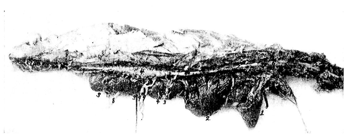
Fig. 50.—Photograph showing spinal cord and relative positions of the kidney, lung and heart. 1, Heart. 2, Lung. Note the indentations made by the ribs. 3, The anterior, middle and posterior lobes of the kidney. The lobules are plainly visible. 4, The lumbo-sacral segment of the spinal cord. 5, Terminal cord filament. 6, The cervico-dorsal segment of the spinal cord. 7, The brachial nerve plexus. 8 and 9, The lumbo-sacral plexus. 8, The ischiadic nerve. 9, The lumbar nerves.
The urinary canals are divided into two kinds, namely, the outer and the inner tubular systems. The outer canals, called the tubulæ uriniferi corticalis, are very small in caliber and are located in the 171lobules. The renal artery (Fig. 53, No. B, 3) breaking up into arterioles in the kidney, and finally reaching the cortical portion of the lobules, form capillary plexuses in the shape of minute spheres, which are the glomerules (Fig. 53, No. C, 3 and B, 5). Around each glomerule there is formed a capsule called Bowman’s capsule, which is the beginning of the uriniferous tubule. This entire mass is called the Malpighian body, or renal corpuscle. This capsule then extends as the urinary, or secreting tubule, being at first constricted, then convoluted, and terminates into a second portion, the descending limb of Henle. This portion of the tubule becomes constricted. It then forms the loop of Henle and ascends as the second portion, or second limb of Henle, which is again of greater diameter. The top of the secreting tubule is slightly wavy and empties into the collecting tubule along with many others. These collecting tubules in turn merge into large tubules which finally empty into the ureter (Fig. 53, No. 14). These collective bundles correspond to the pyramids of the kidneys of mammals.
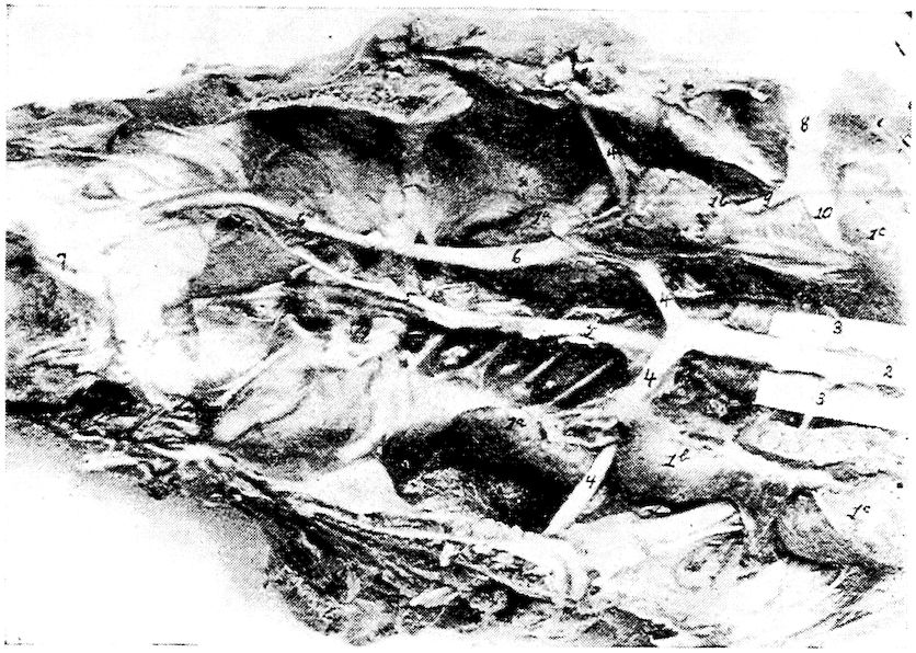
Fig. 51.—The kidneys. 1a, The posterior. 1b, the middle and 1c, the anterior lobes of the kidney. 2, The posterior aorta. 3, The external iliac or crural artery. 4, The ischiadic artery. 5, The sacralis media artery. 6, The ureter which empties into the cloaca at 7. 8, The external iliac vein. 9, The internal iliac vein. 10, The iliacus communis.
172
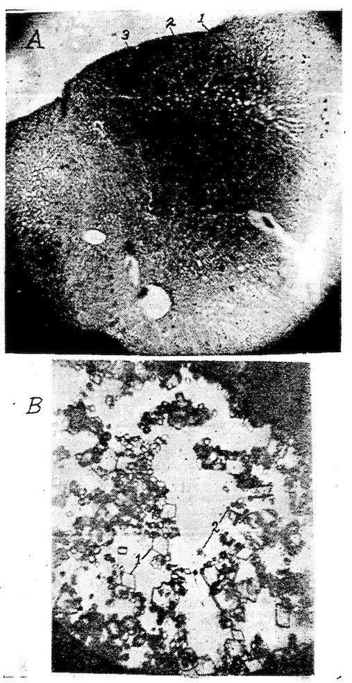
Fig. 52. A. Photomicrograph of a section of a kidney showing three lobules. 1, Cortical portion of lobule showing glomeruli. 2, The central or medullary portion of the lobule. 3, The outer surface of the kidney. B. Salts from the urine of a hen. 1, Uric acid crystals. 2. Sodium urate crystals.
173
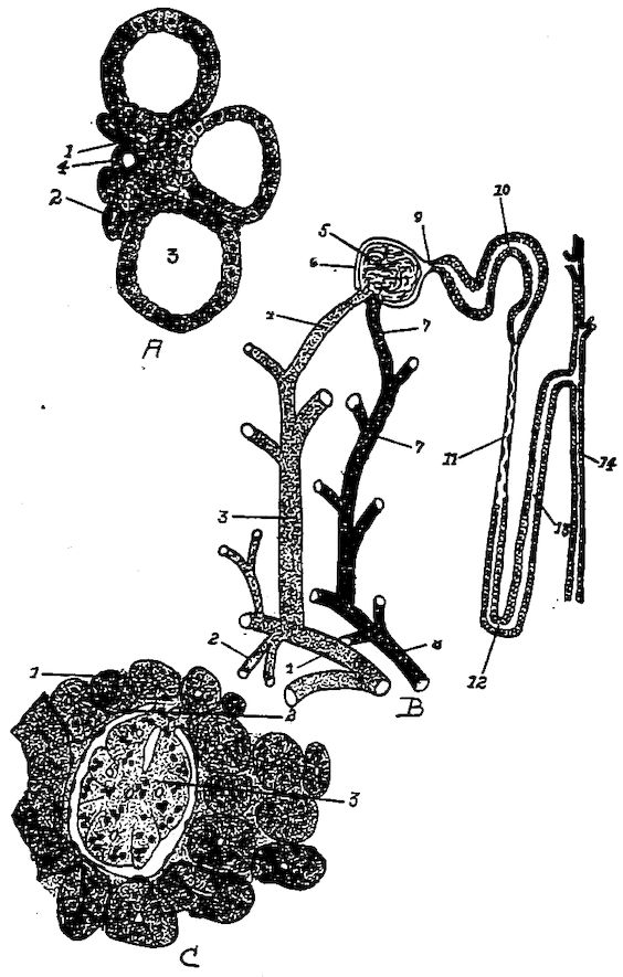
Fig. 53.—The renal structure. A. 1, Capillary blood-vessel. 2, Descending limb of Henle. 3, Collecting tubule. 4, Ascending limb of Henle. B. 1, Branch of renal artery. 2, The descending or medullary branch. 3, The ascending branch. 4, The arteriole taking part in the formation of the glomerule. 5, The glomerule. 6, Bowman’s capsule. 7, Blood-vessel extending from the glomerule. 8, The vein of the medullary part of the lobule. 9, The neck. 10, The convoluted tubule. 11, The descending limb of Henle. 12, Henle’s loop. 13, The ascending loop of Henle. 14, The collecting tubule. C. 1, Section through a convoluted tubule. 2, Bowman’s capsule. 3, The glomerule.
The neck of the uriniferous tubule as it emerges from Bowman’s capsule is short and narrow and is lined with a few cuboidal cells. Toward the glomerular end the cells are of a transitional form gradually merging into the flat squamous type peculiar to Bowman’s capsule. The first convoluted tubule is lined with irregular cuboidal or pyramidal epithelial cells. The descending limb of Henle’s loop is narrow and is lined with a simple layer of flat epithelial cells. In the loop the epithelium changes from the flat type in the descending limb to the cuboidal in the ascending limb. The ascending limb of Henle’s loop again becomes broader and is lined with low cuboidal cells. The second convoluted, or tortuous, tubule is lined with low 174cuboidal cells as are also the collecting tubules. The collecting tubules originate from every part of the internal substance of the lobules, and extending to the gyrations, uniting in the pinniform structure and traversing to the margin of the lobules, following along the uneven surface, infero-laterally and toward the median body line, finally empty into the ureter.
There are many arterial branches given off from the arteria renalis. The arteries which supply the kidneys are given off from the posterior aorta and ischiadic artery. As soon as these arteries enter the kidney they break up into two systems. One system supplies the kidney substance with nourishment in the form of nutrient blood; the second system supplies the glomerules with what may be considered functional blood (Gadow). The second system of arteries branches into the small arteria interlobularis, which pass between the lobules of the kidneys where they give off side branches which penetrate the cortical portion of the lobules and form the capillary plexus, the glomerule.
The arteriole that enters into the structure of the glomerule is lined with endothelial cells and is surrounded by a few muscle fibers and a fine network of connective tissue. The vessel that carries the blood away is similarly constructed.
Function.—In the glomerules the liquid portion of the urine is filtered out of the blood, which urine flows through the uriniferous tubules and passes from the kidney through the ureter. In the cubical cells are extracted the solid portions of the urine which also pass through the tubules with the liquid. The urinary secretion, as found in the ureter, does not contain much liquid, but, on the contrary, is made up of a pasty material consisting of salts which are, for the most part, uric acid crystals and sodium urate (Fig. 52, B). This material becomes hard, like cement, soon after being exposed to the atmosphere. This secretion may be noted as a whitish pasty material on the outer parts of the feces, or droppings, voided by the birds.
The Ureter. Location.—The ureter extends along the inferior surface of the kidney. It has its origin near the anterior extremity of the kidney, and passing posteriorly the entire length of the kidney receives tributary collecting tubules, and terminates in the upper wall of the cloaca in the urodeumal portion.
Shape.—The ureter gradually enlarges in diameter until it reaches 175the posterior border of the kidney, and then maintains about the same caliber throughout the rest of its course.
Structure.—The ureter wall is made up of three coats as follows: mucous, muscular and fibrous.
Function.—The ureters serve as a passage way for the urine from the kidneys to the cloaca.
THE MALE GENERATIVE ORGANS (Fig. 54)
The male generative organs in the fowl consist of two testicles and an excretory apparatus, the vas deferens, for each.
The Testicles. Location.—The testicles are located in the sublumbar region of the abdominal cavity, behind the lungs, below the anterior extremity of the kidneys, and opposite the last three ribs.
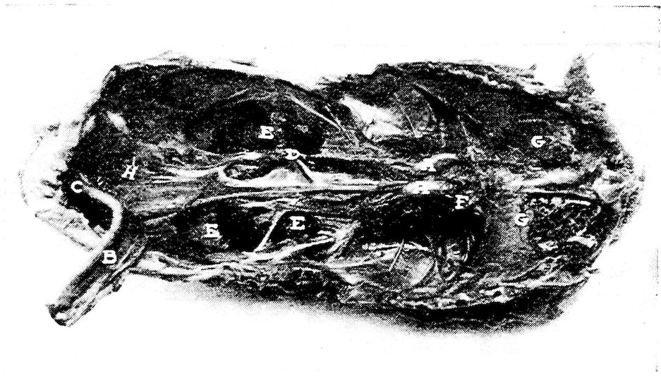
Fig. 54.—The pelvic organs of a cockerel. A, Testes. B, Rectum. C, Cloaca. D, Vas deferens. E, Kidney. F, Adrenal gland. G, Lungs. H, Ureter.
Shape.—The testicles are ellipsoid in shape; their size varies with the different species of birds. The two in the same bird are usually of the same size, though one testicle may be slightly larger than the other. In a summary of a large number of weights of the testicles of ten months old cockerels, the average weight was 0.021 pound each. The average measurements were 2 inches in the major diameter and 1 inch in the minor diameter. In the cockerel before sexual maturity, which is denoted by the male bird’s crowing, the testicles are very small. They resemble, in shape, a navy bean and are yellowish-white in color.
176Structure.—The testicle is surrounded by a thin fibrous capsule, which, in the mature cock, is very vascular. This capsule sends into the interior of the gland, septa which form the framework, or supporting structure. This framework forms the spaces in which are located the glandular substance. The glandular portion consists of the tubuli seminiferi, which are lined with cubical cells. The framework supporting these tubules gives passage to arterial branches of the spermatic artery, which furnish an abundant blood supply; the framework also supports the veins returning the blood from the testicle. The seminiferous tubules end in blind extremities in the epididymus or globus minor, and unite in the seminiferous canals. All arteries, veins, lymphatics, and seminiferous tubules enter or leave through the globus minor at the attached portion of the testicle. The epididymis is made up largely of convoluted tubules which are the continuation of the secreting tubules of the globus major. The walls of the tubules of the globus minor become thicker and are provided with smooth muscle cells in addition to the connective tissue and endothelial lining. The convoluted tubules empty into the vas deferens. The epididymus is covered by the fibrous capsule; which corresponds to the tunica albuginea of mammals and may be considered as a reflection or modification of the visceral peritoneum.
The substance of the testicle is very soft; in fact, it may be said to be of the consistency of encephaloid material. It is made up of secreting tubules in which are found the spermatozoa; it also contains cells which produce an internal secretion, or hormone. There is also produced some fluid in which the spermatozoa float, the whole material manufactured constituting the semen.
The spermatozoa of the fowl are provided with long cylindrical bodies, which may be straight or wavy (Fig. 55, A). The body of the spermatozoon is obtuse anteriorly, and posteriorly, tapers into a filimentary tail, or flagellum, of varying length, by the aid of which the spermatozoon moves about in the fluid.
The histological structure of the testicles of the baby chick at hatching is approximately one-half white fibrous connective tissue. The seminal tubules are small and widely distributed among the connective tissue. The cells of these tubules possess rather large nuclei, round in shape, with linin network and chromatin granules, typical of resting germ cells. As the bird develops the testicles grow, the seminal tubules become larger, and the amount of connective tissue correspondingly less.
177
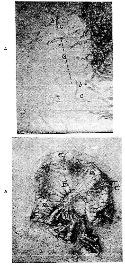
Fig. 55. A. Spermatozoa of a cock. a, The spermatozoa. b, The head. c, The tail. B. The oviduct of a hen removed. A, The oviduct. B, The superior ligament. C, The inferior ligament. Note the oviduct thrown in folds and the anastomosing blood-vessels.
178In addition to the tubules, the spermatogenic cells, and the connective tissue, there is also in the testicles more or less fat. There is a small amount of connective tissue between the seminiferous tubules, in which locations there are also clusters of polyhedral cells, with round nuclei, others are the interstitial cells.
The Vas Deferens (Fig. 54, D). Location and Shape.—Extending from the epididymus, is the vas deferens which runs backward on the infero-internal surface of the kidney and to the outside of the ureter. It is very tortuous passing on the infero-lateral surface of the kidney in company with the ureter and becoming somewhat expanded posteriorly it terminates in the upper wall of the cloaca in a rather small papilla located in the uro-genital portion of the cloaca anterior to the mouth of the ureter. This papilla is the organ of copulation and in ducks is very large, and spirally elongated, and retractile forming a kind of penis. The papilla is traversed by a furrow on the upper surface through which the semen flows.
Structure.—The vas deferens is covered and supported by the peritoneum. Its wall is made up of a fibrous structure in which may be found smooth muscle fibers. The wall does not possess glands. It is lined with columnar epithelium. The posterior end is expanded and terminates into a papilla. The base of the papilla is surrounded by a plexus of arteries and veins, which serve as an erectile organ during the venereal orgasm, when the fossa of the turgid papilla is everted, and the semen brought into contact with the similarly everted orifice of the oviduct of the female, along which the spermatozoa pass by undulatory movements of their ciliary appendage, or tail.
THE FEMALE GENERATIVE ORGANS (Fig. 56)
The female generative organs consist of one ovary and an oviduct.
Location.—The ovary is located similarly to the testicles of the male bird, in the sublumbar region of the abdominal cavity, just at the anterior end of the kidneys, posterior to the lungs, and slightly to the left of the center.
Shape.—In the pullet the ovarian mass appears somewhat like a bunch of grapes, being made up of from 3500 to 4500 small, whitish spheres, which represent the undeveloped ova, and which in the 179active state are developed, one by one, into yolks with their blastoderms. From the blastoderm the fetus may later be developed. In the active ovary of the laying hen the ovarian mass is of considerable size, as it contains ova in different stages of development. Only one ovum is completely developed at a time, though occasionally there may be only a few hours between the maturity of successive ova. The ova receives nourishment from the blood-vessels of the capsule, which vessels are branches of the ovarian artery.
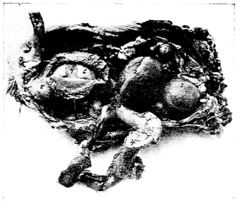
Fig. 56.—Functionating female generative organs of a hen. 1, Ova in process of formation of yolk. 2, Stigmal line at which point the capsule ruptures when ovum is mature. 3, The funnel end of the oviduct. 4, The oviduct torn loose and laid to one side, the albumin-secreting portion. 5, The shell membrane secreting portion. 6, The albumin. 7, The yolk. 8, The shell-secreting portion. 9, The cloaca. 10, The rectum.
Structure.—The ovary contains very vascular cellulofibrous tissue. The ovum as it develops is attached to the ovarian body by means of a delicate white fibrous pedicle. When the yolk is mature it escapes from the enveloping fibrous capsule by a cleavage of the capsule. The cleavage line is called the stigmen (Fig. 56, No. 1802). The yolk is surrounded by a very delicate membrane called the vitelline membrane. The empty sac now shrinks and finally disappears.
The Egg.—The principal divisions of the egg are the yolk, the albumin outside of the yolk content, the shell membranes and the shell (Fig. 58). As stated, the yolk is formed in the ovary, leaving the other three portions to be formed in the oviduct.
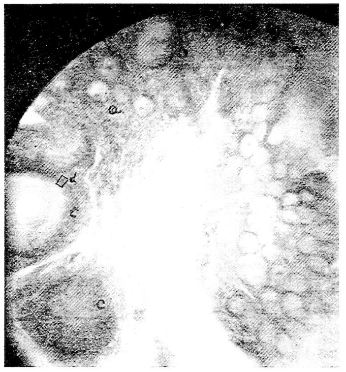
Fig. 57.—Section through the ovary of the hen. a, The ova. d, An ovum beginning to receive deposits of yolk material. c, Ova farther advanced in yolk formation.
The albumin may be subdivided into, first, a thin layer of albumin lying close around the yolk; second, a thick layer of albumin lying at the outer periphery; and third, a modification of the albumin called the chalazæ. The chalazæ are twisted, dense cord-like structures at either pole of the yolk, one end of which is adherent to the vitelline membrane and the other to the inner membrane surrounding the albumin. The chalazæ thus act as stays to this structure, which is carrying a delicate burden, the blastoderm.
181The shell membrane consists of two layers, an inner delicate, and an outer thicker layer. When the egg is just laid these two membranes are in all parts closely adherent to each other, and the egg content completely fills the shell cavity. As soon as the content cools there is a slight contraction; the two shell membranes separate at the large end of the egg, forming an air cell which gradually enlarges as the evaporation of liquid through the pores of the shell takes place.
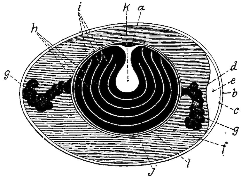
Fig. 58.—A diagram of the parts of the egg. a, The blastoderm. b, The shell. c, The outer shell membrane. d, The inner shell membrane. e, The air-sac at the large end of the egg. f, The albumin. g, The chalaza. h, The yellow layers of yolk. i, The white layers of yolk. k, The flask-shaped portion of white yolk. l, The vitelline membrane.
The shell consists of several layers. Three are easily distinguishable: first, an inner mammillary layer, consisting of minute conical deposits of calcareous material; second, a middle spongy layer, composed of a thick network of fibers; third, an outer delicate, cuticle-like structure. In certain breeds of poultry a pigment may be added; for example, in ducks a pea green, in turkeys a spotted brownish material, and in fowls pink and various shades of brown. The egg shell is porous to admit the free exchange of air during the process of incubation.
An average sized hen egg weighs about 2 ounces, of which 11 per cent. is shell, 32 per cent. yolk, and 57 per cent. white. The principal chemical constituents of the egg are as follows: ash, or mineral matter, 9 per cent.; fat, or hydrocarbon, 9.3 per cent.; proteids, or nitrogenous matter, 11.9 per cent.; and water, 65.5 182per cent. There is apparently no constant proportion of weight between the yolk and albumin. There is also a variation in the weight of the shell due to its variation in thickness.
In an examination of ten eggs of average size, the yolk constituted 31 per cent. of the total weight of the egg.
The following is the result of the analysis of twelve eggs of average size. This analysis included the shell and all other parts taken together.[6]
Moisture 64.25 per cent.
Dry matter 35.75 per cent.
100.00 per cent.
Parts per hundred including shell
Protein 10.2500
Fat 10.6200
Phosphorus .3020
Calcium .6080
Magnesium .0985
Iron .0103
Sulphur .3950
Chlorine .1506
Potassium .0103
Sodium .2000
The Oviduct.—The three separate and distinct portions of the egg, albumin, shell membranes, and the shell, are constructed in different parts of the oviduct.
Location.—The oviduct of the hen extends along the left side of the bodies of the vertebræ, and the roof of the pelvic cavity and lies dorsal to the abdominal air-sac. It extends from the posterior border of the ovary and empties into the cloaca (Fig. 56, No. 3 and 8) through a transverse slit.
Shape.—In a well-developed Plymouth Rock pullet, but one whose reproductive organs have never become active, the oviduct is about 4½ inches long; in the fully developed and active state it is from 18 to 20 inches long, and in a collapsed state about ½ inch in diameter. It is held in position by two ligaments, a dorsal and a ventral (Fig. 55, B) to be described later. The oviduct is tortuous in its course, forming three principle convolutions before reaching 183the cloaca, and like the ovarian mass, in its active state, pushes the abdominal viscera downward and toward the right side.
Structure.—The oviduct consists of three coats: first, the serous, located on the outside, which is a reflection of the peritoneum; second, the muscular middle tunic; third, the mucous coat, which in a resting state is thrown into folds.
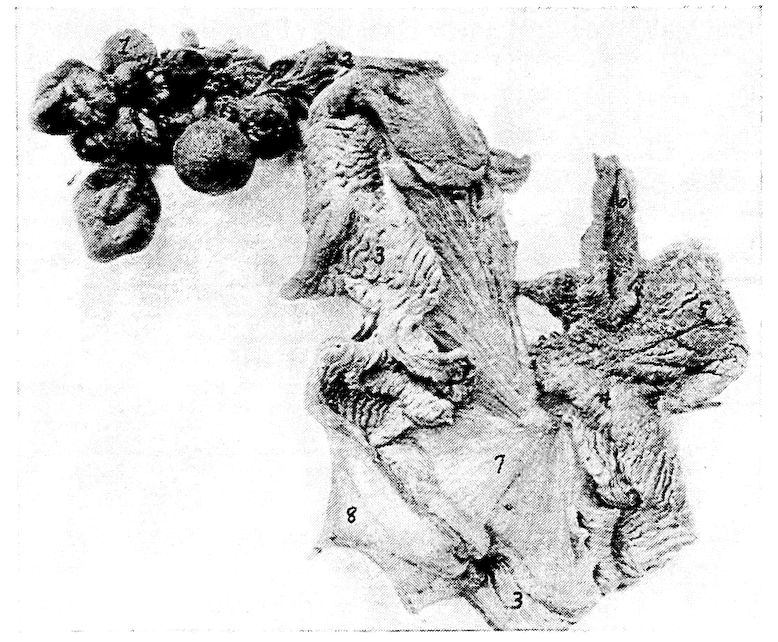
Fig. 59.—The active oviduct of a hen laid open. 1, The ovary. 2, The funnel. 3, The albumin-secreting portion. 4, The isthmus. 5, The shell=gland portion. 6, The vagina. 7, The superior ligament. 8, The inferior ligament.
Parts of the Oviduct.—The parts of the oviduct are as follows: the funnel, the albumin-secreting portion, the isthmus, the shell-gland portion, or uterus, and the vagina (Fig. 59).
184
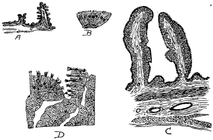
Fig. 60.—The mucous lining of the oviduct. A. Transverse section of the oviduct wall in the region of the neck of the funnel showing primary and secondary folding of the epithelium (after Surface). B. Showing the type of gland cells of the funnel region. C. Transverse section through the wall of the uterus showing the deep folds. D. Section from the albumin-secreting portion showing the opening of a tubular gland and also showing the character of the cells.
The funnel is the trumpet-shaped portion, the ostium tubæ abdominale, whose mouth or fimbriated opening faces the ovary, and lies ventrally to receive the ovum, or yolk, as it is discharged from the ovary. Its thin wall, expanded in the anterior portion, is provided with fimbriæ-like projections. This funnel-shaped portion soon converges to form a constricted portion. This portion of the active oviduct is from 3 to 4 centimeters long. The mucous membrane occurs in folds forming low longitudinal spiral ridges (Fig. 60, A). The fimbriæ are continuous with the dorsal and the ventral ligament of the oviduct; and from this point, where the ridges of the mucous membrane are almost nil, they gradually increase in height as they extend down the tube. These ridges are continued in those of the second, or albumin-secreting portion (Fig. 60, D). At this point they increase in height very rapidly. Here the bundles of muscular fibers of the middle coat are thin and distributed among bundles of connective tissue. The muscular fibers consist of two layers, an outer longitudinal and an inner circular. At places in this portion the inner bundles may be noted to extend longitudinally. In embryological development the epithelial layer has an origin different from the outer layers of the oviduct. In the fetal development the Müllerian duct arises as a thickening along the Wolffian body just ventral to the gonad. This Müllerian duct is at first a solid cord of cells. It later develops a lumen, and grows posteriorly until it connects with the cloaca. At the time of this posterior growth, mesenchyme cells migrate in from the surrounding tissue and form a layer about the duct. From this layer of mesenchyme cells there are developed the outer 185layers of the oviduct, which layers later develop the muscular structure and the connective tissue. The epithelium and its derivatives, which represent the glandular structures, are formed from the walls of the old Müllerian duct. Thus the two sets of tissues, having different origins, likewise have different functions. The epithelium is concerned entirely with secretion, and the derivatives of the mesenchyme are concerned with supporting and muscular function.
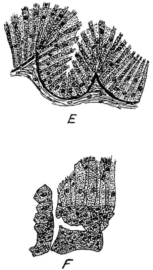
Fig. 60. A.—The mucous lining of the oviduct. E. Section of the epithelium from the vagina showing unicellular glands. F. A section from the isthmus showing opening of a tubule.
To summarize, six layers of tissue occur in the funnel region, namely, an outer serous covering, an outer longitudinal muscular layer, a layer of connective tissue, an inner circular layer, a second layer of connective tissue, and an inner mucous layer. The mucous layer is made up of glands, as follows. The unicellular glands occur between the ciliated cells of the epithelium. These glands are found only in the posterior half of this division of the oviduct. The glandular grooves are made up of an accumulation of gland cells at the bottom of the grooves between the secondary folds of the epithelium. These are found in all parts of this division except the extreme posterior part. In the posterior part we find the third type of glands, the tubular variety.
The second division of the oviduct, as stated above, is the albumin-secreting portion. The funnel division gradually merges into the second portion. These two portions are distinguishable from each other. The walls of the albumin-secreting portion are much thicker and the longitudinal ridges are higher. This section is the longest of the five divisions, measuring from 40 to 42 centimeters in length, or more than half the length of the oviduct. The albumin division terminates rather abruptly into the third division, the isthmus (Fig. 59, A).
186It is probable that the secretion of albumin is not confined to the cells of the second division; yet we are safe in saying that the major portion is formed here. The folds of mucous membrane in this division are thicker and higher than in the funnel, due to their containing cells of the high columnar type, and to the fuller development of the glands which are of the tubular variety. The muscular layer is heavier, and therefore the muscular power to force along the tubes’ contents is greater. In the formation of the mucous folds we find, in transverse sections, that the central core is made up of connective tissue which carries blood-vessels and nerve filaments, as in other similar glandular structure. The epithelium contains glandular cells of two varieties, namely, the ciliated, columnar variety, and the unicellular, goblet variety (Fig. 36, No. B, 2). These two kinds of glandular cells are rather evenly distributed throughout the epithelium of this section of the oviduct. The unicellular gland cells are more numerous at the mouths of the ducts leading from the tubular glands. The nuclei of the ciliated cells are oval and lie near the middle of the cells or a trifle toward the base from the middle. The protoplasm of the cells is finely granular. Strong cilia in considerable number surmount each cell. In some cases the goblet or mucous cells have pushed apart the ciliated cells, and their prolongations extend farther than the surface of the ciliated cells. The nuclei of the goblet cells are round, and lie nearer the proximal end than those of the ciliated cells.
The third division of the oviduct, the isthmus, continues from the albumin-secreting portion and terminates in the expanded portion called by some anatomists, the uterus. Toward the posterior end of the albumin-secreting portion the longitudinal folds of mucous membrane become lower, making, at the juncture of this and the isthmus, a clear fine of demarcation. For a distance of 2 or 3 centimeters the folds are low, after which they gradually become higher, but never reach the height or thickness of those in the albumin-secreting portion.
The clear-cut line between the albumin-secreting portion and the isthmus is partly due to a zone in which the long tubular glands are lacking. The core of the folds of mucous membrane in this zone contain much more connective tissue. The cells are both ciliated and unicellular. The rest of the histological structure of the isthmus is the same as that of the albumin-secreting portion.
187The function of the isthmus is to secrete, or to form, the shell membrane, the membrana testacea.
The fourth division of the oviduct is the uterus, or the shell-gland portion (Fig. 60, C). There is no clear line of demarcation between the isthmus and shell-gland portion, the walls gradually expanding. In this region the folds of mucous membrane become leaf-like and of considerable length, extending into the lumen, thus affording a greater cellular surface. The same coats of the duct are present here as in other parts; but the outer longitudinal muscular layer is thicker and possesses more strength. The quantity of connective tissue is about the same. The glands are of a tubular type and the same two varieties of epithelial cells are found here as elsewhere, namely, the ciliated high columnar and the unicellular mucous variety.
In the active glandular cells of the shell-forming region the nuclei are small, dark staining, and lie toward the center of the cell. The chromatin granules and nucleoli take on a comparatively deep basic stain, but they do not show the intense stain found in the albumin and the isthmus region. The cytoplasm of the uterine tubular glands does not present as heavy a granular appearance as that of the albumin portion. These cells are diffusely granular, the granules appearing of one size and taking the stain faintly. The function of this portion is to secrete, or form, the hard calcareous covering which has been described at the beginning of this section.
The fifth division of the oviduct is the vagina. There is located, at the juncture of the shell-gland portion with the vagina, a strong sphincter muscle. The vagina is that constricted portion of the oviduct extending from this muscle to the cloaca. The mucous membrane forms low narrow folds with secondary folds, which appear continuous with those of the shell-gland portion. The core of these folds is composed of connective tissue. The vagina in the hen of average size measures from 12 to 13 centimeters long (Surface). The inner or circular muscular layer is well developed; it is much thicker than in any other part of the oviduct. This extra development gives the power necessary to successfully expel the egg. The outer longitudinal layer is not so well developed; its bundles are scattered throughout the connective-tissue layers. The egg is caused to move along in the oviduct by a successive series of contractions of the circular muscular fibers posterior to it. There are no tubular glands in this portion, but a simple layer of high 188ciliated columnar epithelium, and some goblet, or mucous cells. The cells on the surface generally are long and slender; in the grooves between the mucous folds the cells are shortest, reaching their greatest length at the tops of the folds.
The function of the vagina is the secretion, or formation, of the outer shell cuticle commonly called the bloom, and also in certain breeds, as indicated above, the tint.
The cloaca furnishes a passage way from the vagina to the external world by way of the anus. The walls of the cloaca contain glands.
The Ligaments of the Oviduct.—It is held in position by two ligaments, one dorsal and one ventral. The dorsal ligament of the oviduct is formed by a double layer of peritoneum with a very small amount of connective tissue interposed. The peritoneum is also reflected over the oviduct. The ventral ligament of the oviduct is narrower than the dorsal, but is similarly constructed. Both ligaments are rather veil-like in appearance. During the first four or five months of the growth of the young female fowl the development of the oviduct and its ligaments is in proportion to that of the body. With the elongation that takes place about the time of functionation, as described above, the ligaments enlarge in proportion to the enlargements of the oviduct. The dorsal ligament maintains a line of attachment to the body wall from the caudal end of the body cavity to the fourth thoracic rib. The ventral ligament elongates only slightly during this developing period. It becomes thicker and stronger and early develops a muscular coat. It also grows in width except at the caudal end. At this point the ligament is simply a mass of muscular tissue of the smooth or involuntary type. These ligaments are fan-shaped.
The muscle fibers of the dorsal ligament of the laying hen, have their origin in a line near the medial side of the dorsal margin. At this point the bundles of fibers are quite thick, but are spread out thinly toward the margin of the oviduct. Frequent anastomoses are noted. The muscular fibers become continuous with the circular ones of the oviduct.
The ventral ligament of the oviduct of the laying hen is largely a muscular cord 3 to 6 centimeters in diameter. The caudal end is thicker, becoming gradually thinner toward the anterior portion. The bundles of muscular fibers extend toward the oviduct blending with the circular fibers of that viscus. The ligaments terminate 189anteriorly in such a manner that they aid in forming the serous ovarian pocket, which guides the yolk into the fimbriated portion, or funnel, of the oviduct. The walls of the ovarian pocket are formed by the left abdominal air-sac, a part of the intestine, and the mesentery. The dorsal portion is formed by the roof of the abdominal cavity, and the ventral portion is formed by the dorsal wall of the air-sac. The medial, the anterior, and the lateral limit of the pocket are formed by a fusion of the wall of the air-sac to the mesentery and to the body wall. Posteriorly, the wall consists of the transverse part of the small intestine and the caudal portion of the left cæcum with their attached mesentery.
190