THE RESPIRATORY APPARATUS (Figs. 50 and 61, A)
Reading edition
Owen says: “Notwithstanding the extent and activities of the respiratory function in birds, the organs subservient thereto manifest more of a reptilian than of the mammalian type of formation.”
By the action of the respiratory organs certain chemical and physical changes take place in the blood. The chief of these consists in absorption of oxygen from, and giving off carbon dioxide to, the atmospheric air, the former changes being necessary for the elaboration of the fluid, the latter for the elimination of a substance which, if retained, would prove injurious. The organs of respiration are invariably adapted to the wants of the animal and the medium in which it lives.
In the bird, which breathes through its nose, the organs of respiration are nostrils, nasal chambers, pharynx, superior larynx, trachea, inferior larynx, bronchi, bronchial tubes, lungs, and air-sacs.
The Nostrils and the Nasal Chambers.—The nostrils of the bird open externally by two small elliptical openings, which pierce the upper mandible. Within each nasal chamber (Fig. 26, A) are three turbinated laminæ, or turbinated bones. The inferior one is a simple fold adhering to the lower and anterior part of the nasal septum. The middle turbinated bone is the largest. It is of infundibular form, and adheres by its base to the septum and externally to the side wall of the nose. It is convoluted with two and a half turns. The superior bone, of bell shape, adheres superiorly to the frontal bone. The internal turbinated bone extends toward the orbit; the external terminates in a cul-de-sac behind the middle turbinal (Fig. 26, No. A, 1 and 2, and G).
The nostrils are separated by a partition which is partly bony and partly cartilaginous. The posterior nares is represented by a long slit in the hard palate.
The Pharynx and the Superior Larynx.—A transverse row of horny, filiform papillæ marks the anterior border of the pharynx, where in other animals the soft palate, or velum, is located. (For further description of the pharynx see special chapter.)
The supero-posterior border of the larynx, at the juncture of the 194larynx and the esophageal margin, is marked by a second transverse row of horny, filiform papillæ, which point backward. There is no epiglottis. The superior part of the larynx is pierced by an oval, slit-like opening, the glottis, which is provided with two lips. These when brought together, tightly close the glottis so that nothing can fall through into the larynx in the act of deglutition. The margin, or rim, of this opening is called the rami glottis. The glottis is controlled by two pair of muscles.
The superior surface of the larynx is somewhat triangular with the apex directed forward. A few delicate, filiform papillæ are upon its surface. The bird has, as already indicated, two larynxes, the superior larynx located at the upper end of the trachea, and the inferior larynx at the bifurcation of the trachea. The inner surface of the superior larynx is smooth and does not contain vocal cords; it is in these animals simply a passage for air. It is joined to the trachea inferiorly by a ligament, the crico-trachealis, and lies at the base of the tongue supported by two cornua of the os hyoideum.
The cartilages forming the principal support of the superior larynx, consist of four pieces, as follows: one unequal ventral piece, two side pieces, and one unequal dorsal piece. The cartilaginous, flat, ventral cricoid, early in the bird’s life, often becomes bony. The side pieces are separated from it, only exceptionally fusing with it. The dorsal cricoid piece also often becomes bony. The two arytenoid cartilages, joined with the cricoid superiorly, are three-sided, and are united to each other in a sharp angle. They form the superior opening of the superior larynx.
The Trachea.—The trachea is cylindrical and varies in length in different kinds of birds in accordance with the length of the neck. It consists of from 90 to 120 cartilaginous rings, complete with the exception of the two uppermost, which rings are held together by intercartilaginous ligaments. The tracheal rings are constructed of hyaline cartilage and the ligaments of fibrous tissue. It is lined with a mucous membrane covered by columnar epithelium. The trachea is a passage for air alone and terminates in the inferior larynx.
The Inferior Larynx.—The inferior larynx, called the true larynx because it is the organ of voice, is located at the inferior end of the trachea and the superior ends of the bronchi. By some anatomists this organ has been called the larynx broncho-trachealis. The larynx is flattened laterally in fowls. It contains two membranous folds, 195which in the production of sound are caused to vibrate. These folds are half-moon shaped elastic structures, located in the bony, arrowlike way, intero-inferiorly. These structures are called the membrana tympana interna. In the duck this inferior larynx is represented by a drum-like cartilaginous and bony structure, called the bulla tympaniformis. This bulla is a resonant apparatus which serves to strengthen the voice.
In song birds there is a double glottis, usually produced by a bony bar, called the pessulus, or os transversale, which traverses the lower end of the trachea from front to rear. It supports a thin membrane which ascends into the tracheal area, and, terminating there by a free concave margin, is called the membrana semi-lunaris. This is most developed in singing birds, and being vibratile, forms an important part of their trilling vocal apparatus. The air passes on each side of the membrana semi-lunaris and its sustaining bone to and from the bronchi and lungs.
The last ring of the trachea usually expands as it descends, with its fore and posterior parts produced, and the lower lateral borders concave; the extremities of the pessulus, butts against the angle thus formed and expands to be attached, also with the fore and posterior terminations of the first half ring of the bronchus, strengthening and clamping together the upper part of the vocal framework. The second bronchial half ring is flattened and curved with the convexity outward, like the first, but is more movable. The third half ring is less curved and further separated from the second, to the extremities of which its own are connected by a ligament, and, for the intervening extent, by a membrane; its inner surface supports the fibrous cord, or fold, which forms the outer lip of the glottis of that side; it is capable of rotary movements on its axis, and is an important agent in the modulation of the voice. All these parts just described are bony.
The Bronchi and the Lungs.—The bronchi, two in number, are provided with only incomplete cartilaginous rings. They enter the inferior face of the lungs, toward their anterior and middle thirds and break up into primary bronchi, which give off at right angles, secondary bronchi, and these latter in turn give off tertiary branches.
196
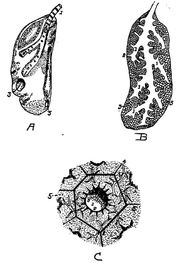
Fig. 61.—The lung. A. The outer surface of one lung. Note the flattened oval shape. It is not divided into lobes. 1, The bronchus. 2, Primary tubules showing openings leading from the primary tubules to the secondary tubules. 3, Openings of two of the large tubes into the diaphragmatic and abdominal air-sacs. B. Sectioned surface of lung. 1, Secondary tubules. 2, Tertiary tubules. 3, Interlacing capillaries and air cells. C. 1, Cavity of tubule. 2, Its lining membrane supporting blood-vessels with large areolæ. 3, Perforations in the membrane at the orifices of the lobular passage. 4, Interlobular space containing the terminal branches of the pulmonary vessels supplying the capillary plexus, 5, to the meshes of which air gets access by the lobular passage.
The lungs occupy only about one-seventh of the thoracic space. They are long, flattened, and oval, extending along each side of the spine from the second dorsal vertebra to the anterior end of the kidneys, and laterally to the juncture of the vertebral with the sternal portions of the ribs. They present two faces, a superior convex and an inferior concave; two borders, an external and an internal; and two extremities, an anterior and a posterior (Fig. 61). The convex surface is also called the dorsal, costal, or superior face. It is moulded on the walls of the thorax and occupies a part of the intercostal space, pushing the intercostal muscles outward. When the surface of the lung is examined it is seen to be furrowed where the ribs pressed during life. These furrows are as deep as the ribs are 197thick. The sides of the lungs are covered with connective tissue which attaches them to the costal walls.
The concave, or inferior, face also called the diaphragmatic or visceral face is directed downward. The diaphragm separates it from the abdominal viscera. The surface is covered by connective tissue which closely attaches it to the diaphragm. It is perforated by the five tubules which bring the posterior air-sac into communication with the lungs.
The borders of the lungs extend parallel to the long axis of the body. The internal border is rectilinear, thick, and rounded. The external border is convex, thin, and sharp.
The anterior extremity terminates in a sharp point which occupies a space formed by the ribs externally and the inferior spines internally. The posterior extremity is somewhat rounded and extends as far back as the anterior border of the kidneys.
As soon as the bronchi enter the lungs they become broadened, the cartilaginous rings disappear, and they continue as membranous channels whose diameters gradually decrease, as they extend backward, to the point where they terminate in the ostium caudale, at which point they are surrounded by a cartilaginous ring. The ostium caudale brings the tubules into communication with the ventral air-sacs.
Twelve air tubules have their origin from each common bronchus, or trunk. Four are given off from the internal wall of the main bronchus by a series of openings arranged in a row. Seven are given off from the external wall by a second series similar to that of the first. The twelfth extends from the inferior wall, and immediately takes a course downward and outward and communicates with the posterior diaphragmatic air-sac. This may be considered as the terminal branch of the trunk.
All of these secondary canals, except the last, pass toward the periphery of the lung. They divide and subdivide at the periphery, covering it with their ramifications. The canals extending from the inner wall are distributed to the inferior face of the lung. Those extending from the outer wall are distributed to the outer face of the lung. The first constitute the diaphragmatic and the second the costal bronchial tubes.
The four diaphragmatic bronchial tubes are numbered in the order in which they are given off. The first is carried forward horizontally, the second transversely inward, the third obliquely inward and backward, 198and the fourth directly backward. They have, by some anatomists, been called the anterior, the internal, and the posterior diaphragmatic bronchial tubes. There are two posterior diaphragmatic bronchial tubes; the larger called the great posterior, and the smaller, which passes directly backward, the small posterior.
The costal bronchial tubes, seven in number, are numbered from the front backward in the order they are given off. Parallel at their origin, and side by side, like pipes of an organ, they soon spread out in fan-shape like the preceding. They extend from their central origin to the periphery. The first extends obliquely upward and inward to the anterior extremity of the lung. All branches from this bronchus extend from its anterior wall. The first branches are inflected to reach the external border of the lung. The succeeding branches are directed forward and the last forward and inward. They all meet those from the anterior diaphragmatic bronchus, but do not anastomose with them.
The second, the third, and the fourth costal bronchi extend in a transverse manner and ramify on the inner border of the lung.
The fifth and the sixth are directed toward the posterior extremity of the lung. The seventh, very small, reaches this extremity, where it disappears.
The first costal bronchus is the largest; those following it gradually become smaller. At their points of origin they adhere closely to the ribs. They are all imperforate, which is a distinguishing feature from those occupying the opposite face.
The canaliculi or tertiary tubules given off by these secondary bronchial tubes do not differ greatly in caliber in the various parts. They are given off at right angles from the pulmonary wall of each bronchus, and extend perpendicularly into the lung substance. Thus we find three kinds of conduits, the primary, the secondary, and the peripheral, or tertiary. The first are like the barbs of a feather on its shaft; and the second and parenchymatous are implanted on the pulmonary walls of the first, like the hairs of a brush on their common base. Thus instead of the branching of the bronchi being dichrotomous, as in mammals, it is piniform.
The canaliculi, or finer tubules, communicate with one another. The inner microscopic appearance of the canaliculi indicate that they are divided into areola, which gives them a cellular aspect. These tertiary bronchi open on a dense labyrinth of blood capillaries 199(Fig. 61, C). At this point the ciliated epithelial cells give way to simple squamous epithelium.
Thus we find three kinds of bronchi, or their ramifications, as follows: the primary, the secondary, and the tertiary.
The Air-sacs (Fig. 61, A).—The air-sacs are bladder-like structures consisting of a delicate cellulo-serous membrane, an extension from the bronchial tubes, in some places strengthened by an external envelope of elastic fibrous tissue. Long thin blood-vessels are distributed in the substance of these walls. They are branches from vessels of the general circulation and not extensions from those of the lungs. No lymphatics have been found in the air-sacs.
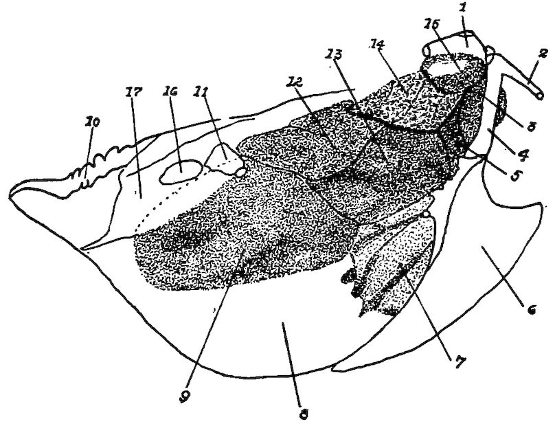
Fig. 61, A.—Diagram of air-sacs and their location. 1, The proximal end of the humerus. 2, The proximal end of the right clavicle. 3, The cervical air cell. 4, The right coracoid bone. 5, The anterior thoracic air cell. 6, The right side of the sternum. 7, The right side of the liver. 8, The peritoneum. 9, The right abdominal air cell. 10, The coccyx. 11, The proximal end of the right femur. 12, The right supero-posterior air-sac. 13, The right infero-posterior air-sac. 14, The right lung. 15, The axillary extension of the air-sac. 16, The obturator foramen. 17. The pelvis.
These sacs do not communicate with each other and normally they are not fully inflated. In some locations they extend into the bones and are in communication with the extensions of the bronchial tubes. In fact, by some anatomists they have been called “bladder-like, extra-pulmonary expansions of the bronchial tubes, free from cartilage.” The air-sacs make the bird’s body lighter, thus making 200long-continued flight possible. They are best developed in those birds which fly most. There are four pairs of cells and one single cell from which all other expansions and extensions are made. These sacs are as follows: a single anterior thoracic, and, in pairs, cervical, anterior diaphragmatic, posterior diaphragmatic, and abdominal.
The Anterior Thoracic Air-sac.—The anterior thoracic air-sac is located above the clavicles and the interclavicular space, in the cavity of the thorax. It is related superiorly with the trachea and the esophagus; laterally with the lungs and the cervical air-sacs; inferiorly with the sternum, the clavicle, and the interclavicular aponeurosis; posteriorly with the heart and the anterior diaphragmatic reservoir; and anteriorly with the integuments of the neck. It contains the inferior larynx and the two primary bronchi, and large vascular trunks from which are given off vessels supplying the neck and the wings.
Three prolongations, called subpectoral, subscapular, and middle, or humeral, arise from the lateral walls of this air-sac. These prolongations cross the walls of the thorax and pass around the articulation of the shoulder.
The subpectoral prolongation extends from the thoracic reservoir by an orifice situated behind the coracoid, and passes beneath the tendon of the great pectoral muscle. When the pectoralis major contracts, this contraction dilates the subjacent cell and draws into it a greater quantity of air.
The subscapular and the humeral prolongations communicate with the thoracic air cell by a common opening situated behind the small adductor muscle of the humerus. The subscapular prolongation, after leaving this point, spreads under the scapular and the subscapular muscle, which it separates from the ribs and corresponding intercostal muscles, and extends in a longitudinal direction.
The humeral prolongation, smaller than the subscapular, occupies the axilla, and is in shape triangular. It has from its summit into an infundibular fossa, an extension which enters the canal of the humerus. The walls of this cell form the lining of the air space in the humerus.
The thoracic air-sac thus possesses numerous membranous folds which divide its cavity. The contiguous structures which it overlies, as the trachea, the esophagus, the muscles of the inferior larynx, as well as the arteries and veins, make its outer walls irregular. 201This orifice is dilated during inspiration, by the contraction of the two first fasciculi of the diaphragm.
The Cervical Air-sacs.—The two cervical air-sacs are located just above the thoracic air-sac at the inferior part of the neck and in front of the lungs. They are cone-shaped with the base directed forward and the apex backward. They are related superiorly with the cervical muscles, and inferiorly with the thoracic air-sac from which they are separated by the trachea, the esophagus, the pneumogastric nerve, and the jugular veins. The walls touch each other internally, and form a median septum which includes in its substance the two common carotid arteries. Externally they are related to the origin of the cervical nerves, to each of which they contribute a small sheath. They surround the vertebral artery, and are connected with the subcutaneous muscles and the skin. The summits communicate with the anterior diaphragmatic bronchus. Prolongations extend from their bases which conduct the air into all the vertebræ of the neck and the back, into all the vertebral ribs, and into the spinal canal. Parallel with and adjacent to the vertebral arteries, and lodged in the canals excavated in the transverse processes of the cervical vertebræ, are two cervical prolongations, one on each side, which extend to the cranium from the base of the cervical reservoirs. From their sides, at the last six cervical vertebræ, are six extensions in the form of diverticuli, which, lying against each other, pass from each side into the muscles of the neck. They are surrounded by a thin fibrous envelope, a continuation of the mucous lining of the sac, and apparently form a canal in the inferior part of this region. These prolongations are better developed in palmipedes than in chickens. On the internal side of these prolongations, one or more foramina penetrate the vertebral segment, which allow the extensions of the prolongations into the spinal canal. Chauveau states that “as the medullary tissue is replaced by air in the bones of birds, so is the subarachnoid fluid replaced by air around the spinal cord.”
The prolongations extending from the cervical air-sacs, having entered the thorax, terminate by passing into the first dorsal vertebra. After permeating every part of this vertebra, it escapes by a lateral opening and forms a small sac located between the first two ribs, near the origin of the first dorsal nerve. From this sac an 202extension is given off, which enters the second vertebral segment at the antero-lateral part; from this point it passes back, forming a new air-sac between the second and third ribs. It now passes in the same manner into the third vertebra and extends through the third intercostal sac, and so on till the last dorsal vertebra has been served. At the same time that these sacs receive the air from the vertebræ preceding them, and transmit it to those which follow, they communicate it to all the vertebral ribs. The aerial currents which leave the cervical air-sacs do not communicate with those of the cranium. Experiments show that the cranial bones have apparently no communication with the respiratory apparatus.
The Anterior Diaphragmatic Air-sac.—The two anterior diaphragmatic or supero-posterior air-sacs are related with the lungs anteriorly, and with the abdominal viscera posteriorly. Anteriorly also is the thoracic air-sac, posteriorly are the posterior diaphragmatic air-sacs, and laterally the ribs and the intercostal muscles and internally is the esophagus. The lungs communicate with these air-sacs through circular openings from the great posterior diaphragmatic bronchus and frequently by a second opening from this same tube. These are the only sacs which receive air from the lungs through two openings.
The Posterior Diaphragmatic Air-sac.—The two posterior diaphragmatic, or infero-posterior air-sacs are oval in shape and located between the thoracic and the abdominal cavity. They are related anteriorly with the anterior diaphragmatic air-sacs. These two sacs form a vertical transverse partition. The posterior diaphragmatic air-sacs are related posteriorly with the abdominal air-sacs from which they are separated by the diaphragm. They are related below with the lateral parts of the sternum and the sternal ribs, and externally with the ribs and the intercostal muscles. These air-sacs communicate with the lungs through openings located in the middle part of the external border of the lung, into the extremity of voluminous bronchial tubes which follow the direction of the largest air tubes.
The Abdominal Air-sacs.—The two abdominal air-sacs located on each side of the abdominal cavity, when inflated with air, form enormous bladder-like structures. They are related laterally with the abdominal wall and internally with the abdominal viscera. The anterior extremities are in communication with the mesobronchi and are somewhat inflected to pass under the fibrous arches extending 203from the spine to the pelvis. Anteriorly these sacs adjoin the diaphragm, the testes in the male, and ovary in the female, and to the parietes of the abdomen and those of the pelvis. Below and in front, they rest on a fibrous septum, which in all birds divides the abdominal cavity into two smaller cavities: one anterior, representing the abdomen and containing the liver; the other posterior, representing the pelvis and containing the gizzard and the intestines. The anterior portion overlies the posterior part of the lobes of the liver, the proventriculus, the spleen, and the gizzard. The kidneys are located above these air-sacs. Dorsal to the sacs is also a part of the intestines and in the female the oviduct. The abdominal air-sacs are attached by a ligament-like structure in their medial, their anterior, and their lateral margin. The posterior, the dorsal, and the ventral margin are free. Mesially this attachment is to the mesentery, connecting the left cæcum to the dorsal margin of the gizzard, and also to the mesentery of the proventriculus. The anterior attachment is to the body wall and extends in front of the end of the ovary and the adrenal glands. At the antero-lateral part of the body cavity the attachment extends in a widening band along the lateral side of the ovary and of the oviduct, as far back as the caudal margin of the sac. The lateral attachment is related to the kidney, the dorsal ligament of the oviduct, and the abdominal wall.
Each of these abdominal sacs has three extensions: one suprarenal and two femoral.
The suprarenal extension leaves the principal sac at the postero-external part of the kidney, extends upward, and forward, and expands over the surface of the kidney. At the internal border of the kidney, this prolongation extends between the transverse processes of the sacral vertebræ, reaches a height of the first dorsal vertebra, forms a triangular canal located above the sacrum in the sacral channel, and is separated from its fellow by a series of corresponding spinous processes.
The two femoral extensions, an anterior, small, and a posterior, large, extend from the abdominal air-sac at the cotyloid cavity, leave the pelvis through the bony passage occupied by the crural vessels, extend around the coxo-femoral articulation, and terminate in a blind extremity. In some birds, particularly in birds of prey and ostriches, there are prolongations extending into the femur, entering through a foramen at the anterior part of the great trochanter.
204Summary of Bones Supplied by Each Air-sac.—The thoracic air-sac communicates on each side of the thorax with twelve bones, including the four sternal ribs. It supplies air to the clavicles, which are perforated at both their extremities, and to the coracoids, which are perforated just below their scapular extremity. The sternum is supplied through two series of openings, the middle ones that conduct air into the sternal ridge and the lateral ones, eight in number and very small, correspond to the intercostal spaces. The sternal ribs are penetrated by small foramina at their inferior extremities. From the subscapular extension the scapulæ receive air through one or two foramina at their anterior extremity. The humeral prolongation supplies the humerus through a foramen located at the upper edge of the humeral fossa, at the infero-internal part of the articular head.
The cervical air-sac furnishes air to all the cervical vertebræ, to all the dorsal vertebræ, and to all the vertebral ribs. The anterior parts of the vertebræ of the neck are supplied with air through the passage accommodating the vertebral artery. The posterior parts of the vertebræ are supplied by extensions from the interspinal canal. The first extensions obtain entrance to the anterior segments by one or more openings of the inner wall of the intertransverse canals; the median extensions penetrate the posterior segments by two openings, a right and a left, situated on the inner wall of those segments. The first dorsal vertebra is supplied with air in the same manner, by the middle and the lateral canals of the neck. This air, after passing through the first vertebra, leaves by a lateral exit to enter a small air-sac. From this it passes into the superior part of the second vertebra, escapes from this through its lower portion, to be received into a lateral sac, and so on to the last dorsal vertebra. These sacs also supply the vertebral ribs with air, which enters them by very small openings located on their spinal extremities.
The diaphragmatic air-sacs do not have communications with the bones.
The abdominal air-sacs communicate with the sacrum, the coccygeal vertebræ, the iliac bones, and the femurs. The air passing through the sacrum, the coccyx, and the ilium comes directly from the suprarenal extensions; the air which fills the femoral cavity comes from the femoral extensions.
In some birds these air spaces are more greatly developed than in 205others. The bones that are always aerated in all birds are the cervical and the dorsal vertebræ, the sternum, and the humeri. Those aerated in some kinds only are the furculum, the scapulæ, the vertebral and the sternal ribs, the sacrum, the coccyx, and the femurs. Those that are never aerated are the bones of the forearm, the hand, the leg, and the foot.
The service of air to the bones in most parts of the body by the air-sacs, as just shown, is in special cases otherwise rendered. The Eustachian tubes furnish air to the bones of the cranium and to the upper jaw; while the lower jaw receives air from the pneumatic foramen situated upon each ramus behind the tympanic articulation, and from an air cell which surrounds the joint.
The cavities of the embryonic bones, which afterward become pneumatic, are filled with marrow. Selenka states that the invasion of the bones by the air is a late development, and that in the humerus this invasion occurs after the twenty-second day in the life of the chick.
Hunter and Compar, who have made extensive researches, consider the function of the air-sacs as threefold.
First, the air-sacs are subsidiary respiratory organs, which aid in ridding the blood of waste products and in taking in oxygen.
Second, they aid mechanically the actions of respiration in birds. During the act of inspiration the sternum is depressed, the angle between the vertebral and the sternal ribs is made less acute, and the thoracic cavity proportionately enlarged; the air then rushes into the lungs and into the thoracic receptacles, while those of the abdomen become flaccid. When the sternum is raised, or approximated toward the spine, part of the air is expelled from the lungs and the thoracic air-sac through the trachea, and part is driven into the abdominal receptacles, which are thus alternately enlarged and diminished with the expansion and the contraction of the thorax. Hence the lungs, notwithstanding their fixed condition, are subject to due compression through the medium of the contiguous air receptacles, and are affected equally and regularly by every motion of the sternum and of the ribs.
Third, they reduce decidedly the specific gravity of the whole body. This must necessarily follow from the large spaces filled with air as well as from the absence in the bones of marrow and other fluids. The air-sacs by their position also render equilibrium more stable.
206