NEUROLOGY
Reading edition
The Nervous System.—The nervous system is an apparatus by means of which animals appreciate and become influenced by impressions from the outer world. Animals react on these impressions, and thus are enabled to adapt themselves to their environment. This system is the organic substratum of life, sensation, and motion. Broadly stated, the nervous system connects the various parts of the body with each other, and to coördinate the parts into a harmonious whole in order to carry on the bodily functions methodically and to control the physiological division of labor throughout the organism.
The nervous system consists of two parts. The first is the cerebro-spinal system, which comprises the central nervous axis, including the brain and the spinal cord, and the peripheral nerves, including the cranial and the spinal nerves. The second is the sympathetic nervous system. The two parts of the system are closely linked together, and both terminate in peripheral nerve endings, including those of special sense, of sensation, and of motion.
The cerebro-spinal nerves especially preside over the special senses, motion and sensation; and the sympathetic over the digestive, the pulmonary, and the vascular apparatus.
From a structural standpoint, the nerve system consists of cell elements peculiarly differentiated from all other tissue cells in that their protoplasm is extended in the form of processes, often to great distances from the nuclear region. The cell elements are held in place by supporting tissue and receive an abundant blood supply; they are partly of ectodermal and partly of mesodermal origin.
The cell element of the nerve system, called a neurone, is the developmental, structural, and functional unit of the nervous system. It is a single cell presenting unusual structural modifications. It comprises not only the nerve cell body with its numerous protoplasmic processes, or dendrites, but also the axone, which may vary in length from a fraction of a millimeter to fully half the bird’s length. The bulk of the axone is many times the bulk of the cell body.
265Certain non-medullated axones are surrounded by a delicate, homogeneous, nucleated sheath, called the neurilemma, or sheath of Schwann.
THE CRANIAL NERVES
The cranial nerves have their origin in the brain and leave the cranial cavity in pairs. They are numbered numerically from before backward, there being twelve pairs in all. The following is a tabulation according to their number, name, and function:
No. Name Functional Nature
I. Olfactory Smell-sense
II. Optic Visual-sense
III. Oculomotor Motor to muscles of eyeball and orbit
IV. Pathetici Motor to superior oblique muscle of eyeball
V. Trifacial Mixed: Sensor to face and tongue. Motor to face
VI. Abducentes Motor to External rectus of eyeball
VII. Facial Motor to muscles of head and face
VIII. Auditory Hearing-sense
IX. Glosso-pharyngeal Mixed: Tongue, pharynx and muscles of throat
X. Vagus Mixed: Sensori-motor to respiratory tract and part of alimentary tract
XI. Spinal accessory Motor to muscles of pharynx, neck and heart
XII. Hypoglossal Motor to muscles of the tongue
Olfactorius.—Nervus olfactorius (Fig. 75, C, 16). This is the nerve of smell, one of the nerves of special sense. The organ of smell consists of five layers as follows:
First, a layer of olfactory fibers extending in different directions and consisting of a dense plexiform arrangement of the axones of the olfactory cells. From this layer the fibers pass into the layer of olfactory glomeruli where their terminal ramifications mingle with the dendritic terminals of cells lying in the more dorsal layers, to form distinctly outlined spheroidal or oval nerve fiber nests, the olfactory glomeruli.
Second, a fine granular layer of basic substance containing round cellular structures, the stratum granulosum.
Third, broader granular, or molecular layer, having on its inner surface a row of large pyramidal cells which are both small and large and which send their dendrites into the olfactory glomeruli. Their points are directed outward.
Fourth, a layer of round cells tightly pressed together and measuring about 5 microns in diameter. Between these cells are very fine nerve fibers.
266
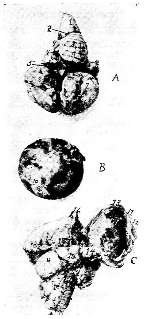
Fig. 75.—The brain of a hen. Photograph. A. Upper surface of the brain. 1, Medulla oblongata. 2, Calamus scriptorius. 3, Cerebellum. 4, Optic lobes. 5, Transverse fissure. 6, Longitudinal fissure. 7, Upper surface of the left cerebral lobe. 8, Upper surface of the right cerebral lobe. 9, Lateral pillar of the cerebellum. B. The posterior surface of the eyeball. 10, The sectioned surface of the optic nerve. C. The inferior surface of the brain. 11, Corneo-scleral juncture. 12, The cornea. 13, The sclera. 14, The optic nerve. 15, The optic chiasm. 16, The olfactory lobes. 17, The medulla oblongata. 18, Tuber cinererum et infundibulum.
267Fifth, a layer of epithelial cells.
The peripheral fibers and the nerve cell layers near the hemispheres disappear so that the basic substance of the trabecula with the hemispheres form the entire lobe mass. On the lower surface of the hemispheres there is a long bundle of nerve fibers which enter into the substance of the olfactory lobes and there disappears. These fibers are medullated. Non-medullated fibers enter into the makeup of the olfactory trunk.
The original center of the olfactory nerve is not in the hemisphere, but in the same location as the optic nerve. This nerve trunk consists of very fine non-medullated fibers. The nerves of smell are therefore not peripheralistic nerves. The nerve fibers are distributed to the mucous surface of the turbinated bones. Toward the front they form an expanded prolongation.
The olfactory nerve, as it emerges from the cone-like anterior tip of the cerebrum, is of considerable thickness and extends along with the median dorsal artery of that region above and to the inside of the orbit, under the thin bony structure. Before its termination into the posterior turbinated bones, it is crossed by the superior maxillary division of the fifth pair of cranial nerves. It extends as far as the pituitary membrane of the turbinate bones upon which its filaments are distributed radially.
The Opticus.—The nervus opticus (Fig. 75, No. C, 14) the second cranial, is the nerve of sight. The optic lobe, or tuberculum bigeminum lies at the base of the brain on each side of the optic tract.
From the optic lobes the two trunks pass forward along the under surface of the cerebri, forming the optic chiasm at the hypophysis (Fig. 75, No. C, 15), at which point the nerve fibers originating from the right side pass to the left, and vice versǎ. From the chiasm the true optic nerves pass forward to the posterior surface of the eyeball. They are composed of a bundle of very fine, marrow-like nerves. These fibers enter into the ganglionic cells of the retina. By removal of the finely adherent neurilemma, the optic nerve is seen to be composed of parallel, longitudinal lamellæ, the margins of which are mostly free on one side.
The Motor Oculi.—The motor oculi (Fig. 62, No. C, 10), the third cranial nerve is a motor nerve. It originates close to the base of the brain behind the position of the hippocampus of mammals on the inner side of the crus cerebri, and also on the inner 268somatic column close below and somewhat aside from the aqueduct of Sylvius. The nerve leaves the brain cavity through a distinct foramen near the foramen opticum, and supplies the following eye muscles: first, after entering the orbit, it sends a branch upward to the inferior portion of the inferior rectus; then, it gives off the thick ramus ciliaris. The trunk then extends under the optic nerve and passes forward to innervate the inferior rectus, the internal rectus, and the inferior oblique. A ciliary ganglion forms on the trunk.
The Patheticus.—(Fig. 62, No. C, 11) is a small motor nerve, originating close to the sulcus centralis, in the circle of the center brain over the valve of Vieussens, between the posteriors of the optic lobes. It extends in a dorsal direction between the cerebellum and the lobus opticus, to the posterior of the latter of which it then forms a loop ventrally. Lying close to the optic foramen it passes through a fine opening into the eye cavity and supplies the superior oblique muscle of the eye. During its course it passes dorsally over the optic nerve, and then crosses dorsally over the ophthalmic division of the fifth pair of cranial nerves and the internal rectus muscle.
The Trifacialis.—(Fig. 62, No. C, 12). This, the fifth cranial, is a mixed nerve and is divided into two parts, portio major and portio minor.
The portio major originates in the ganglion cells of the posterior part of the brain. Commencing near the medulla oblongata it passes through the posterior part of the brain, then upward and outward; and along its route it forms the Gasserian ganglion, which lies partly in the cranial cavity or in its wall.
The portio minor consists of the downward passing fibers containing the motoric elements which are distributed to the muscles of the jaw and of the eye. The roots are found in the ganglion from the center and back brain, close below the pathetic nerve origin, where the pathetic passes between the lobus opticus and the pars peduncularis; it then takes a lateral course between the two.
The fibers of the portio minor do not take part in the formation of the Gasserian ganglion, but are only partly surrounded by it.
There are three nerve trunks given off of the trifacialis: the ophthalmic, the superior maxillary, and the inferior maxillary.
The ophthalmic division of the fifth nerve is the smallest of the three branches. It emerges directly from the Gasserian ganglion, 269and passes through a narrow bony canal in the base of the brain, below the pathetic and the abducens, and through the foramen ophthalmicum. It then enters the eye cavity above the optic nerve, and on the wall of the cavity extends downward, and with regard to the eyeball, dorsally lying close to the rectus internus muscle and close to the surface of the eyeball. It passes along the olfactory nerve, extends under the superior oblique muscle, and finally reaches the inner angle, or canthus, of the eye. It here divides into the recurrent externa and the ethmoidalis. The ethmoidalis branch is a straight continuation of the ophthalmic. It extends along close to its fellow of the opposite side, and, over the vomer, splits into two branches. The smaller of these branches breaks through the bone cells of the jaw, continues upon its ventral surface in a furrow extending forward, and terminates in the gum region. It supplies the gum and point of the beak. The larger branch enters into the cell substance extending to the tip of the beak, in its course sending out a number of fine filaments, which spread out along the outer surface. At this point these two branches may fuse. The larger branch is endowed with the sense of touch.
The recurrent externa divides shortly after leaving the main trunk of the eye cavity passes over the lacrimal gland. It gives two branches to this gland and one to the membrana nictitans, and innervates the upper eyelid. It then emerges from the eye cavity, passing over the os lacrimale, and gives one or more branches to the integument of this region, including the comb. This branch is large in birds with a large comb. Branches pass in front of the lacrimal bone through the outer nasal cavity and into its deeper structure.
Shortly after the ophthalmic branch has entered into the eye cavity and before it crosses the optic nerve, it gives a fine branch to the motor occuli.
The second and the third branches of the fifth cranial nerve are mixed. They contain elements of both the portio major and the portio minor. These two branches come from the lower part of the outer region of the ganglion, and pass together through a cavity which is located between the os petrosum and alæ and basis sphenoid, and then branch.
The ramus secundus, or superior maxillary division, of the trifacial, is the second branch and passes into the orbit below the optic nerve and the eyeball. It is called the recurrent infra-orbitale. 270It gives an ascending branch to the gland of Harder, one to the conjunctiva, one to the membrana nictitans, and one to the eyelids. It also gives a branch to the skin below the eye and to the angle of the mouth. This latter branch is called the recurrent subcutaneous. These two branches communicate with the recurrent nasal ciliaris of the first trigeminus branch. The second branch passes below the nasal opening, then passes on and forms the alveolar nerve on the side between the gums. It sends several recurrent posterior branches to the elevations on the back part of the mouth. It extends forward to the point of the beak.
The inferior maxillary division of the fifth is larger than the other two. It is directed downward and outward then upward to the temporal region. At the temporal region it divides into five parts. It gives a branch to the temporalis muscle, one to the pterygoid muscle, and one to the mylo-hyoideus. The main branch gives twigs to the parotid gland region and enters into the dental canal of the lower jaw. Numerous filaments break through the lower jaw bone, and then spread out on the skin and rim of that bone. The largest branch, called the recurrent maxillaris externa, comes out near the coronoid process. The rest of the trunk extends to, and comes out of, several foramina at the anterior point of the jaw bone.
These filaments are contained in grooves or cavities of the lower jaw and terminate in touch buds.
The trifacialis fuses with the other cranial nerves and with the sympathetic nervous system. Some of these fusions are as follows: The recurrent ophthalmica fuses with the orbito-nasalis ganglion. Indirect fusion of the recurrent maxillary division of this nerve takes place near the Gasserian ganglion through the sympathetic nerves, the temporo-lacrimalis, the facial nerves, the large cervical ganglion, and indirectly with the glosso-pharyngeal and the vagus. This fusion has been called the superior recurrent branch of the trigeminus, or trifacialis.
There is a direct fusion of the superior recurrent maxillary division of the fifth just before entering the upper jaw. This fusion is with the spheno-palatine ganglion and the sympathetic carotidis cephalica, and also with the large cervical ganglion.
The Abducens (Fig. 62, No. C, 13).—This nerve originates in the somatic and motoric column along with the other nerves of the eye muscles. Its nucleus lies in the circle of the pars commissuralis of 271the cerebellum. The nerve then passes ventrally, as does the motoris oculi and leaves the brain along the median line. This is a comparatively large nerve and passes somewhat laterally and ventrally from the foramen opticum through a canal in the sphenoid. It then enters the orbital cavity. Some muscular twigs are given off to the quadratus and to the pyramidalis muscle. This nerve also innervates the external rectus muscle of the eye. The abducens anastomoses with the ramus ciliaris and the ophthalmic nerve and gives fine nerve twigs to the ramus ciliaris externus of the ciliary ganglion.
The Facialis and the Acusticus.—The facial nerve originates with the auditory (Fig. 62, No. C, 14 and 15) in a very vaguely known manner, from the cerebellum. It divides into three parts.
The first of these probably comes from the complex ganglion with the posterior roots of the auditory. This part belongs to the somatic sensory group of nerves. From this same group originates the auditory, which spreads out into the cochlea and takes the impressions of sound. This nerve is short and thick, and, at the point where it loses its medullary covering on entering the cochlea, there is developed a ganglion. This ganglion is similar to the spinal ganglia.
The second part is provided with one root which originates mesially and ventrally, from the deeper ganglion cells. Some of the fibers from this root constitute the vestibular branches. They accompany the auditory and supply the anterior part of the ear labyrinth and the semicircular canals. The larger part of the fibers of this trunk make up the intermediate part of the facial. The geniculate ganglion is formed at their fusion. The sympathetic spheno-palatine nerve emerges from this ganglion, coming out of the aqueduct of Fallopius.
The third part, called the portio dura, is the main facialis. It is located opposite the auditoria intermedia. Its roots may be traced to the complex ganglion, from which they take a direction ventrally from the median portion.
The facialis, after emerging from the aqueduct of Fallopius, takes a curved course and partly fuses with the sympathetic temporo-lacrimalis on the upper posterior wall of the ear drum. It is here accompanied by the carotid and the cephalic arteries. It leaves the ear drum through an opening in the quadrate bone, giving off a large branch to the digastricus muscle and a small one to the stapedius of the columella auris. The facialis trunk is quite large. It 272then passes downward along the quadrate bone, where it receives a large branch from the glosso-pharyngeus. This nerve also gives branches to the mylo-hyoideus and the stylo-hyoideus muscle, crosses laterally the glosso-pharyngeus, and finally fuses with branches of the subcutaneous and with the first, the second, the third, and the fourth cervical nerves. After this fusion the nerves innervate the skin of the anterior of the neck and the constrictor colli muscle.
The facialis anastomoses as follows: with a fine branch of the sympathetic temporo-lacrimalis, indirectly with the ramus trigeminus, with the large cervical nerve ganglion and with the spheno-palatine ganglion and nerve.
The Vagus Group.—The ninth, the tenth, and the eleventh cranial nerves are by some anatomists called the vagus group, first so called by Willis. They are made up of both sensory and motor nerves.
The Glosso-pharyngeus.—The roots of the glosso-pharyngeus (Fig. 62, No. C, 16; Fig. 76, No. 13) emerge, along with those of the vagus, from the medulla oblongata, and enter, as a short trunk, the foramen jugulare et caroticum. Between these two nerves there is usually found a thin portion of bone. The trunk of the glosso-pharyngeus forms a ganglion in the foramen jugulare et caroticum where it receives connecting branches from the near-by ganglion radicis and vagus nerve. The glosso-pharyngeus passes out of the foramen jugulare et caroticum above the large superior sympathetic nerve ganglion, with which it communicates. The glosso-pharyngeus passes diagonally over the ramus temporo-lacrimalis of the sympathetic system, and then receives a very strong branch from the vagus (Fig. 76, No. 12). It sends a short branch to the recurrent lacrimalis of the sympathetic system. At this point there is formed a reddish-yellow ganglion, the petrosal ganglion, which is similar to the petrosal ganglion of mammals. Frequently it is found close below the large superior cervical nerve ganglion. The petrosal ganglion is frequently connected by special fibers with the cervical nerve ganglion, the large superior cervical nerve ganglion and the ganglion radicis vagi. The glosso-pharyngeus is divided into the following branches.
First, the recurrent pharyngeus which gives branches to the upper part of the throat and which is tortuous in its course. It receives a branch from the superior cervical nerve ganglion and gives off branches to the salivary glands and papillæ of the posterior tongue region.
273Second, the recurrent lingualis, which passes with the lingual artery over the hyoid bone to the base of the tongue and to the papillæ. Another branch passes below the tongue bone and supplies the tongue and the pharynx. The glosso-pharyngeal is a mixed nerve.
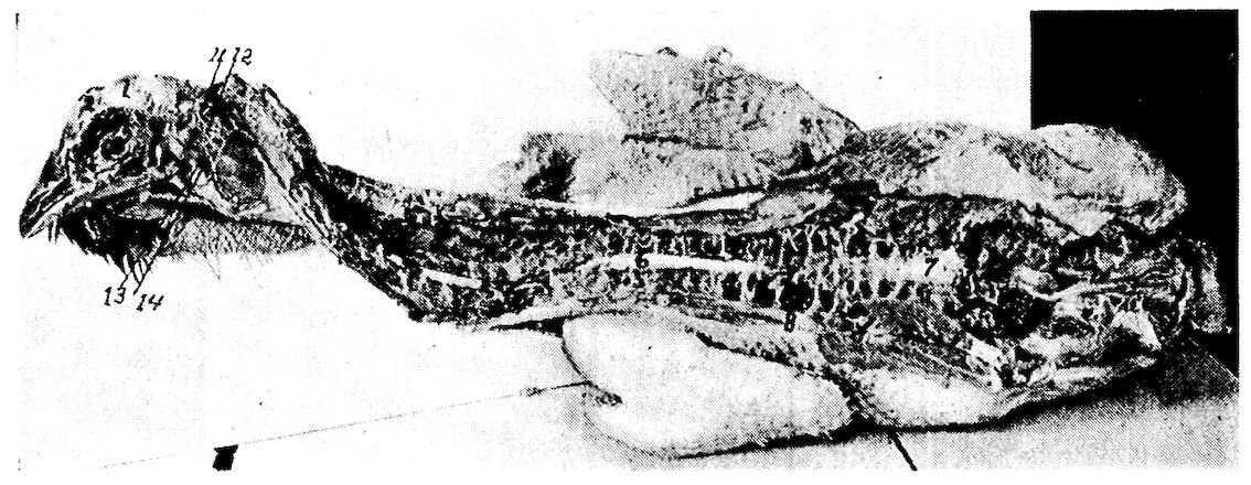
Fig. 76.—Photograph of spinal cord of a hen. 1, Cerebrum. 2, The olfactory nerve terminating at 3, the turbinated bones. 4, The superior maxillary division of the fifth nerve dividing. 5, The eye with the upper part laid open. 6, The cervical segment. 7, The lumbo-sacral segment. 8, The lungs. 9, The kidneys lying above the peritoneum. 10, The terminal filament of the spinal cord. 11, The hypoglossal nerve. 12, The vagus nerve. 13, The glosso-pharyngeal nerve. 14, The branch from the vagus.
The Vagus.—The vagus, or pneumogastricus (Fig. 18, No. 5; Fig. 62, No. C, 17; Fig. 76, No. 12), a mixed nerve, forms a ganglion 274in the foramen through which it passes. The foramen lies between the os petrosum and os occipitale, close to the foramen jugulare, above and to the inside of it.
The ganglionic root of the vagus communicates with the superior cervical nerve ganglion. The vagus, after coming out of the foramen, takes up branches of the spinal accessory, passes to the superior nerve ganglion, crosses the carotid artery, and then, accompanied by the internal jugular, it connects by a branch with the hypoglossal. It then communicates with the petrosal ganglion, and receives a long branch from the superior cervical ganglion. It then extends along the neck with the jugular vein and often fuses with the sympathetic ganglionic plexuses. As it passes, it is interwoven with the glosso-pharyngeus, but each nerve element retains its own individuality. Extending down the neck, on entering the thoracic cavity, it lies between the plexus brachialis and the carotid artery. Then it passes below the subclavian artery, and between the bronchial tubes, the aorta pulmonalis, and the subclavian vein. Then ventrally it rests upon the glands, and the right and the left vagi fuse or unite. From here they radiate down to the stomach in fan-shape, and, continuing, they fuse with the sympathetic system.
The other branches of the vagus are:
The first branch is the recurrent laryngeus, which supplies the lower end of the bronchial tubes, and the esophagus, then enters above the bronchus near the origin of the subclavian artery as the recurrent cardiacus.
The second are the recurrent pulmonale which pass into the lungs, each fusing with its fellow from the opposite side, and giving off branches inferior to the vena cava including a branch to the heart.
The third are the recurrent hepatici which pass through the diaphragm and are distributed to the liver.
The Accessorius Spinalis.—This is a very small motor nerve (Fig. 62, No. C, 18). It comes out between the dorsal and the ventral root of the third cervical nerve. It lies close to the neck and extends anteriorly, receiving roots from the first and the second cervical nerve. It passes through the occipital foramen into the brain cavity and then enters the ganglion radicis vagi. It passes out of the cranial cavity through the foramen jugulare. It then partly fuses with the vagus and partly, as a fine branch, with the subcutaneous colli.
The Hypoglossus.—The hypoglossus is a motor nerve (Fig. 62, No. C, 19). It originates anterior to the eleventh pair of cranial 275nerves, and from the same ganglion as the abducens and the motor oculi. It leaves the medulla oblongata from its ventral surface, and passes with a posterior and an anterior branch out of the cranial cavity through two separate foramina in the os occipitale laterale. The posterior branch, much smaller than the anterior, passes between the cranial bones and the cervical sympathetic nerve; it then passes at right angles through the rectus capitis anticus, and, while closely following the carotid artery, it fuses with the anterior branch. The larger, anterior branch, much stronger than the posterior, sends a short strong branch to the complexus muscle. The rest of this branch, which probably has sympathetic elements, is disposed as is a spinal nerve. It crosses the sympathetic nerve, and at this point forms a typical cervical sympathetic ganglion. It also forms a short loop with the recurrent ventralis of the first cervical plexus. From this loop, after it has given off several branches to the muscles of the neck, it gives off one or two strong branches, which fuse with each other and with the posterior thin branch, thus forming the trunk of the hypoglossus. It receives many elements from the first cervical nerves.
The hypoglossus sometimes communicates with the vagus, crosses over the latter, and divides itself into two main branches, as follows:
First, the recurrent laryngo-lingualis, which passes between the cornua of the os hyoideum and the larynx to the anterior part, and furnishes the principal tongue muscles. It extends along the inferior surface of the tongue and fuses with the one of the other side and extends to the free tip of the tongue. This nerve probably receives sensory elements from the second root and from the confluent of the first cervical nerve. This form of anastomosis is well marked in birds with thick tongues, as ducks.
Second, the recurrent laryngeus furnishes the muscles of the superior larynx and of the tongue skeleton. It extends downward and also furnishes the muscles of the trachea. It follows the course of the jugular vein and at the entrance of the chest supplies the furcula. The nerve passes into the thorax, downward along the side of the bronchial tubes, and supplies innervation to all the muscles of the inferior larynx.
THE SPINAL CORD
Structure of the Cord.—The spinal cord is called the myelon or medulla spinalis. It is a comparatively large, white, irregularly 276cylindrical cord, flattened from above downward. It extends from the foramen magnum at the base of the medulla to the caudal portion of the spinal canal where it terminates in a fine filament. In order to allow considerable motion of the spinal column without danger of injury to the spinal cord, the cord is loosely suspended in the canal. The meninges, or coverings, are continued from the brain. In addition to these there are to be found arteries, veins, and nerves entering and others making their exit from the canal and from the cord. At the cervico-dorsal juncture, where the brachial plexus is given off, the cord is enlarged. There is another enlargement at the point where the lumbo-sacral plexus is given off. At this point, superiorly, there is a longitudinal cavity called the sinus rhomboidalis. This sinus contains a gelatinous substance. There is a pair of nerves given off at each intervertebral foramen. Thus there are as many pairs of spinal nerves as there are vertebral segments. The inferior root is the motor root and carries the impulses from the cord to the periphery. The superior root is the sensory branch and carries the impulses from the periphery to the cord, and thence the impulse is carried to the centers in the brain. The sensory and the motor roots are of about equal size. The inferior have more numerous filaments. The ganglion on the superior or sensory root is relatively large. In the sacral region the sensory and the motor branches pass through their own bony canal.
White rami communicantes are given off at each intervertebral foramen to the sympathetic system and gray rami are received from the sympathetic ganglia. Thus there is established a direct communication between the sympathetic and the spinal system. There is a ganglion on the superior root just outside the spinal cord.
The ganglionic portion, or gray matter, is arranged in the center of the cord in two comma-shaped parts forming an X. The white matter, or the material forming the fibers, is arranged around the central ganglionic portion.
The spinal cord may be divided into two lateral symmetrical halves. There are two longitudinal fissures, one on the upper and the other on the lower half. The upper called the superior median fissure is narrow but deep. The one on the inferior side, called the inferior median, is usually more pronounced. The superior parts of the gray matter are called the superior cornua, or horns; the inferior parts the inferior cornua, or horns. The center of the cord 277is pierced by a central canal which communicates with the fourth ventricle at the calamus scriptorius.
STRUCTURE OF THE NERVE TRUNKS AND GANGLIA
The protoplasmic processes called dendrites have a similar structure to the cell body. The dendrites branch dichotomously, become rapidly smaller, and usually end at no great distance from the cell body.
The axone or axis cylinder process, differs from the cell body and from the dendrites. It does not contain chromophilic granules. It consists entirely of neurofibrils and perifibrillar substance. It emerges from the cell at an enlargement known as the axone hill. This hill is free from chromophilic bodies.
Nerves are divided into two kinds, medullated and non-medullated.
Medullated nerves are divided into two kinds: medullated nerves with a neurolemma, and medullated nerves without a neurolemma.
Medullated nerves with a neurolemma consists of an axone, a medullary sheath, and a neurolemma. A delicate membrane called an axolemma, or periaxial sheath envelops the axone. The medullary sheath called myelin, is semifluid, somewhat resembling fat. The outer covering is the neurolemma, or sheath of Schwann. It is a delicate, structureless membrane which incloses the myelin. There is under this sheath an occasional oval nucleus. At intervals there are constrictions, or nodes, called the constrictions of Ranvier. The part between the nodes is called the internode.
The medullated axones without a neurolemma are the medullated nerve fibers of the central nervous system.
The non-medullated axones, or non-medullated nerve fibers, are divided into non-medullated axones with and those without, a neurolemma.
The non-medullated axone without a neurolemma is merely a naked axone. They are confined to the gray matter and to the beginnings and endings of sheath axones, all the latter being uncovered for a short distance after leaving the nerve cell body and also just before reaching their terminations.
The long axones serve to make connections with the peripheral, or distant, nerve cell, muscle cell, or gland cell; while the shorter axones of certain neurones divide into terminal branches in the immediate vicinity of its cell body, presumably to come into relation with other nerve cells in the same or adjacent groups.
278
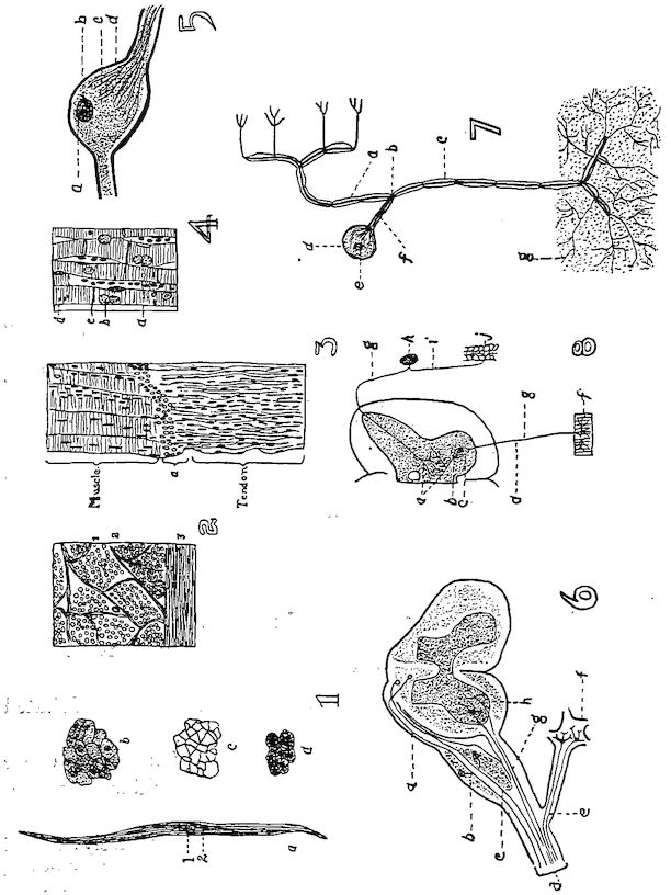
Fig. 77.—Histological structure of tissues. 1. Non-striated or involuntary muscle cell. a. 1, Nucleus; 2, Protoplasm. b. Transverse section showing nuclei in the center of the cells that are cut through the nuclear zone. c. A cross-section of the connective tissue which binds together the muscle cells. d. A section showing the so-called intercellular bridges. 2. A cross-section of the rectus abdominalis. 1, The endomysium which binds the cells into primary fasciculi or bundles. 2, The perimysium which surround the bundles of fasciculi. 3, The epimysium which surrounds the muscle. These binding structures are white fibrous connective tissue. 3. A longitudinal section at the juncture of a muscle and tendon. a, The279 juncture between the muscle and tendon. Note the many nuclei in both muscle and tendon. This is voluntary or striated muscle which make up the dermal, dermo-osseous and skeletal muscles. 4. A longitudinal section of heart muscle. a, Connective-tissue cell. b, Nucleus of a muscle cell. c, Cement line between the muscle discs. d, The cell or segment. 5. A bipolar ganglionic nerve cell. a, The nucleus. b, The nucleolus. c, The fibrillar structure. d, The medullary sheath. 6. A diagram showing the scheme of the peripheral nerve trunk. a, The neuraxis of the peripheral sensory neurone. b, The spinal ganglion of the superior or sensory root. c, The dendrite or peripheral nerve fiber of the sensory nerve. d, The nerve trunk. f, The sympathetic nerve ganglion connected with the spinal cord through the white and the gray ramus communicans. e, The neuraxis of the sympathetic neurone. g, Neuraxis or trunk of the motor neurone or nerve cell. h, The anterior horn of the gray matter of the spinal cord. 7. A diagram of a peripheral sensory neurone. a, The neuraxis which ends in the spinal cord or brain. b, The T-shaped division of Ranvier. c, The dendrite or sensory nerve fiber in the nerve trunk. d, The nucleus and nucleolus of the cell e., f. the axis cylinder process of the cell. g, The telodendrions or terminal branches of the dendrite or axis cylinder. 8. A schematic diagram of the sensory motor reflex. a, The telodendria. b, The dendrite. c, Nerve cell of the motor neurone. d, The motor neurone. f, The muscle fiber. g, The neuraxis of both sensory and motor neurones, the upper being the sensory. h, The nerve cell in the sensory ganglion. i, The sensory neurone or axis cylinder (nerve fiber). j, The skin with peripheral telodendrion of sensory neurone.
Neurones are devoted to the maintenance of functions. Reproductive neurones are so arranged as to receive afferent nerve impulses from other tissues; emissive neurones give off efferent nerve impulses. The former are sensory neurones; the latter are motor neurones if connected with muscles, and excito-glandular if connected with gland cells. The basis, then, of the nerve system is a series of neurones, with projecting and association processes, coördinated for the purpose of performing specific actions manifested either by motion, by trophic changes, or by the apperception of stimuli of a chemic, mechanic (tactile and auditory), thermal, or photic nature.
The whole of the nerve structure is composed of the nerve tissue and supporting connective tissue. The neurones constitute the nerve tissue, while the supportive tissue is composed of neuroglia and of white fibrous tissue derived either from the investing membrane or from the sheaths of its numerous vascular channels.
The neurones, or nerve cells, exhibit marked variations as to external characters, dimensions, and form. The neurone presents a swollen cell mass and a nucleus; it is known as the ganglion cell. From this cell body are given off a number of processes of two distinct kinds: first, protoplasmic processes, which are commonly branched, called dendrites; second, a single, thinner, and paler process; the axis cylinder process.
280The bodies of cells vary in size from 4 to 100 microns or more in diameter. The largest cells occur in the inferior horn of the spinal cord, in the spinal ganglia, in the large pyramidal cell layer of the cerebral cortex, in the Purkinjean cell layer of the cerebellum, and in Clark’s column of the spinal cord. Very small cells occur in the olfactory bulbs, in the granular layer of the cerebral and cerebellar cortex, and in the gliosum cornuale of the cord.
Nerve cells are classified according to the number of processes arising from the cell body, and neurones are referred to as unipolar, bipolar, and multipolar.
Unipolar cells are met with frequently in early stages of embryonic development, but are rare in adults, occurring only in the retina, in the olfactory bulb, and within the baskets of the Purkinjean cells of the cerebellum. The cells of the cerebro-spinal ganglia, except the cochlear and vestibular, are apparently unipolar, but they are developmentally and functionally of bipolar nature.
Bipolar cells are found almost exclusively in the peripheral sensory system, as in the olfactory membrane, in the retina, in the cochlear and vestibular ganglia, and in the cerebro-spinal ganglia of the embryo.
Multipolar cells are the most numerous and form the principal elements of nerve centers throughout the system. They are termed multipolar because of the greater or lesser number of dendrites given off in addition to the single axone.
The body of the nerve cell consists of a mass of protoplasm surrounding a nucleus. The cytoplasm of the nerve cell consists of two distinct substances: first, neurofibrils; second, perifibrillar substance. In most nerve cells there is a third substance called chromophilic bodies.
The neurofibrils, extremely delicate, are continuous throughout the cell body and all of its processes. Within the body of the cell they cross and interlace and probably anastomose.
The perifibrillar substance is a fluid or a semifluid substance which both in the cell body and in the processes surrounds and separates the neurofibrils.
The chromophilic bodies are granules or groups of granules which occur in the cytoplasm of all the larger and of many of the smaller nerve cells.
281
THE SPINAL NERVES
The first pair of spinal nerves come out between the atlas and the occipital bone. Each nerve divides into three branches. The first is the anterior branch which innervates the dorsalis, the biventer cervicis, the cervicales, and the caput posticus muscles. The second is the recurrent ventralis which passes mesially and downward from the recurrent communicans. It innervates the rectus capitis anticus muscle and joins a branch of the hypoglossal nerve. There is given off a white ramus communicans to the sympathetic nerve.
The second spinal nerve emerges from the opening between the first and the second cervical vertebræ and is similarly disposed as the first cervical. It gives off a branch called the recurrent ventralis which fuses with branches of the first cervical and the hypoglossal nerve. This nerve innervates the complexus muscle.
The succeeding cervical nerves emerge in a similar manner down the neck and are distributed to the muscles and other structures of the cervical region. The second, the third, and the fourth cervical nerve gives off branches which form anastomosing loops with the facial nerve.
According to Gadow the last cervical nerve passes between the last cervical and the first dorsal segment. There are thus fifteen pair of cervical nerves.
The first pair of dorsal nerves pass out between the first two dorsal vertebræ. The first few dorsal nerve branches innervate only the trunk muscles. Other branches supply the skin and the other adjacent integument. The ventral branches of the cervical nerves often communicate with the ventral branches of the first dorsal nerves. These branches are larger than the superior branches and aid in the formation of the brachial plexus. Smaller inferior branches are distributed to the scalenus and other muscles, and extend as far posterior as the intestines.
The dorsal branches of the spinal nerves in the lumbo-sacral region are very small, on account of the lack of extensive development of the muscles of this region. The elements entering into these nerve trunks are largely vasomotor nerves. In addition to the upper twigs supplying the skin and the other integument of the region, other branches descend into the abdominal cavity.
The caudal spinal nerves are not well developed and disappear in the region of the caudal vertebræ. The dorsal branches innervate 282the levator muscles, and the ventral branches the depressor muscles.
There is thus given off, throughout the vertebral column, a pair of nerves between each two vertebral segments, making as many pairs of nerves for the region as there are vertebral segments of that region.
The Brachial Plexus.—The brachial plexus (Fig. 73, No. 19) arises principally from the roots of the last three cervical and the first dorsal spinal nerves. These branches anastomose beneath the deep face of the scapulo-humeral articulation.
The brachial plexus divides into two distinct parts. The first (Fig. 73, No. 20) the dorsal called the superior thoracic is distributed to the serratus and the rhomboideus muscle. The second is the inferior thoracic (Fig. 73, No. 21), which consists of the main plexus, and which gives off muscular branches especially to the sterno-coracoideus and to the main portion passing on as the nerve trunk to the structures of the wing.
From the superior thoracic plexus there are several secondary plexuses formed.
The dorsalis or serratus plexus. This plexus is located adjacent to the anterior part of the main plexus and is formed of from two to four spinal nerve branches. The rhomboideus is supplied by branches which may be traced to the first root of this plexus. This nerve is called the rhomboideus superficialis. Another nerve is given off from the middle part of the dorsal side of the plexus and is called the rhomboideus profundus. A third, the superficialis serratus, is the largest of the branches. It breaks up into terminal branches, one going to each serration, or digitation, of the serratus muscle. Branches from the anterior portion of this plexus are distributed to the patagii muscles.
The superior brachial plexus gives off the following nerves:
The subcoraco-scapularis, which is purely a motor nerve, springs from the anterior roots of the plexus. It gives off a branch to the following three muscles: subcoracoideus, subcoracoido-scapularis, and scapulo-humeralis.
The scapulo-humeralis is distributed to the scapular humeral region.
The latissimus dorsi is located on the peripheral border of the scapulo-humeralis or supraspinatus. It originates from the second, and the fourth nerve roots of the plexus. It is located on the dorsal 283side of the plexus. It divides into two main branches, one going to the latissimus dorsi and the other to the scapulo-humeralis muscle. From the side are given off branches which supply the patagii muscle and enter into the formation of the dorso-cutaneous plexus.
The axillaris springs from the second and the third root of the brachial plexus, extends in a lateral direction, passing the ventral and distal rim of the insertional part of the posterior scapulo-humeralis or teres et infraspinatus and enters near the capsular ligament of the shoulder-joint. It gives off to this joint a small branch, called the recurrent articularis, which passes outward between the triceps brachialis and the humerus. It lies on the inner side of the major deltoid, and patagii muscles, and also the skin of the lateral shoulder and the upper arm region. The recurrent axillaris communicates with the main branch of the radial nerve.
The cutaneus brachii superior is a small nerve which springs from the last brachial plexus root and passes between the skin and the triceps brachii muscle on the dorsal surface of the upper arm, or brachial, region. It extends down over the elbow region, where it gives off numerous branches to the skin of these regions, to the meta-patagium and to the extensor muscles of the upper arm.
The brachialis longus superior is a large nerve trunk which springs from most of the other plexus roots. It extends around to the dorsal side of the upper arm and supplies the skin, the feathers, and the muscles of the forearm and the hand. Branches from this nerve trunk supply the triceps brachii and other muscles of the region. The main branch passes between the radius and the ulna, and, passing the elbow-joint, gives off branches to that joint. Passing on, it divides into two branches. One of these extends superficially over the upper part of the condylus ulnaris and supplies the extensor digitorum communis and the extensor metacarpi ulnaris, or flexor metacarpi radialis muscles, and, continuing superficially to the ulnar side, is distributed to the skin of the region. The second, a deep branch, extends on the ulnar side of the radius over the extensor indicis longus, and innervates the latter muscle, the extensor pollicis longus, the extensor pollicis brevis, the adductor pollicis, the interosseous palmaris, and the flexor digitorum.
The inferior brachial plexus gives off the following branches:
The supra-coracoideus, a large nerve which springs from the 284first main root of the plexus, and passes outward through the foramen coracoideum of the sternal ligament.
The sterno-coracoideus, a small nerve which extends downward from the plexus.
The somewhat large posterior coraco-brachialis, nerve which springs from one or two middle roots of the plexus and accompanies the pectoral nerve.
The anterior thoracic, a large nerve which springs from two or three of the posterior roots of the plexus and extends to the shoulder cavity where it branches. The anterior branch is distributed to the patagium and the front part of the pectoral muscles. The posterior branch supplies muscles along the side of the thorax and extends into the abdominal muscles.
The anterior coraco-brachialis, a small nerve which springs mostly from the inferior longus brachialis, passes to the distal end of the tuberculum humeralis radii, then passes backward between the front part of the humerus and the biceps, where it supplies the anterior coraco-brachialis.
The cutaneous brachialis et inferior brachialis, a small nerve which springs from the posterior roots of the plexus. It is distributed to the skin of the region and branches are given off to the patagii and the ventral wing surface. A few branches extend as far as the upper arm.
The brachialis longus inferior (Fig. 68, No. 2), a continuation of the main trunk of the inferior brachial nerve, which comes out of all the plexus roots except the first. It gives off some branches to the pectoral region and then enters the shoulder cavity. It gives off the anterior coraco-brachialis, passes down the humerus in an S-shape, and divides into two branches, the ulnar and the median.
The ulnar nerve (Fig. 67, No. 3) divides into numerous branches which are distributed to the ulnar side of the forearm and the hand. The ulnar nerve passes below the skin of the ulnar outside rim of the forearm and gives branches to the carpi ulnaris, finally supplying the flexor digitorum; it then passes with the tendon of this muscle downward to the interosseous dorsalis, the abductor indicis, or flexor minimi brevis, the flexor pollicis, and the abductor pollicis, or extensor proprius pollicis.
The median nerve (Fig. 67, No. 2) extends down the arm and gives branches to the biceps muscle, and to the patagii region. Continuing, the median nerve supplies the pronator muscle and the brachialis 285inferior, or brachialis anticus, and then divides into two branches. The first and largest branch passes between the two pronator muscles giving branches to the pronator profundus, or pronator longus, and the flexor digitorum profundus muscle. This nerve then passes anteriorly along the tendon to the base of the digit, and, on the dorsal side, joins the other branch of the median nerve. At this point there is often a plexus, and at the point of fusion of the ulnar nerve on the ulnar side there may also be a small plexus. These plexuses mainly supply the skin of these regions. The second branch, passes forward, crosses the extensor carpi radialis and lies just below the pronator profundus, or pronator longus, giving branches to the skin. It passes downward on the radial side of the ulnar and supplies the muscles of that part; it then continues down the hand. Branches are given off to the extensor proprius pollicis, the interosseous dorsalis, the flexor digitorum, and to the skin between the thumb and the forefinger.
The intercostal nerves (Fig. 70, No. 18) are given off from the spinal cord of the dorsal region. The superior branches are small and supply the superior dorsal region. The inferior branches lie one behind each rib, innervate the intercostal muscles, and give a few twigs to the superficial thoracic muscles.
The Lumbo-sacral, or Crural, Nerve Plexus.—The crural plexus (Fig. 70, No. 35 and 36) is made up of trunks from the last two lumbar and first four sacral spinal nerves. There are two portions of this plexus separated by a considerable distance.
The anterior portion (Fig. 70, No. 35) consists of the lumbar nerves and a portion of the first sacral nerves. The fusion takes place on the bony ridge that separates the lumbar from the sacral region.
The posterior portion (Fig. 70, No. 36) consists of a part of the first sacral and all of the three succeeding nerves.
The anterior nerve of this plexus is distributed to the abdominal muscles. An anterior branch is given off to the sartorius. A large cutaneous branch enters between the sartorius and the ilio-trochanteric eminence, and supplies the outer and the upper surface of the upper thigh region. Several short branches are given off from the middle, or main, mass of the crural plexus to the ilio-trochanteric, or gluteal, muscle. Another nerve passes over the side of the sartorius adjacent to the femoro-tibialis, or extensor femoris, and supplies the ilio-tibialis, or gluteus primus. The rest of this 286section of the crural nerves pass in a distal direction over the inner and front side of the cutaneous nerve and enter the ilio-femoralis, or gluteus medius, the ambiens and the femoro-tibialis muscle.
The furcalis nerve comes out between the last two lumbo-sacral vertebræ.
The obturator nerve springs from several roots. The anterior root of this nerve comes from the main trunk of the crural plexus, and its last root from the furcalis nerve. The obturator nerve extends in a ventral direction from the plexus, then horizontally on the inner surface of the abdominal cavity. It gives off twigs to the obturator muscle and then passes through the obturator foramen. After leaving the abdominal cavity it gives branches to the accessorius, the obturator, and, the pubio-femoral, or adductor longus muscle.
The crural plexus gives off another large nerve trunk which extends downward. Shortly after emerging from the plexus it gives off a muscular branch to the ilio-femoralis, or gluteus medius muscle. It then passes between this muscle and the shaft of the femur, to the inner posterior surface of the thigh, and to the inner surface of the knee. It gives off a branch to the structures of the knee. At the knee this nerve terminates in several branches, some of which pass to the inner surface of the tibial head, the internal lateral ligament, the periosteum, the internal condyle, and finally to the upper part of the head of the gastrocnemius muscle.
The main part of this nerve passes downward as the cutaneous nerve, along the inner surface of the lower thigh.
The ischiadic plexus (Fig. 64, No. 41) has five to six roots, which fuse into the ischiadic trunk. This main trunk extends out of the abdominal cavity, through the ischiadic foramen, close behind the anterior trochanter of the ilium (Fig. 65, No. 1). It gives a branch to the external ilio-femoral or gluteus medius, and a branch to the post-acetabular portion of the ilio-tibialis, or gluteus primus muscle. The main trunk (Fig. 69, No. 13) passes downward to the lower portion of the thigh. In this course there are two branches which run parallel to the femoral artery and the femoral vein. It gives off a small muscular branch to the accessorius, and further down gives off a long slender branch to the outside of the knee-joint. It gives off a lateral cutaneous branch (Fig. 66, No. 7) to the posterior outer portion of the lower thigh. It supplies motor fibers to the external head of the gastrocnemius muscle.
At the knee region the ischiadica divides into three parts. The 287largest branch (Fig. 69, No. 14) passes with the tendon of the ilio-fibularis, or biceps femoris muscle, through a loop, the biceps band, and lies on the upper lateral surface of the fibula. It is covered by the external head of the gastrocnemius muscle. It innervates the three posterior muscles of the lower thigh, and then divides into two branches. One, the superficialis peroneus (Fig. 69, No. 16) passes with the profundus nerve, forming a double trunk and occupying the tibio-fibular groove on the antero-lateral side. It passes over the transverse ligament and the tibio-metatarsal joint, and after sending small branches to the structures of the tibial side of the metatarsus, it ends as a cutaneous nerve on the upper side of the third and the fourth toe.
The other branch is the peroneus profundus, which separates from the peroneus superficialis and passes downward in company with the tendons, under the transverse ligament, and then along the anterior upper surface of the metatarsus, where it innervates the muscles of that region. It gives branches to the malleolus, to the tendons of the third toe, and to the median part of the second toe, and supplies the cutaneous structures of the third and the fourth toe.
The third branch of the ischiadica, a long nerve, is given off just after the ischiadica passes through the loop. It passes downward between the two peroneal nerves. It is covered by a sheath. It passes over the posterior outside rim of the intertarsal joint and innervates the tendon sheath. The main portion of this nerve is located on the anterior surface of the tendon Achillis, and passes down on the plantar side, and innervates the periosteum and all plantar foot muscles. It finally radiates to the plantar surface of the three anterior toes.
The integument of the toes is sparingly supplied with nerves.
The nerve trunks that do not pass through the biceps band, or loop can be divided into a medial (Fig. 69, No. 15) and a lateral (Fig. 69, No. 17) portion. The medial portion (Fig. 69, No. 15) soon divides into numerous branches which supply the muscles of the posterior and the inner portion of the thigh. A rather large, long branch passes downward along the tendon of the plantar muscle, which lies on the posterior median edge of the tibia, and gives off twigs to the median and posterior part of the intertarsal joint, supplying the periosteum and other structures and the adjacent skin. It passes downward along the outer side of the medial metatarsal insertion of the tendon Achillis.
288The fifth branch is given off from the second trunk and lies laterally. It is covered by the external head of the gastrocnemius, passes along the vena saphena, and gives off a short main branch to the inner side of the intertarsal joint. The main part passes the tibial side of the joint, becomes subcutaneous, and finally innervates the two plantar muscles. A lateral ascending inner branch innervates the flexor perforatus digitorum, and, in company with an outer branch, the external head of the gastrocnemius, and also the flexor pedis perforatus.
The plexus pedundus is formed from the spinal nerves coming out of the plexus ischiadicus. These fibers emerge caudalward and are directed horizontally. They frequently anastomose with each other, especially on the pubic rim and on the outside of the plexus ischiadicus. These branches are deeply imbedded in the kidney substance and innervate the pubio-coccygeus, or depressor coccygis lateralis, the ilio-coccygeus, the transversalis, the sphincter, and other muscles of this region, and the skin of the anal region.
THE BRAIN
The Brain Coverings.—The cerebro-spinal axis of birds is similar to that of mammals. The meninges of the brain are three in number, dura mater, arachnoid, and pia mater. The dura mater is the thickest. It is constructed of white fibrous connective tissue, and lines the cranial cavity. Thus it serves as an internal periosteum. It is continuous with the spinal dura mater at the foramen magnum, and is also prolonged as a sheath of the nerves. In birds of flight where the air-sacs and reservoirs are developed to the highest state, Sappy finds that, “just as the medullary tissue is replaced by air in the bones of birds, so might it be imagined that the subarachnoid fluid is also replaced by air around the spinal cord,” and observations justify the correctness of this statement. The dura mater measures exactly the volume of the marrow in birds; so that there does not exist between the fibers and the nervous surface any space for an accumulation of liquid. This anatomical fact is sufficient to demonstrate the absence of subarachnoid fluid in the bird. In denying the existence of this fluid, it ought to be added that in this class of vertebrates, the spinal prolongation is covered by a triple envelope; and that between the pia mater and the dura mater is a thin transparent membrane, which is lubricated by a 289serous fluid. This fluid however does not collect; it only moistens the arachnoid membrane.
The arachnoid is located between the dura mater above and the pia mater below. The pia mater adheres closely to the nerve tissue.
The falx cerebri exists in fowls and in turkeys. It has the form of a segment of a circle, and extends from the middle of the interval of the openings for the olfactory nerves to the tentorium cerebelli. The falx cerebelli is absent. The tentorium is small and is sustained by a bony plate, and there are in addition two folds, one on each side, that separate the hemispheres from the tubercula quadrigemina. Owing to the absence of the falx cerebelli, the meninges lie close together. The falx cerebri is ossified in birds.
The Brain Structure.—The brain (Fig. 62, C and Fig. 75, A and C) is made up of three principal parts: the cerebrum, the cerebellum and the medulla oblongata. In a fowl of medium size the brain weighs about 150 grains.
The pons varolii is absent in birds. The crura cerebelli (Fig. 75, No. 9) are immediately connected with the corpora restiformia. The lower face of the isthmus is convex posteriorly; in front, the tubercula bigemina (Fig. 75, No. 4) are united to each other by a comparatively large transverse cord, formed by the optic nerves intercrossing in the median line. The superior face of the medulla oblongata is depressed above to constitute a fourth ventricle; in front of this ventricle are the tubercula bigemina, or optic lobes. These two voluminous tubercles are separated from each other above, where they embrace the cerebellum, and are salient on the sides of the lower face. They are hollow internally, and communicate with the aqueduct of Sylvius. The thalami optici are not well developed.
A large transverse fissure divides the cerebrum from the cerebellum (Fig. 75, No. 5). The optic chiasm (Fig. 75, No. 15) behind which lies the hypophysis (Fig. 62, No. C, 20) covers the region of the middle brain. The large transverse fissure is the dividing line between the hemispheres and the optic lobes. If the hypophysis be removed there will be observed a slit surrounded by gray matter which is called the tuber cinereum et infundibulum (Fig. 75, No. 18). On the pars commissuralis and the after brain there are visible the roots of the fifth, the ninth, the tenth, and the twelfth pairs of cranial nerves. Close beside the median furrow which extends to 290the front part of the long furrow of the spinal cord there is observed the third pair of cranial nerves, which have their origin in the middle brain. The fourth pair of cranial nerves extend from the roof of the middle brain on both sides between the middle brain and the optic lobes. These nerves finally emerge and become visible on the ventral surface.
The sixth nerve is visible near the middle furrow and almost in the middle of the pyramids, and near the roots of the fifth, the seventh, the ninth, the tenth, and the twelfth pair of cranial nerves.
THE DIVISIONS OF THE BRAIN
(Thalamus
(Pineal body
(Infundibulum
(Hypophysis
Forebrain (Optic tract and chiasm
(Cerebral hemispheres
(Olfactory lobes
(Third ventricle
(Lateral ventricles
(Peduncles of the cerebrum
Midbrain (Optic lobes
(Aqueduct
(Medulla oblongata
Hindbrain (Cerebellum
(Fourth ventricle
The medulla oblongata (Fig. 62, No. C, 1; Fig. 75, A and C) terminates anteriorly in the pars commissuralis and the pars peduncularis. As the spinal cord approaches the head there is a gradual swelling, or lateral thickening, which merges into the medulla oblongata. The superior and the inferior surface of the medulla oblongata (Fig. 75, No. A, 1 and C, 17) are flattened. There is a shallow furrow and a slight swelling at the point where the hypoglossal nerve emerges. The central canal of the spinal cord gradually comes closer to the upper surface and communicates with the fourth ventricle, at which point the posterior raphe is shortened and the sulcus longitudinalis posterior becomes shallow. At the point where the central canal terminates in the fourth ventricle there is a V-shaped point called the calamus scriptorius (Fig. 75, No. 2). The fourth ventricle is located on the upper wall of the medulla oblongata below the cerebellum, and is bounded laterally by the peduncles of the cerebellum. It is marked posteriorly by the calamus scriptorius and anteriorly by the valve of Vieussens.
291The valve of Vieussens is located at the posterior end of the aqueduct of Sylvius. The posterior part of the fourth ventricle is marked by grooves or furrows. Extending along the central part of the floor of the fourth ventricle there is a sulcus, or groove, called the sulcus centralis, which divides the superior pyramids. On the median lateral sides there are two points of gray substance which form the alæ cinereæ. The roots of the pneumogastric, the glosso-pharyngeal, and the spinal accessory may be traced to the alæ cineræ and the gray matter of the ridges of the medulla oblongata. The sixth pair of the cranial nerves emerge from the medulla near the sulcus centralis and to the side of the auditory nerve. Part of the trigeminus emerges from the rim of the furrow next to the ridge. At the outer border of the medulla are observed the thick ends of the roots of the pneumogastric, the spinal accessory, the glosso-pharyngeal, and the auditory facialis.
On both sides of the sulcus longitudinalis inferior are found the inferior pyramids, or pyramidal columns. These pyramids become expanded near the origin of the third pair of nerves and merge into the cerebral peduncles, or crura cerebri.
The pyramidal fibers may also be traced to the optic lobes. The roots of the abducens are found at a point between the crura cerebri and the optic lobes. The third pair lies to the side of this, and the roots of the trigeminus are adjacent to those of the third. A bundle of fibers from this region pass into the cerebellum and form the crura cerebelli; others pass into the cerebellum from the side, spreading out in fan-shaped radiation, and forming the white central substances peculiarly arranged, called the arbor vitæ, or tree of life. A third bundle fuse with the crura cerebelli anteriorly, and pass into the peduncles of the cerebrum, or crura cerebri.
The gray ganglionic substance forms columns which extend the whole length of the medulla oblongata and into the cerebri. The medulla oblongata contains numerous centers which preside over various visceral functions as deglutition, respiration, thermotactic, secretory, cardiac, and digestion.
In the medulla as in the spinal cord there are five main groups of cells. First, the posterior or upper horns, from which come the somatic sensory nerves. Second, Clark’s cells, located centrally, which are the origin of the ganglionated splanchnic nerves. Third, cell groups of the lateral horns which are the center for the non-ganglionated splanchnic nerves, and certain other nerves for the 292viscera, including the enteric muscles. Fourth, cell groups of the anterior horns, which are the center of all somatic voluntary muscles. Fifth, groups of single cells probably belonging to the posterior horns which are probably centers for other splanchnic nerve fibers.
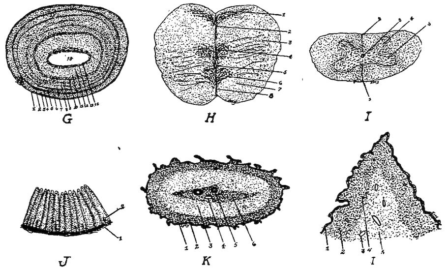
Fig. 77, A. G, A section through the tuberculum bigeminum. 1, Pia mater. 2, Nerve fibers. 3, Fine granular ground substance. 4, Thin layer of small cells. 5, Fine granular ground substance. 6, A second thin cell layer. 7, Fine granular ground substance. 8, Third thin cell layer. 9, Fine granular ground substance. 10, Fourth widely extended cell layer. 11, A fine granular layer. 12, A thick layer of spindle cells. 13, A layer of medullated nerve fibers. 14, Ependemal cells. 15, The sinus. H, A transverse section through the medulla oblongata, 1, 3, 4 and 6 are ganglionic centers. 2, The dorsal groove. 8, The ventral fissure. 5, The central canal. 7, Fiber tracts. I, A transverse section through the cervical portion of the spinal cord. 1, Ventral septum. 2, Dorsal septum. 3, The central canal. 4, The dorsal horn. 5, The ventral horn. J, A section through the wall of the oil gland, or rump gland. 1, The stroma. 2, The tubular glands some of which are branched. K, A section through a spike of a comb of a cock. 1, The epithelial layer. 2, A dense fibrous subepithelial layer. 3, A second fibrous vascular layer. 6, A denser central core supporting the large arteries and veins. 4, A vein. 5, An artery. L, A section through a lobe of the wattle of a hen. 1, The epithelial layer. 2, A dense vascular layer. 3, A vein. 4, An artery. 5, A loose fibrous vascular structure.
The gray matter in the cord is in the form of two commas placed with their backs together with the central canal passing between 293them. The central canal is located in the center of the cord. As the cord nears the medulla oblongata the form of the gray matter changes. The second, the third, and the fifth ganglion centers are arranged laterally, and show a distinct side-horn group. The central canal separates the two Clark’s columns; later the side-horn group enlarges and extends ventrally around the anterior horn group and joins on the other side in half-moon shaped formation.
The central ganglionic mass of the spinal cord, the center for the enteric visceral system, is divided into three complex parts. First, there are cell groups on both sides of the posterior raphe. Second, the half-moon shaped ventral groups extending parallel peripherally described above. Third, lying between groups one and two a complex group, which possesses a large collection of cells. These furnish splanchnic nerve centers, the anterior supplying motor nerves and carrying motor impulses outward, and the superior sensory which carry impulses to the centers. The cell group of the superior horns takes a sidewise peripheral position (Gadow).
The cerebellum (Fig. 62, C, 2) is located above the medulla oblongata and posterior to the cerebrum. The cerebellum consists principally of a median lobe called the worm. The lateral lobes are conical and rudimentary. The under part of the worm forms the roof of the fourth ventricle.
On the upper surface of the cerebellum are numerous transverse markings (in the hen 13 or 14) which divide the lobe into leaves. When the cerebellum is cut lengthwise there is observed on the sectioned surface the peculiar arrangement of the white and the gray matter, the arbor vitæ, mentioned above. There is a small cavity in the cerebellum which communicates with the fourth ventricle.
The cerebellar cortex shows four parts, namely: First, the central part composed of white medullated fibers, between which are arranged neuroglear cells. Second, a rather reddish layer of cells of different sizes. These cells are embedded in a fine ground substance and are about 0.003 mm. in diameter. Third, a plain layer of large pear-shaped ganglion cells, the cells of Purkinje. Fine branches extend outward from the apex of these cells. Fourth, an outer gray layer which consists of small multipolar nerve cells with some neuroglear cells. The color of these three layers, according to Gadow, depends upon the color of the plumage; that is, if the plumage is dark, this layer is dark; if light, the layers are 294light. The marked coloring is said to be most distinct in the second layer.
The anterior portion of the cerebellum consists of medullated nerves whose fibers run crosswise. These are the extensions of the true cerebellar fibers, which become fewer and fewer as they proceed anteriorly. Finally, a few remaining fibers running crosswise fuse as the commissura Sylvii, and extend into the lobi optici. A layer of nerve cells extends under these medullated nerve fibers.
A bundle of fibers extending from the medulla oblongata. These fibers extend into the optic lobes, form the crura cerebri, or peduncles of the cerebri, and also form the inferior wall, or floor, of the aqueduct of Sylvius. The crura are sometimes spoken of as the partes pedunculares. These contain gray matter. The following division is made of the ganglion of this region. First, a group of cells near the base of the peduncles, which are divided from the ventral rim by the arciform fibers. This ganglionic formation may be considered an extension from the medulla oblongata. Second, a group of the ganglion cells of the lobus optici. Third, a group of cells lying near the lobus opticus, which give rise to the ascending roots of the trigeminus. Fourth, a group of ganglionic cells from which spring the roots of the motores occuli, and which lies near the middle line and under the sulcus centralis. Many ganglion cells are found to the right and to the left of the sulcus centralis, which show different arrangement of the cells at different levels. Fifth, a group of irregular cells which extends centrally on the superior walls of the third ventricle and to the superior part of the lobus opticus.
The microscopic structure of the roof of the lobus opticus, or corpus bigeminum, shows that the layers are arranged parallel to the pia mater and are divided into the following parts:
Externally, the pia mater.
An outer layer of very fine nerve fibers, which lies just below the pia mater.
A layer of fine granular ground substance.
A thin layer of small cells the diameter of each of which is 0.038 mm.
A layer of fine granular ground substance.
A second thin cell layer.
A zone of fine granular ground substance.
A third thin cell layer.
295A fine granular ground substance.
A fourth widely extended cell layer.
A fine granular layer.
A somewhat thick layer of spindle-shaped cells.
A layer of medullated nerve fibers.
This last is the inner layer of nerve fiber. On the inner surface of this layer we find the ependymal cells, the cells that line the cavity of the optic lobes. The commissura Sylvii form the covering of the lobi optici and join these two lobes. This commissure is formed from the upper and the lower layer of nerve fibers. In the commissure these fibers cross each other.
The hypophysis (Fig. 62, No. C, 20) lies back of the chiasm, or optic commissure, and below the middle of the third ventricle. The infundibulum, a pedicle-like structure, connects the hypophysis with the third ventricle of the cerebri. It contains a cavity and forms an extension of the third ventricle. The hypophysis is divided into two lobes, an anterior and a posterior.
The third ventricle communicates anteriorly with the lateral ventricles of the cerebri through the foramen of Monro, and posteriorly with the fourth ventricle through the aqueduct of Sylvius. The posterior walls of the third ventricle are relatively thick and form the posterior brain commissure. The wall is thin from the commissure to the chiasm. Within it lie two ridges, which connect the two hemispheres with the corpus callosum, and, farther anteriorly, with the anterior cerebral commissure. The space from the third ventricle and to the anterior commissure is called the laminæ terminales.
The pineal gland, located between the cerebri and the cerebellum, lies against the choroid plexus, which covers the highest point of the third ventricle.
The cerebrum is divided by a deep longitudinal fissure into two hemispheres. The cerebrum is shaped somewhat like the heart on a playing card. The lower face is somewhat flattened. The upper and lateral sides are not provided with convolutions or sulci, but are smooth. The fissure of Sylvius is faintly marked on the inferior face. The olfactory lobes appear well developed and are relatively large for the size of the brain. They lie close together on the extreme anterior portion in the median line.
The olfactory cerebral crura emerge from the cranium at the upper angle between the posterior wall, the roof, and the septum of 296the orbit, and pass through the olfactory foramina and in grooves on the upper part of the septum; then passing forward they penetrate the frontal structure and are finally distributed over the turbinated mucous membrane.
The corpus striatum is large; it occupies nearly the entire floor of the ventricles.
The anterior commissure is found between the hemispheres. Its middle part lies in the lamina terminalis on the division between the anterior and the middle brain, and close to the thalami optici. Its side extensions are rounded masses called the nervi amygdales.
The corpus callosum, quite rudimentary, lies on the posterior dorsal rim of the anterior commissure.
The corpus striatum forms a thin broad ridge, which passes ventrally. This structure consists of twenty-five large pyramidal cells which lie in the posterior lateral dorsal section, and of from ten to fifteen pyramidal cells which are located in the rest of the section. In addition to these two groups there are many cells about 5 millimeters in diameter. These lie, six or more, in a nest imbedded in neuroglia. There is a thin layer of spindle cells near the ventral line. The outer nerve nest, or nerves amygdalis, lies in the posterior lateral ventral part. It is covered by a thin membranous layer. Its cells are pyramidal in shape and are from 10 to 15 millimeters in diameter. The cells terminate in spindle form toward the periphery. Nerve fibers extend from the anterior commissure.
The outer wall of the corpus striatum consists of the following layers:
An outer white layer consisting of fine medullated nerve fibers imbedded in a ground substance containing numerous nerve cells.
A layer of ganglionic cells, consisting of pyramidal cells from 10 to 15 millimeters in diameter, of other round cells 20 millimeters in diameter, and finally of cells only 5 millimeters in diameter. This layer forms a reddish line in the dorsal portion of the corpus striatum.
These layers form part of the median and the posterior cerebral wall of the lateral ventricle. The lateral ventricle is closed except for a slit-like opening behind the posterior commissure. This is the foramen of Monro, through which the lateral ventricle communicates with the middle, or third, ventricle.
The choroid plexus is found at the base of the lateral ventricle.
The wall of the lateral ventricle becomes thinner near the region of the transverse commissure on the surface toward the middle 297brain. At this point the pia mater and the ependyma, or the lining cells of the ventricular membrane, form the major part of the wall.
The domestic fowl does not have the hippocampus.
The wall of the middle ventricle consists of an outer white layer, which is arranged similarly to that of the corpus striatum. The ventral rim of the wall is formed by a spiral band which consists of fine medullated fibers, in which are imbedded a few cells.
The processus cerebri mammillares are also called the tubercula olfactoria. From these structures extend forward the olfactory nerves, the posterior roots of which may be traced to the walls of the lateral ventricles.
The structure of the processus cerebri mammillaris is made up of five layers as follows:
An outer layer, which consists of colorless olfactory fibers extending in all directions.
A granular ground substance layer, in which are imbedded a few cells.
A thicker granular layer, on the inner edge of which are twenty large pyramidal cells with processes pointing outward.
A layer of closely packed cells, in which are fine medullated nerve fibers. These cells are about 5 millimeters in diameter.
An innermost layer of ependymal cells.
Near the hemisphere the peripheral nerve fibers and nerve cells disappear, leaving only the ground substance of the processus to aid in the formation of the hemisphere.
There is a long bundle of fibers on the lower surface of the hemisphere, which blends with the substance of the olfactory fibers.
The peduncles of the cerebrum (Fig. 62, No. 21) are the slightly diverging columns of nerve tissue, which form the anterior continuation of the medulla oblongata and disappear under the optic tracts and the chiasm.
THE SYMPATHETIC NERVOUS SYSTEM
The sympathetic nervous system commences anteriorly at the large superior cervical nerve ganglion. This ganglion, anteriorly, brings into communication the glosso-pharyngeal, the pneumogastric, or vagus, and the sympathetic system.
The following nerve trunks emerge from the superior cervical nerve ganglion: a large trunk fusing with the vagus and directed 298downward, accompanying and surrounding the carotid artery; a second large trunk fusing with the recurrent pharyngeus and the glosso-pharyngeus; a third trunk which merges with the hypoglossal; finally several trunks which are distributed to the head.
The temporo-lacrimalis, one of the large sympathetic nerves of the head, as it extends from the cervical nerve ganglion, receives branches from the ganglion radicis vagi. This nerve, passing between these two ganglions, extends horizontally forward and outward through a foramen, crossing the glosso-pharyngeus. Near the Fallopian canals it crosses the facial nerves, lies supero-laterally, and receives a short branch from the facial nerve whose fibers are traceable to the geniculate ganglion. It also receives a branch from the recurrent maxillaris.
This nerve accompanies the external ophthalmic artery forming around it a network of fibers, called the external ophthalmic plexus. It lies outside and downward from the optic nerve, sends fine branches to the external ophthalmic artery and to the masseter artery, and extends along with a small branch of the superior maxillary nerve to the skin of the outside rim of the eye cavity.
The ophthalmic plexus, a second trunk extending to the head, enters in its course, anteriorly, the lacrimal plexus. Its fibers also supply the lacrimal gland, and finally anastomose with the second recurrent branch of the trigeminus.
The sympathetic caroticus cephalicus nerve, a third trunk extending to the head, after emerging from the large superior cervical nerve ganglion, receives some small branches from the glosso-pharyngeal ganglion, and then enters, in a horizontal manner, a foramen, the canalis caroticus externus, located in the lower part of the basi-occipital and the sphenoid bone. The anterior opening of this foramen, or canal, is close to the posterior part of the pterygoid bone. Inside this canal the sympathetic caroticus cephalicus receives a small branch which extends in a straight line from the basal part of the facial nerve. After receiving this branch, the nerve trunk, passing to the ear drum, lies close to the petrosum and the sphenoid. It is covered by the masseter muscle. The carotic ganglion is located at the point of the fusion of the caroticus cephalicus with the main trunk. At this point the caroticus cephalicus divides into two branches. The first is the superior recurrent nerve, which lies close to the upper surface of the alæ of the sphenoid, passes between the obliquus externus and the orbital wall, around the eyeball, 299and finally into the orbital septum and the internal muscles of the eye. It communicates with the ophthalmic nerve and sends twigs to the lacrimal gland, the gland of Harder, the upper eyelid, and the nose. The orbito-nasale ganglion is located where these nerves communicate near the nasal region. The second branch of the sympathetic caroticus cephalicus is the inferior recurrent nerve. This branch passes forward and dorsally from the pterygoid bone to a point where the upper rim of the rising wings of the jaw bone meets the sphenoid rostrum. In this course, some branches are given off to the pharynx, one branch near the lacrimal gland, and a branch which communicates with the superior maxillary nerve just before the nerve enters the jaw bone. The spheno-palatine ganglion is at the point of fusion.
The terminal branches of the spheno-palatine are distributed to the hard palate, the nose, and the lacrimal gland.
From the large cervical nerve ganglion and near the roots of the caroticus cephalicus are given off a few small nerve fibers which pass alone to the pharynx or accompany the jugular nerve branches, and fuse with the main trunk of the caroticus cephalicus nerve.
From the large superior cervical nerve ganglion the sympathetic trunk extends downward toward the thorax. It is covered deeply with muscles. This portion is known as the cervical sympathetic nerve trunk. A trunk lies on each side of the cervical vertebræ. The large thoracic nerve ganglion, the inferior cervical, is located along this trunk at the entrance of the thorax. From this ganglion there is given off the recurrent cardiacus which supplies the heart. The end branches of the sympathetic nerve trunk blend with the pneumogastric nerve. The inferior nerve ganglion is the ganglion cardiacum. Near this ganglion is found a nerve plexus in which there are imbedded peripherlistic ganglia. This thoracic plexus, accompanying the collica artery, passes to the abdominal region, supplying the intestines and taking part in the formation of the abdominal plexus. The abdominal plexus is located near the anterior portion of the kidneys. Its fibers are directed mainly downward to the visceral organs. The large intestine, the rectum and the copulatory organs receive branches. These latter branches take part in the formation of the pedunda nerve plexus. Some branches of this plexus, follow the branches of the posterior mesenteric artery.
The thoracic trunk (Fig. 64, No. 42) of the sympathetic is double. 300The anterior portion gives off an anterior splanchnic nerve, or plexus (Fig. 64, No. 43) which accompanies the celiac axial artery to the gizzard and liver, communicating with the pneumogastric. The posterior splanchnic nerve is intimately combined with the adrenal body, and the testes, or the ovary (Fig. 64, No. 45). Intestinal branches accompany those of the mesenteric arteries (Fig. 64, No. 46). Other branches supply the kidneys, and communicate with long branches of the spinal nerves destined for the cloaca and adjacent parts, and thus form a plexus similar to that found in mammals.
FUNCTIONS OF THE NERVOUS SYSTEM
According to function, the nerves composing the trunks are divided into afferent and efferent.
The afferent nerves are those that convey the impulses from the periphery of the body to the nerve center, which are located in the brain or in the spinal cord. The impulses conveyed are those of special senses, as sight, hearing, taste, touch, and smell. Impulses producing sensation pleasurable or painful come from the skin, the muscle, or the viscera.
The efferent nerves are those which convey impulses from the nerve centers to the periphery. These impulses may be motor as those going to the muscle cells of the skeletal muscles, the viscera, or the blood-vessels. These motor impulses make movements in these organs possible. In the blood-vessel they result in the control of the caliber of the vessel. These impulses may be of an inhibitory character, as in slowing the heart. They may be secretory impulses stimulating the gland to activity or regulating metabolism.
The ganglia are nerve centers which receive and generate impulses; the nerve trunks are filaments which convey impulses. The gray matter of the cord described above is the ganglionic portion, and the outer white matter is made up of nerve fibers which convey impulses from one part of the cord to another, or to and from the brain.
The nerves that have their roots in the spinal cord superiorly, are sensory; that is, they convey the sensory impulses from the periphery to the cord ganglion and to the brain. They have a ganglion just outside the cord.
The nerves that have their roots in the spinal cord inferiorly; that is, they convey motor impulses from the nerve centers to the 301periphery. The function of the inferior roots is to supply all the voluntary muscles as well as the oviduct, the intestines, and other hollow viscera, including the blood-vessels with the power of movement. Many of these fibers pass to the sympathetic ganglion and are distributed as sympathetic nerve fibers.
The cord is divided into different tracts, that is, certain groups of fibers convey certain kinds of impulses.
The superior column of the cord conveys to the cerebrum such impressions as temperature, pressure, and muscular tension.
The fibers of the lateral columns carry sensations of pain.
The fibers of the direct cerebellar tract carry impulses which result in the maintenance of the equilibrium of the body.
All the voluntary impulses originate in the cerebrum, pass through the cerebellum and travel direct to the bulb; they then pass over to the opposite side, and travel by the crossed pyramidal tract to the multipolar cells of the inferior horn of the spinal cord, and transmit the impulse through motor fibers that originate at that point in the cord.
All sensory impulses enter the brain on the side opposite their origin, and all motor impulses leave the brain on the side opposite that to which they are distributed. Injury of the motor area of the right side of the brain leads to paralysis of the left side of the body.
An impulse of the vasomotor nerve travels in the lateral column of the cord.
A nerve impulse may originate in the brain and be modified in passing through a ganglion in the spinal cord or in the sympathetic system.
The system of reflex action is as follows: first, an efferent nerve which conveys the impulse from the periphery to a nerve center; second, a ganglion, or nerve center, to receive the impulse and generate other impulses; third, an efferent nerve to convey the impulse from the nerve center to the periphery.
The following is an example of reflex action. The foot of a fowl is pierced with a pin. The sensory impulse is conveyed by sensory nerve fibers to the nerve centers. In this center the ganglionic nerve cells generate a motor impulse which is sent back through the motor nerve fiber to the muscles controlling the part. The result is that the muscle contracts and jerks the foot.
There are many functional centers located in the medulla oblongata. Destruction of this part of the system results in instant death 302of the bird. In addition to furnishing a path for fibers carrying impulses from the body to the cerebrum, it furnishes a large number of centers for such functions as respiration, swallowing, secretion, temperature, and vasomotor and cardiac activity.
The function of the cerebellum is principally that of coördination. It brings about harmony and rhythm in muscular movements. If the cerebellum be removed, the bird can no longer walk. It has lost, with this removal, all power to coördinate.
The cerebrum of the bird has no convolutions, and the gray, or ganglionic, portion is thin, indicating low power of intelligence. There are certain areas presiding over other functions, as motor areas, sensory areas, and so on.
A careful study of the brain of the fowl shows us that the centers presiding over sight and smell are well developed.
The olfactory bulbs are the centers of the sense of smell.
The optic thalamus is the center of the sense of sight.
The sympathetic nerve system transmits impulses to the involuntary muscular structure of all organs, including those of the intestinal tract, the blood-vessels, and perhaps also the glands.
303