OSTEOLOGY
Reading edition
Osseous Structure.—Bone is structurally modified connective tissue which has become hard by being impregnated with calcium salts.
Kinds of Bone Tissue.—There are two kinds of bone tissue: substantia compacta, or compact bone tissue; and substantia spongiosa, or spongy, cancellous bone tissue.
Compact Bone Tissue.—The compact bone tissue forms the hard outer layer of all bones. It is thickest in the shaft and becomes thin toward the extremities. Through the compact bone tissue approximately parallel with the longitudinal axis of the bone, run canals called Haversian canals, through which pass blood and lymph vessels for the nourishment of the bone and nerves. The Haversian canals are surrounded by concentric lamellæ. The spaces between the cylinders thus formed are filled with interstitial lamellæ; and both the exterior surface of the bone and the interior surface surrounding the medullary canal, are built up of peripheral, or circumferential lamellæ. Between the lamellæ, somewhat irregularly placed, are minute reservoirs, called lacunæ, which contain bone corpuscles. From the lacunæ radiate minute canals, or canaliculi, which maintain circulation through the bone substance, and which communicate with the Haversian canals. Complex anastomoses exist among the canaliculi. Still other channels for the passage of blood-vessels are Volkmann’s canals which pierce the peripheral lamellæ, thus allowing vessels to pass from the periosteum to the Haversian canals. Similar channels afford communication between the inner Haversian canals and the medullary cavity.
The entire structure composed of an Haversian canal, its surrounding 18lamellæ, lacunæ, and canaliculi, with their contained vessels, is called an Haversian system.
Cancellous Bone Tissue.—The cancellous bone tissue forms the bulk of the short, flat, and irregular bones and of the extremities of the long bones. It consists of delicate bony plates and spicules, which intercross in various directions. The spaces between these plates and spicules, called cancelli, are occupied by marrow except in the bones that are pneumatic. The blood-vessels, lymphatics, and nerves course through this marrow but are not arranged in an Haversian system.
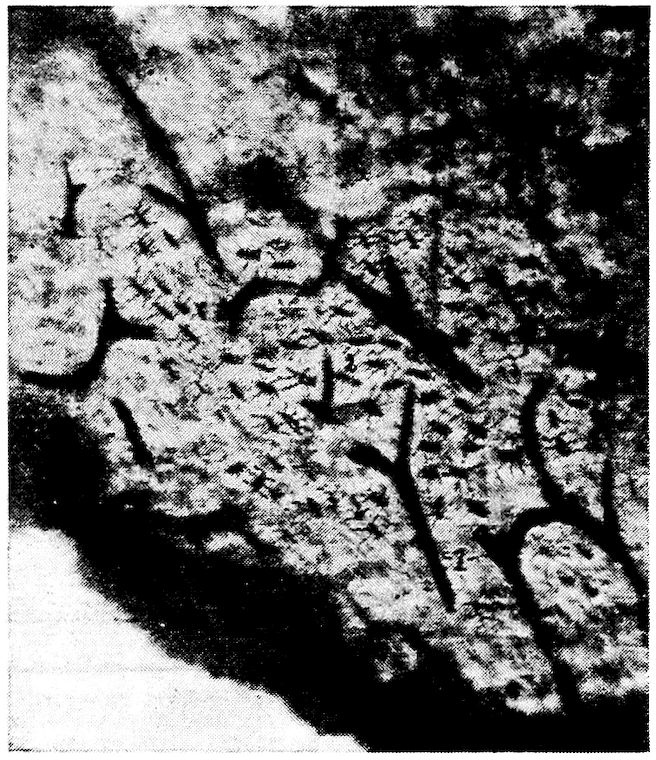
Fig. 1.—Longitudinal section of compact bone of the femur of the hen. 1, Haversian canals. 2, Lacunæ with their canaliculi.
The Periosteum.—Covering the surface of bone, except at the articular surface where it is covered with cartilage, is a membrane, the periosteum, which consists of two layers: an outer, fibrous, protective layer, and an inner, cellular, osteogenic layer. The outer layer consists principally of white fibrous tissue. The inner layer contains many more connective-tissue cells, which gradually become more closely aggregated as we proceed toward the osseous surface; but there is no sharply defined line of demarcation between the two periosteal layers.
19
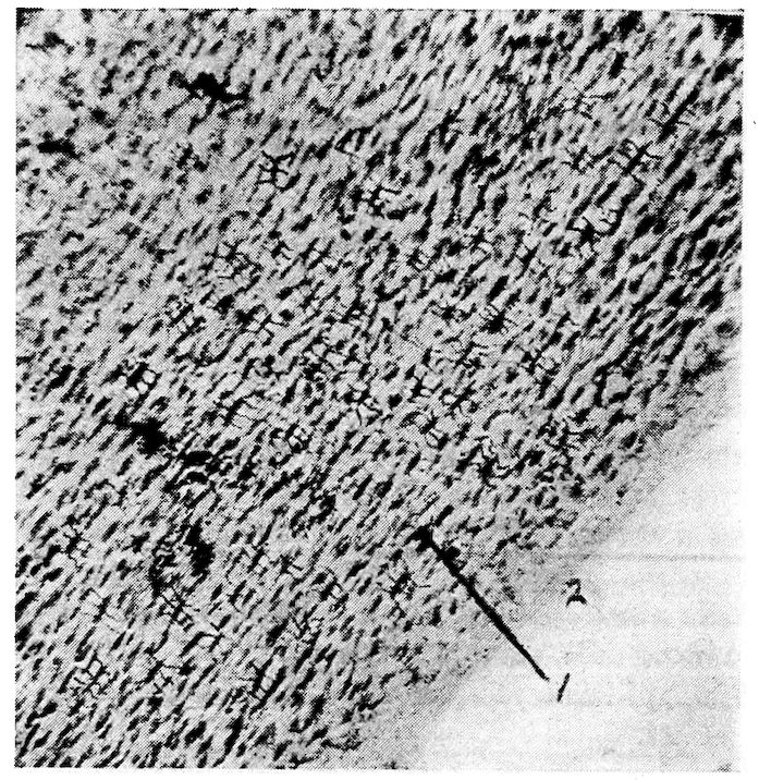
Fig. 2.—Transverse section of compact bone of the femur of the hen. 1, The lacunæ and canaliculi. 2, The periosteum.
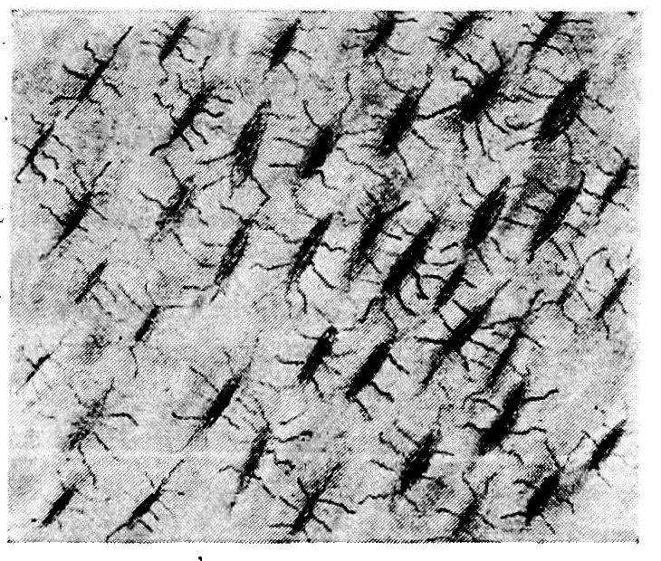
Fig. 3.—Transverse section of compact bone of the femur of the hen showing the lacunæ and canaliculi under high magnification.
20The periosteum is firmly attached to the bone by trabeculæ of fibrous tissue, called the fibers of Sharpey. These fibers of Sharpey penetrate the bone at right angles to its surface and carry blood-vessels.
Marrow.—There are two kinds of marrow: yellow, or medulla ossium flava, and red, or medulla ossium rubra.
The yellow marrow occurs in all bones except the femur and proximal portion of the tibia of adult fowls. It is composed of a network of fibrous tissue carrying blood-vessels, fat cells, and myelocytes, or marrow cells.
The red marrow is found throughout the femur and the proximal portion of the tibia, and in a few of the pelvic bones and vertebræ in the adult fowl, and in certain other bones of the baby chick. Red marrow consists of a delicate network of connective tissue supporting a dense capillary plexus, a small amount of fat, and numerous cells. The cellular elements of red marrow consist of marrow cells which contain large nuclei and possess ameboid movement, red blood cells, giant cells containing one or more nuclei, and various kinds of leucocytes, including eosinophiles, mast cells, and also osteoclasts.
Growth of Bone.—In the baby chick, only the shaft and a portion of the extremities of the long bones are thoroughly ossified, the extreme ends, and of the femur most of the articular head, being cartilaginous. The bones grow in length by an increase in the cartilage, the cartilage gradually becoming ossified. Growth in diameter is accomplished by the constant deposition of new layers of bone beneath the periosteum. During this process the osteoclasts absorb the bone from within. The formation of the marrow cavity is thus effected.
Classification of Bones.—The bones of the fowl are classified as long, short, flat, and irregular.
Long Bones.—The long bones occur in the legs and wings, where they serve as levers to sustain weight and make locomotion possible. A long bone consists of a shaft and two extremities. The superior is called the proximal and the inferior the distal extremity. The expanded articular surfaces in forming joints with adjoining bones afford ample space for the attachment of ligaments. The shaft is cylindrical and hollow.
Short Bones.—Short bones occur in the feet and in the wings. Their structure is similar to that of the long bones.
21Flat Bones.—The flat bones occur where extensive protection is needed, as in the cranial region; or where large surface for muscular attachments is needed, as in the costal and pelvic regions. Flat bones are made up of two thin layers of compact bone with a variable amount of cancellous tissue interposed.
Irregular Bones.—The irregular bones include the vertebræ, the patellæ, and the carpal bones.
Composition of Bone.—Bone consists of organic and inorganic matter. Organic matter gives toughness and elasticity to the bone, and inorganic matter hardness. The organic substance of bone is called ossein. When boiled in water ossein is resolved into gelatin. The following tables[1] give the results of an analysis of the femur, fresh, of a mature hen.
Fresh femur:
Water 18.23 per cent.
Dry matter 81.77 per cent.
Dry matter:
Organic matter 63.09 parts
Inorganic matter 18.68 parts
Salts in dry matter:
Calcium 6.970 per cent.
Magnesium 0.283 per cent.
Potassium 0.004 per cent.
Sodium 0.276 per cent.
Iron 0.020 per cent.
Phosphorus 3.210 per cent.
Sulphur 0.085 per cent.
Chlorine 0.520 per cent.
Carbon dioxid 0.550 per cent.
The inorganic matter of the femur of the hen consists of 18.68 parts or 22.84 per cent. of dry matter, and the organic matter of 77.16 per cent. Stated in other words the femur, including its contained marrow, consists of organic and inorganic matter in the ratio of 3.4, approximately, to 1.
The Skeleton of the Fowl.—The skeleton of a bird is remarkable for the rapidity of its ossification. It is worthy of note that other parts of the bodies of adult birds also become ossified. Among such parts are the tendons of the muscles of the legs, of the feet, and of the 22neck; the plates of the corneal margin of the sclerotic tunic of the eye; and the stapes of the ear. Ossification in birds at the attachments of the semi-lunar valves of the aorta and of the pulmonary artery has been reported by Owen.
The bony structure is compact, and the bones contain a greater proportion of phosphate of lime than do the osseous structures of mammals. Especially is this the case in those parts of the skeleton which are permeated by air.
The Divisions of the Skeleton
( The Skull ( Cranium
( ( Face
The Axial skeleton ( ( Cervical region
( ( Dorsal region ( Ribs
( The Vertebral column ( ( Sternum
( Lumbar region
( Sacral region
( Coccygeal region
( Scapula
( Shoulder girdle ( Coracoid
( ( Clavicle
(
( ( Arm ( Humerus
( (
( Fore limb ( Forearm ( Radius
( ( ( Ulna
( (
( ( ( Carpus
The Appendicular skeleton ( ( Hand ( Metacarpus
( ( Phalanges
(
( Pelvic girdle ( Ilium
( (Hip bone) ( Ischium
( ( Pubis
(
( ( Thigh ( Femur
( Hind limb (
( Leg ( Tibia
( ( Fibula
(
( Foot ( Metatarsus
( Phalanges
The bodies of birds contain many air reservoirs to make them light that flying may be more easy. Many bones have their weight in proportion to size and strength thus greatly reduced. In very young birds the cavities of bones contain, instead of air spaces, loosely arranged red marrow, which is in most bones later absorbed. 23The air reservoirs in bones are most capacious in the best flyers. In the non-flyers more of the bones retain their red marrow.
The bones supplied with air spaces are relatively larger than in mammals, and are provided with small transverse osseous columns which cross in different directions and from side to side. These cross beams give stability to the thin wall of the bone. The membranes lining these cavities are very vascular.
The Axial Skeleton
THE SKULL
The skull is divided into the cranial and facial portions. In these parts we find present 31 bones: one occipital, two parietal, two frontal, one ethmoid, one sphenoid, and two temporal; all of which constitute the cranial group; two premaxillary, two maxillæ, two nasal, two lacrimal, two palatine, two pterygoid, two zygomatic, one vomeral, the two jugal, and two quadrato-jugal, which constitute the facial group; two quadrati and one inferior maxillary, which constitute the inferior jaw group.
The peculiarities of the skull are the long os incisivum and the single condyle located on the occipital bone just below the foramen magnum. The condyle articulates with the atlas.
The head of the bird is small in proportion to the size of the body, and in front it is conical in shape.
The Cranium
The cranial cavity, or cavum cranii, incloses the brain with its membranes and vessels.
The dorsal wall, or roof, is formed by the frontal and the parietal bones. In the median line of the cerebral portion is the internal parietal crest. The roof of the cerebellar portion is marked centrally by a groove.
The posterior wall of the cerebellum is formed by the occipital bone.
The lateral wall is formed chiefly by the temporal bone. It is marked by a ridge which divides the cavity into the cerebral and cerebellar compartments. The cerebral portion is marked by a depression which receives the optic lobes. A crest divides this cavity from the optic portion. The walls are marked by digitations and vascular grooves.
24
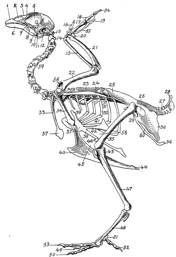
Fig. 4.—The skeleton of the domestic fowl. 1, Os incisivum. 2, External nasal opening. 3, Os nasale. 4, Os lachrymale. 5, Lamina perpendicularis. 6, Os dentale. 7, Os palatine. 8, Os quadrato-jugal. 9. Os pterygoideum. 10, Os quadratum. 11, Os articulare. 12, External auditory canal. 13, Atlas. 14, Os carpi radiale. 15, Radius. 16, First finger. 17, Os metacarpus. 18, Second finger. 19, Third finger. 20, Os carpi ulnare. 21, Ulna. 22, Humerus. 23, Thoracic vertebræ. 24, Scapula. 25, Os ilium. 26, Foramen ischiadicum. 27, Caudal vertebræ. 28, Pygostyle. 29, Foramen obturatum. 30, Os ischium. 31, Processus uncinatus. 32, Vertebral rib. 33, Os claviculum or furculum. 34, Os coracoideum. 35, Os femoris. 36, Os pubis. 37, Body of sternum. 38, Lateral internal process of sternum. 39, Costal process of sternum. 40, Sternal crest, cristi sterni, or keel of breast-bone. 41, Sternal rib. 42, Lateral external process of sternum. 43, Os fibula. 44, Xyphoid process of sternum. 45, Internal notch. 46, Os patella. 47, Os tibia. 48, Os metatarsus. 49, Second toe. 50, Fourth toe. 51, Os metatarsale. 52, First toe. 53, Second toe. 54, Second phalanx of second finger. 55, Os metacarpus. 56, Air opening in humerus. 57, Hypocledium. 58, External notch. 59, Cervical vertebræ. 60, Foramen oblongum.
25The ventral wall, floor, or basis cranii interna, may be regarded as forming five fossæ. These are: one anterior, one middle, one posterior, and two lateral. The anterior fossa supports the frontal and olfactory parts of the cerebrum. It lies at a higher level than the middle fossa. The middle fossa, or fossa cranii media, is circular in outline and extends into the lateral fossæ which lodge the olfactory lobes. Just back of the middle fossa is the sulla turcica, upon which rests the pituitary body. The posterior fossa, or fossa cranii posterior, lodges the medulla oblongata.
Bones of the Cranium (Figs. 4, 6, 7, and 8).—The bones of the cranium fuse early in the chick’s life. The sutures uniting the bones can usually be seen in the fetus or in the baby chick soon after it emerges from the shell. The major portion of the cranial bones become entirely fused. Each cranial and each facial bone ossifies from a distinct center or centers.
The cavity for the cerebrum is much larger than the cavity for the cerebellum. The cranial cavities in birds are relatively larger than in mammals. The bones are designated as in mammals. There are three single bones, the occipital, the ethmoid, and the sphenoid. Those in pairs are the frontal, the parietal, and the temporal.
The Occipital. Location.—The occipital bone or os occipitale, is situated at the posterior part of the cranium, of which it forms the posterior wall. This part is called the base of the cranium. The occipital bone articulates with or touches inferiorly, the sphenoid, laterally, the temporal, and superiorly, the parietal.
Development.—The occipital bone is developed from four centers of ossification; the dorsal, or os occipitale superius, two lateral, or ossa occipitales laterales, and the ventral, or os occipitale inferius, all of which may be seen distinct in the baby chick (Fig. 5, Part II, No. 1).
26
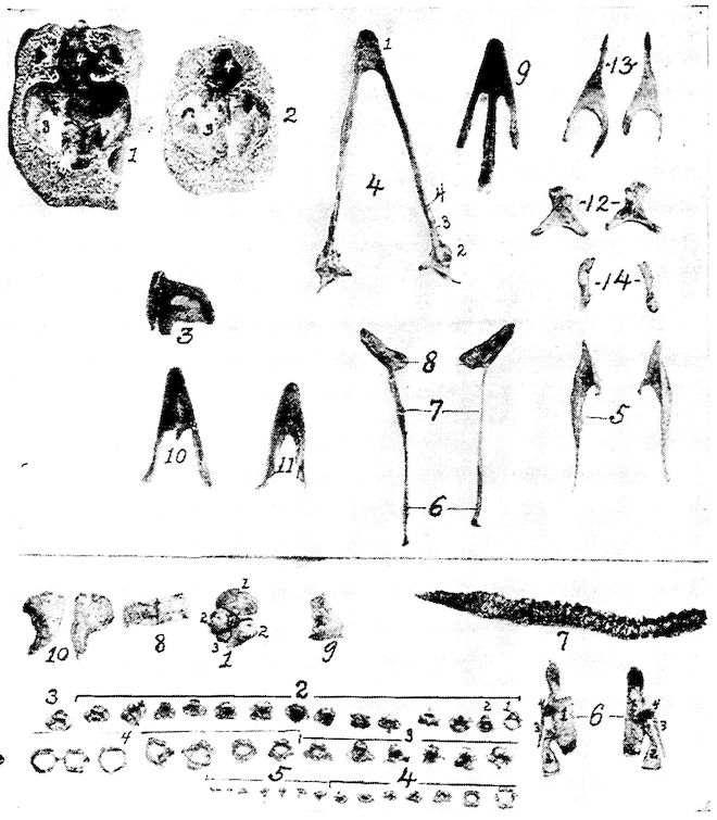
Fig. 5.—Bones of the head and vertebra. Part I.—1, The cranium. 2, The skull cap. 3, The ethmoid bone. 4, The inferior maxilla. 5, The palatine bone. 6, The quadrato-jugal. 7, The jugal. 8, The superior maxilla. 9, The premaxilla. 10, The horny covering for the premaxilla. 11, The horny covering for the os dentale. 12, The os quadratum. 13, The nasal bone. 14, The pterygoid. Part II.—1, The os occipitale (1, dorsal; 2, two lateral; 3, ventral portions and 4, foramen magnum). 2, The fourteen cervical vertebræ. (1, atlas; 2, axis). 3, The seven dorsal vertebræ. 4, The fourteen lumbo-sacral vertebræ. 5, The seven coccygeal vertebræ. 6, The pelvis (1, ilium; 2, ischium; 3, pubis; 4, cotyloid cavity or acetabulum). 7, The vertebral column complete from the baby chick. 8, The parietal bone. 9, The temporal bone. 10, The frontal bone.
27Description.—In the center of the occipital bone is the foramen magnum. Through this foramen the spinal cord extends into the cranial cavity and connects with the medulla oblongata. The occipital bone has a single condyle, which is located just below the foramen magnum and articulates with the atlas. At the base of the condyle a small subcondyloid fossa receives the body of the atlas during extreme flexion of the head. In the center of the lateral wing of the occipital bone there is a small foramen through which passes the hypoglossal nerve. Somewhat laterally from this foramen there is an opening through which the vagus, or pneumogastric nerve passes. Laterally from these is the canalis caroticus et jugularis. Between the os occipitale superius, or dorsal portion, and the ossa parietalia is a space to which ligaments are attached, called the fontanel.
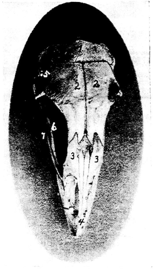
Fig. 6.—View of the frontal region of the skull of a hen. 1, Parietal. 2, Frontal. 3, Nasal. 4, Premaxilla. 5, Temporal. 6, Inferior jaw. 7, Jugal bone.
The Ethmoid. Location.—The ethmoid, or os ethmoidale, forms the anterior wall of the cranium, and the orbital septum. It is related anteriorly with the vomeral, superiorly with the nasal 28and frontal, posteriorly with the temporal, and inferiorly with the sphenoid, and palatine.
Description.—The ethmoid consists of a perpendicular and a horizontal lamina. The perpendicular lamina, located between the orbital cavities, is called the septum interorbitale. On each side of the septum interorbitale and near the superior orbital roof are two foramina for the passage of the olfactory nerves. In the horizontal plate, which forms the anterior cranial wall, are located the optic foramina through which pass the optic nerves.
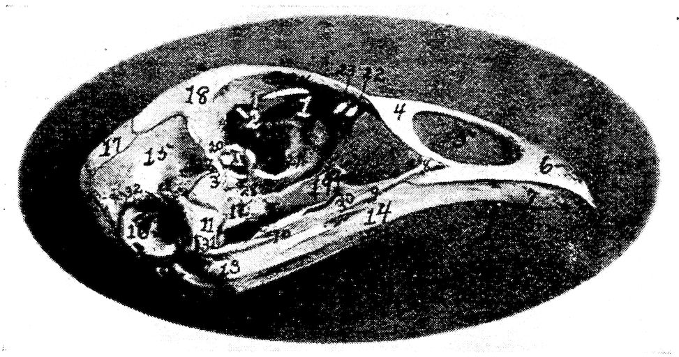
Fig. 7.—Side view of the skull of a hen. 1, Lamina perpendicularis. 2, Foramen for the passage of the nerve of smell. 3, Foramen for the passage of the optic nerve. 4, Nasal bone. 5, External nasal opening. 6, Premaxilla. 7, Os dentale. 8, Superior maxilla. 9, Os jugal. 10, Os quadrato-jugal. 11, Os quadratus. 12, Pterygoideus. 13, Os articulare. 14, Inferior maxilla. 15, Temporal. 16, External auditory canal. 17, Parietal. 18, Frontal. 19, Palatine. 20, Orbital process, posterior to which is the processus zygomaticus. 21, Interorbital foramen and optic foramen for passage of optic nerve. 22, Attachment for inferior oblique; 23, for superior oblique; 24, for levator palpebræ superioris; 25, for internal rectus; 26, for superior rectus; 27, for external rectus; 28, for inferior rectus; 28, for inferior rectus; 29, for depressor palpebræ inferioris; 30, for orbicularis palpebrarum; 31, for tensor tympani; 32, for circumconcha.
The Sphenoid. Location.—The sphenoid, or os sphenoidale, forms the floor of the cranial cavity and articulates with or touches posteriorly the occipital, laterally the temporal, and anteriorly the palatine, and ethmoid.
Description.—The sphenoid bone, the largest part of the cranial floor, is formed by the fusion of the nasal and the cranial parts. It is a three-cornered bone with two, thin, broad wings. These wings are divided into two portions, orbital wings, or alæ orbitales, and 29temporal wings, or alæ temporales. The temporal wing forms a cover for the Eustachian tube trumpet, and for the canal coming from the sella turcica, which latter gives passage to the intracranial carotid artery. The orbital wing forms the lower portion of the posterior wall of the orbital cavity, and lies directly before the os petrosum or temporal bone where the second and third branches of the fifth pair of cranial nerves emerge from the cranial cavity.
Anteriorly the sphenoid has a foramen for the passage of the Eustachian tube, the tuba auditiva, and also a sharp-pointed projection, the nasal portion, called the rostrum.
The Frontal. Location.—The frontal bones, or ossa frontales, two in number, form the forehead, a portion of the nose, and a portion of the roof of the orbital cavities. They are related posteriorly with the parietal, laterally with the temporal and zygomatic, and anteriorly with the nasal and premaxillary. They touch inferiorly the ethmoid.
Description.—Each of these bones has a processus orbitalis which is seen at the outer margin of the posterior and upper orbital wall and just in front of the zygomatic process of the squamous portion of the temporal. The frontal bone forms the anterior portion of the superior wall of the cranial cavity. The two bones are thin, flat, and meet at the median line of the forehead. The external surface is convex. The inner surface has a ridge located longitudinally and in the center the bone becomes narrow anteriorly.
The Parietal. Location.—The parietal bones, or ossa parietalia, two in number, form the posterior part of the roof of the cranial cavity. They meet in the median line and are related posteriorly with the occipital, anteriorly with the frontal, and laterally with the temporal bones.
Description.—The parietal bones are short and very broad (Fig. 5, Part II, No. 8). Each bone is quadrilateral in outline and has two surfaces, four borders and four angles. The external parietal surface is convex and smooth and the internal, cerebral surface is concave.
The Temporal. Location.—The temporal bones, or ossa temporales, two in number, are located at the lateral portion of the cranium, and aid in the formation of the cranial wall. The temporal bone is related posteriorly with the occipital, superiorly with the parietal and frontal, externally with the quadratus, anteriorly with the ethmoid, and inferiorly with the sphenoid.
30Description.—The temporal bones consist of the fused squamous and petrous temporals; they contain the essential organs of hearing.
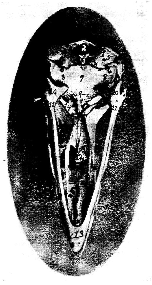
Fig. 8.—Inferior view of the skull of a hen. 1, Occipital. 2, Foramen magnum. 3, Occipital condyle and just below the basi-occipital. 4, Foramen for the passage of the hypoglossal nerve. 5, Foramen for the passage of the vagus. 6, Canalis caroticus and jugulare. 7, Sphenoid. 8, Temporal wings of the sphenoid. 9, Foramen auditiva. 10, Os articulare. 11, Os angulare. 12, Vomer. 13, Os dentale. 14, Pterygoid.
The squamous portion of the temporal bone possesses the long thin zygomatic process sometimes called the posterior orbitalis. It forms a small flattened tongue, directed forward, sometimes free, and at other times united by its superior border to the summit of the orbital process. This is especially true in the turkey. This process is seen near the lower outer portion of the posterior orbital wall. The squamous portion is also provided antero-laterally with an articular facet which articulates with the quadrate bone. The large portion of the temporal bone lies on the side of 31the cranium superior to the ala sphenoida temporale. It extends outward and anteriorly over the rims of the petrosum and ala sphenoida orbitale. The squamous part forms the upper three-fourths of the cochlea, the inner auditory canal, the upper part of the fenestra ovalis, the anterior vertical and the outer semicircular canal, and the lower part of the posterior vertical semicircular canal.
The petrous portion forms the posterior wall of the foramen ovale and the fenestra ovalis in which lies the columella. The fenestra ovalis and the fenestra rotunda are the only two entrances into the labyrinth. In this region may be seen the fusion line between the os petrosum and the os occipitale. Superiorly and posteriorly the petrous portion touches the external parietalia and occipitalis; infero-laterally it unites with the basi-sphenoid. The outer rim of the foramen ovale is broadened by the ala sphenoida and mesially by the basi-sphenoid. This foramen gives exit to the second and third divisions of the fifth pair of cranial nerves.
The lateral surface of the temporal bone presents a short tube, the external acoustic process, or processus acusticus externus, to which is attached the concha of the ear. The process is directed outward. Its lumen, the external acoustic meatus, or meatus acusticus externus, conducts to the cavity of the middle ear in the bare skull, but is separated from it by the tympanic membrane in the natural state.
Bones of the Face
The bones of the face, or ossa faciei, are the premaxillary, or upper mandible, the maxillæ, the nasal, the lacrimal, the palatine, the pterygoid, the zygomatic, the vomeral, the jugal and the quadrato-jugal. The vomer is single, the others are paired. In the inferior maxillary group there are the quadrati, and the inferior maxillary, or lower mandible. The turbinated and hyoid bones are also discussed with the bones of the face.
The beak consists of the anterior portion of the upper and lower mandibles which are covered with a horny structure.
The Premaxilla. Location.—The premaxillæ, or ossa incisiva, or mandibular structures, are located in the extreme anterior facial region.
Description.—The premaxillæ are long and the anterior end is pointed. Each consist of two lateral halves which become fused before the chick is hatched. They partly circumscribe the openings 32into the nose. This bone is the base of the upper portion of the beak and determines its form. It forms the anterior walls of the nasal cavity. It has between the nasal bones, two processes which extend back to the anterior point of the cerebral cavity. The posterior part of the incisivum and nasale are flat, thin, and elastic. The extensions then are the processus maxillaris, the processus palatinus, and the processus frontalis, the first of which forms part of the jaw rim, the second, which aids in forming the gum plate, and the last which reaches to the anterior portion of the cranial wall.
The Maxilla. Location.—The superior maxillaries, or ossa maxillares, two in number, form the floor of the upper beak, a part of the palatine roof and nasal walls.
Each maxilla borders laterally the premaxilla and the nasal; inferiorly, the anterior point of the palatine; and posteriorly, the jugale.
Description.—The maxillæ are thin, flat, bones. They have three borders and three angles. The palatine processes of the two bones do not meet in the median line, which results in a cleft in the median palatine region. The bone-like gums are formed partly by the palatine processes but more largely by lateral wings of the os incisivum. Each maxilla has posteriorly a yoke-like extension, superiorly a small extension, and also a palatine process. The latter articulates by a facet with the vomer.
The Nasal. Location.—The nasal bones, or ossa nasalia, two in number, are located in the lateral facial region. The nasal bone articulates posteriorly with the frontal; laterally, on the inner border, with the processus frontalis of the premaxilla, and on the outer border with the lacrimal; and inferiorly with the premaxilla, maxilla, jugale, and vomer.
Description.—The nasal bone, or os nasale, is broad, flat, and elastic and forms the posterior wall of the nasal opening. Under this bone is located the infraorbital sinus.
There are three extensions: first, the processus intermaxillaris, which forms the upper wall of the nasal cavity; second, the processus maxillaris anterior, directed downward and forward, which fuses with the maxillary bone and forms the posterior rim of the nasal cavity; and third, a posterior processus frontalis, which lies parallel with the ethmoid.
The Lacrimal. Location.—The lacrimal bones, or ossa lacrimalia, 33two in number, are located at the outer border and at the junction of the processus frontalis of the nasal, with the frontal bone.
Description.—The lacrimal bone is small, and rather filiform. They become fused with the nasal and the frontal bones, forming part of the margin of each.
The Palatine.—Location. The palatine bones, or ossa palatina, two in number, enter into the formation of the inner part of the bony gum and hard palate, or roof of the mouth. They form the support for the hard palate. Each palatine articulates posteriorly with the rostrum, or nasal portion of the sphenoid, and with the pterygoid; and anteriorly with the maxilla.
Description.—The palatine bone is curled posteriorly and is thin at the upper portion and thick at the lower border. Anteriorly it has a long rather filiform projection.
The Pterygoid. Location.—The pterygoid bones, or ossa pterygoidea, two in number, are located back of the region of the posterior nares. They extend diagonally outward and backward from the median region of the sphenoid rostrum to the quadrate bone. They articulate anteriorly with the sphenoid rostrum and the palatine, and posteriorly with the quadrate.
Description.—The pterygoid bones are slender and cylindrical, and are expanded at the ends into an articular facet.
The Zygomatic. Location.—The zygomatic, or ossa zygomatica, two in number are situated below the orbital cavity and extend from the maxilla to the quadrate bone.
Description.—The zygomatic is small, slender, rod-shaped, and forms the lateral portion of the upper jaw. The anterior portion of the zygomatic represents the jugal and is fused with the maxilla and with the anterior processus maxillaris of the nasal bone, the maxilla, and the posterior portion, the quadrato-jugal, which articulates with the quadrate bone.
The Vomer. Location.—The vomer is located in the median nasal septum. It articulates with the rostrum of the sphenoid, being connected to it by a mass of ligaments. It touches anteriorly the posterior portion of the maxilla.
Description.—The vomer is a median bone and aids in the formation of the septum nasi. It consists of a thin plate, thickest posteriorly and diminishing toward the anterior edge.
The Jugal and Quadrato-jugal.—The jugal and quadrato-jugal are united forming a long slender cylindrical bone called the zygomatic, 34lying at the outer side of the upper jaw. They have been described under zygomatic, which see.
The Quadrate. Location.—The quadrate bones, or ossa quadrata, two in number, are located antero-laterally to the temporal bones. Each articulates inferiorly with the posterior articular portion, or pars articulare, of the inferior maxilla and infero-laterally with the quadrato-jugalare portion of the zygomatic. It articulates antero-internally with the pterygoid and supero-posteriorly with the temporal.
Description.—The quadrate bone is anvil-like in shape. It has an anterior process, the orbital process, for muscular attachments, and posteriorly it affords attachment to the ear drum.
The Inferior Maxilla. Location.—The inferior maxilla, lower jaw, or os maxillare inferius, also called the mandibular bone, or pars dentis, is analogous to the lower jaw of mammals. It articulates posteriorly with the quadrate bone.
Description.—The inferior maxilla is the largest bone of the face. It is made up of a right and a left limb which are separate in the fetus and which unite subsequently anteriorly, forming the inferior portion of the beak. Each limb of the jaw is developed from five elements: the pars articularis, which forms the jaw-joint and, expanded, articulates with the quadrate bone; the pars angularis, lying just in front of the pars articularis; the pars supra-angularis, a slender bone lying just above the angularis; the splenial, a thin plate of bone, lying along the inner surface of the mandible; and the pars dentalis, which forms the anterior portion of the jaw.
The Turbinate Bones. Location.—The turbinate bones, or ossa turbinata, six in number, are attached to the lateral walls of the nasal cavity (Fig. 26, No. A, 1 and 2). In each nasal cavity there are three turbinate bones, one anterior and two posterior. Of the two posterior the upper one lies supero-posterior to the inferior one. The turbinate bones are attached to the lateral nasal walls, project into the cavity and thus greatly diminishing its extent.
Description.—Each turbinate bone is composed of a very thin lamina, finely cribriform in many places, and in the fresh state, covered on both sides with mucous membrane. These bones are curled and partly membrano-cartilaginous structures which give greater surface in the nasal passage for mucous membrane in which the olfactory nerve terminal filaments are distributed.
35The Nasal Cavity.—The nasal cavity, or cavum nasi, is a longitudinal passage which extends through the upper part of the face. It is divided into right and left halves by a median septum nasi. Its walls are made up of the premaxilla, maxilla, nasal, vomer, and palatine bones.
The Hyoid Bone. Location.—The hyoid bone, or os hyoideum, is situated chiefly between the rami of the mandible, but its upper part extends around the outer margin of the base of the skull. The hyoid bone does not articulate with any bones of the skull, but is firmly attached by strong fibrous structure.
Description.—The hyoid bone consists of the following parts: the body, or basi-hyal, which is subcylindrical and presents in front a trochlear articular surface, convex transversely, and concave vertically for articulation with the ewer-shaped portion of the glosso-hyal. The anterior free portion, or lingual process, is called the glosso-hyal, or entoglossal bone. The lingual process gives support to the muscular and fibrous structures of the tongue. On either side of the basi-hyal there is a limb passing posteriorly along the side of the superior larynx, extending upward along the outer border of the occipital bone. This is the cornu of the os hyoideum and is divided into two elements, first, the basi-branchial which is bone and articulates with the basi-hyal, and the second, the cerato-branchial, cartilaginous in structure. In the center and projecting backward from the body of the os hyoideum is a spur process called the uro-hyal, partly bony and partly cartilaginous, and which rests upon the superior larynx.
THE VERTEBRAL COLUMN
The vertebral, or spinal column consists of 42 bones, as shown in the following table:
Cervical region 14
Dorsal region 7
Lumbo-sacral region 14
Coccygeal region 7
Total 42
Many of the bones of the dorsal and lumbo-sacral regions do not have free articulations. The cervical and coccygeal alone have free movements.
The Cervical Vertebræ (Fig. 4, No. 59; Fig. 5, Part II, No. 2). Location.—The cervical vertebræ form the neck of the fowl.
36
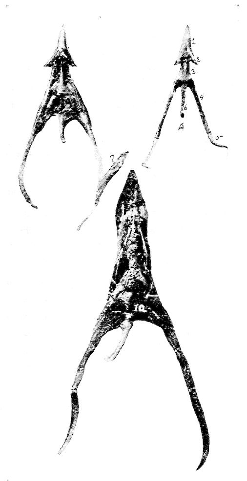
Fig. 9.—Os hyoidum and tongue muscles. 1, Glosso-hyal. 3, Basi-hyal. 4, Basi-branchial element of the cornua. 5, Cerato-branchial element of the cornua. 6, Uro-hyal or spur process. 7, Genio-hyoideus. 8, Cerato-hyoideus showing the slender tendon. 9, Hyoideus transversus. 10, Hyoideus transversus.
37Description.—The long cervical section of the spinal column is S-shaped and is made up of fourteen vertebræ. The anterior segments move freely forward, the middle ones backward and the posterior ones forward, allowing the S-shaped curve of the neck. The neck is flexible so that it is possible for the beak to reach the coccygeal oil gland. The bird is enabled to reach the feathers on all parts of the body to cleanse and oil them.
The Atlas. Location.—The first cervical vertebra is called the atlas. Anteriorly it articulates with the single condyle of the occipital bone. Posteriorly it articulates with the axis, or second cervical vertebra.
Description.—The atlas is the smallest of the cervical vertebræ and is ring-shaped. The anterior articular surface, half-moon in shape, forms a deep articular cavity. The joint, called a ball-and-socket joint, makes possible movements in all directions. The condyle of the occiput also touches an articular end of the odontoid process of the axis, or second cervical vertebra. Posteriorly there projects from the atlas two small lateral wings possessing articular facets which articulate with similar facets on the lateral wings of the axis. Posteriorly the body of the atlas is also provided with an articular surface which articulates with a similar surface on the anterior portion of the body of the axis.
The Axis. Location.—The axis is the second cervical vertebra. It articulates anteriorly with the atlas, and by a facet on the extreme anterior end of the odontoid process, with the condyle of the occipital bone. Posteriorly it articulates with the third cervical vertebra.
Description.—The body of the axis is short. The upper anterior portion of the body of the axis is provided with a tooth-like process called the odontoid process. There are two anterior lateral wings provided with small articular facets which articulate with similar facets of the atlas. The anterior surface of the body of the axis forms a true articulation with the body of the atlas.
The axis is provided with a superior and an inferior spine. There are two posterior articular processes which articulate with the prezygapophyses, or anterior articular processes, of the third cervical vertebra. The posterior part of the body of the axis forms a true articulation with the body of the third cervical vertebra.
Other Cervical Vertebræ.—Beginning with the axis, the body of which is relatively short, the body of each succeeding vertebra 38is longer than that of the preceding. The articulations of each vertebra with adjoining vertebræ are effected by means of diarthrodial facets, convex in one direction and concave in the other.
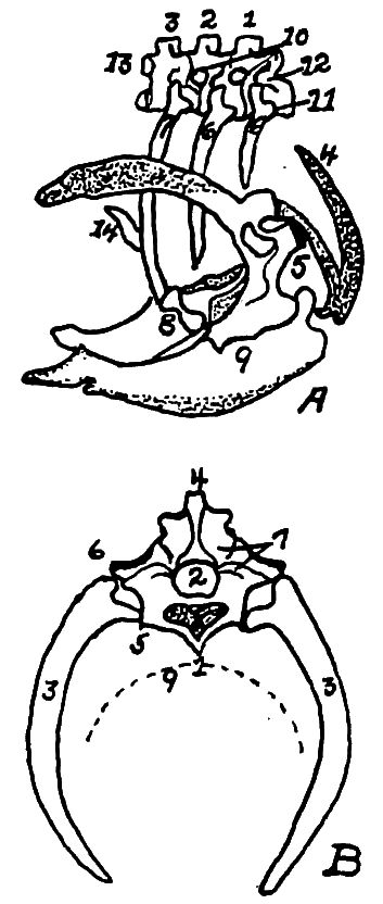
Fig. 10.—A. Diagram of three first dorsal vertebræ and scapular arch-side view. B. Diagram of section through the hemal arch. A. 1, First dorsal segment. 2, Second. 3, Third. 4 and 5, Hemal arches. 6, The pleurapophyses or floating ribs. 7, The third pleurapophyses or dorsal vertebral rib articulating with the hemapophysis or sternal rib, 8, and this in turn with the wing of the sternum form a hemal arch. 9, The sternum. 10, Intervertebral foramen through which the nerves pass. 11, Articulation with the body of vertebra parapophyses. 12, Articulation with transverse process or diapophyses. 13, Oblique process or zygapophysis. 14, The epipleural appendage. B. 1, Hemal or inferior spine (hypapophysis). 2, The neural arch. 3, The pleurapophyses or rib. 4, The superior or neural spine or neurapophysis which aids in the formation of the anapophyseal ridge. 5, Parapophyseal surface for head of rib. 6, Tubercle of rib articulating with the diapophysis. 7, Lamina of neurapophysis. 8, Centrum. 9, Hemal arch.
Between the bodies of the vertebræ are pads of fibrous cartilage. Above these bodies and inferior to the neural spines extends throughout the spinal column the large neural canal which is occupied by the spinal cord. Between the vertebral segments the neural canal is exposed superiorly, since here the neural arches do not completely bridge the canal. These spaces are protected by intervertebral ligaments.
In addition to the superior neural spines and the inferior spines, 39from the body of the vertebra project lateral processes, and anterior and posterior oblique processes.
All cervical vertebræ except the atlas and the axis are made up of the following parts: a body, or centrum, a neural canal, a neural arch, a superior dorsal, or neural spine, or neurapophysis, which in most cervical vertebræ is only slightly developed, two oblique processes, or zygapophyses, two transverse processes, or diapophyses (Fig. 10, No. 12) and in some vertebræ two tubercles, or anapophyses above the posterior zygapophyses. The prezygapophyses are directed upward and inward; the postzygapophyses are directed downward and outward. The inferior spines are only well developed in the last two or three cervical vertebræ.
Between vertebral segments, except the central sacral portion, on each side, there is an intervertebral foramen (Fig. 11, No. A, 4), through which pass the spinal nerves. In the central portion of the sacrum where the vertebræ have fused, there are two foramina on each side for each original vertebra, one located above the other, the upper giving passage to the sensory branch, the lower to the motor branch of the nerve.
The Dorsal Vertebræ (Fig. 4, No. 23; Fig. 11, A). Location.—The dorsal, or thoracic vertebræ, or vertebræ thoracicales, aid in forming the roof of the chest cavity.
Description.—The dorsal vertebral section is made up of seven vertebræ, with strong short bodies. The first and sixth dorsal segments articulate as do the cervical, by the bodies and oblique processes. (Fig. 11, No. A, 3, illustrates the oblique processes.) The seventh dorsal is fused with the first lumbo-sacral vertebra. The second to the fifth inclusive of the vertebræ are fused together, and the superior and inferior spinous processes are fused into a prominent plate-like ridge.
The transverse processes of the dorsal vertebræ, from the second to the sixth, are well developed and are bridged over with a thin layer of bone. The ventral spines are partly fused and form a very prominent and continuous ridge. (Fig. 11, No. A, 1, shows the fused superior spines; No. 2, the fused inferior spines; and No. 4, the intervertebral foramina.)
The Lumbo-sacral Vertebræ (Fig. 5, Part II, No. 4). Location.—The fused lumbo-sacral section of the spinal column forms the roof of the pelvic cavity.
40
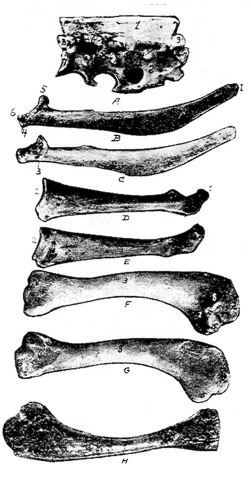
Fig. 11.—Bones from the scapular arch. A. Dorsal vertebra. 1, Superior spinous ridge, 2, Inferior spinous ridge. 3, Oblique processes. 4, Intervertebral foramina. 5, Articular facette for head of the rib. 6, Articular facette for tubercle of the rib. 7, Articular portion of the body. B. Outer surface of scapula. C. Inner surface of scapula. 1, Thin caudal end. 2, Articular head. 4, Processus furcularis. 5, Processus humeralis. 6, Processus coracoideus. D. Outer surface of coracoid. E. Inner surface of coracoid. 1, The furcular tuberosity. 2, Articular facette for articulation with the sternum. 3, Articular surface for humerus and scapula. F. Outer surface of humerus and G the inner surface of the same. 1, Trochanteric fossa. 2, Oval articular head at proximal end. 3, Shaft. 4, Distal end showing articular condyles. 5, Trochanter. 6, Trochlea for ulna. 7, Trochlea for radius. 8, Depression or fossa. H. Section through the median plane of the humerus showing the delicate cross partitions illustrating provisions made for bones provided with air-sac extensions.
41Description.—The lumbo-sacral region consists of fourteen vertebræ which are distinct in the body of the newly hatched chick, but which become fused soon after hatching. With these vertebral segments there are fused the last dorsal and first coccygeal vertebral segments. This fusion is so complete that the segments are indicated only by the intervertebral foramina on the sides through which the spinal nerves pass, and by transverse markings on the inferior surface of the bodies.
The lumbo-sacral vertebræ, called the sacrum, and the ilia are fused. The dorsal spines of the vertebral segments are indicated only in the anterior portion where they are fused, forming a plate. There are no prominent ventral spines.
The Coccygeal Vertebræ (Fig. 4, No. 27). Location.—The coccygeal or caudal vertebræ, or vertebræ coccygeæ, constitute the bones of the tail.
Description.—There are seven coccygeal vertebræ. The last segment, shaped like a plough share and therefore called the pygostyle, is the largest and is supposed to have been formed by the fusion of several original distinct segments. It supports the coccygeal oil gland and the row of rudder feathers, or rectrices, which are arranged fan-wise.
All of the coccygeal vertebræ except the first are freely movable, thus allowing the tail to be used as a rudder during flight. The lateral spines are long and well developed, and the superior spines are bifurcated, thus giving increased surface for muscular attachment. The first coccygeal segment is fused with the last lumbo-sacral vertebra.
THE THORAX
The dorsal vertebræ superiorly, the ribs laterally, and the sternum, or breast-bone inferiorly, form the skeleton of a large cavity called the thorax. The dorsal vertebræ have been described.
The Ribs (Fig. 4, No. 32; Fig. 10, A and B). Location.—The ribs form the lateral bony wall of the thorax, articulating superiorly with the dorsal vertebræ.
Description.—The ribs are arranged in order of length, the ultimate rib being the longest. From anterior to posterior, they approach more nearly a horizontal position. The ribs are divided into the true and the false. The true ribs articulate with the sternum. The false ribs do not touch the sternum; they are floating. The true ribs are composed of two parts, a vertebral, or dorsal, and a sternal, 42or ventral. The vertebral part, or extremitas vertebralis, is provided with an articular head, or caput costæ; a neck, or collum costæ; and an articular tubercle, or tuberculum costæ. The head and tubercle articulate with the dorsal vertebra. Below the head of each rib is a pneumatic foramen.
The distal extremity of the dorsal section of the rib articulates with the proximal end of the sternal section by a diarthrodial articulation.
The articulations of the true ribs with the sternum is diarthrodial and each articulate by two small ridges with a double sternal facet. The first, the second, and the seventh ribs are floating, or false ribs. The first rib articulates with the quadrate part of the last cervical vertebra and also with the first dorsal vertebra. The seventh, the last rib, articulates with the under side of the anterior alar part of the ilium. This rib is situated similarly to the true ribs; but, instead of articulating directly with the sternum, the lower end lies against the sternal segment of the rib just anterior to it.
From the posterior edge of the second, the third, the fourth, and the fifth ribs, and near the middle of the dorsal segment, are flat, uncinate, bony processes which project upward and backward, overlying in each case, the succeeding rib and giving greater surface for muscular attachments and greater stability to the thorax.
The Sternum (Fig. 4, No. 40; Fig. 10, No. A, 9). Location.—The sternum, or breast-bone forms the inferior portion, or floor of the thoracic cavity.
Description.—The sternum is a quadrilateral, curved plate with processes projecting from each angle and from the middle of the anterior and posterior borders. The posterior medial projection, or metasternum, is the longest, and has a tall, plate-like ridge—the sternal crest, crista sterni—running along its ventral surface. The crest serves the important function of increasing the bony area for the attachment of the powerful muscles which move the wings. The anterior medial projection, or rostrum, is short, and pierced at its root by an opening from which extend two elongated, saddle-shaped depressions into which the end of the coracoid bones are received.
The plate-like process of bone, the posterior lateral process, which projects from the caudal angles of the sternum soon divides into two parts. The shorter of these, the oblique process, broadens toward its free end and covers the sternal segments of the last two ribs. The 43sides of the sternum are thus provided with an external and an internal process forming an external and an internal notch. These notches are bridged over by a broad ligament, to which the muscles are attached. In poor flyers, as the domestic fowl, these notches are large. The posterior end of the sternum is called the xiphoid process or processus xiphoideus. Anteriorly the sternum is provided with lateral external processes, the costal processes. The lateral borders of the sternum are pitted by four depressions into which the sternal segments of the ribs are received. The dorsal, or inner surface of the bone is pierced by openings by which the air-sacs communicate with the interior.
The Appendicular Skeleton
The appendicular skeleton consists of the shoulder girdle, the fore limb, the pelvic girdle, or hip bone, and the hind limb. The shoulder girdle consists of the scapula, the coracoid, and the clavicle. The fore limb consists of the arm, forearm, and the hand. The arm consists of the humerus, the forearm of the radius and the ulna; and the hand consists of the carpus, the metacarpus, and the phalanges. The pelvic girdle consists of the ilium, the ischium, and the pubis. The hind limb consists of the thigh, the leg, and the foot. The thigh consists of the femur, and the leg consists of the tibia and the fibula. The foot consists of the metatarsus and the phalanges.
THE SHOULDER GIRDLE
The shoulder girdle consists of the scapula, the clavicle and the coracoid.
The Scapula. Location.—The scapula (Fig. 11, A, B) lies on the outer and superior rib surface, extending parallel with the dorsal vertebræ.
Description.—The scapula is a thin, sword-like bone, becoming thicker as it approaches the shoulder-joint. The scapula expands and becomes thin near the free end, which reaches nearly to the antero-lateral portion of the ilium (Fig. 4, No. 24). The scapula articulates with the coracoid. A pneumatic foramen is located at the base of the acromion process. The anterior part of the scapula is provided with an articular head and is provided with an inner process, called the processus furcularis, which lies near the furcula and coracoid. An outer stronger processus humeralis forms the 44posterior half of the glenoid cavity, or cavitas glenoidalis, in which the humerus articulates and the processus coracoideus.
The Clavicle. Location.—The clavicles, commonly known as the wish bone, are located in the anterior chest region.
Description.—The clavicles are long, slender bones uniting below in the hypocledium, a laterally flattened process. They are joined to the upper end of the coracoid by fibrous cartilage. The hypocledium is joined to the anterior point of the sternum by the claviculosternal ligament. The clavicles, as united by the hypocledium, form a v-shaped structure called the furculum, or, popularly, the wish bone.
The forks play the part of an elastic spring, whose office it is to prevent the wings from coming toward each other during contraction of the depressor muscles. The conformation of this bone is, therefore, like the sternum, related to the extent and power of flight; and for this reason it is that, in swift flyers, the two branches of the furculum are thick, solid, widely separated, and curved like a U; while in those that fly heavily and with difficulty, these branches are thin and weak, and join at an acute angle. The latter formation greatly diminishes its strength, and lessens, in a singular manner, the reactionary power of the bony arch it represents.
The Coracoid. Location.—The coracoid is located just back of the clavicle and at the side of the entrance of the thoracic cavity. It is the strongest bone of the shoulder girdle, extending upward, outward, and forward. It articulates inferiorly with the sternum and superiorly with the humerus and the scapula, and is attached to the superior end of the furcular limb by a fibrous cartilage.
Description.—It is thinnest in the center or shaft and broadens toward the inferior extremity. The upper hook-like part of the coracoid forms the fore part of the glenoid cavity, and together with the scapula and furcula form the foramen triosseum through which passes the tendon of the elevator muscle of the wing. The upper end flattens out into three tuberosities, the tuberositas furcularis which is thick and to which attaches the limb of the furcula, the tuberositas scapularis which unites to the scapula, and the tuberositas humeralis which lies between these and articulates with the humerus.
THE FORE LIMB
The bones of the fore limb are humerus, ulna, radius, carpus, metacarpus, and phalanges.
45The Humerus. Location.—The humerus constitutes the arm, which, when at rest, lies parallel to the dorsal vertebræ. It articulates superiorly with the glenoid cavity, a shallow ball-and-socket joint, formed by the scapula and the coracoid; and inferiorly with the ulna and radius.
Description.—The proximal extremity of the humerus is provided with a trochanter (Fig. 11, G, No. 5) and a large oval head or caput humeri, which articulates in the glenoid cavity. The head is an elongate, semi-oval convexity, with the long axis transverse from the radial to the ulnar side and with the ends continued into upper and lower crests. The upper crest of the head of the humerus is on the radial side and the lower crest on the under side. Under this latter crest there is a pneumatic fossa (Fig. 11, F, No. 1), at the upper end of which there is an opening into the bone, the pneumatic foramen, which brings the air-sac into communication with the air space of the bone. The shaft, or corpus humeris, is irregularly cylindrical and slightly S-shaped.
The proximal part of the shaft, which is expanded on the palmar side, is concave across and convex lengthwise. The distal part is slightly flattened. The shaft of the humerus is almost cylindrical.
The distal extremity of the humerus is provided with two articular condyles, one of which articulates with the ulna and the other with the radius. On the radial side of the palmar surface there is a ridge; and on the ulnar side of the same surface there is a second ridge diverging to the opposite tuberosity. The radial surface is a narrow, subelongate convexity, extending from the middle, approximately, of the palmar surface, obliquely to the lower part of the radial tuberosity. The two articular convexities, or trochlea, at the distal end of the humerus are bent toward the palmar aspect, the anconal aspect is the side on which the elbow is situated. The inner convexity is the larger and articulates with the ulna. To the outside is the processus cubitalis humeri. The outer articulates with the radius and is so arranged that the radius makes a greater flexion than the ulna. At the lower end of the humerus there is a depression which receives the anconeus of the ulna during flexion and extension of the forearm. On the shoulder- and elbow-joint are found grooves over which the tendons glide, at which places sheaths are provided. This arrangement also aids in keeping the capsular ligament in place.
46The Ulna. Location.—The ulna, larger than the radius, is bent and articulates with the radius only at the ends. The two bones are bound together by a ligamentous band. It also articulates superiorly with the distal end of the humerus, and inferiorly with the carpus. When the wing is folded the radius is superior and a trifle to the inner side of the ulna.
Description.—The proximal end is most expanded, and is obliquely truncate for the articular excavation adapted to the ulnar tubercle of the humerus. A short angular process behind the cavity represents the olecranon, which is the processus olecranalis coracoideus ulnæ to which the ligamentum capsulare cubiti attaches. On the inner side of the head is the internal tubercle of the ulna and on the external side the external tubercle of the ulna. An excavation is noted on the radial side of the proximal end for the lateral articulation of the head of the radius. On the upper side near the articular part is located the sigmoid cavity.
The shaft, or corpus ulnæ, decreases in size near the distal end. It is slightly curved, flattened laterally with an internal and an external ridge.
The distal end of the ulna is slightly expanded into a trochlea which is sharply convex and articulates with two free carpal bones, the scapho-lunar, or os carpi radiale, and the cuneiform, or os carpi ulnare. The scapho-lunar is placed on the radial side, and the carpiulnare on the ulnar side. The distal extremity of the ulna is provided with a styloid process and on the dorsal side with a tubercle.
The Radius. Location.—The radius lies beside the ulna with which it articulates at each extremity. At the inferior extremity the articulation is rotary. It also articulates superiorly with the humerus and inferiorly with the os carpi radiale.
Description.—The radius, cylindrical in shape, is more slender than the ulna.
The proximal end is expanded, subelliptical, with a concavity for the oblique tubercle and a thickened convex border for articulation with the ulna. This end is provided with the tuberositas radii.
The shaft, or corpus radii, is slender, subcompressed, and has a slight bend upward from the ulna. A nutrient foramen occurs in this shaft.
The distal end is expanded and rather flattened with two grooves on the anconal side for passage of tendons. For articulation with 47the scapho-lunar the radius is provided with a terminal transverse convexity produced palmad, which also articulates with the ulna laterally. There is a tuberosity on the radial side of the expansion and inferiorly the inferior tuberculum ossis carpi radialis and superiorly a superior tuberculum ossis carpi radialis.
The Hand.—The hand is made up of the carpus, the metacarpus, and the phalanges.
The bones of the hand are so arranged as to allow abduction and adduction, or flexion in the ulno-radial plane, movements which are necessary in the outspreading and folding of the wing. Thus the hand of the fowl moves in a state of pronation, without the power of rotation. The carpal bones are so placed between the anterior arm and metacarpus as to reduce the abduction which is necessary to hold and extend the wing, so that the hand or wing be in a fixed position.
The Carpus.—The carpus in the domestic fowl is represented by two bones, ossa carpi, called the scapho-lunar, or os carpi radiale, and the cuneiform, or os carpi ulnare. The scapho-lunar is the smaller and is located between the radius and metacarpus. The cuneiform is the larger and is located between the ulna, the radius, and the metacarpus. The cuneiform is somewhat anvil-like in shape, being provided with a body and two prongs.
The Metacarpus.—The metacarpal bones, or ossa metacarpi, two in number, are separated at their middle portion, and consolidated at their extremities. The upper proximal base of the metacarpus is provided with a tubercle, the tuberculum muscularis, and externally the tuberculum ulnare ossis metacarpi. The distal end of the metacarpal bone is provided with a tuberculum articulare.
The Phalanges.—The first three fingers only are represented, and these are rudimentary.
The first finger called the pollex or thumb, consists of but one joint. It is located on the proximal and outer end of the metacarpal bone. It has on its proximal end a tubercle, the tuberculum articulare.
The second finger, the best developed, consists of two phalanxes. These are the main bones extending from the metacarpus. Each articular end is provided with a tuberculum articulare.
The third finger, small, cylindrical in shape, is located at the distal and inner side of the metacarpal bone.
48THE PELVIC GIRDLE (Fig. 4, No. 25; Fig. 5, Part II, 6)
The pelvic girdle is made up of three bones as follows: the ilium, the ischium, and the pubis, all of which are fused in adult life. They are separate in the baby chick (Fig. 5, Part II, 6). The pelvis together with the lumbo-sacral vertebræ forms a thin, irregular, shell-like structure extending superiorly from the tail to the thoracic region. The sacrum is broad posteriorly and together with the ilium forms the pelvic roof. The ilium and ischium are its lateral walls.
The top surface of the pelvis shows the fovea ilio-lumbalis dorsalis, which is bounded mesially by the crista ilii. Between it and the spina lumbalis there is a broad furrow, the bottom of which is formed by the dorsal surface of the lumbar vertebræ. The sulcus ilio-lumbalis dorsalis is formed by the rims of the ilia, so that a ridge is observable. The cavum ilio-lumbale dorsale is formed by the iliac rims on either side. Anteriorly is the canalis ilio-lumbalis, which is formed by the ilium and the lumbar vertebral spines. It is located longitudinally to the vertebræ. The anterior opening of this canal is the larger. Posterior to the acetabulum is the post-acetabular ridge.
The under part of the pelvis presents three distinct regions. The cavity is divided into the fovea lumbalis, or anterior part, the fovea ischiadica, or mesial part, and the fovea pudendalis, or posterior part. Posterior to these is the region called the planum coccygeum.
The fovea lumbalis contains the anterior lobe of the kidney, and is circumscribed anteriorly by the last rib-carrying vertebra, and posteriorly by the transverse process of the last lumbar vertebra.
In the fovea ischiadica which follows, lie the nerve plexus ischiadicus and the middle lobe of the kidney. Its posterior boundary is the linea arcuata. The linea arcuata is a line drawn from the acetabulum of the one side to the acetabulum of the other side.
The fovea pudendalis contains the posterior lobe of the kidney. The posterior boundary is the anterior border of the first coccygeal vertebra. It gives passage to the nerve plexus pudendo-hemorrhoidalis.
The ilium, the ischium, and the pubis join to form the cotyloid 49cavity, or acetabulum, in which articulates the head of the femur. The floor of the cavity is perforated by a relatively large round foramen.
The Ilium. Location.—The ilium, together with the lumbo-sacral vertebræ, forms the roof of the pelvic cavity. It articulates at its inner border with the lumbo-sacral vertebræ, postero-laterally with the ischium and at the cotyloid cavity with the pubis.
Description.—The ilium is remarkable for its development in the long axis of the vertebral column. It is long and narrow; and mesially, where it is thickest it forms the upper wall of the acetabulum. Anterior to the acetabulum it is outwardly concave, and posterior to the acetabulum it is convex. The ilium fuses with the last dorsal vertebra and with the lumbo-sacral vertebræ and is excavated on its internal face. This surface is irregular and lodges the kidneys. This inner margin of the renal part enters with the square extensions of the posterior excavation passing into the posterior iliac spine. The external margin is the extension of the crista transversa and forms the processus ischiadicus. Posteriorward the renal part of the ilium joins the ischium.
The ilia converge at the summits of the anterior sacral spines forming the ilio-lumbar spines.
The ilium is joined to the square extensions of the last sacral vertebra by the symphysis ilio-sacralis, to the larger part of the lumbar vertebræ by the sutura ilio-lumbalis, and to the transverse processes of the last sacral vertebræ by the symphysis ilio-sacri.
The Ischium. Location.—The ischium is located in the postero-inferior part of the pelvis. It joins superiorly with the ilium, and inferiorly with the pubis.
Description.—The ischium is smaller than the ilium and is a flattened, triangular-shaped bone, thickest where it forms the posterior part of the acetabulum, becoming thinner and broader as it extends backward. Posteriorly it forms the caudal extension. The inferior border is turned slightly outward and is fused with the pubis. Between these bones there is located the large oval ischiadic foramen, through which passes the ischiadic nerve. This bone aids the ilium in the formation of the obturator foramen through which passes the tendon of the internal obturator muscle. The lower part of the ischium which separates the ischiadic foramen from the obturator foramen is called the ramus ascendens ossis ischii.
50The Pubis. Location.—The pubis is located along the inferior margin of the ischium and extends further back than the posterior border of the ischium.
Description.—The pubis is a long, slender, rib-like bone. It forms the lower and front portion of the acetabulum. The ischiadic foramen formed by the ischium and pubis is single and nearly circular in fowls, double in pigeons, and in geese and ducks it is elongate.
THE HIND LIMB (Fig. 4, Nos. 35, 47, 48, 49, 50, 51, 52, and 53; Fig. 12)
The pelvic, or hind, limb supports the body. The bones of each leg are the femur, which constitutes the thigh, the tibia and the fibula, which represents the leg, and the metatarsus and the phalanges, which form the foot.
The Femur (Fig. 12, A and B). Location.—The femur, or os femoris, the first bone of the hind extremity, extends obliquely downward and forward, articulating with the acetabulum above and the tibia, the fibula, and the patella below.
Description.—The femur, one of the largest, thickest, and strongest bones of the body, belongs to the class of cylindrical bones, and presents for examination a shaft and two extremities.
The superior, or proximal extremity, is provided with a head, caput femoris; the neck, collum femoris; and the trochanter. The head is relatively small, and is marked by a depression above for the round ligament which fills the cavity in the acetabular wall. Its axis is nearly at right angles with the shaft. A neck joins it to the body at the proximal end. External to the head of the femur is the trochanter. The trochanter presents an outer convexity over which the tendon of the gluteus maximus extends to become inserted below. The trochanteric ridge, which is opposite the articular head, presents an outer flattened surface which possesses impressions for muscular attachments. The trochanteric fossa, or fossa trochanterica, is shallow.
The shaft, or corpus femoris, is shorter than the tibia, is in general cylindrical, bent forward, and the lower half is flattened and expanded transversely. A nutrient foramen is located in the median portion of the shaft. The shaft shows muscular linear ridges.
The distal extremity is large in both directions and comprises the trochlea in front and two condyles behind, one internal and one external. 51The condyles are separated by the intercondyloid fossa, or fossa intercondyloidea, which is marked with pits for the attachment of ligaments; and above this is the epitrochlear fossa.
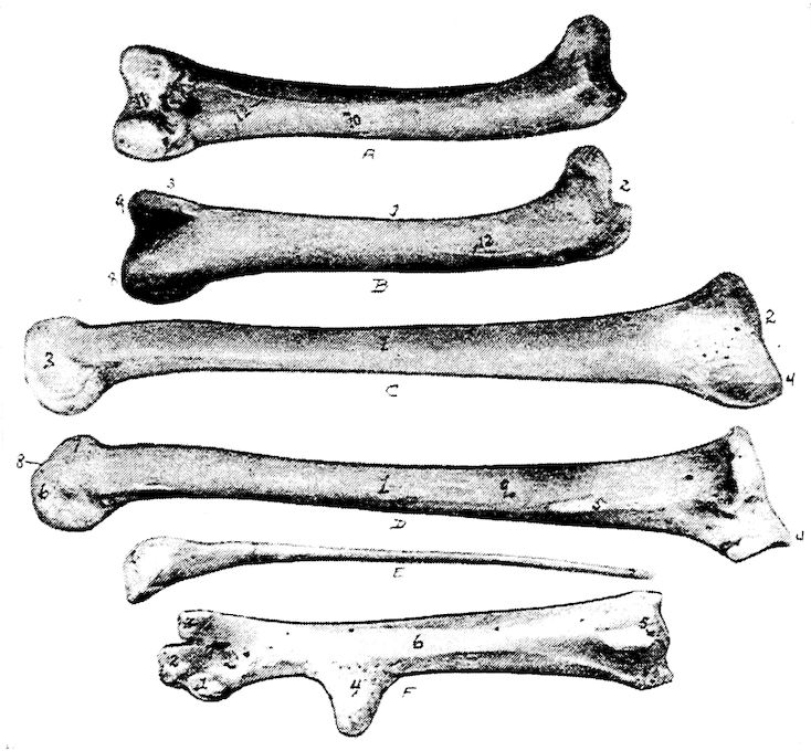
Fig. 12.—Bones of the hind extremity. A. Posterior view of femur. B. Anterior view. 1, Shaft. 2, Proximal extremity. 3, Distal extremity. 4, Articular head. 5, Trochanter major. 6, Shallow trochanteric fossa. 7, Convexity over which the tendon of the gluteus maximus glides. 8, External condyle. 9, Internal condyle. 10, Nutrient foramen. 11, Intercondyloid fossa. 12, Muscular linear ridges. 13, Epitrochlear fossa. C. Internal view of tibia. D. External view. 1, Shaft. 2, Articular head. 3, Distal end. 4, The rotular process. 5, The fibular ridge. 6, External condyle. 7, Internal condyle. 8, Intercondyloid space. 9, Nutrient foramen. E. Fibula, lateral view. 1, Articular head. 2, Its attenuated portion. F. Posterior view of metatarsus. 1, Trochlea for inner or second digit. 2, Trochlea for middle or third toe. 3, Trochlea for outer digit. 4, Bony core for spur. 5, Bony canal for tendons. 6, Groove for tendons. 7, Fossa intercondyloidea.
The inner condyle begins anteriorly as a ridge, and expands into a convexity which attains its greatest breadth posteriorly where it 52becomes more flattened. The inner side of the inner condyle is flattened and is provided with a tuberosity at its mid-part and a second just above the posterior part of the condyle.
The outer condyle is formed in the same manner as the inner condyle. It is indented at its broad, lower end by an angular groove, which, winding divides the posterior part of the condyle into two convexities. The more external convex ridge and the groove dividing it from the outer condyle are adapted to the head of the fibula. There is in this part a fibular ridge and above this ridge a tuberosity.
The Tibia. Location.—The tibia (Fig. 12, D and C) extends obliquely downward and backward from the knee-joint to the hock. It articulates above with the femur and, by its procnemial process with the patella, below with the metatarsus, and laterally with the fibula.
Description.—The tibia is the longest bone in the posterior limb, and possesses a shaft and two extremities. It is largest at the proximal end and presents three faces.
The proximal extremity presents a semi-oval articular surface, not quite at right angles with the shaft, which articulates with the condyles of the femur. The margin is raised toward the anterior of the bone. The head of the tibia, or caput tibiæ, extends into a rotular process which extends transversely, and is truncate. From the anterior of this process there descends two vertical ridges; one near the angle of the rotular process, the procnemial ridge; the other from the outer fibular angle, the ectocnemial ridge. On the outer side of the intercondylar tuberosity there is a surface for the ligamentous union with the head of the fibula; and a short distance below this there is a vertical ridge for the close attachment, almost a fusion, with the fibula, called the fibular ridge.
The shaft, or corpus tibiæ is straight and the upper two-thirds subtrihedral; the lower third oval. A nutrient foramen occurs near the upper postero-internal portion of the middle third of the bone.
The distal extremity is much smaller than the proximal one; it is quadrangular in form. The expanded inferior end of the tibia forms two articular condyles above which posteriorly there is the epicondyloid fossa. The inner condyle, the larger, has a groove near the lower end of the anterior part of the shaft, which deepens toward the intercondyloid space. This intercondyloid fossa in young birds is covered by a strong ligament, which in older birds, becomes 53ossified. On the lateral side of each condyle, there is a depression for the attachment of ligaments.
The Patella. Location.—The patella, or knee-cap, thin and wide, articulates with the procnemial process of the tibia, and with the deep trochlea of the distal end of the femur.
Description.—The patella is irregular in shape with three faces and three borders. The posterior surface is articular. The other two surfaces are rough for tendinous attachment. The patellar ligaments in old birds may become ossified.
In order to turn the foot in and out, the tibia not only turns around the inner condyle of the femur, but also around the patella, so that the posterior surface turns outward and thus turns outward the metatarsus and all the toes.
The Fibula. Location.—The fibula lies at the outer border of the tibia. It articulates superiorly with the outer condyle of the femur, and laterally with the tibia.
Description.—The fibula is rudimentary; it is largest superiorly and tapers to a slender point. The head is compressed laterally, and furnishes an upper and an inner articular surface.
The Tarsus.—There exist, during fetal development of the chick, two rows of tarsal bones which later become fused. The upper row fuses with the tibia and the lower with the metatarsus. Therefore, in the adult, there is no tarsus.
The Metatarsus. Location.—The metatarsal bone extends downward and forward. In birds it consists of one bone, which articulates superiorly with the inferior extremity of the tibia. On the distal end it has a threefold trochlear arrangement which articulates with the three principal digits.
Description.—The proximal extremity posteriorly has a process which may be considered as a consolidation of originally separate metatarsal bones. The process at the supero-posterior part of the metatarsus is called the hypotarsus of the tarso-metatarsus, through which extends a canal called the hypotarsal canal, and which gives passage to flexor tendons.
The proximal end of the metatarsus is ossified from one center of ossification forming an epiphysis which caps the ends of the three original metatarsal bones that coalesce, first with one another, then with the epiphysis, thus forming a single compound bone. Above and just to the inside of the metatarsus there may occur a small bone which is imbedded in ligaments and articulates with the 54inner proximal surface of the metatarsus. This has been called by Gadow a sesamoid bone.
The shaft shows tendonal grooves which are best marked on the posterior surface. The shaft is rounded at the sides and flattened on the anterior and posterior surfaces. At the juncture of the middle and inferior thirds of the metatarsus there is a conical process turned slightly backward, which serves as a base for the spur. The spur is a horny structure.
At the distal extremity occurs the threefold trochlear arrangement mentioned above, which incloses the fossa intercondyloidea. The inner trochlea is the broadest, and the outer the narrowest. The inner trochlea articulates with the proximal end of the second, the middle with the third, and the outer with the fourth toe.
The Phalanges. Location.—Most domestic fowls are provided with four digits, or toes; the Houdan and Dorking are provided with five. In fowls with four toes the three principal toes, the second, the third, and the fourth, are directed forward, and the first, or hallux, is directed backward.
Description.—The last phalanx of each toe, called the ungual phalanx, is slightly curved downward, is pointed anteriorly, like the claw of the cat or dog, and is covered with a horny sheath. The articular ends of the joints of the phalanges are enlarged. The base of the basal phalanx has two enlargements, the superior-inferior tubercles, or tuberculum superius et inferius, between which is located the fossa articularis transversa. Laterally the head has two condyles, condyli laterales, which are divided by the sulcus longitudinalis. The bodies of the phalanges vary in form; they are superiorly rounded, but inferiorly rounded, flat, or even somewhat concave. The distal extremity has an articular trochlea.
The first toe, or digit, called the great toe, or hallux, is composed of three phalanges, or segments. The first segment, or basal phalanx is considered a rudimentary metacarpal bone; it is attached by a fibrous cartilaginous tissue to the inner posterior surface of the inferior extremity of the metacarpal bone just below the spur.
The second toe, likewise composed of three phalanges, is directed forward. It articulates with the inner trochlea located on the inferior metacarpal bone.
The third toe, made up of four phalanges, is the middle of the 55three forward toes. It articulates with the middle trochlea of the inferior extremity of the metacarpal bone.
The fourth toe, is composed of five phalanges; it is, however, of approximately the same length as the third, the segments being shorter. The fourth or outer toe articulates with the outer trochlea of the distal extremity of the metacarpal bone.
56