ANGIOLOGY
Reading edition
The Circulatory Apparatus.—The circulatory apparatus consists of two tubular systems: the blood vascular system and the lymphatic system.
The blood vascular system consists of the heart, the arteries, the veins, and the capillaries.
The heart is the central, propelling organ. The arteries form a series of efferent tubules, which, by branching, constantly increase in number and decrease in caliber, and which serve to carry the blood from the heart to the tissues. The capillaries are extensions from these latter tubules into which the arteries empty, and through the walls of which the interchange of elements between the blood and the other tissues takes place. The veins form a system of converging tubules which receive the blood from the capillaries, decrease in number and increase in size as they approach the heart, and return the blood to that organ.
The lymphatic system consists of capillaries and veins alone. As in the blood system, the lymph capillaries collect the effete material and pour it into the lymph veins, and these in turn, carry it to the large blood veins adjacent to the heart.
Both these systems have one and the same continuous lining, which consists of a single layer of endothelial cells. In the heart this lining is called the endocardium, and in the vessels, the endothelium. It forms a perfectly smooth surface.
THE HEART (Fig. 21, No. 7)
The heart of the domestic fowl is located in the median line of the thoracic cavity. It is more anterior and mesial than in mammals. Its axis is parallel with the axis of the trunk. The lungs being confined to the dorsal part of the trunk, the lower part of the heart is not surrounded by them, but extends backward, the apex resting in the anterior part of the anterior median fissure of the liver.
The heart has the form of an acute cone (Fig. 50, No. 1), the apex of which is bluntly rounded.
207The heart is surrounded by a sero-fibrous sac, the pericardium. This sac adheres to the cervical air reservoirs anteriorly and to the diaphragmatic septum posteriorly. It is composed of two membranous layers: the parietal, external, dense, and fibrous; and the visceral, internal, and serous. The pericardial sac has no direct attachment to the heart, except at the upper extremity where it surrounds the large vessels emerging from it. The serous layer is reflected over the outer portion of the heart, where it is called the epicardium. The function of the pericardium is to prevent friction during the beating of the heart. It contains a small amount of serous fluid for perfect lubrication. This fluid is called the liquor pericardii.
Internally the heart has four cavities: two auricles and two ventricles. The right ventricle is more crescent-shaped than in solipedes, and in a manner envelops the left ventricle in front and to the right, though it does not reach the point of the heart. The right auricle is larger than the left. The auriculo-ventricular valve is not tricuspid as in mammals. This valve instead of being formed as usual by a membranous curtain, with margins retained by cords fixed to the walls of the ventricles, is composed of a wide muscular leaf, which appears to be a portion of the inner wall of the ventricle detached from the interventricular septum. This septum is convex; and the auriculo-ventricular orifice is an oblique slit situated between it and the muscular valve in question; so that, when the heart wall contracts at the systole, the valve is applied against this septum and closes the passage. The bicuspid, or auriculo-ventricular valve of the left side usually has two segments, though occasionally there may be three. The fossa ovalis is a depression behind the posterior semi-lunar valve in the septum of the heart. The membranous septum closing the foramen ovale is complete and strong but thin and transparent. The right auricle receives the blood from the two venæ cavæ coming from the anterior extremity, and from the posterior vena cava. These empty into a sinus. The left auricle has two vessels, the pulmonary veins which bring blood to it from the lungs.
Structure of the Heart.—The heart is lined by a serous membrane, the endocardium, which is a continuation of the endothelium of the blood-vessels. There are a few muscular pillars in the inner wall, called the columnæ carnæ. To give the heart its pumping power, it is made up of contractile tissue, a specialized kind of muscle 208called heart muscle. It is involuntary-striated and occupies an intermediate position, both morphologically and embryologically, between smooth involuntary muscle and striated voluntary muscle (Fig. 74, No. 4). It, like striated voluntary muscle, is both transversely and longitudinally striated. Heart muscle cells are short, thick cylinders, which are joined end to end to form long fibers. By means of lateral branches the cells of one fiber anastomoses with cells of adjacent fibers. Each cell of heart muscle contains one centrally located nucleus. There is no distinct sarcolemma, but the sarcoplasm is more dense near the surface of the cell, which gives it the appearance of an enveloping cell wall. There is a zone free from fibrillæ around the nucleus. The longitudinal fibrillæ, which make up the cell, are held together by a cement-like substance.
The main mass of the heart wall, called myocardium, consists of the specialized muscular tissue just described. The myocardium differs in thickness in different parts of the heart wall. It is thickest in the left ventricle and thinnest in the auricles. The left ventricle forces the blood through the systemic circulation and hence must be thicker to give it more power than is needed for the right ventricle, which forces the blood only through the lungs. The auricles are thinnest of all; for they receive the blood and pass it only to the chambers below. The auricular appendages at the base of the heart in fowls are not so well marked as in mammals. The auricular muscles consist of an outer coat common to both auricles, the fibers of which are transverse and of an inner coat, independent for each auricle, the fibers of which are longitudinal. Between the two coats, occur bundles of muscle the fibers of which run in various directions. The disposition of the muscle tissue of the ventricles is much more complicated. It is composed of several layers of fibers intricately interwoven.
The endocardium, covering the inner surface of the myocardium, forms a serous lining of all the chambers of the heart. At the arterial and venous openings it is continuous with and similar in structure to the intima of the vessels. The endocardium consists of two layers, an external layer closely attached to the myocardium and consisting of mixed fibers, including those of elastic tissue and smooth muscle cells; and an inner, single layer of endothelial cells, spoken of above.
The heart is supplied with nutrient blood by the two coronary 209arteries, which are given off from the common aorta just above the semi-lunar valves.
A right, or anterior, and a left, or posterior, coronary from their point of origin, turn in a ventral direction between the root of the aorta and the pulmonary artery, the right going to the right coronary groove and the left to the left coronary groove in the crown furrow. From here they send branches into the heart. The anterior, right, coronary, or coronaria dextra, the larger, is given off from the inferior wall of the aorta. It divides into a ramus superficialis and a ramus profundus. The ramus superficialis enters the crown furrow and divides into two or three branches on the right heart wall. These branches extend to the apex of the heart. Twigs from this artery along its course extend into the muscular wall reaching the posterior of the coronary groove where they anastomose with those of the left coronary, the ramus profundus, and with other branches from the same artery. The ramus profundus, larger than the preceding, gives off fine branches into the walls of the aorta and of the pulmonary artery, then enters from behind into the right wall of the septum ventriculorum, extends into the apex of the heart, and supplies the septum, or right inner chamber wall with the last branch, this breaking through the posterior wall of the auricular appendage.
The posterior, left coronary, or coronaria sinistra, originates from the dorsal wall of the aorta, proceeds as one branch on the upper surface of the left auricular appendix, and then extends between the left appendix and the pulmonary artery to the ventral surface of the heart. On the left side it supplies the wall of the pulmonary artery and gives off a ramus profundus. It sometimes divides into two parts and supplies the ventral wall of the right chamber and then extends to the left wall of the septum medium. The rest of the coronaria sinistra enters into the left crown furrow as the ramus superficialis, which provides the left and dorsal upper surface of the left chamber to the apex. From this furrow it extends into the left chamber and the left appendage, and finally fuses with the ramus superficialis of the coronaria dextra.
THE BLOOD-VESSELS
The blood-vessels consist of arteries, veins and capillaries.
The Structure of the Capillaries and Arteries.—The capillaries are minute vessels which connect the arterioles, or terminal arteries, 210with the venules, or terminal veins. They are only from 6 to 14 microns in diameter. Their walls consist of a single layer of endothelial cells, which are somewhat elongated in the long axis of the vessels. Their edges are serrated, and are united by a small amount of intercellular cement-like substance. Capillaries branch without diminution in caliber, and these branches anastomose to form capillary networks, the meshes of which differ in size and shape in different tissues and organs. The largest meshed networks occur in the serous membranes and in the muscles; and the smallest occur in the glands, such as the liver.
The walls of the arteries are thick and stand open when empty, owing to the elastic tissue contained in their walls, while the walls of the veins collapse when empty, owing to their containing a smaller amount of elastic tissue. The arterial wall is provided with three coats: tunica intima, or inner coat; tunica media, or median coat; and tunica adventitia, or outer coat.
The tunica intima consists of a single layer of endothelial cells, continuous with and similar to that forming the walls of the capillaries. In passing from the capillaries to the arterioles, there is first a thin coat, or sheath-like layer, of connective tissue around the outside of the endothelial tubes. Further along, isolated smooth muscle cells arranged in a circular manner occur between the endothelial layer and the layer of connective tissue, this structure forming vessels called precapillary arteries. Further along still, the muscle cells form a complete layer; in this section the vessels are called arterioles and are made up of three coats: the inner endothelial, the middle muscular, and the outer fibrous.
In arteries of medium size the intima consists of the endothelial layer, a layer of delicate white and elastic fibers, connective-tissue cells, and the membrana elastica interna, or an outer layer, the elastic layer, of the intima.
The media consists of a thick coat of circularly arranged smooth muscle cells, its thickness depending largely upon the size of the vessels. There is also a small amount of fibrillary connective tissue, which supports the muscle cells. Elastic tissue is present in the media, the amount depending on the size of the vessel, the larger the vessel the more elastic tissue there is present. In the large arteries coarse elastic fibers intermingle with the finer ones. When much elastic tissue is present the muscle cells are separated into more or less well-defined groups.
211The adventitia is composed of loose connective tissue with some elastic fibers. A few smooth muscle cells are present, which, as are also the elastic fibers, are arranged longitudinally. The adventitia, blending with the connective tissue surrounding the arteries, serves to anchor the vessels to the surrounding structure.
Structure of the Veins.—In many respects the walls of the veins resemble those of the arteries. The same three coats exist and the same elements enter into their structure. The transition from capillary to small veins, and from those to larger veins, is similar to the transition from the arteries to capillaries, in inverse order. The walls are not so thick as those of arteries. The elastic tissue is much less in quantity and in smaller veins disappears. There is not a clear line of demarcation between the intima and the media.
The veins of birds differ from those of mammals in that they have fewer valves. The valves are also less perfect, and often permit a backward flow of blood.
The walls of the arteries and of the veins are supplied with nutrient blood-vessels. These are called the vasa vasorum, or blood-vessels of the blood-vessel wall. They are mostly in the adventitia. They may arise from the vessel to which they are distributed or take origin from an adjacent vessel. These small arteries supplying the vessel coat after terminating into capillaries form small veins through which the blood, from the structure of the vessel wall, is returned.
The walls of the blood-vessels are supplied with both medullated and non-medullated nerves. The non-medullated nerve fibers are axones of the sympathetic neurones and control the caliber of the vessels. These fibers are called the vasomotor nerves. They form plexuses in the adventitia, from which are given off branches which penetrate the media and terminate on the muscle cells. The medullated nerves are the axones of the spinal nerves. The larger fibers are found in the connective tissue outside the adventitia and give off branches to the media where they divide repeatedly, lose their sheath, and terminate in the media and at times in the intima.
The Arterial Trunks
The common aorta is short; it originates from the left ventricle (Fig. 61B, No. K, 14), and is guarded by three semi-lunar valves. The aorta breaks through the pericardium just to the right of the pulmonary arteries in a ventral direction; it then turns upward dorsally and to the right of the inferior bodies of the vertebræ. It is then 212directed anteriorly, and dorsally to the right bronchus, between the right bronchus and the right lung. The right and the left coronaries are given off from the common aorta; they have been discussed. There is next given off the left brachio-cephalic or brachio-cephalic sinister artery (Fig. 61B, No. K, 10), which is just above the border of the base of the heart. This artery passes upward and slightly forward, over the center of the inferior larynx. Just beyond this point the subclavian artery is given off; this artery later becomes the axillary and the axillary the brachial artery. Then there is given off the anterior and the posterior thoracic arteries; and finally, the pectoral, which supply the pectoralis muscles and later terminate in the carotid, the vertebral, and the cervical arteries. The carotid artery gives off an esophageal artery. The other arterial trunk given off from the common aorta is the right brachio-cephalic or brachio-cephalic dexter (Fig. 61B, No. K, 18). The right brachio-cephalic artery gives off the subclavian, which continues as the axillary, and continues as the brachial artery. There is given off the anterior and the posterior thoracic, the right carotid (Fig. 61B, No. K, 11), the vertebral, and the dorsal. The last continues as the cervical.
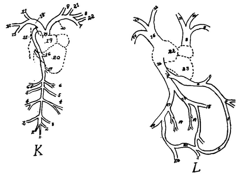
Fig. 61B. K. The arterial trunks. 1, The middle sacral artery. 2, The hypogastric artery. 3, The posterior mesenteric artery. 4, The ischiadic artery. 5, The femoral artery. 6, The renal artery. 7, The left pectoral artery or thoracico caudalis. 8, The left axillary artery. 9, The left carotid artery. 10, The left brachio-cephalic artery. 11, The right carotid artery. 12, The right brachial artery. 13, The right pectoral artery. 14, The common carotid artery. 15, The posterior aorta. 16, The celiac axis. 17, The anterior mesenteric artery. 18, The right brachio-cephalic artery. 19, The auricular portion of the heart. 20, The ventricular portion of same. 21, Sterno-clavicular artery. 22, Anterior thoracic. L. The venous trunks. 1, The caudal vein. 2, The coccygeo-mesenteric vein. 3, The posterior mesenteric vein. 4, The anterior mesenteric vein. 5, The gastro-duodenal vein. 6, The portal vein. 7, The hepatic vein. 8, The left anterior vena cava. 9, The left pectoral vein. 10, The left brachial vein. 11, The left jugular vein. 12, The right jugular vein. 13, The right brachial vein. 14, The right pectoral vein. 15, The right anterior vena cava. 16, The posterior vena cava. 17, The common iliac vein. 18, The femoral vein. 19, The renal vein. 20, The internal iliac vein. 21, The hypogastric vein. 22, The auricular portion of the heart. 23, The ventricular portion of same.
213The pulmonary arterial trunk is given off from the conus arteriosus of the right ventricle. It is guarded at its origin by three semi-lunar valves similar to those of the aorta. The trunk divides into two pulmonary arteries, which are short, one called the pulmonalis dexter and the other the pulmonalis sinister. The former goes to the right and the latter to the left lung. Each branch penetrates the lung near the bronchus. These arteries divide, or branch, similarly to the bronchi. Following the branchings of the bronchi, they finally terminate into the lung capillaries, forming networks on the bronchi and the air-tube terminals.
BRANCHES OF THE BRACHIO-CEPHALIC ARTERY
( Thyroidea
( ( Cervicalis inferior
( Vertebralis ( Vertebralis anterior
( ( Vertebralis posterior
( ( Arteria cervicalis ascendens
( Bronchiales
( Truncus ( Inferior esophageal
( caroticus ( Subcutaneous colli
( ( ( Carotis cerebralis
( ( Carotis ( Cervicalis superior
( ( communis ( Occipitalis
( ( ( Carotis externa
( ( Spinalis anterior ( Basilaris ( Cerebelli inferior
Brachio-cephalic (
( ( Clavicularis
( ( Sterno-clavicularis ( Sternalis
( ( ( Acromialis
( (
( ( ( Subscapularis
( ( ( ( Ulnaris
( Subclavia ( Axillaris ( Brachialis ( Brachialis profunda
( ( ( Radialis
( ( Humeralis ( Circumflex humeralis
( ( ( anterior
(
( ( Thoracica externa
( Thoracalis ( ( Arteria thoracica
( Thoracica inferior ( longa
(
( Mammaria interna
( Circumflex humeralis posterior
Brachialis profunda ( Collateral ulnaris
( Collateral radialis
214The cord-like remnant of the ductus botalli, or embryonal connection between the lung arterial trunk and the anterior aorta, has been observed but is rare in grown birds.
The arteries, the veins, and the lymphatic vessels of birds anastomose far more frequently than those of mammals.
BRANCHES OF THE ARTERIA CAROTIS CEREBRALIS INTERNA
( Occipitalis ( Occipitalis sublimis
( ( Occipitalis profunda ( Meningea
( ( Temporalis
( ( ( Rete temporale
( Ophthalmica ( Recurrent ophthalmicum ( Rete ethmoidalis
( externa ( Ethmoidalis
( ( Plexus palpebralis
( ( Plexus alveolaris inferior
( ( Plexus muscularis
Carotis cerebralis ( Plexus temporalis ( Plexus lacrimalis
( ( Ramus ciliaris posticus
( ( Meningea media
(
( ( Sphenoidea ( Ethmodalis
( ( Spheno-maxillaris ( Ethmoidalis ( externa
( ( ( Ophthalmica ( ( Ethmoidalis
( ( ( interna ( ( interna
( Cerebralis ( (
( Ramus ( Sylviæ
( anterior ( Cerebralis profunda ( Choroid plexus
( Arteria centralis retinæ
( Ramus posterior
BRANCHES OF THE ARTERIA CAROTIS EXTERNA
( Hyoidea
Carotis externa ( Laryngea superior
( Facialis
( Lingualis
BRANCHES OF THE ARTERIA CAROTIS FACIALIS
( Auricularis
Facialis ( Facialis interna ( Alveolaris inferior ( Mentalis
( Maxillaris interna
( Facialis externa
215
Branches of the Carotid Trunk
The carotis communis artery springs from the carotid trunk of the brachio-cephalic. It is directed horizontally, ascending to the ventral side of the neck. It then extends downward to the inferior median neck region. Just after leaving its origin it gives off several small branches to the bronchi and to the esophagus, and extends toward the head. The carotid lies on the thyroid gland and at this point touches the jugular vein (Fig. 21). At this point the thyroid arteries are given off from the carotid trunk. The thyroid gland also receives blood from the bronchialis artery.
Dorsalward and near the thyroid gland the carotid artery gives off a branch (the bronchialis) which accompanies the recurrent laryngeal nerve, along the inferior larynx and the bronchi, and supplies the lung substance and that part of the esophagus in this region.
The vertebral artery is given off from the carotid trunk, dorsalward to the thyroid gland, and on the left side. The right vertebral artery may be given off from the brachialis dextra or right brachialis.
The inferior esophageal artery is given off from the ventral side of the carotid, supplies the esophagus, extends then to the skin of the neck and to the trachea, is directed anteriorly toward the head and anastomoses with the vertebral artery. This artery accompanies the vagus nerve and forms a collateral artery to the carotid and the vertebral artery.
The subcutaneous colli springs from the carotid artery near the thyroid gland and communicates with the inferior cervical artery, which, in turn springs from the vertebral artery.
The Common Carotid.—Near the last cervical vertebra the two carotid arteries occupy the same channel, to which they are attached by fascia, and, lying close to the inferior surface of the bodies of the cervical vertebræ, are covered by the colli muscles. Near the third or the fourth cervical vertebra, the two carotid arteries separate. Near the atlas each carotid divides into the carotis cerebralis and carotis externa. Near this division the superior cervical artery branches off. This latter artery extends down the neck in company with the pneumogastric nerve and the jugular vein, supplies the skin and the neck muscles, and anastomoses with the inferior cervical artery and the subcutaneous colli artery.
216The occipital artery originates either from the carotis communis or sometimes from the superior cervical.
Branches of the Carotis Cerebralis
The branches of the carotis cerebralis are as follows:
1. The occipitalis (Fig. 72, No. 18), which in turn gives off, first, the occipitalis sublimis. The occipitalis sublimis supplies the outer and the middle portion of the digastricus and the posterior mylo-hyoideus muscle. It also gives off, second, the occipitalis profunda which supplies the inner portion of the digastricus muscle and becomes the meningeal artery, which passes through the foramen vagi, entering the brain cavity, where it supplies the coverings of the brain as far as the sella turcica. Superficial branches of this artery are distributed to the muscles in the region of the atlas and anastomose with the vertebral artery.
2. The ophthalmica externa, which passes below the articulation of the quadrate bone, around the tympanic cavity, enters the canalis caroticus, and passes into the cranial cavity. The ramus occipitalis, passing out of the same foramen, again enters the diploë of the cranium; then after passing dorsally over the upper outer semicircular canal of the ear, passes backward through the occipital bone.
The ophthalmica externa (Fig. 72, No. 9) forms a main trunk and gives off two branches: the temporal artery and the recurrent ophthalmic. The recurrent ophthalmic gives off the rete temporalis, rete ethmoidalis, a branch to the orbital gland, and finally anastomoses with its own branches and with those of the internal ophthalmic at the olfactory foramen and aids in forming the ethmoidal artery. These arteries supply blood by giving off branches to the muscles of the eye, sclerotic coat, iris, choroid coat, and the ciliary bodies. It gives off another branch to Harder’s gland, and finally anastomoses with the ethmoidalis.
3. The plexus temporalis, rete mirabile ophthalmicum, or wonderful network (Fig. 72, No. 8), is formed between the second and the third trunk of the trigeminus nerve and the rete ophthalmicum.
Inferior to this is the alveolar plexus, which plexus accompanies the third branch of the trigeminus into the lower jaw. Two main branches form this plexus, one coming from the carotis facialis, which anastomoses with the alveolar artery.
217The palpebral plexus lies between the trunks of the trigeminus, and supplies mainly the lower eyelid.
The plexus muscularis extends to the fifth portion of the temporal muscle.
The plexus lacrimalis forms on the posterior orbital wall on the ramus ethmoidalis and supplies the lacrimal gland and the upper eyelid. It anastomoses with branches from the facial artery.
The ramus ciliaris posticus supplies the inferior rectus and the external rectus muscle of the eyeball. It anastomoses with the ophthalmica externa.
The median meningeal artery passes through the foramen occupied by the second branch of the trigeminal nerve. Passing into the cranial cavity, it supplies the dura mater. Before passing into this foramen, small branches are given off, which supply the skin and the temporal muscle; and some branches anastomose with the superior cervical and the ethmoidal arteries.
4. The Cerebral Artery.—Each cerebral artery enters the cranial cavity through the canalis caroticus located in the sphenoid bone, passes forward medially from the cochlea, dorsally from the Eustachian tube, and passes through a small canal which opens on the inner surface of the sella turcica. Originating in this foramen, the sphenoid artery divides into two branches which anastomose with the pterygoidean and the pterygo-pharyngeal artery and which supply the upper jaw and the throat regions.
The spheno-maxillaris artery supplies the gums.
The right and the left cerebral arteries unite at the sella turcica. After this union they divide again immediately and pass to the base of the brain, where they give off twigs to the optic nerve, to the optic chiasm, and where also is given off the arteria retinæ centralis. Passing posteriorly each cerebral artery gives off the ramus posterior, and then passes to the side of the cerebellum.
The basilar artery, a continuation of the anterior spinal artery, is located ventrally and mesially to the cerebellum. Laterally and inferiorly the basilar artery gives off the inferior cerebellar artery.
The ramus anterior is given off from the cerebralis artery and continues as the internal ophthalmic artery. The ramus anterior also gives off the sylvian artery, which supplies the sides of the cerebrum and the middle brain. In the fissure between the hemispheres and the optic thalamus is located the arteria cerebri profunda. This artery passes along the median surface of the cerebrum and in 218its course supplies the adjacent parts. It enters into the formation of the choroid plexus of the lateral ventricle.
The internal ophthalmic artery extends out of the cranial cavity, through the optic foramen, into the orbital cavity. It passes upward along the interorbital wall and supplies the optic nerve, the trigeminus nerve trunk, and the eye muscles, and dorsally anastomoses near the olfactory nerve with the rete ethmoidale and the external ophthalmic artery, and continues as the ethmoidal artery.
The ethmoidal artery supplies the supra-orbital gland and the gland of Harder. It gives off twigs to the rete ophthalmicum and other branches to the skin of the frontal region, where it anastomoses with branches of the external facial artery. These branches in the frontal region are rather large. Large branches are given off to the comb.
The arteria ethmoidalis externa originates in the nasal cavity from a division of the ethmoidal artery.
The external ethmoidal artery passes below the lacrimal bone, extends forward, supplies the walls of the nasal cavity, and finally sends a branch anteriorly into the upper median portion of the jaw bone, and other branches to the septum nasi and other parts of the nasal cavity.
There are frequent anastomoses between the ethmoidalis interna, the facialis, and the spheno-maxillary artery.
The internal ethmoidal artery supplies principally the posterior turbinated bones and septum nasi.
Branches of the External Carotid Artery
Branches from the external carotid and the facial artery supply the tongue and its muscles, the larynx, the lower jaw bone, the gums, and the upper lateral facial region.
The external carotid (Fig. 62, No. A, 8) gives off the following branches:
First, the hyoid artery (Fig. 72, No. 12) which supplies the inner portion of the depressor mandibular and also the cornua of the os hyoideum, and extends to the tip of the tongue.
Second, the superior laryngeal artery (Fig. 72, No. 16) which supplies the sterno-brachialis and gives off branches which extend downward to the trachea and to the esophagus. The main artery passes along the left side of the esophagus and anastomoses with branches of the inferior esophageal, forming collateral circulation in that region.
219
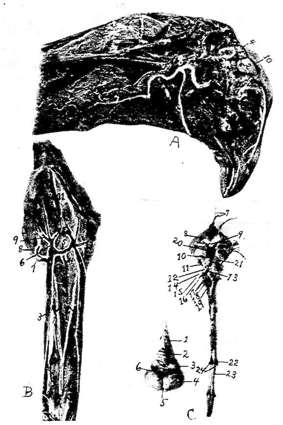
Fig. 62. A. Blood-vessels of the head. 1, The cerebellum. 2. The cerebrum. 3, The semicircular canals. 4, Sinus occipitalis superior. 5, Sinus temporalis sphenoideus. 6, Sinus transversus dexter. 7, Superior esophageal artery. 8, Carotis externus. 9, Lingual artery. 10, Sinus longitudinalis. B. Veins of the antero-inferior part of the head and neck. 1, The right carotid artery. 2, The left carotid artery. 3, Vena jugularis dexter. 4, Vena vertebralis dexter. 5, Transverse vein. 6, Vena cephalica dexter anterior. 7, Vena lingualis. 8, Vena cephalica posterior. 9, Vena facialis externa dexter. 10, Vena infrapalatina. 11, Posterior nares. 12, Vena facialis interna. 13, Vena facialis externa sinister. C. The brain. 1, Medulla oblongata. 2, Cerebellum. 3, Optic lobe. 4, Cerebrum. 5, Longitudinal fissure. 6, Transverse fissure. 7, Olfactory nerve. 8, Optic nerve. 9, Optic commissure. 10, Motor oculi. 11, Patheticus. 12, Trigeminal, or trifacialis. 13, Abducens. 14, Facialis. 15, Auditory, or acousticus. 16, Glosso-pharyngeus. 17, Pneumogastric. 18, Spinal accessory. 19, Hypoglossus. 20, Hypophysis. 21, Crus cerebri. 22, Ganglion on superior nerve trunk. 23, Spinal cord. 24, A pair of spinal nerves.
220Third, the lingualis artery (Fig. 62, No. A, 9) which lies between the mylo-hyoideus and the posterior part of the hyoid bone. It supplies the tongue muscles. It passes to the median surface of the lower jaw bone, where are given off small branches which enter the jaw and anastomose with the inferior alveolar artery.
Fourth, the Facial Artery.—The facial artery divides into the following branches:
The auricular artery is given off near the articulation of the os quadratum with the os zygomaticum. It is located at the outer auditory canal, and its branches are distributed to the parotid region and to the depressor mandibular, or digastricus, muscle.
The external facial artery is located between the os quadratum and the masseter muscle. It gives off a branch to the lower jaw bone and to the skin of that region, and then passes to the lacrimal bone and supplies branches to the commissure of the mouth, the auditory canal, the masseter muscle, the three eyelids (upper and lower eyelids and the membrana nictitans), the nose cavity, and the skin in the frontal region. It communicates on the other side with the ethmoidal artery.
The internal facial artery passes over and through the os pterygoideum, supplies all the jaw muscles, and is continued as the inferior alveolar artery.
The inferior alveolar artery enters the canal of the lower jaw bone along with the mandibular nerve. It is finally continued as the mental artery, leaving the canal, and passing to the outer jaw surface.
The facialis continues as the internal maxillary artery.
The internal maxillary artery (Fig. 72, No. 15) supplies the pterygoid muscle, the upper part of the pharynx, the commissure of the mouth, the salivary gland region, the gums, and finally anastomoses with branches of the spheno-maxillaris artery.
Branches of the Vertebral Artery
The vertebral artery is given off, dorsally near the thyroid gland, from the carotis communis or from the carotid trunk. The vertebral artery passes horizontally and dorsally into the canals formed in the transverse processes of the cervical vertebræ. After it leaves the carotid, it is divided into anterior and posterior branches.
221The posterior vertebral artery passes in the canals of the transverse processes of the fifth or sixth dorsal vertebra. It sends branches into the vertebræ, into the spinal canal, and into the intercostal muscles between the ribs and anastomoses with the intercostal arteries.
The anterior vertebral artery is larger than the posterior. It extends laterally along the side of the neck in the foramen of the transverse processes of the cervical vertebræ, and lies along the course of the inferior vertebral vein and the deep imbedded trunk of the sympathetic nerve. It continues to the head. In its passage it gives off to each vertebral segment a dorsal and a ventral branch. Twigs from these branches pass into the bodies of the vertebræ and the spinal canal, giving nutriment to the bony structure and to the spinal cord and its coverings. Other twigs are distributed to the muscles of the neck and some finally anastomose with branches of the carotis communis. This artery, reaching the head, gives off a long anastomosing branch which passes between the atlas and posterior part of the occipital bone and joins the ramus profundus and the occipital artery, thus again communicating with the carotid artery. The remainder of the vertebral artery is small and passes through the foramen magnum into the cranial cavity where it anastomoses with terminal branches of the cerebral artery. At the base of the neck and before the vertebral artery enters the canal of the cervical vertebræ, it gives off the arteria cervicalis ascendens, superior artery of the crop (Fig. 73, No. 18) which branches out on the upper surface of the crop is also distributed to the neck muscles, and later subdivides into the transverse cervical arteries which supply the skin and the muscles of the base of the neck, and the shoulder region.
On the ventral side of the neck there are given off two subvertebral carotid arterial branches. They lie in a shallow furrow on the ventral side of the cervical vertebræ and close to the median line.
Branches of the Subclavian Artery
The subclavian artery (Fig. 20, No. 13; Fig. 73, No. 11) gives off the following branches:
1. Sterno-clavicularis (Fig. 73, No. 9) which originates on the upper part of the subclavia, between the carotid artery and external thoracic (Fig. 73, No. 3), divides into many branches. The sterno-clavicularis 222gives off the sternal artery, which enters by the side of the supra-coracoid muscle and the anterior rim of the sternum. It is distributed to the inner surface and over the air-sac. Another outside branch passes the posterior end of the crista sterna and supplies the large breast muscles.
The clavicular artery (Fig. 73, No. 8) accompanies the clavicle to the shoulder-joint.
The acromial artery is given off from the sterno-clavicularis on the ligament near the shoulder-joint.
2. The thoracic artery branches from the subclavian artery (Fig. 70, No. 4). The thoracic artery gives off the following branches:
The internal mammary, or internal thoracic artery (Fig. 73, No. 16) arises on the inner side of the sternum and extends downward and backward, giving nutrient branches to the anterior vena cava and to the diaphragm. At the point of the costo-sternal muscle it divides into an inner and an outer branch.
The inner branch supplies the costo-sternal muscle and extends along the ribs and along their juncture with the breast-bone. It finally extends posteriorly, giving numerous branches to the abdominal muscles. The outer branch is distributed in a similar manner, giving off branches to the diaphragm and to the abdominal muscles, and anastomoses, on the surface of the abdominal muscles, with the epigastric artery.
The external thoracic artery (Fig. 73, No. 3), ramus superior, supplies principally the large breast muscles.
The inferior thoracic artery, external ramus inferior, passes along the outside of the pectoralis major muscle, extends downward, and gives off the arteria thoracica longa (subcutaneous thoracic). This latter artery supplies the skin of the breast region. Other branches are given off to the muscles of the breast and to the skin of the region, and some finally anastomose with the branches of the sternal artery.
3. The axillary artery (Fig. 73, No. 12) extends out of the thoracic cavity along with the brachial nerve plexus. It gives off the subscapularis which supplies the muscles of the scapular region. The axillary artery terminates as the brachialis which passes between the biceps brachii and anconeus muscles downward along the humeral shaft. It gives off the anterior humeral circumflex artery and the brachialis profunda artery (Fig. 68, No. 3). It gives off near the elbow-joint the ulnar and radial arteries.
223The anterior circumflex humeral artery passes through the short head of the biceps and gives off a branch to the biceps muscle, supplying the insertional part of the breast muscles.
Branches of the Brachialis Profunda
The brachialis profunda artery (Fig. 68, No. 3) continues as the posterior circumflex humeral artery. This artery supplies the muscles of the posterior part of the humerus, the skin of the wing, and the muscles of the brachial region. The trunk of the arteria brachii profunda passes downward along the anconeus muscle, supplying that muscle and giving off the collateral ulnar artery. On the ulnar olecranon it anastomoses with the recurrent ulnar, thus establishing, at this region, collateral circulation. The rest of the brachialis sends branches to the biceps muscle and to the skin of the upper arm, and finally gives off the arteria collateralis radialis, which supplies the condylo-ulnaris. It then anastomoses with the end branches of the recurrent radialis artery.
The radial artery (Fig. 67, No. 4) passes downward on the outer rim of the forearm. It continues down to the carpal region, where it supplies the muscles and the skin of the carpal region it gives off the recurrent radial, which passes on the middle finger and finally anastomoses with the collateral radialis artery.
The ulnar artery (Fig. 68, No. 6 and 9) passes downward on the inner surface of the ulna to the carpal region where it gives off a branch to the wing plexus and divides into two branches, the smaller branch supplying the thumb (Fig. 68, No. 7). This branch passes down the radial side of the middle finger to the last joint of the second finger. The larger (Fig. 68, No. 8) lies between the second and the third finger bones, and passes through a slit between these two bones to the flexor side of the hand and extends to the last finger-joint, supplying the structures in the region. The ulnar artery gives off a small arterial twig to the papilla of each large wing feather (Fig. 67, No. 6).
The recurrent ulnar passes backward to the olecranon of the ulna and supplies the muscles, the skin, the feathers, and other structures of the region, and terminates in end collateral ulnar branches (Fig. 67, No. 5).
224BRANCHES OF THE POSTERIOR AORTA[7]
( Esophageal
( Intercostales
( Dorsal
( Lumbars
( Spermatics
( Renals
( Ovarian
( Celiac ( Recurrent esophageal
( axis ( Recurrent intestinalis ( Recurrent ilio-colicus
( ( Posterior or recurrent sinister ( Renalis
( ( Anterior or recurrent dexter ( Splenics
( ( ( Hepatic
( ( Hepatica dextra ( Gastric
(
Posterior ( Sacralis media ( Coccygeæ laterales
aorta ( ( Coccygea media ( Coccygeæ laterales
(
( Anterior mesenteric ( Recurrent ilio-celiacus
( ( Recurrent superior hemorrhoidal
( Posterior mesenteric ( Median hemorrhoidal
( ( Recurrent renalis
( ( Tibialis postica
( Ischiadica ( Tibialis antica ( Peroneal ( Anterior tibial plexus
( ( Arteria ovarialis
( Pudenda communis ( Renalis
( ( Hemorrhoidalis intima
( ( Pudenda externa
(
( Crural ( Internal pelvic (umbilical)
( (External iliac) ( Circumflex femoris
( ( Femoralis
The posterior aorta (Fig. 63, No. 2) passes backward along the inferior part of the bodies of the dorsal and lumbo-sacral vertebræ. In the thoracic cavity it lies dorsal to the esophagus. It gives branches (esophageal) only to the esophagus during its passage as far as the seventh dorsal vertebra. Following this point there are given of several pairs of arteries which divide into a superior and an inferior branch. The superior branch extends upward and anastomoses with branches of the vertebral artery and other twigs coming from the intercostal arteries. These latter arteries given off are small dorsal arteries. The inferior branches extend downward between the ribs and supply the intercostal muscles. The intercostal arteries 225do not take their origin from the aorta in numerous and regular branches as in mammals; they consist originally of but few vessels, which are multiplied by anastomoses with each other and with the arteries which come out of the spinal canal. An arterial plexus is thus formed around the head of each rib, from which a vessel is sent to each of the intercostal muscles and ribs and are continued into the muscles upon the outside of the body and its integuments. The anastomosis of the intercostal arteries round the ribs is similar to the plexus, which is produced by the great sympathetic nerve in the same location. The lumbar arteries are given off farther posteriorly and along the lumbar region. The lumbar, like the dorsal branches are given off in pairs. These arterial branches pass along the thigh and the upper sacral vertebral region, and supply those parts. Ventrally they are covered by the kidneys and pass into the abdominal muscles. The posterior aorta gives off the spermatic arteries and the ovarian artery. In the male the former supply the testes. The ovarian artery of the female gives off a twig to each calyx containing an ovum. Each calyx is voluminously supplied with blood. Next the renal arteries are given off to the kidneys.
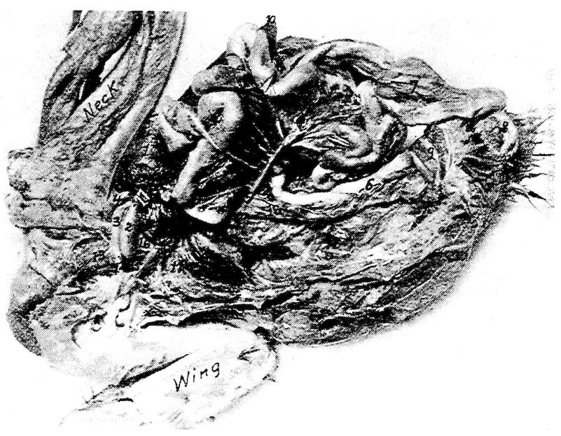
Fig. 63.—The vascular system injected. 1, The heart. 2, The posterior aorta. 3, The right brachio-cephalic artery. 4, The left brachio-cephalic artery. 5, The anterior mesenteric artery showing its many branches and anastomoses near and on the intestines. It is accompanied by branches of the mesenteric vein. 6, The rectal branch of the posterior mesenteric artery. 7, The duodenal loop and pancreas showing the pancreatic artery. 8, The anus. 9, The cloaca. 10, The liver. 11, The lungs. 12, The right subclavian artery. 13, The right carotid artery. 14, The right anterior vena cava. 15, The subclavian vein. 16, The right jugular vein. 17, The carotid trunk. 18, The posterior vena cava.
226
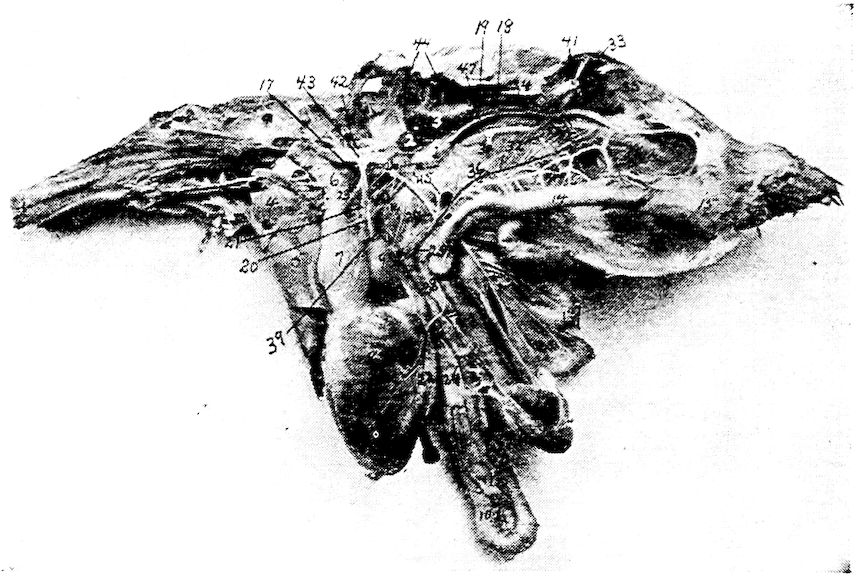
Fig. 64.—The splanchnic arteries, veins and nerves. 1, A portion of the left testes. 2, Adrenal gland. 3, Anterior lobe of kidney. 4, Heart. 5, Liver. 6, Second portion of the esophagus. 7, Proventriculus. 8, Gizzard. 9, Spleen. 10, Duodenal loop. 11, Pancreas. 12, Blind extremity of cæcum. 13, Floating portion of small intestine. 14, Rectum. 15, Cloaca. 16, Anus. 17, Celiac axis. 18, Ureter. 19, Vas deferens. 20, Recurrent sinister artery. 21, Anterior recurrent dexter artery. 22, Arteria hepatica dextra. 23, Recurrent esophageal artery. 24, Recurrent intestinalis artery. 25, Ilio-colicus artery. 26, Anterior mesenteric artery. 27, Recurrent branches of the same. 28, Ilio-cœliacus artery. 29, Recurrent superior hemorrhoidal artery. 30, Posterior mesenteric artery. 31, Crural arteries. 32, Sacralis media artery. 33, Recurrent renalis. 34, Middle renal lobe. 35, Posterior renal lobe. 36, Vena mesentericus communis. 37, Vena hemorrhoidalis. 38, Vena pancreatico-duodenalis. 39, Vena proventriculo-leinealis. 40, Vena portalis dextra. 41, Ischiadic nerve. 42, Thoracic sympathetic trunk. 43, Anterior splanchnic plexus. 44, Spinal nerves. 45, Posterior splanchnic nerve plexus. 46, Intestinal nerve trunk. 47, Vena renalis magna.
The celiac axis (Fig. 64, No. 17) originates near the seventh sternal vertebra and to the right of the esophagus, breaks through the diaphragm, and, near this point, gives off a recurrent esophageal (Fig. 64, No. 23). The celiac axis gives off three main branches: The anterior or recurrent dexter, the posterior or recurrent sinister, 227the recurrent intestinalis, all of which lie to the right of the spleen, under the left lobe of the liver, and along the left side of the stomach. The recurrent sinister (Fig. 64, No. 20) gives arterial branches to the proventriculus, the gizzard, the pyloris, and the left lobe of the liver. The anterior or recurrent dexter (Fig. 64, No. 21) gives off a renal artery and from four to six splenic arteries.
The recurrent intestinalis artery (Fig. 64, No. 24) arises on the posterior of the stomach within the duodenal loop, and supplies the duodenum and the pancreas (Fig. 63, No. 7). It gives off a branch, called the recurrent ilio-colicus (Fig. 64, No. 25), which supplies the large intestines, including the cæca.
The arteria hepatica dextra (Fig. 64, No. 22) is a branch from the celiac axial trunk. It gives several branches to the right lobe of the liver and to the gall-bladder. The gastric branches pass to the muscles of the stomach. The anterior and posterior recurrent branches often anastomose.
The anterior mesenteric artery (Fig. 63, No. 5; Fig. 64, No. 26) originates from the posterior aorta near the generative glands. It is directed downward, and divides into many branches which pass in the mesentery toward the intestines. Recurrent branches (Fig. 64, No. 27) are given off, which anastomose, forming mesenteric arches. From these arches are given off branches which supply the intestinal walls.
The anterior mesenteric artery gives off the recurrent ilio-celiacus (Fig. 64, No. 28) which is distributed to the cæca; other of its branches are distributed to the small intestine.
The anterior mesenteric artery extends along the small intestine and ends in the recurrent superior hemorrhoidal arteries (Fig. 64, No. 29) which anastomoses on the surface of the rectum with a branch of the posterior mesenteric artery.
Branches from the anterior mesenteric artery also anastomose with branches of the celiac axis.
The posterior mesenteric artery (Fig. 64, No. 30) is given off from the posterior aorta posterior to the origin of the anterior mesenteric and near the arteries of the thigh and is distributed to the lesser mesentery supplying the large intestine. Branches of the superior hemorrhoidal arteries anastomose with branches of the posterior mesenteric artery. Branches from this source are called the median hemorrhoidal (Fig. 63, No. 6).
The posterior mesenteric artery (Fig. 64, No. 30) also gives off a 228long branch to the cæca. Branches of the posterior mesenteric artery anastomose with branches of the anterior mesenteric.
The crural arteries (Fig. 64, No. 31) are given off in a pair from the posterior aorta. They pass through the mass of the lobes of the kidneys, at about the median region, and extend out of the pelvic cavity in front of the spine and ilio-pubic ligament.
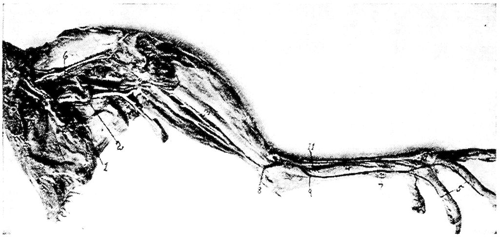
Fig. 65.—Blood-vessels and nerves of the hind extremity. Inside view of leg. 1, Ischiadic nerve. 2, Ischiadic artery. 3, Posterior tibial vein. 4, Vena metatarsa dorsalis interna. 5, Vena metatarsalis plantaris profunda. 6, Vena cruralis. 7, Vena metatarsalis interna, vena magna. 8, Vena tibialis postica. 9, Vena metatarsa magna. 10, Nerves of the toes. 11, Vena metatarsalis dorsalis profunda.
The crural, or the external iliac artery, is divided into three branches as follows:
First, the internal pelvic, or umbilical artery, is given off just as the cruralis leaves the pelvic cavity. It passes, as a long vessel, on the inner surface of the lumbo-sacral bones and supplies the obturator internus muscle and extends into the umbilical region. It gives off branches to the abdominal muscles, and, in the female, a branch is distributed to the ligament of the oviduct.
Second, the arteria circumflex femoris, which passes between the sartorius and vastus internus and extends dorsalward, supplies the sartorius muscle, the vasti muscles, and the ilio-trochanteric region.
Third, the femoral artery, which passes beside the vena cruralis and extends down the posterior surface of the thigh to the knee-joint, supplies the upper thigh muscles with the exception of the adductor muscles.
229
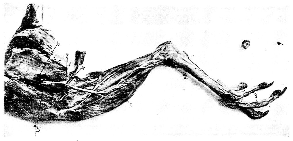
Fig. 66.—Blood-vessels and nerves of the posterior extremity. Outside view. 1, Anterior tibial artery. 2, Metatarsal artery. 3, Digital arteries. 4, Vena cutaneous crurus. 5, Ischiadic artery. 6, Vena cruralis. 7, Lateral cutaneous branch of the ischiadic nerve.
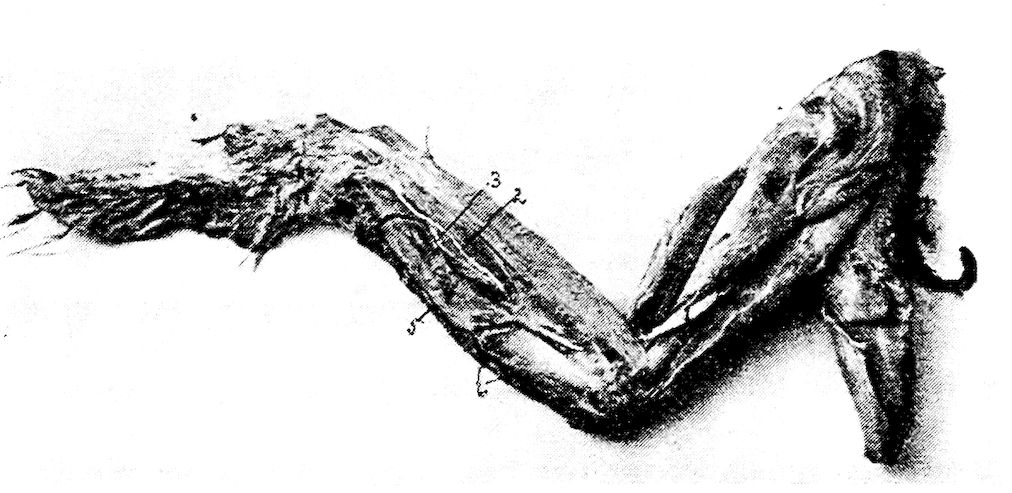
Fig. 67.—Blood-vessels and nerves of the fore limb. Outside view. 1, Median nerve. 3, Ulnar nerve. 4, Radial artery. 5, Recurrent ulnaris artery. 6, Twigs of ulnar artery to wing feathers.
The ischiadic artery (Fig. 69, No. 9) forms the main artery of the posterior extremity. This artery, the largest vessel of that region, is given off from the posterior aorta and passes ventrally over and between two of the main lobes of the kidney. The continuation of the posterior aorta is called the sacralis media (Fig. 64, No. 32). The ischiadic artery gives off a recurrent renalis on the posterior lobe of the kidney (Fig. 64, No. 33). On the left side it gives off a branch to the oviduct and to the ligament of the oviduct. The main trunk leaves the cavity with the ischiadic nerve (Fig. 65, 230No. 1) through a foramen formed by the os ilium and the os ischium. It sends branches into the adductor muscles of the upper and the lower thigh. It gives off anastomotic branches which unite with those from the femoral artery. At the flexure of the knee-joint it gives off two branches to that region (Fig. 69, No. 10). It gives off another branch to the gastrocnemius muscle and one to the flexor perforans digitorum. It terminates in the anterior and posterior tibial arteries.
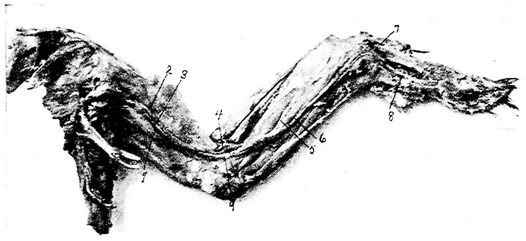
Fig. 68.—Blood-vessels and nerves of the fore limb. Inside view. 1, Vena humeri profunda. 2, Brachialis longus inferior. 3, Brachialis profunda. 4, Radial artery. 6, Ulnar artery. 7, Ulnar arterial branch to thumb. 8, Digital branch of ulnar artery. 9, Ulnar artery.
The first, the tibialis postica (Fig. 69, No. 12) passes between the gastrocnemius and the deep flexors giving off branches in its course to the skin and to other parts, and disappears shortly below the intertarsal joint.
The second, the tibialis antica (Fig. 66, No. 1; Fig. 69, No. 11), is often the larger of the two arteries. The anterior tibial artery gives off branches to the knee-joint and to adjacent structures. It gives a large branch to the head of the gastrocnemius muscle. It passes to the posterior surface of the tibial head and gives off the peroneal artery, which passes through the membrana interossea located between the tibia and fibula giving branches to the anterior side of the membrane of the patellar region of the knee-joint. It gives branches to the anterior side of the lower thigh and finally terminates subcutaneously in the anterior tibial plexus.
The main portion of the anterior tibial artery passes downward 231along the posterior surface of the membrane located between the tibia and fibula, and sends branches for the flexor digitorum communis et profundus. It then breaks through the membrane, reaches the anterior side and there communicates with the anterior tibial plexus. It gives off branches to the muscles on the anterior side of the lower thigh and the skin, and passes between the outer and the middle malleolus of the tarsal bones. It passes to the plantar surface and divides between the toes. In addition to the plantar branches there are given off dorsal branches between the third and the fourth toes. These branches form the direct continuation of the dorsal vessels of the anterior tibial plexus.
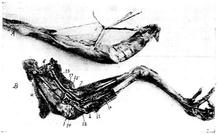
Fig. 69.—Blood-vessels and nerves of the femoro-tibial region. A section of the tibia removed. A. Veins, arteries and nerves dissected as in B. B. V, Vein. A, Artery. N, Nerve. 1, Vena poplitealis. 2, The three veins forming the vena poplitealis. 3, Vena femoris interna profunda. 4, Vena femoris anterior. 5, Vena cruralis. 6, Vena cutanea abdominalis femoralis. 7, Vena cutaneous cruralis. 8, Deep vein of the knee-joint. 9, Ischiadic artery. 10, Branches of ischiadic artery to flexure of knee. 11, Anterior tibial artery. 12, Posterior tibial artery. 13, Ischiadic nerve. 14, Trunk (a branch of the ischiadic) which gives off the superficialis peroneus and peroneus profundus. 15, Median branch of ischiadic nerve. 16, Superficialis peroneus. 17, Lateral branch of the ischiadic nerve.
The arteria pudenda communis (Fig. 70, No. 20) passes to the depressor coccygeus muscle, gives off a branch to the caudal part of the kidney, crosses the ureter, provides the ischio-pubic and pubio-coccygeus, or depressor coccygis lateralis, muscles, and on 232the lateral rim of the latter muscle it gives off the arteria hemorrhoidalis intima. This artery passes to the bursa of Fabricius and to the end of the cloaca. It enters into the ischio-coccygeus muscle, and divides into the arteria pudenda externa, and in ducks, the arteria profunda penis. These branches supply the vas deferens, the ureter, the cloaca, the penile structure, and the muscles of these parts.
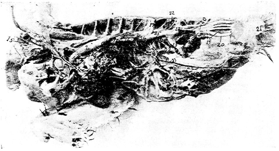
Fig. 70.—Blood-vessels of a Cornish cock. 1, The left pulmonary artery. 2, Right pulmonary artery. 3, Left brachio-cephalic artery. 4, Left subclavian artery. 5, Left carotid artery. 6, Right brachio-cephalic artery. 7, Posterior aorta. 8, Posterior vena cava. 9, Left pulmonary vein. 10, Right pulmonary vein. 11, Celiac axis. 12, Anterior mesenteric artery. 13, Ischiadic artery. 14, Crop. 15, Superior artery of the crop and vein of same name. 16, Testicular or ovarian artery. 17, External iliac artery. 18, Intercostal nerve. 19, Median sacral artery. 20, Arteria pudenda communis. 21, Anus. 22, Vena hypogastrica. 23, Right vena hepatica magna. 24, Left vena hepatica magna. 25, Vena iliaca interna. 26, Vena iliaca communis. 27, Vena iliaca externa. 28, Vena coccygo-mesenterica. 29, Vena umbilicalis. 30, Vena suprarenalis. 31, Origin of the pars renalis. 32, Lumbar veins. 33, Vena epigastrica. 34, Lumbales (arteries). 35, Anterior division of the lumbo-sacral plexus. 36, Posterior division of the lumbo-sacral plexus. 37, Left anterior vena cava. 38, Right anterior vena cava.
The Sacralis Media.—The median coccygeal artery (Fig. 70, No. 19) forms the single extension of the sacralis media. It gives off lateral branches between the caudal vertebræ which supply the dorsal muscles of that region and the skin. The third pair are the largest. These are the arteriæ coccygeæ laterales, and are located on the dorsal side of the tail, they supply the tail glands and rudder feathers, the main tail feathers, or rectrices. A small arterial twig is given off to the papilla of each rectrix.
233
The Venous Trunks
The venous blood enters the lungs from the right ventricle through the two pulmonary arteries (Fig. 70, No. 1 and 2).
The two pulmonary veins (Fig. 70, No. 9 and 10) collect the arterial blood from the lungs, and empty it into the left auricle.
There are three venæ cavæ which collect the systemic blood and empty it into the right auricle. These venæ cavæ are two anterior (Fig. 70, No. 37 and 38) and one posterior (Fig. 70, No. 8).
Each anterior vena cava is formed by the union of a vena jugularis, a vena vertebralis, and a vena subclavicularis (Fig. 63, No. 15).
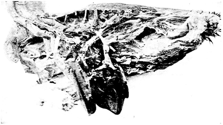
Fig. 71.—Veins of the liver of a fowl. 1, Vena mesenterica communis. 2, Vena portalis propria. 3, Anterior vena mesentericus. 4, Vena portalis dexter. 5, Vena portalis sinister. 6, Posterior vena cava. 7, Celiac axis. 8, Base of the heart. 9, The liver. 10, Hepatic veins.
The jugular vein is formed by the union of the vena cephalica anterior, and the vena cephalica posterior.
The jugular vein (Fig. 62, No. B, 3) passes along the side of the neck and lies near the trachea, the esophagus, and the pneumogastric nerve. Near the base of the skull the two jugular veins, the right and the left, are connected by a transverse vein. By this anastomosis part of the blood from the left jugular vein is sent into the right. Therefore the right jugular vein is larger than the left. The jugular veins collect the blood from the tongue region, the thyroid, the esophagus, the trachea, the crop, and other structures along its course through the cervical region.
234The vertebral veins are divided into the anterior and the posterior, or the inferior and the superior. The anterior vertebral vein is located in the cervical region and collects the blood from the brain and the inner part of the head. The vertebral vein passes along the dorsal side of the spinal cord.
BRANCHES OF THE VENÆ CAVÆ ANTERIORES
( Venæ linguales
( Vena occipitalis lateralis ( Vena occipito-collores
( Vena ascendenes lateralis
( Venæ colli cutineæ
( Venæ esophagealeæ
( Venæ tracheales
( Vena ( Vena subscapularis
( jugularis ( Venæ glandularum thyroidearum
( ( ( Vena occipitalis ( Sinus foraminis
( ( ( interna ( occipitalis
( ( Vena (
( ( vertebralis ( Vena vertebralis posterior ( Vena intercostales
Vena cava ( ( ( Vena vertebralis anterior
anterior ( ( Vena vertebralis
( Vena vertebralis lateralis dorsalis ( Venæ intercostales
(
( Venæ coronariæ
( Vena subclavicularis
( Vena thoracica interna ( Vena intercostales
( Vena pharyngea superior
( Vena muscularis depressoris mandibularis
Vena facialis ( Vena muscularis colli anterior superioris
communis ( Vena lingualis
( Vena sublingualis et sphenoidea
The posterior vertebral vein is located in the dorsal region, passes backward and receives the blood from the dorsal neck region and from the intercostal veins and from the vertebral segments and adjacent regions.
The anterior and the posterior vertebral vein form one trunk, the vertebral vein, and this trunk empties into the vena jugularis just before the subclavian and the jugular unite.
The subclavian vein collects the blood from the anterior extremities. It unites with the vena jugularis of the same side.
The left vena cava receives the coronary veins from the heart.
235
THE VEINS OF THE HEAD
Branches of the Vena Facialis Interna
The vena facialis interna (Fig. 62, No. 12) lies dorsal on the pterygoid bone and receives the vena maxillaris, the vena ophthalmica, the vena mandibularis interna, the venæ pharyngeæ superiores, and the vena retis mirabilis temporale.
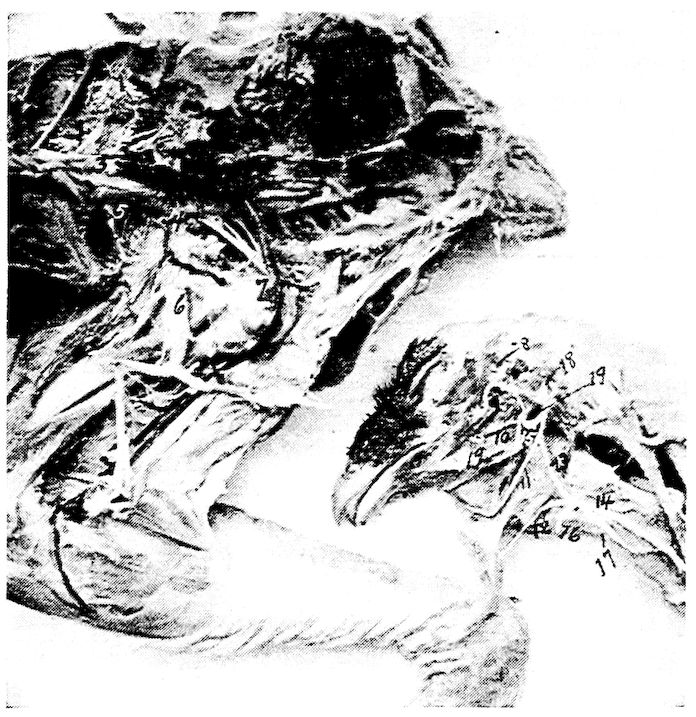
Fig. 72.—Blood-vessels and nerves of the abdominal cavity and hind extremity and head. 1, Vena renalis. 2, Vena intervertebrales sacrales. 3, Posterior aorta. 4, External iliac. 6, Ischiadic nerve. 7, Ischiadic artery. 8, Arterial rete (plexus temporalis). 9, Ophthalmic artery. 10, Palatine artery. 11, Lingual artery. 12, Hyoid artery. 13, 14, External carotid artery. 15, Internal maxillary artery. 16, Superior laryngeal artery. 17, Superior esophageal artery. 18, Occipital artery. 19, Internal carotid artery.
The vena maxillaris comes out of the upper beak with the recurrent trigeminus nerve and then passes backward between the jugular and palatine bones. It lies medio-ventrally to the eyeball and communicates at this point with the ophthalmic vein. It is covered ventrally with the pterygoid bone. The maxillary vein collects the blood from the gland of Harder, the upper beak, and 236receives blood from the vena supra-palatina and from the lower jaw bone and the external mandibular vein. It also receives blood from the external sublingual vein, and the sublingual gland. It follows the inner rim of the jaw bone. The maxillary vein receives an anastomosing branch from the cutaneous facial vein, at a point near the commissure of the mouth.
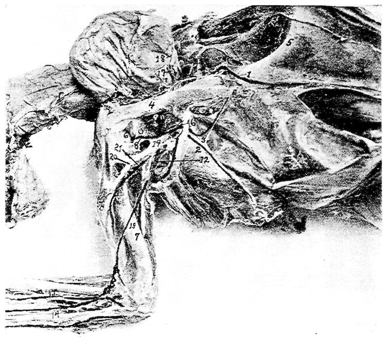
Fig. 73.—Photograph of blood-vessels and nerves of the thorax. 1, Vena sternalis. 2, Vena thoracico-externa. 3, External thoracic artery. 4, Coracoid bone. 5, Sternum. 6, Shoulder-joint. 7, Humerus. 8, Clavicularis artery. 9, Sterno-clavicularis artery. 10, Thoracico-humeralis artery. 11, Subclavian artery. 12, Axillary artery. 13, Brachialis profundus artery. 14, Ulnar artery. 15, Radial artery. 16, Internal mammary artery. 17, Superior vein of the crop. 18, Superior artery of the crop. 19, Brachial nerve plexus. 20, Anterior division of brachial nerve plexus. 21, Posterior division of brachial nerve plexus.
The vena ophthalmica lies close to the point where the olfactory nerve passes out of the cranial cavity. The largest branch it receives is the recurrent ophthalmo-temporalis, which accompanies the lateral side of the optic nerve. It collects the blood from the base of the brain, from Harder’s gland, from the eye muscles, from the 237inner eye, the nose, the lacrimal gland, the skin of the frontal region, and the comb.
BRANCHES OF THE VENA JUGULARIS
( Vena maxillaris
( Vena ophthalmica
( Vena facialis ( Vena mandibularis interna
( interna ( Vena retis mirabilis temporalis
( Vena cephalica ( ( Venæ pharyngeæ superiores
( anterior or (
( facialis ( ( Vena facialis cutinea
( communis ( ( Vena temporalis
( ( Vena facialis ( Vena auricularis
Vena jugularis ( ( externa ( Vena palpebralis
or cephalica ( ( Vena transversus
communis ( ( Vena retis mirabilis temporalis
( Vena subclavia
( Venæ vertebrales
( ( Sinus cranii et cerebri
( ( Vena carotis
( (
( ( Vena auris ( Sinus foveæ cerebelli
( ( ( Vena occipitalis externa
( Vena cephalica ( Vena jugularis prima
( posterior ( Vena occipitalis
( Vena lingualis
( Vena mandibularis externa
Vena maxillaris ( Vena sublingualis externa
( Vena supra-palatina
Vena ophthalmica ( Vena ophthalmo-temporalis, recurrent
Vena facialis communis ( Vena cephalica externa
Vena occipitalis externa ( Vena occipitalis interna
Vena retis mirabilis temporalis ( Vena spheno-temporalis
Sinus foveæ cerebelli ( Sinus occipitalis externa
The trunk of the internal facial vein receives the blood of the internal mandibular vein. The internal mandibular vein emerges from the alveolar canal of the inferior jaw bone and receives branches from the muscles of mastication.
The vena retis mirabilis temporalis passes out of the eye cavity and lies between the internal facial vein and the external facial vein, and passes around the os quadratum and the os pterygoideum. It receives veins from the outer ear region, lacrimal gland, eyelids, and the spheno-temporalis.
238The venæ pharyngeæ superiores are the veins on the dorsal surface of the pharynx. These veins form the small plexuses which unite with the two trunks of the facial veins. This forms an anastomosis between the left and the right facial veins.
Branches of the External Facial Vein
The vena facialis externa (Fig. 62, No. B, 9) lies behind the quadrate bone and is partly covered ventrally by the inner extension of the lower jaw bone. It collects the blood principally from the upper region of the cranium and the face, including the comb.
The vena facialis cutinea receives branches from the muscles in the region of the jaw, the lower jaw bone, the muscles of the eyelids, the frontal region, and the anastomotic branches from the vena maxillaris.
The vena temporalis is made up of veins from the skin of the temporal region, the masseter and the tongue muscles, and from the sides of the upper throat region.
The vena palpebralis is made up from veins from the three eyelids and the temporal region near the eye. Its trunk passes laterally over the temporo-mandibularis ligament. The vena palpebralis collects blood from the lacrimal gland, the eyelids, and the outer ear region.
The following veins empty into the trunk of the vena facialis communis: venæ pharyngis superiores, vena muscularis depressoris mandibulæ, vena muscularis colli anterior superioris, vena lingualis et hyoidea. The right terminal branches of the vena lingualis empty into the vena cephalica posterior.
The venæ pharyngis superiores and vena lingualis et hyoidea collect blood from the muscles of the tongue and posterior tongue region.
The vena lingualis et hyoidea (Fig. 62, No. B, 7) collects the blood from the muscles of the tongue region, the lower tongue glands, and the upper throat region.
Between the two trunks of the vena facialis communis and anterior to the entrance of the vena cephalica posterior there is an anastomosis, the vena transversus (Fig. 62, No. B, 5). Through this anastomosis which lies crosswise the two jugular veins are influenced in different ways. At this point both jugular veins are of the same size. The anastomotic branch lies crosswise. Through this anastomosis the 239right receives some blood from the left head region indicating that the blood flows from left to right. This, as stated before, makes the left jugular vein the smaller.
Branches of the Vena Cephalica Posterior
The left vena cephalica posterior (Fig. 62, No. B, 8) communicates with the transverse vein (Fig. 62, No. B, 5). The left receives the smaller veins of the skin region, the esophagus, and the trachea. These vessels communicate with their fellow of the opposite side and with the vertebral vein of the same side, which causes a gradual reduction of the size of the jugular vein.
BRANCHES OF THE VENA OCCIPITALIS EXTERNUS
( ( Venæ choroideæ
( Sinus ( Venæ cutaneæ et frontales
( longitudinalis ( Venæ nasales
Vena ( ( Venæ ophthalmiæ
occipitalis (
externus ( ( ( Sinus occipitalis
( Sinus ( Sinus ( Sinus temporo-sphenoideus
( semicircularis ( transversus ( Venæ cerebellares
( ( ( Sinus petrosus sphenoideus
Sinus petrosus sphenoideus ( Retis mirabile temporalis
Sinus temporo-sphenoideus ( Sinus annularis venosus basilaris
Sinus foraminus occipitalis ( Venæ medullares
( Venæ cerebrales
Sinus ( Vena cerebralis basilaris ( Sinus venosus annularis anterior
venosus (
annularis (
basilaris ( Vena ophthalmica
( Vena medullaris mediana longitudinalis
The vena cephalica posterior (Fig. 62, No. B, 8) is formed from the veins of the sinuses of the cranium, the brain, the vena auris, the vena jugularis prima, the vena carotis, and the vena occipitalis. It collects most of the blood from the cranium and the posterior part of the head and from the tongue.
THE VENOUS SINUSES OF THE HEAD
The sinus longitudinalis (Fig. 62, A, 10) extends from the dorsal median line of the internal occipital protuberance to the olfactory 240nerve. It collects the blood from the choroidea of the brain and from the skin of the frontal region. It receives laterally the veins of the nose, the vena ophthalmica, and the sinus transversus.
The sinus occipitalis (Fig. 62, No. A, 4) forms the continuation of the transverse sinus. It is located in the posterior cerebral cavity, and is wing-shaped, extending both to the right and to the left.
The sinus foraminis occipitalis a continuation of the sinus occipitalis, lies transversely on the ventral side of the cranial cavity and receives the veins from the medulla oblongata. One branch passes through the os occipitalis basilare, and then extends sidewise to the foramen magnum and anastomoses with the vena occipitalis.
The sinus transversus (Fig. 62, No. A, 6) extends in a pair from the internal occipital protuberance between the cerebrum, cerebellum, and the corpus quadrigeminum. It receives vessels from the upper surface of the cerebellum. The sinus has three branches, all of which empty into the sinus semicircularis.
The sinus semicircularis extends along the dorsal petrosal rim, anterior to the inner ear, and from this point backward. Near the foramen magnum it passes through the os occipitale and empties into the external occipital vein.
The vena occipitalis externus extends near the foramen magnum, surrounds the basis occipitalis, and receives the semicircular sinus and through the semicircular sinus the blood from the sinus transversus.
The sinus petrosus sphenoideus, passing in company with the occipital sinus, extends from the transverse sinus to the cavity near which the trigeminus lies. The sinus dividing into two branches, one of these extends to the hypophysis and unites with the basal veins of the brain; the other passes with the rete out of the cranial cavity along with a branch of the trigeminus, and communicates with the rete mirabile temporale.
The sinus temporo-sphenoideus (Fig. 62, No. A, 5) extends forward and outward from the transverse sinus, and, between the corpus quadrigeminum and the cerebrum, unites with the sinus venosus annularis basilaris.
The sinus venosus annularis basilaris lies in the region of the optic nerve and surrounds the hypophysis, forming a complete circle. It receives small vessels from the cerebrum, the corpora quadrigemina, 241and anteriorly the longitudinal basilar vein or vena basilaris media, of the cerebrum, which comes out of the anterior annular venous sinus. The anterior annular venous sinus surrounds the base of the olfactory nerves. It communicates with the sinus petrosus sphenoideus, which emerges from behind the optic lobe, the sinus temporo-sphenoideus, which emerges from in front of the optic lobe, and posteriorly the median longitudinal vein of the medulla oblongata or vena basilaris. Laterally and anterior to the optic lobe it receives the vena basilaris lateralis, and also a branch of the vena ophthalmica.
The blood received by the sinus annularis basilaris comes from the brain cavity through three pairs of veins, as follows: first, the two ophthalmic veins which pass through the posterior orbital wall between the olfactory and the optic nerve; second, two other branches of the ophthalmica which pass with the optic nerve; third, two veins which pass through a foramen by the side of the sella turcica to the hypophysis together with the carotis cerebralis, and then leave the cerebral cavity ventrally, at which point they are called the venæ carotes. These last accompany the cerebral artery backward, and pass through a foramen in the base of the cranium. Each one then passes through the cranial wall close to the external auditory canal, and empties into another vein in the posterior region of the head.
The sinus fovæ hemispherii cerebelli lies in the hollow between the os petrosum and the semicircular canals. It receives blood from the external occipital sinus and from the cerebellum, and empties into the vena auris interna.
VEINS OF THE BRAIN CAVITY
The vena auris interna passes through a bony canal along the outer rim of the posterior semicircular canal and then along the external semicircular canal. It receives vessels from the labyrinth, and extends along the posterior rim of the tympanum to the outer ear canal.
The vena occipitalis interna, extending from the sinus foraminis occipitalis, passes through the side of the atlas and divides near the condyle into two branches. One of these branches the internal occipital, forms the root of the vena vertebralis, and the other empties into the vena occipitalis externa.
242Near the outer and upper part of the condyle it forms two large veins and collects the blood from the vena occipitalis media. These veins collect the blood from the rectus capitis anticus, and communicate with the vena cephalica anterior.
The vena occipitalis externa, sometimes called the vena occipitalis collateralis, extends from the vena auris, and receives vessels from the neck region. It receives on the side of the occipitoatloid joint the vena occipitalis interna. The vena occipitalis externa forms a junction with the vena vertebralis emptying into the transverse vein.
THE VEINS OF THE NECK
Branches of the Vena Jugularis
The trunks of the vena jugularis or vena cephalica communis pass subcutaneously on both sides near the trachea and the esophagus. Near the height of the thyroid gland they almost touch the carotid artery. They then cross, dorsalward, the trunk of the subclavian artery, and in their course receive the vertebral vein and unite with the subclavian vein toward the side of the vena brachiocephalica. The union on the right side lies to the right of the anterior aorta, and the union on the left side lies to the left of the pulmonary artery.
The jugular vein (Fig. 18, No. 4) receives in its course the following veins:
The venæ linguales (Fig. 62, No. B, 7), which at times empty into the vena cephalica posterior.
The vena occipito-collares, which arises on the ventral side from the muscles of the neck and the vertebræ; empties into the vena occipitalis lateralis and also communicates with the vena jugularis by anastomoses.
The venæ ascendentes laterales, which collect blood from the lateral sides of the neck.
The venæ colli cutineæ, which enter the jugular vein laterally. The upper branches are directed crosswise, and are shorter than the lower ones. The lower branches are located near the thorax, are directed upward, and anastomose by fine terminal twigs. Plexuses are frequently formed in the inner side of the skin of the neck.
243BRANCHES OF THE VENA SUBCLAVIA
( Vena profunda ulnaris
( Vena brachialis ( Vena profunda radialis
( Truncus venæ ( ( Vena profunda humeri
( axillaris (
( ( Vena basilica or cutinea ulnaris
(
( ( Vena cutinea abdomino-pectoralis
Truncus venæ ( Vena pectoris ( Vena infrascapularis anterior
subclaviæ ( externa ( Vena thoracica externa
( Vena coracoidea
( Vena sternalis
The venæ esophageæ are numerous smaller veins formed in closely woven plexuses in the region of the esophagus along the neck. Some pass upward and some downward.
The venæ tracheales are located on the supero-lateral side of the trachea and along the edge of the lateral tracheal muscle. They have numerous anastomoses transversely with the longitudinal vein of the cervical region, anteriorly with the vena lingualis, and on the left side with the left jugular vein.
The vena subscapularis passes along the median line of the scapula to the side of the jugular vein, and anastomoses with the vertebral vein.
The venæ glandularum thyroidearum are several small, short veins coming from the thyroid gland.
The vena vertebralis, originating near the atlas, is a continuation of the internal occipital vein. The internal occipital vein receives the bulk of the blood from the brain and communicates with the lateral veins of the cervical region and collects blood from the cervical vertebra. The vertebral vein accompanies the vertebral artery and the deep sympathetic nerve trunk in the vertebral canal. The vein either leaves this canal with the vertebral artery at a point near the last two vertebral nerve trunks of the brachial plexus, or it leaves the canal one vertebra in front of the vertebral artery.
THE VEINS OF THE DORSAL REGION
The vena vertebralis lateralis dorsalis collects blood from the cervical vertebræ, from the dorsal vertebræ, and from the intercostal region.
244The blood from the neural canal, including the spinal cord, is collected in one long vein extending from the head to the tail on the upper side of the cord. This vein has sinus-like expansions. Between each two vertebræ it anastomoses with the vertebral vein on each side. In the lumbo-sacral region these anastomotic branches empty into the hypogastric veins.
The vena intercostalis is formed in the costal region and anastomoses with the vein extending longitudinally between the capitulum and the tuberculum of the rib.
The vena vertebralis posterior at the height of the first and the second dorsal vertebræ, empties into the main trunk of the vertebral vein. Its lower roots come out near the side of the kidney, and unite, forming a large ascending trunk uniting with the vena vertebralis anterior forming one large trunk, which extends into the chest cavity.
The posterior vertebral vein also receives a large vein from the skin of the outer tarsal region and a few vessels from the muscles of the outer abdominal and the outer costal region, from the skin, from the pectoralis muscles, and from the shoulder region.
VEINS OF THE THORAX
Branches of the Vena Subclavia
The vena thoracica externa (Fig. 73, No. 2) is made up of veins mainly from the pectoral group of muscles. The posterior branch anastomoses with the vena cutinea. Its anterior branch is made up mainly of branches from the furcular region. It empties into the vena pectoralis externa and the vena pectoralis externa empties into the vena subclavia.
The vena coracoidea comes out of the region of the shoulder-joint, passes downward along the inner surface of the coracoid, and receives small branches from the walls of the arteria brachia cephalica and from the pericardium.
The vena sternalis (Fig. 73, No. 1) is made up of two branches. The outer branch comes from the muscles of the subclavian region passes over the cristi sterni, medially, to the sterno-coracoid joint and into the breast cavity, where it receives the inner branch, which drains the inner surface of the breast-bone.
The vena thoracica interna empties into the vena cava anterior. It extends from the abdominal muscles where it communicates with 245the epigastric vein. It then passes on the inner side of the thoracic cavity close to the breast-bone and receives many intercostal veins.
The vena cava sinistra receives first, the vena proventricularis communis, which collects the blood from the walls of the proventriculus, and second, the vena coronaria cardis magna. This latter vein originates close to the apex of the heart and collects blood principally from the walls of the left ventricle. It connects in the left sulcus transversus with the vena cardis superior and ends at the base of the left upper vena cava. Its exit is not guarded by a valve. The veins of the right ventricle are partly on the surface. They collect blood along the sulcus transversus dexter and enter directly into the right ventricle. The veins of the front part of the heart are small. They collect behind the sulcus coronalis and end either directly into the right auricle or into the vena cardis magna.
VEINS OF THE FORE LIMB
The vena radialis profunda accompanies the radial artery on the dorsal anterior rim of the index-finger and passes, on the dorsal side, over the carpal region. It passes through the interosseous ligament between the ulna and the radius, and reaches the ventral surface of the arm. At this point it passes upward and anastomoses with the vena ulnaris. It collects the blood from the skin of the anterior wing region and the flexor muscles of the anterior arm. It empties into the vena brachialis.
The vena humeri profunda (Fig. 68, No. 1) emerges at the height of the elbow, and collects blood from the skin of the dorsal surface of the wing. It also receives veins from the muscles of the posterior side of the upper arm. It passes subcutaneously and dorsally over the dorsal portion of the humerus in company with the external radial muscle and passes with it around the external part of the humerus between the long and short heads of the triceps. It takes a diagonal course to the shoulder cavity and at that point empties into the brachial trunk.
The profundus ulnaris originates at the volar surface of the hand, proceeds in company with the ulnar artery, and sends on the base of the hand small anastomotic veins to the vena cutanea ulnaris or basilica. It passes along the anterior arm and between the flexor carpi ulnaris and the pronator profundus muscle to the elbow-joint. On the median surface of the biceps it passes upward and anastomoses 246with the radial vein. The ulnar vein, in the region of the elbow-joint, receives a large lateral branch which extends around the end tendon of the biceps and anastomoses above with the vena basilica.
The vena basilica or cutanea ulnaris is a long vein which originates from the subcutaneous dorsal surface of the index-finger. Near the base of the hand it receives an anastomosing branch from the vena radialis and the vena ulnaris, and then passes upward along the posterior rim of the ulna. It receives numerous branches from the roots of the flight feathers. It crosses below the elbow-joint and reaches the volar surface of the arm. It receives a large branch from the ulnar vein, and then, passing in a diagonal and median direction to the triceps, extends to the shoulder cavity where it empties into the axillary vein.
The outer breast veins unite forming a trunk which crosses ventrally to the subclavian artery, and empties into the subclavian vein.
The vena cutanea abdomino-pectoralis collects the blood from a large skin area of the abdomen, the upper thigh, the breast, and the intercostal region. In the skin of the abdomen it forms a network.
BRANCHES OF THE ILIACS
( Vena metatarsalis ( Vena metatarsalis
( Vena ( interna or magna ( plantaris profunda
( tibialis ( Vena tibialis antica
( postica ( Vena metatarsalis ( Venæ metatarsales
( ( externa
(
( Vena ( Vena metatarsalis dorsalis profunda
( Vena ( tibialis ( Vena metatarsalis dorsalis interna
( poplitealis ( antica ( Vena peronealis
( ( Vena cutanea cruralis
( ( Venæ surales
Vena (
iliaca ( ( Vena cutanea abdominalis femoralis
externa ( Vena ( Vena femoralis interna profunda
( cruralis ( Vena femoralis anterior
( ( Vena epigastrica
The main trunk passes in the median abdominal line forward and then upward, along the outer edge of the pectoralis major, over the first sternal rib; receives blood from the infrascapular vein, and empties into the pectoralis externa.
The vena brachialis is located in the middle of the humerus and 247the triceps muscle. It is formed by the vena ulnaris, and the vena radialis. It passes with the median nerve and the brachial artery over the inner surface of the humeral joint. Posterior to the humeral head it receives the vena profunda humeri. At the shoulder cavity it receives the vena cutanea ulnaris or vena basilica.
The trunk of the axillary vein is very short and is formed by the veins of the shoulder and the wing. The deeper wing veins accompany the large arterial and the nerve trunks.
BRANCHES OF THE VENA ILIACA INTERNA
( Vena intervertebralis lumbalis
( Pars truncalis ( Vena renalis magna
( (
( ( ( Vena hypogastrica caudalis sinistra
( ( Vena renalis ( Vena hypogastrica caudalis dextra
( ( ( Vena portalis
( ( Vena coccygea ( Vena cutanea et pudenda
Vena iliaca ( ( Vena coccygea mesenterica
interna or ( ( Vena cutanea pubica
vena hypogastrica ( Pars caudalis ( Vena cutanea caudalis
( ( Vena pudenda ( Vena spermatica
( ( Vena caudalis muscularis
( ( Venæ sacrales
( ( Venæ intervertebrales sacralis ( Venæ renales
( ( Venæ renales
( Pars renalis ( Vena ischiadica
( Vena obturatoria
( Vena suprarenalis externa ( Azygos sacralis
THE POSTERIOR VENA CAVA
The posterior vena cava (Fig. 63, No. 18; Fig. 70, No. 8) has its origin in the posterior half of the body of the bird, somewhat to the right of the posterior aorta, near the anterior lobe of the kidney, by the union of the right and the left vena iliaca communis. It receives the blood from all of the posterior half of the body including the posterior limbs, of the visceral organs, of the abdominal and the pelvic cavity. The posterior vena cava passes dorsally through the right lobe of the liver and through the diaphragm and ends in a short, broad trunk on the posterior dorsal side of the right auricle of the heart. Its opening into the heart is guarded by two half-moon 248shaped valves. The basal part of the trunk reaches from the right auricle of the heart to the upper anterior rim of the liver where it receives three large trunks, first, the right and second, the left vena portalis hepatica magna, and third, smaller vessels from the liver substance.
THE VEINS OF THE POSTERIOR EXTREMITY
In the skin region on the inner sides of the toes near their bases the small veins collect into five metatarsal veins. The largest vein collects the blood from the first, the second, and the third toe, passes up the tarsus and is called the vena metatarsalis interna, or vena magna (Fig. 65, No. 7). It is located just beneath the skin on the inner surface of the metatarsal bone. It passes in a circle, around the condyle of the tibia and becomes the vena tibialis postica (Fig. 65, No. 3 and 8). The vena tibialis postica passes under the tendon Achillis and the tendon of the flexor digitorum brevis, lies subcutaneously upon the latter, and reaching the knee-joint crosses over the upper surface of the ischiadic nerve and becomes the vena poplitealis, at which point it receives the vena tibialis antica.
BRANCHES OF THE VENA CAVA POSTERIOR
( Vena ovariana
( Venæ testiculæ
( Vena proventricularis communis
( Vena suprarenalis revehentis
( Vena portalis magna sinistra
( Vena portalis magna dextra
( Venæ innominatæ
( Venæ hepaticæ
Vena cava ( Vena cardis coronaria magna
posterior ( Vena proventricularis inferior
( ( Vena iliaca interna ( Vena hypogastrica
( ( Vena iliaca externa ( Vena renalis
( ( Vena suprarenalis externa
( Vena iliaca ( Vena ischiadica
( communis ( Vena renalis
( ( Vena intervertebralis lumbalis
( ( Vena renalis magna
( Vena mesenterica communis ( Vena coccygo-mesenterica
Vena ( ( Vena mesenterica anterior
portalis ( Vena pancreatico-duodenalis
dextra ( ( Vena proventricularis
( Vena proventricularis lienalis ( Vena splenica
Vena coccygo-mesenterica ( Vena hemorrhoidalis
249On the dorsal side of the metatarsus are two veins. The vena metatarsalis dorsalis profunda (Fig. 65, No. 11), which extends under the tendon of the extensors of the toes, along with the artery and the nerve. It collects the blood from the third and the fourth toe and in the middle of the metatarsus receives the vena metatarsalis dorsalis interna (Fig. 65, No. 4), which connects the vena metatarsalis dorsalis profunda and the vena magna. The two dorsal veins anastomose with the vena magna, at the intertarsal joint. They pass transversely under the ligament and form the main trunk of the vena tibialis antica which lies close to the anterior surface of the tibia. Near this point there is formed a plexus of veins which again form a trunk and communicates with the vena peronealis and enters between the tibia and fibula with the tibialis antica. It extends along the flexure of the knee and the posterior part of the lower thigh.
The vena metatarsalis externa passes subcutaneously on the outside of the fourth toe and the metatarsus, and above the intertarsal point joins the tibialis postica.
The vena metatarsalis plantaris profunda (Fig. 65, No. 5) lies on the ventral side of the foot and forms several anastomosing arches with the other veins of the toes. Below the intertarsal joint it enters the vena metatarsalis magna (Fig. 65, No. 9).
The vena cutanea cruralis (Fig. 69, No. 7) originates at the height of the tarsal region and passes subcutaneously on the outer posterior surface of the lower thigh region to the vena poplitealis.
The venæ surales or inferior muscular branches of the vena poplitealis consist of many veins. One branch comes from the region of the shank and from the gastrocnemius muscle; another as a main branch from the posterior surface of the lower thigh; and a third from the outer surface of the muscles and skin of the upper thigh. The three branches together with the anterior and posterior tibial (Fig. 69, No. 2) unite at the flexure of the knee forming the vena poplitealis (Fig. 69, No. 1).
In the region of the upper thigh, between the knee and the abdominal cavity, the following four veins form the vena cruralis: (Fig. 66, No. 6; Fig. 69, No. 5). First, the vena cutanea abdominalis femoralis (Fig. 69, No. 6) which comes out of the side of the abdominal 250wall, draining the skin of the inner surface of the upper portion of the thigh, the adductor muscles, and the region of the abdominal and the breast border. It crosses the ischiadic artery in a diagonal direction, and enters the vena cruralis in the middle of the crural region.
Second, the vena femoralis interna profunda (Fig. 69, No. 3) forms a communication between the end of the suralis near the knee. It lies on the median portion of the flexor cruris internus muscle.
Third, the vena femoralis anterior (Fig. 69, No. 4) is formed from branches from the sartorius and adjacent structures, and anteriorly empties into the crural vein near where the latter enters the abdominal cavity.
Fourth, the vena epigastrica (Fig. 70, No. 33) is formed by branches from the abdominal wall and branches from the walls of the abdominal air-sacs. It passes along the median surface of the os pubis and ends into the vena cruralis near the spine of the ilio-pubica or at a point where these join with the vena hypogastrica.
VEINS OF THE CAUDAL REGION AND OF THE PELVIC CAVITY
The vena iliaca interna or the vena hypogastrica (Fig. 70, No. 22) collects most of the blood from the tail. The vena iliaca interna collects most of the blood from the pelvic cavity, and the adjacent intestines. It unites with the vena iliaca externa (Fig. 70, No. 27) and receives the vena renalis magna and forms the trunk of the vena iliaca communis (Fig. 70, No. 26).
The vena coccygea originates between the coccygeal vertebræ, and collects the blood from the region of the tail, including the tail feathers, the tail muscles, the tail gland, and the skin of the region. These small collecting vessels form a trunk on each side of the coccyx. The right and the left pass laterally, each one taking up a vena cutanea et pudenda, and frequently anastomosing with the vessels on the other side. These often unite into one vessel. Both trunks are connected by a transverse, or anastomotic, vessel. At this anastomosis there empty into it the vena coccygea mesenterica and the vena portalis. There also communicate at this point the right and the left hypogastrica caudalis. In its course it is partly imbedded in the kidney and passes anteriorly to the vena iliaca communis. This circle is called the arcus hypogastricus. Thus the 251veins of the abdominal cavity have many anastomoses forming many arcs, making possible two outlets for the blood.
The azygos sacralis empties into the arcus at about its middle. From here it extends forward to the inside and under the kidney and empties into the vena suprarenalis externa.
The vena cutanea pubica originates on the lower portion of the abdomen, collects the blood from the muscles of the distal part of the ischium, and enters the pelvic cavity between the ischium and the ilium. It joins with the vena cutanea caudalis.
The vena cutanea caudalis originates from branches which drain the skin and other parts of the ventral coccygeal region. The vena cutanea pubica also communicates with the vena caudalis muscularis and with the vena pudenda, thus forming the caudal trunk of the vena hypogastrica.
The vena pudenda originates in the walls of the cloaca in the region of the generative organs.
A small vena spermatica accompanies the lower end of the vas deferens and the ureter, and empties medially into the vena pudenda.
The pars renalis of the vena hypogastrica extends from the middle of the arcus to the union of the vena hypogastrica and the vena cruralis.
The vena hypogastrica communicates with the pars renalis. The vessels that empty into the pars renalis are as follows:
The venæ sacrales collect blood from the dorsal wall of the abdominal cavity and enter the pelvis through the foramen sacralis. They pass between the pelvic wall and kidney, and at times pass through the kidney tissue. They empty into the pars renalis of the hypogastric arch.
The venæ intervertebrales sacrales (Fig. 72, No. 2) originate in the region of the roots of the plexus of sacral nerves and pass through the kidney substance or on the dorsal surface and empty into the pars renalis.
The venæ renales (Fig. 72, No. 1) are very numerous and originate in the kidney substance, forming two main and several minor branches, which pass posteriorly, and empty partly into the vena hypogastrica, partly into the vena renalis magna, and also into the vena intervertebralis; other branches empty into the trunk of the iliaca communis.
The vena suprarenalis externa (Fig. 70, No. 30) is located near the anterior rim of the kidney and is connected with the vena 252hypogastrica. It also receives on the medial side, short branches which come out of the upper kidney surface and sacral vertebræ.
The vena ischiatica originates by the union of several venous branches which come from the muscles of the pelvis and upper thigh region. The ischiatic vein enters the pelvic cavity along with the ischiatic nerve and artery, and communicates with the vena hypogastrica at about the level of the anterior lobe of the kidney. It is always smaller than the vena cruralis.
The vena obturatoria originates mainly from vessels from the obturator muscles. It enters the pelvic cavity through the obturator foramen. Another branch is sometimes found which comes out of the inner surface of the peritoneum which covers the obturator muscles and the walls of the abdominal air-sacs and empties between the vena ischiatica and the vena vertebralis into the vena hypogastrica.
VEINS OF THE TRUNCUS VENA ILIACA COMMUNIS
The vena intervertebralis lumbalis, which comes out of the lumbar region, the spinal canal, the lumbo-sacral nerve plexus, and several small venous branches from the lobes of the kidney. It passes dorsalward through the kidney and empties into the iliac vein. There are also communications with the vena intervertebralis thoracica.
The vena renalis magna (Fig. 64, No. 47) forms the main descending vein to the middle and posterior lobes of the kidney. It lies ventrally and mesially on the middle lobe and partly on the inner part of the posterior lobe. It sometimes has on each side two main trunks. The vein receives some small vessels out of the anterior kidney lobe, also other small veins from that part of the peritoneum which covers the kidney, from the rectum and finally small veins from the ureter.
VISCERAL VEINS OF THE POSTERIOR VENA CAVA
The venæ testiculæ, or the vena ovariana drain the blood from the testicles of the male and ovary of the female. The size of these veins change with the enlargement of the testes or of the ovary during reproductive activity.
The venæ suprarenales revehentes are short, thick trunks coming from the adrenal glands. The left empties into the left side of 253the posterior vena cava and the right into the right dorsal side. These receive veins from the testicles in the male and from the ovary in the female.
The vena proventricularis inferior drains the stomach wall. One branch of this vein enters the left side of the posterior vena cava; the other enters the vena proventricularis communis, which in turn empties into the trunk of the anterior vena cava sinistra.
The venæ hepaticæ consist of one large and several small veins from each lobe of the liver, and empty into the posterior vena cava.
Some small vessels come from the pericardium and the peritoneal covering of the liver, and pass in the mediastinum to the trunk of the posterior vena cava.
In the region of the vena portalis:
The liver receives almost all the blood from the stomach, the intestines, the pancreatic gland, the spleen, partly from the liver itself, and partly from the abdominal air-sacs. This blood enters into the liver through the vena portalis dextra, the vena portalis sinistra, and the vena portalis propria.
In both lobes of the liver these veins divide into numerous small branches and collect again into two large short trunks, the vena hepatica magna dextra (Fig. 70, No. 23), coming out of the right lobe of the liver and the vena hepatica magna sinistra (Fig. 70, No. 24) coming out of the left lobe. These two vessels empty inferiorly into the posterior vena cava.
The vena portalis dextra receives the blood from the vena mesenterica communis.
The vena mesenterica communis (Fig. 64, No. 36) receives the blood from the vena coccygo-mesenterica (Fig. 70, No. 28) which comes from the arcus hypogastricus and receives the vena hemorrhoidalis (Fig. 64, No. 37). This drains the cloaca and the bursa of Fabricius, and it also receives veins from the rectum and from the base of the cæca.
The vena mesenterica anterior (Fig. 71, No. 3) accompanies the anterior mesenteric artery, and collects the blood from numerous vessels from the small intestine.
The vena pancreatico-duodenalis (Fig. 64, No. 38) comes out of the duodenum and the pancreas, along the right side of the stomach and along both cæca.
The vena proventriculo-lienalis (Fig. 64, No. 39) comes out of 254the dorsal side of the proventriculus on the left side of the gizzard, passes along the hilus of the spleen, and takes up several splenic veins.
The vena portalis dextra (Fig. 64, No. 40; Fig. 71, No. 4) receives a vein near the base of the gall-bladder. This branch enters the right lobe of the liver and unites with the vena portalis sinistra.
The vena portalis sinistra (Fig. 71, No. 5) enters the left lobe of the liver and there forms a sinus. It receives vessels which come from the muscles of the gizzard, the inferior vena proventricularis, and from the proventricular wall.
The vena portalis propria receives small veins which come out of the walls of the abdominal air-sacs and from the fat of the abdominal walls.