EMBRYOLOGY OF THE CHICK
Reading edition
That a new individual may be brought into existence, there must be accomplished the union of the male element, or spermatozoon, with the female element, the ovum. This union is called fertilization. In the fowl this fertilization is accomplished at the anterior portion of the oviduct, after the calyx has ruptured and discharged its yolk, and before the albumen has been formed around it. The blastoderm is found on the surface of the yolk. One spermatozoon is all that is required; in fact, only one can be used in this union.
Spermatogenesis.—The spermatozoa are formed by the seminiferous tubules of the testis. From these cells, called the spermatogonia, are formed other cells called spermatocytes, which in turn form the spermatids, the immediate forerunners of the spermatozoa. During the period of multiplication the spermatogonia divide repeatedly by mitosis. Numbers of small cells are thus produced, each containing in its nucleus the number of chromosomes typical of the somatic cell of that fowl. In the second period the cells become larger and spermatocytes of the first order are formed. Then comes the period of maturation, during which two succeeding divisions rapidly occur. The first division results in the formation of two cells exactly alike. These are spermatocytes of the second order. They differ from the somatic cells in that they contain only one-half the typical number of chromosomes. The second division produces two similar spermatids from one spermatocyte of the second order. Therefore four spermatids exactly alike may be formed from one spermatocyte of the first order. From these spermatids the spermatozoa are formed (Fig. 55). The heads of the spermatozoa contain the nuclei derived from the spermatids; the necks contain the centrosomes; and the tail, consisting of three parts is probably formed from the protoplasm. Three parts of the tail are as follows: first, the pars conjunctionis, which unites the tail to the neck; second, the pars principalis, which constitutes the main length of the tail; and, third, the pars terminalis, which consists of an axial filament which transverses the entire tail and is surrounded by a protoplasmic sheath.
319Oögenesis.—The ovum during its formation passes through three stages.
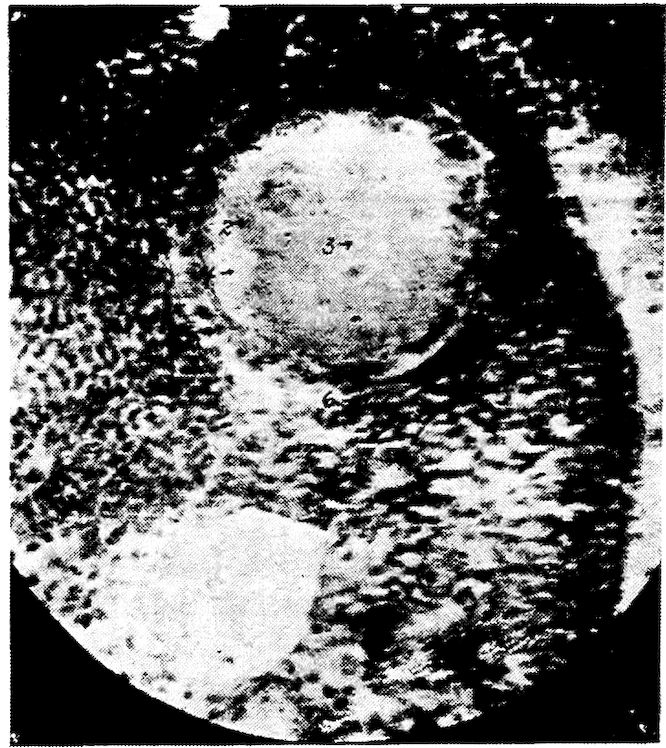
Fig. 81.—Section of ovum in a hen. 1. Nucleolus. 2. Nucleus. 3. Liquor folliculi. 4. Stratum granulosum. 5. Follicular cells. 6. Theca folliculi. 7. Peripheral stroma.
The first stage, that of division, takes place before the chick is hatched, and, according to Bradley, comes to an end about the time of hatching. This stage consists of the rapid formation of ova in the female chick. In the second stage, which begins about the time of hatching, there is an increase in the size of the units of the ovary, accompanied by yolk formation. At this time each ovum is in its own follicle (Fig. 81), and is surrounded by a layer of cuboidal cells and a theca. The theca is formed from the adjacent fibrous stroma. The third stage, that of maturation, commences during the development of the yolk and is complete after it has escaped to the oviduct. Maturation consists of each cell’s dividing into two unequal parts. In each division the cell is split into a small cell known as the polar body, which is cast off and disappears, and a larger cell, which is the ovum proper. In this process half of the original chromosomes are cast off.
Fertilization.—The sperm travels rapidly; experiments have shown eggs to be fertile laid twenty-four hours after service by a male. 320When the ovum is discharged into the ovarian pocket it is surrounded by spermatozoa.
After the male pronucleus has united with the female pronucleus in the single-celled ovum, there is a cleavage of the cell in the long axis of the egg, making two cells; and then a cleavage at right angles, which progressively continues, makes the mulberry-like mass. The remaining content of the egg consists of food for the development of the embryo. From this mass of cells before the egg is laid the blastoderm is formed. The cells of the blastoderm are differentiated into two layers. The superficial layer is the ectoderm and the lower layer is the entoderm. In the newly laid egg the blastoderm may be observed. It is about 4 millimeters in diameter. It has a transparent central area, the zona pellucida, which is located over the subgerminal region. There is a peripheral, less transparent area called the zona opaca.
In the fertile egg, as soon as it is subjected to the proper temperature, cell multiplication in the blastoderm begins. The first signs of such change are noted in the pellucid area of the blastoderm where embryonal traces appear in the form of the parallel lines called the plicæ primitivæ, which diverge to form the cephalic dilatation. At about this time takes place the formation of the myelencephalous columns, in which the blood lakes expand in the surrounding halones and in the tracts along which pass colorless blood particles. These tracts extend from below the cephalic expansion to the peripheral sinuses, as the proto-vertebræ, which begin to appear at the sides of the myelon. The red color is acquired by the blood, and the heart by its movements, is made more manifest as the punctum saliens. A distinct membrane, the serous layer, is formed upon the germ and the blastoderm. The cephalic end of the embryo rises from the surface of the blastoderm, and then, curving down, sinks into it, forming for itself a kind of hood of the serous layer. This hood gradually extends from the margin of the fossa over the body, and, meeting a similar fold formed by the projecting and incurved tail, closes over the germ on the upper side, making a circumscribed cavity, which is the amnion. The progress of differentiation of layers of the blastoderm has, meantime, gone on beneath. The serous layer is in part reflected from the vascular and from the mucous layer. The mucous layer is concerned in the formation of the intestinal canal; and beyond this part, which is at first an open groove, the mucous layer expands over the yolk, 321which it ultimately incloses, the margins of the vitellicle so formed contracting and uniting at the side opposite the embryo at a sort of cicatrix, to which the last part of the abdominal yolk adheres. The vitellicle is richly vascular, and the surface next to the yolk is augmented by rugæ.
The fowl’s egg, at about the fortieth hour, shows the buds from which the limbs are developed. A vesicle is seen to protrude near the anal end of the intestine, which, rapidly expanding, spreads over the embryo, acquiring a close adhesion to the amnion, but remaining distinct from the vitellicle, over which it spreads. It finally encloses the albumen and interposes itself between the latter and the lining membrane of the shell. Umbilical vessels are associated with this membrane. Hunter called this membrane the allantois from its containing urine, and Owen states that the sac which surrounds the albumen acts as the chorion or placenta; for it is most probable that from this surface the albumen is absorbed and the chick supported on its developmental food. The external part of the sac apparently acts as lungs as it comes into contact with the shell of the egg through pores of which there is an exchange of air. Oxygen is consumed and carbon dioxide is given off. The blood in the vessels of this membrane is in color more like arterial blood and that in the interior more like venous blood.
The embryonic mass of the incubating egg always floats to the top side. As the embryo grows it turns upon its left side, exhibiting a profile view; it then indents the yolk, and finally almost divides it into two parts.
The peripheral layers of cells rise from the margin of the germ mass, and extend and contract toward the opposite pole. This tract of germ substance is the primitive streak. Along the median line it next forms a furrow, which stops short of the ends of the streak. This streak terminates opposite the point from which the germ begins, and swells into the head. The median furrow expands upon it. The cephalic borders are next united by a thin layer of epithelial cells above the furrow, converting it into a cavity, or ventricle. The myelonal furrow is similarly covered by a layer, uniting the lateral columns. The embryonal trace becomes longer, narrower, and bends round the vitellus. A layer of epithelial cells forms a network over the whole dorsal surface of the embryo. Oblique striæ appear in the broadening germ mass radiating from the primitive streak. These indicate divisional segments. These beginnings 322of aponeurotic septa probably accompany and support nervous productions from the myelon columns.
Two transverse constrictions begin to divide the cephalic enlargements into three lobes, the second and the third of which expand into vesicles. An accumulation of cells at the side of the middle expansion appears to add greatly to its breadth. This forms the basis of the eyes.
The differentiation and the confluence of the cell constituents of the primitive streak have led to the formation of a pair of albuminous cords along the sides of the median furrow, forming the myelon proper. The cells exterior to and above them are converted into muscle and fibrous septa; and beneath the column is a jelly-filled cylinder, with a transversely striated sheath, pointed at both ends, forming the notocord. Its anterior point passes a little in advance of the acoustic vesicle. Beneath the notocord and surrounding the blastema is stretched the vegetative, or mucous, layer of cells, in contact with the yolk. Both the head and the tail of the now cylindrical embryo are liberated from the surface of the yolk. A fold of the blastema, reflected from the under part of the head, sinks like a pouch into the yolk, and soon includes the rudiment of the heart, like a bent cord, which begins to oscillate about the seventh day. From the midline of the inferior surface of the embryo, or its mucous layer, two longitudinal plates descend, diverging into the yolk-substance, and form the primitive intestinal groove.
The ophthalmic vesicle elongates and curves outward until the two ends almost come into contact. Between these two ends and beneath the delicate tegumentary layer connecting them the crystalline lens is formed. About the same time, the otoliths appear in the acoustic vesicles, which have now acquired a cartilaginous case. The cerebral lobes begin to be formed by a small fold, rising laterally and overlapping the forepart of the second enlargement, which has expanded to greater breadth. The olfactory cavities appear as small cutaneous follicles.
The two myelonal columns, expanding between the ear sacs and receding so as to show the notocord beneath, bend upward and inward, and unite, to be continued into the posterior of the optic lobes, thus commencing the cerebellar bridge across the epencephalic ventricle. The encephalic vacuities have begun to be filled by the granular basis of the cerebellar substance.
The intestinal groove begins to be converted into a canal at its 323two ends. Beneath the anterior end, and behind the heart, there gradually accumulates the cellular basis of the liver.
The commencement of the development of the organ of hearing is by a superficial depression of the cephalic blastema to meet the process from the encephalon, which forms the acoustic nerve. The lining of the depression becomes, on closure of the slit, the proper tunic of the labyrinth.
The vesicle of the labyrinth swells into four dilatations, of which three are ampullar and the fourth cochlear. The ampullar dilatations extend into very slender canals, at first almost in the same plane, by which they are brought into mutual communication. As the canals expand and elongate, they assume their characteristic relative positions as external, superior, and posterior, the posterior end of the external canal being extended beneath the posterior canal. The cochlear dilatation curves as it elongates. An inner layer becomes distinct from the common membrane and forms the acoustic lamina.
As in the development of the ear, so in the development of the eye, the production of the nerve process from the cerebral center is the first step; the infolding of the superficial blastema to meet the nerve is the next. The so-called cutaneous follicle becomes a circumscribed sac or vesicle, in which the changes and the development next proceed, converting the vesicle into an acoustic labyrinth or into an eyeball. In each case neural elements of two vertebræ become modified to lodge and to protect the sense organs, forming respectively the recess called otocrane and that called the orbit. The one is located between the occipital and the parietal vertebræ, and the other between the frontal and the nasal vertebræ. The part of the outer blastemal layer of the head which sinks to meet the process from the mesencephalic dilatation, rapidly changes its follicular into a vesicular state. The vesicle thus formed elongates, bending around the cell mass in which the crystalline lens is formed, and the meeting of the two ends results in forming the choroid fissure at the lower part of the eyeball.
The mesencephalic process, or optic nerve, expands at the posterior of the circular sac, and, in the course of mutation into eyeball, lines its posterior part with a layer which becomes the retina. The transparent layer covering the forepart of that sac and the inclosed lens is formed into the cornea. Other layers of the sac are formed into the choroid, the ciliary processes, the iris, and the pecten.
324Of the appendages of the eye the membrana nictitans is the first to develop. Then develop the lower lid and, last, the upper lid.
After the development of the essential organs of sense, the skin is developed. Modifications of the skin form the outer ear and the eyelids. Then are formed the maxillary arch, the hyoidean arch, and the scapular arch.
The vessels which return the blood from the vitellicle are the transverse and the longitudinal vitelline veins. The first are so called because these trunks pass to the embryo at right angles to its axis. They are the largest returning canals. The longitudinal veins extend parallel with the axis of the embryo; they are of smaller size. The right anterior longitudinal vein becomes the right precaval and receives the remains of the right transverse vitelline vein, as the right vena azygos. The left anterior longitudinal vitelline vein is also persistent as the left precaval, and enters in the mature bird, as in the embryo, at the posterior or the lower part of the auricle. The left transverse vitelline vein is also subsequently reduced, by receiving only the vertebral veins of that side, to the condition of a so-called azygos vein. The main trunk of the post-caval is the result of the returning vessels from the abdominal viscera and the posterior limbs at a later stage of development. There is but one principal posterior longitudinal vitelline vein, and this anastomoses with the left transverse vein as it enters the embryo.
The auricle, which, by its dilatation of the left side, appears to be double, receives the venous blood at its right division. The left one, subsequently receives the veins from the lungs, is ultimately separated from the left precaval and the right auricle to which that vein is conducted and restricted.
The ventricular part of the heart, at the second day of incubation, is in the form of a bent tube, curving from behind downward, forward, to the right and upward, continuing insensibly into the part representing the aortic bulb, in which the septum first appears, and ultimately dividing the ventricle into two.
At this stage the piers of the maxillary arch appear as buds from beneath the eyeballs. The naso-premaxillary process is above their interspace. The piers of the mandibular arch and those of the hyoidean arch follow in close succession. The blastemal base of the scapular arch projects slightly at the sides of the fovea cardiaca; the piers, now separate, ultimately meet in front of the heart, and accompany it in its retrograde course. The mesencephalon 325is the largest of the segments of the brain which are connected with the eyeballs.
When the heart has assumed its form as such, distinct from the great trunks rising from it, the two arteries from the base of the ventricles appear. The artery to the right bifurcates, one division supplying the head and the wings, the other winding over the right bronchus. That to the left also bifurcates. Its left division arching over the left bronchus and anastomosing with the right arch a little below and behind the apex of the heart. Its right division arches over the back of the heart, bending to the right and anastomosing with the right aortic arch just above the outer ductus arteriosus. Each of these divisions of the left primary arterial trunks sends off a branch to its corresponding lung. As the lung expands, and especially at the beginning of the act of expansion toward the close of the period of incubation, the blood is diverted into the pulmonary vessels, and the channels below them shrink and disappear. The left primary artery is retained as the trunk of the pulmonaries, and, through the changes in the interior of the ventricle, this artery comes to discharge exclusively the ventricle corresponding to the right in mammals. The retained aorta rises from the left ventricle.
The air-sacs begin at the lower point of the lung, appearing like small hydatids, and extend further and further into the abdomen, in front of the kidneys. They are at first full of fluid. Soon after the development of the abdominal air-sacs others are developed.
The lungs are at first free, but afterward begin to be attached to the ribs and to the spine.
In the female embryo we first observe two oviducts, one on each side of the basis, or stroma of the ovarium, which appears in a relation to the primordial kidneys similar to that of the testes in the male. At the period when the permanent kidneys have sent their ureters to the cloaca, the oviducts have been developed as prolongations from that part, and, up to a certain point of development, they are of equal size and length. Subsequently the left oviduct alone continues to grow; the right remaining stationary or shrivels; occasionally it may be discerned as a rudimentary in the mature bird, but usually all trace of it has disappeared before hatching. The left oviduct expands above or at its free end into the infundibular orifice, where its parietes are very thin. As it descends, these increase in thickness, and the efferent tube gradually acquires the texture and form of an intestine. It is attached and supported by a duplicature of the peritoneum.
326
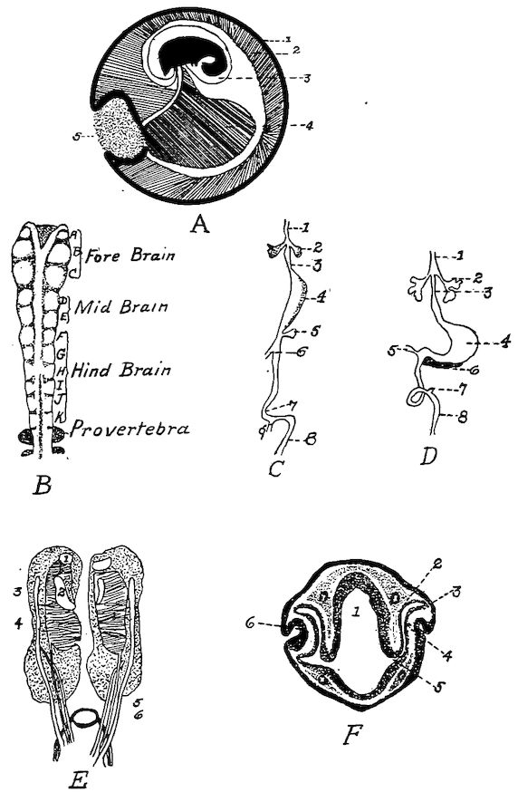
Fig. 82.—Embryological studies. A. 1, The chorion and allantois. 2, The allantoic cavity. 3, The amnion. 4, The yolk sac. 5, A small quantity of remaining albumin. B. Brain tube of a chick 25½ hours old showing partly closed brain tube with eleven folds of neuromeres. C. The development of the alimentary tract. 1, Trachea. 2, Lung. 3, Esophagus. 4, Stomach. 5, Pancreas. 6, Bile duct. 7, V-shaped loop of midgut. 8, Cloaca. 9, Vitello-intestinal duct. D. 1 to 6 inclusive, same as C. 7, Cæca. 8, Cloaca. E. Kidneys, Wolffian bodies, and testes of an embryo chick. 1, The adrenals. 2, The genital. 3, Primitive oviduct. 4, Permanent kidney. 5, Ureters. 6, Duct of primitive kidney which conducts the excretion into the cloaca. F. A transverse section of a chicks’ head 48 hours of incubation. 1, Forebrain. 2, Pigmented layer of retina. 3, Ectoderm. 4, Nervous part of retina. 5, Optic stalk. 6, Invagination of ectoderm to form the lens rudiment.
327At first, the right and the left ovaria are similar in size, but the symmetry is soon disturbed by concentration of development in the left ovary. The right ovary remains stationary and ultimately, in most birds, completely disappears by the time the chick is ready to emerge from the shell.
Three fetal membranes are developed, the chorion, the amnion, and the allantois. There is also developed the yolk sac (Figs. 82 and 83). The amnion is connected to the body wall at the umbilicus. The amniotic fluid is found in this sac. The chorion is at first surrounded by the albumen, but as the albumen is absorbed the chorion comes in contact with the inner shell membrane. It is probable that the chorion consists of ectoderm on the outside and of mesoblast on the inside. The amnion, on the other hand, is formed of mesoblast on the outside and ectoderm on the inner, or embryonal, side. The allantois springs from the embryo soon after the fourth day, and develops from the ventral wall of the primitive gut. By some embryologists, the yolk sac is included in the embryonic membranes. It commences as the splanchno-pleure surrounding the mass of yolk. It becomes smaller as the yolk is absorbed. At first its outline is round but later its walls become folded in. The yolk is dissolved and absorbed by the entodermic lining of the sac, and is carried to the embryo by veins called the vitelline veins (Fig. 83, No. B, 4), which ramify on the walls of the sac.
Some time after the fourteenth day, the chick assumes a position lengthwise within the egg shell so that the head is near the broad end of the egg. The head is bent upon the chest and the beak is usually tucked under the wing. Later the head assumes a position, by a double curve of the neck, so that the beak is in contact with the air cell. About the fifteenth day, the coils of intestine, which heretofore have been outside the abdominal cavity, are withdrawn into the abdominal cavity, as is also the abdominal yolk sac. As the chick pips out of the shell, the umbilicus becomes occluded.
The outer upper part of the tip of the beak is provided with a short, stout, spike-like arrangement, called the egg tooth. On the twentieth day this part of the beak is forced against the wall of the egg and gradually breaks through the egg shell. The breathing by the lungs commences some time before hatching; this is evidenced by the chick within the shell giving chirping sounds.
328
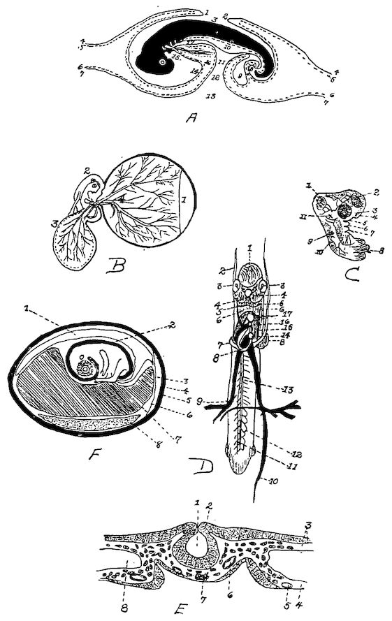
Fig. 83. A. Longitudinal section of chick, 4 days incubation. 1, Cephalic fold. 2, Caudal fold. 3, True amniotic cavity. 4, Epiblast. 5, Somatic mesoblast. 6, Visceral mesoblast. 7, Hypoblast. 8, Future anua still closed. 9. The allantoic vessicle. 10, The mesentery. 11, Intestine. 12 and 13, Yolk sac. 14, Cavity of true amnion. 15, The mouth. 16, Pleuroperitoneal cavity. 17, Fore gut. B. Membranes of the chick at the third day of incubation. 1, Membrane surrounding albumin. 2, Amnion. 3, Allantois. 4, Vitellicle. C. The fore part of an embryo chick at the second day. 1, Mesencephalon. 2, The eye. 3, Olfactory organ. 4, Mandibular arch. 5, Maxillary arch. 6, Maxillary arch. 7, Hyoidean arch. 8, Scapular arch. 9, Depression organ of hearing. 11, Process from encephalon for union with nerve of hearing. D. Primitive blood-vessels at second day of incubation (Owen). 1, Mesocephalon. 2, The right anterior longitudinal vein. 3, The eye. 4, The mandibular329 arch. 5, The hyoidean arch. 6, Scapular arch. 7, The same. 8, The ventricular portion of the heart. 9, The right transverse vitelline vein. 10, Posterior longitudinal vitelline vein. 11, Vertebral vein. 12, Tributaries of same. 14, The auricle. 16, Aortic bulb. E., A transverse section of chick embryo, 29 hours incubation. 1, Neural canal. 2, Neural crest. 3, Somatopleure. 4, Splanchnopleure. 5, Omphalomesenteric vein. 6, Aorta. 7, Notocord. 8, Pleuroperitoneal cavity. F. Chick embryo at the ninth day of incubation. 1, Allantois. 2, Amnion. 3, Air cell at large end of the egg. 4, Egg shell. 5, Outer shell membrane. 6, Inner shell membrane. 7, Yolk sac. 8, Albumin (Bradley).
The circulation of the blood is somewhat like the circulation in the fetus of quadrupeds. The primitive tubular heart (Fig. 83, No. D, 8) is bent in S-shape; the two ends are connected with blood-vessels; and later, by the development of the septa, the cavities of the adult heart are defined. The foramen ovale is found in the median septum. This opening brings the right and the left side into communication. The septa between the cavities of the heart are completed by the end of the sixth day. The two vitelline, or omphalo-mesenteric, veins carry the blood containing the nutrients from the yolk sac to the liver, where it is mixed with the blood drained from the intestines by the portal vein, and this blood is finally carried by the posterior vena cava into the right auricle of the heart. From here it passes through the foramen ovale to the left auricle; from the left auricle it enters the left ventricle, from which it is forced into the aorta, and then out into the systemic circulation. Practically all, if not all, of the blood of the pulmonary artery is sent into the aorta through a connection with this vessel called the ductus arteriosus.
Down appears about the thirteenth day of incubation. There are two kinds of down on the chick, one long, which comes first, about two or three days before hatching; a second, or fine, down forms at the roots of the other. As the embryo develops the air cell, at the large end of the egg which is developed between the two shell membranes, or membranæ putaminæ, gradually enlarges.
330