ARTHROLOGY
Reading edition
Kinds of Joints.—Joints may be movable, immovable, or mixed. In movable, or true joints the articular surface of each bone is covered by cartilage. The bones are held together by ligaments, the capsular one often surrounding the joint and enclosing the synovial membrane. In some joints there is a pad of fibrous cartilage interposed between the two articular cartilages. Such a pad, called a meniscus, adds to the elasticity and the free movement of the joint. Movable joints form the most numerous class; they are for the most part found in the limbs.
In an immovable joint there is only a thin layer of fibrous or cartilaginous material interposed between the bones. The fibrous layer of the periosteum of both bones unite to cover the connecting material and becomes attached to the same, thus serving as a ligament. If the connecting material is fibrous, the joint is called a suture; if cartilaginous, a synchondrosis. These joints are found in the skull and in the pelvis.
The term mixed is used with reference, not to the motion in joints, but to their structure, which partakes of the nature of both the movable and immovable. The bones are firmly joined by a strong interposed pad of fibrous cartilage to which also is adherent the ligaments of the joint. There are no capsular ligaments; the cartilaginous pad or disc is softer toward its center, where occasionally there may be one, or even two, narrow cavities. Authorities differ as to whether such cavities are lined by synovial membrane or not. Since there are really no frictional surfaces in such a joint, motion depends upon the flexibility of the disc. The joints between the vertebral centra afford the best illustration of the mixed class.
Movement of Joints.—The movements admissible in joints may be divided into four kinds: gliding, angular movement, circumduction, and rotation. These movements are often, however, more or less combined in the various joints. It is seldom that there occurs only one kind of motion in any particular joint.
Gliding movement is the most simple kind of motion that can 57take place in a joint, one surface gliding or moving over another without any angular or rotary movement.
Angular movement occurs only between the long bones. By it the angle between the two bones is increased or diminished. It may take place in four directions: forward and backward, consisting of flexion and extension; or inward and outward from the medial line of the body, consisting of adduction and abduction. Abduction of a limb is movement away from the medial line of the body. Adduction of a limb is movement toward the medial line of the body.
Circumduction is that limited degree of motion which takes place between the head of a bone and its articular cavity, whilst the extremity and sides of the limb are made to circumscribe a conical space, the base of which corresponds with the inferior extremity of the limb, the apex with the articular cavity; this kind of motion is best seen in the shoulder- and hip-joint.
Rotation is the movement of a bone upon an axis, which is the axis of the pivot on which the bone turns, as in the articulation between the atlas and axis, when the odontoid process serves as a pivot around which the atlas turns; or else is the axis of a pivotlike process which turns within a ring, as in the rotation of the radius upon the humerus.
Pronation is a form of rotation in which the inferior extremity of the radius passes before the ulna, and thus causes the hand to execute a kind of rotation from without inward.
Supination is a form of rotation in which the movement of the forearm and hand are carried outward so that the anterior surface of the latter becomes superior.
The Ligamentous Structure.—Ligaments are dense, fibrous, connecting structures. They are made up principally of white fibrous tissue and exist in all true joints.
There are four kinds of ligaments associated with true joints:
The first kind, the capsular ligament which encloses all true joints, is thin and consists of interlaced fibers attached to the bone at the edges of the articular cartilages. It either partly or wholly surrounds the joint, enclosing and protecting a synovial apparatus, which, by secreting a liquid resembling serum, lubricates the joint to prevent friction.
The second kind, the binding or lateral ligaments, consist of flattened or rounded cords or bands of fibrous tissue. Such a ligament extends from one bone to the other, and firmly attached to 58their roughened surfaces, holds the bones in place and at the same time allows the required amount of motion.
The third kind, located between the joints, is called interosseous ligament.
The fourth kind, called the annular ligament binds down and protects the tendons.
Ligaments of the Ear[2].—The concha of the ear is provided with a superior and an inferior ligament.
Ligaments of the Jaw.—The articulations of the lower jaw are complex. The freely movable articulation is between the inferior maxilla and the quadrate. Less freely movable articulations are formed by the quadrate with the temporal, the zygomatic and the pterygoid. A lateral ligament of the jaw, the articulo-jugale extends from the posterior border of the inner wing of the os articulare of the inferior maxilla (Fig. 8, No. 10, and Fig. 19, No. 15) to the outer border of the os quadrato jugulare.
A lateral temporo-maxillary ligament extends from the outer surface of the temporal bone to the outer border of the os articulare of the inferior maxilla (Fig. 8, No. 11, and Fig. 19, No. 14).
Ligaments of the Vertebræ.—In each space between the bodies of the vertebræ there is a meniscus intervertebralis. This meniscus is analogous to the annulus fibrosus of mammals (Gadow), which, is possibly formed as a protrusion of the anterior surface of the vertebral body. The meniscus or disc may develop into a ring-shaped structure, the true meniscus, or it may develop to different degrees as an extension of the vertebral body surface, and become, as in the dorsal and lumbar regions, fused with the vertebral segments, in which case it is called the annulus fibrosus.
The vertebral disc which is connected with the atlas and which is fused with that bone, represents the first meniscus, which is called the ligamentum transversum atlantis. This, as well as other ligaments of the spinal column, may become ossified.
The ligament which is located between those vertebral bodies which face each other and is inside the joint cavity, is called the ligamentum suspensorium corporum vertebralium. It passes through the central opening of the meniscus and lies exactly in the long axis of the body of the vertebra.
The first ligament of the neck is the ligamentum suspensorium dentis epistrophei. The ligamentum capsulare atlantico-occipitale 59and the ligamentum capsulare atlantico-epistrophicum odontoideum are two parts of the joint capsule of the vertebral body. Other ligaments of this part are the membranæ obturatoriæ intervertebrales posteriores, which are located between the semicircular rims of the neck vertebræ.
The ligamentum transversum atlantis surrounds the occipital condyle.
The ligamentum nuchæ is a thin, membranous, ribbon-like structure which lies between the muscles of the right and those of the left side of the middle, and the lower part on the posterior of the neck, and ends in attachments to the superior spines of the cervical vertebræ.
The ligamentum elasticum interspinale profundum and the ligamentum elasticum interspinale superficiale and the three last named are the ligaments which keep the neck of the fowl in the s-shape, without the action of the muscles.
The ligamentum capsulare obliquum connects the facets of the oblique processes.
Ligaments of the Ribs.—The thick inferior end of the sternal portion of the true rib has two small articular heads, which articulate with two depressions in the articular surface of the sternum. This articulation is held firm by a capsular ligament.
The upper end of the sternal rib forms an almost perfect right angle with the inferior end of the dorsal rib with which it is connected by a joint provided with a synovial apparatus and a capsular ligament. This arrangement allows free movement outward and inward and is the main joint in respiration.
The upper end of the dorsal portion of the rib articulates with the dorsal vertebra. The joint formed by the articular head with the body of the vertebra is provided with a capsular ligament. The tubercle of the rib articulates with the facet on the transverse process of the dorsal vertebra, and is provided with a transverse ligament, called the ligamentum transversum externum. It is also provided with a capsular ligament.
The ligamentum triangulare connects each processus uncinatus with the succeeding rib. This ligament is in the form of a membranous sheet, or aponeurosis.
Ligaments of the Sternum (Fig. 13).—The external and the internal notches of the sternum are bridged over with a thin membrane 60which gives an extensive surface for muscular attachments. The sternal muscles overlie this portion.
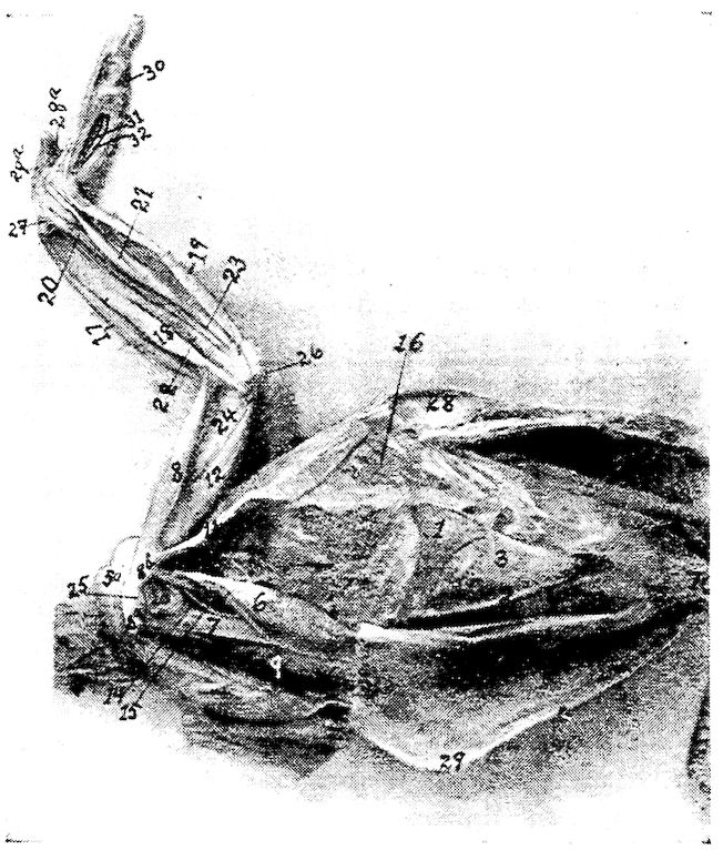
Fig. 13.—Muscles of the fore extremity. Inside view. 1, Lateral external process of sternum. 2, Lateral internal process of same. 3, Ligament of the external notch. 4, Ligament of the internal notch. 5, Pectoralis major. 5a, Its fan-shaped expansion at shoulder-joint laid back. 6, Pectoralis tertius. 7, The coracoid. 8, Biceps. 8a, Its long head. 8b, Its short head. 9, Pectoralis secundus. 10, Rectus abdominis. 11, Teres et infraspinatus. 12, Deltoid. 13, Capsular ligament. 14, Teres minor. 15, Coraco-brachialis. 16, Serratus magnus anticus. 17, Extensor metacarpi radialis longior. 18, Pronator brevis. 19, Flexor carpi ulnaris. 20, Flexor carpi ulnaris brevior. 21, Flexor digitorum profundus. 22, Extensor indicis longus. 23, Extensor ossis metacarpi pollicis. 24, Humerus. 25, Shoulder. 26, Elbow. 27, Carpus. 28, Section through vertebra. 29, Keel of sternum. 28a, Flexor brevis pollicis. 29a, Extensor proprius pollicis. 30, Flexor minimi digiti brevis. 31, Interosseous palmaris. 32, Interosseous palmaris.
The sternum is connected by a fibrous mass with the inferior portion of the clavicle, or hypocledium (Fig. 57), the claviculosternal ligament.
The inferior narrow elongated end of the coracoid forms with the sternum a true articulation, which is provided with a capsular 61ligament, the ligamentum capsulare. Capsular ligaments occur at the articulations between the sternum and ribs.
Ligaments of the Shoulder-joint (Fig. 13).—The shoulder-joint is made up of the scapula, the humerus, and the coracoid. The ends of these three bones form the foramen triosseum (Fig. 15, No. A, 7) through which passes the tendon of the elevator muscle of the wing.
The furcula, independent of the shoulder-joint or girdle, is attached to the supero-internal part of the proximal end of the coracoid by fibrous connective tissue; it is also connected to the other bones of the shoulder-joint by the ligamentum coraco-furculare and the ligamentum furculo-scapulare.
The ligamentum coraco-scapulare extends from the tuberosity of the furcula to the coracoid and to the processus furcularis of the scapula.
The ligamentum coracoido-scapulare externum extends between the external tuberosity of the coracoid, the tuberosity of the scapula, and the humerus.
The ligamentum coracoideo-scapulare inferius extends from the coracoid to the inner tubercle of the scapula.
Another long, broad ligament belongs to the episternal apparatus.
The shoulder-joint is provided with a wide, loose, capsular ligament (Fig. 13, No. 13). Attached to the humerus are four other ligaments, of which three pass from the anterior end of the coracoid and the fourth from the scapula. The latter are as follows: first, the supero-anterior ligamentum humero-coracoideum, which extends from the small tubercle of the humerus to the coracoid bone; second, the antero-inferior ligamentum humero-coracoideum, which extends from the humerus to the coracoid; third, the ligamentum coraco-humerale, which extends from the coracoid to the large tubercle of the humerus; fourth, the ligamentum humero-scapulare which extends between the processus humeralis of the scapula and the head of the humerus.
Ligaments of the Elbow-joint (Fig. 14).—The elbow-joint is made up of the ulna, radius and the humerus. The ligaments of the elbow-joint are as follows: the ligamentum capsulare cubiti, or capsular ligament, which extends from the processus cubitalis of the humerus to the processus olecranalis coracoideus of the ulna and to the tuberositas radii of the radius (Fig. 15, No 5).
The ligamentum laterale cubiti externum connects the outer humeral distal extremity with the head of the radius (Fig. 15, No. 2).
The ligamentum laterale cubiti internum lies between the inner 62distal extremity of the humerus and the tuberculum internum of the ulna (Fig. 14, No. 9).
The ligamentum annulare radii originates on the olecranon, surrounds the head of the radius, and is attached to the tuberculum internum of the ulna (Fig. 15, No. 3).
The ligamentum cubiti teres extends from the head of the radius to the upper end of the ulna (Fig. 15, No. 4).
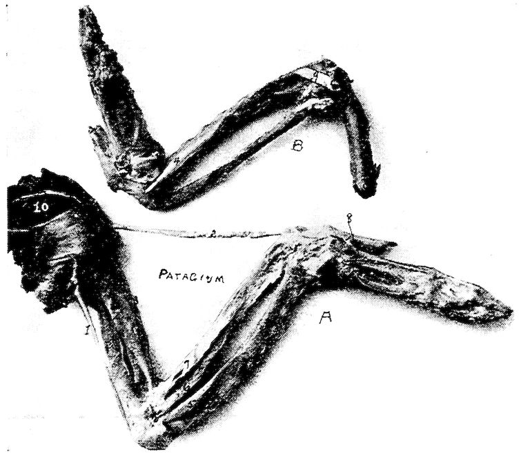
Fig. 14.—Muscles and ligaments of the arm and forearm of a hen. A. External view. 1, Expansor secundarium. 2, Tensor patagii longus. 3, Tensor patagii brevis. 5, Brachialis anticus. 6, Pronator longus. 7, Supinator brevis. 8, Extensor indicis brevis. 10, Scapulo-humeralis. B. Internal view. 9, Ligamentum laterale cubiti internum.
The ligamentum transversum spreads out between the head of the ulna and the radius, thus uniting the two bones, and limiting supination (Fig. 15, No. 1).
Ligaments of the Carpal Joint.—The carpal joint is made up of the ulna, radius, the two carpal bones and the metacarpus.
There are two strong ligamenta obliqua carpi ulnaris which extend 63from the processus styloideus of the ulna to the tuberculum posterius carpi ulnaris of the os carpi ulnaris.
The ligamentum posticum ulnare carpi ulnaris extends from the processus styloideus of the ulna to the os carpi ulnaris.
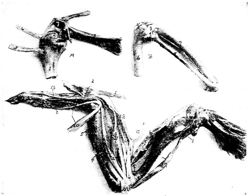
Fig. 15.—Ligaments of the arm and forearm of a hen. A. The scapulo-coraco-humeral articulation. 1, Proximal end of the humerus. 2, Articular head of humerus. 3, The coracoid. 4, Proximal end of the clavicle. 5, Tendon of pectoralis tertius. 6, Tendon of pectoralis secundus. 7, Opening or foramen triossium through which the tendon of the elevator of the wing passes. (Pectoralis secundus.) 8, Broken end of scapula. B. 1, Ligamentum transversum. 2, Ligamentum laterale cubiti externum. 3, Ligamentum annulare radii. 4, Ligamentum cubiti teres. 5, A portion of the ligamentum capsulare cubiti. 6, Distal extremity of humerus. 7, Ulna. 8, Radius. C. Muscles of the outside surface of the arm. 1, Tensor patagii longus. 2, Extensor digitorum communis. 3, Tensor patagii brevis. 4, Anconeus. 5, Flexor carpi ulnaris. 6, Biceps. 7, Triceps. 8. Deltoid. 9, Brachialis anticus. 10, Flexor metacarpi radialis. 11, Extensor ossis metacarpi pollicis. 12, Extensor metacarpi radialis longior. 13, Extensor indicis longus.
The ligamentum ulnare carpi radialis is a short ribbon-like ligament which passes over the above-mentioned ligaments and is attached to the inner surface of the os carpus radialis.
The ligamentum ulnare carpi radialis internum is a strong ligament 64which originates on the inner part of the elbow. It extends to the upper rim of the base of the main digit.
The ligamentum radiale carpi radialis externum originates on the outside of the head of the middle digit and inserts to the superior tuberculum carpi radialis of the radius.
The ligamentum carpi radialis internum, a short ligament, extends from the inside of the head of the main digit to the inner rim of the carpi radialis.
The ligamentum carpi interosseum is located between the carpal bones.
The ligamentum ulnare metacarpi internum is spread out between the processus styloideus of the ulna and the tuberositas muscularis on the upper side of the middle finger base at a point where the second and the third metacarpi separate.
The ligamentum ulnare metacarpi externum extends from the tubercle of the distal extremity of the ulna, on the dorsal side, to the tubercle on the base of the metacarpus.
Ligamentum radiale metacarpi extends from the head of the inner digit to a point near the tuberculum muscularis of the radius.
The ligamentum transversum ossis carpi radialis et metacarpi is located between the inferior tuberculum ossis carpi radialis and the first metacarpal bone.
There is another ligament between the tuberculum superiorus carpi radialis and the first metacarpal bone.
The ligamentum ossis carpi radialis internum et metacarpi extends from the inner surface of the carpus radialis to the tuberositas muscularis of the radius.
The ligamentum ossis carpi ulnaris externum et metacarpi extends from the ulnar carpal bone to the tuberculum of the metacarpal bone.
The ligamentum ossis carpi ulnaris internum et metacarpi extends from the processus uncinatus of the ulnar carpal bone to the tuberositas muscularis of the metacarpal bone.
Ligaments of the Finger.—The ligamentum pollicare connects the thumb with the first metacarpal bone.
The ligamentum anterius ossis metacarpi et primæ phalangis digiti secundi extends from the tuberculum articulare metacarpi, of the proximal end of the metacarpus, to the tuberculum articulare of the first phalanx of the second or large finger. Similarly attached, are also an internal and a posterior ligament, or ligamentum internum and ligamentum posterius.
65The three phalanges are all provided with capsular ligaments, or ligamenta capsularia, which bind together the several phalanges.
The small or third finger is connected to the metacarpus by a capsular ligament, the ligamentum capsulare, and by an interosseous ligament, or ligamentum interosseum digitorum, to the first phalanx of the second finger.
Ligaments of the Pelvis.—The obturator foramen, formed by the ilium and the ischium, is covered by a broad membranous ligament (Fig. 4, No. 29).
The oblong foramen, or foramen oblongum, and the foramen ischiadicum, formed by the ischium and the pubis are covered by broad membranous ligaments (Fig. 4, Nos. 29 and 60).
Poupart’s ligament or ligamentum Poupartii, quite small in fowls, extends from the anterior lower rim of the ilium to the pelvic cavity, and is inserted at the bottom of the acetabulum.
Another broad ligament originates from the posterior rim of the ischium and is attached to the square surface of the first coccygeal vertebra.
The ilio-pubic ligament extends from the pubic spine to the last rudimentary rib.
Ligaments of the Hip-joint (Fig. 25, F).—The hip-joint is made up of the ilium, ischium, pubis and femur and is a deep ball-and-socket joint. The cavity or acetabulum is called a cotyloid cavity.
The hip-joint is provided with three ligaments.
The ligamentum capsulare femoris is attached around the rim of the cotyloid cavity and around the rim of the articular head of the femur (Fig. 25, No. F, 4).
The ligamentum teres, or round ligament, is a very short ligament which closes the hole at the floor of the cotyloid cavity, at which point it is attached. It is also attached to the head of the femur.
The ligamentum ilio-sacrale strengthens the hip-joint capsule. It passes from the lower anterior rim of the os ilii, extending over the capsule to the neck of the femur to which it is attached.
The Ligaments of the Knee-joint (Figs. 16 and 17).—The knee-joint is made up of the femur, patella, tibia and fibula, and is provided with the following ligaments:
The ligamentum extero-laterale genu is a strong ligament extending from the condylus externus femoris, or external femoral condyle to the outer surface of the head of the fibula, or capitulum fibulæ. 66From this head there passes inward, a strong ligament to a point between the femur and fibula (Fig. 25, No. D, 4).
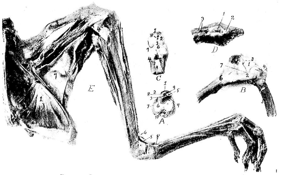
Fig. 16.—Ligaments and muscles of the hind extremity. A. A transverse section through the tibial or tarsal cartilage, front view. 1, Gastrocnemius tendon. 2, Flexor perforatus annularis primus pedis. 3, Flexor perforans digitorum profundus. 4, Flexor perforatus indicis secundi pedis. 5, Extensor longus hallucis. 6, The articular surface. 7, Ligamentum capsulare ossis tarsi. 8, Flexor perforatus medius secundus pedis. 9, Flexor perforatus indicis secundus pedis. 10, Tendons of the extensor muscles. B. Knee-joint. Inside view of femoro-tibial articulation. 1, Anterior ligamentum cruciatum. 2, Internal ligamentum laterale genu. 3, Anterior patellar ligament. 4, Patella. 5, Distal end of femur. 6, Proximal end of tibia. 7, Location of meniscus or pad of fibrocartilage. C. Anterior view of tibio-tarsal articulation. 1, Distal end of tibia. 2, Its articular surface. 3, Ligamentum anticum. 4, Proximal end of metacarpus. 6, Pad of fibrocartilage. D. The dorsal surface of the coccyx. 1, The bilobate oil gland. 2, Its duct. 3, Levator coccygis. E. Inside view of pelvis and thigh. 1, Obturator internus. 2, Ambiens. 3, Vastus internus. 4, Internal ligamentum laterale genu. 5, Tibialis anticus. 6, Loop through which the tibialis anticus passes. 7, Adductors of the thigh. 8, Loop for the extensor tendon.
The ligamentum intero-laterale genu extends from the outer surface of the internal condyle of the tibia, or condylus internus tibiæ. It gives off a thin ligamentous slip which enters the joint and is attached to the inner half-moon shaped pad of fibrous cartilage, or meniscus, which it draws backward by the flexion of the knee (Fig. 16, No. B, 2).
67The ligamentum popliteum arises from the fossa poplitea of the distal extremity of the femur and extends downward to the posterior rim of the head of the tibia.
The anterior ligamentum cruciatum genu originates from the fossa poplitea and extends outward antero-laterally to the rim of the head of the tibia (Fig. 25, No. 6).
The posterior ligamentum cruciatum genu is a short, strong ligament which originates from the cavity of the internal condyle of the femur, or condylus internus femoris, and inserts into the internal glenoid cavity of the tibia (Fig. 25, No. 1) or cavitas glenoidalis interna.
The meniscus of the femoro-tibial articulation is divided into four parts, as follows: first, the internal adhesio cornu antici cartilaginis lunatæ; second, the posterior adhesio cornu antici cartilaginis lunatæ; third, the external cornu cartilaginis lunatæ; fourth, the anterior cornu antici cartilaginis lunatæ. The latter originates between the condyles and passes around the ligamentum cruciatum genu posticum (Fig. 16, No. B, 7).
The inner semi-lunar fibrous cartilage is well developed and is joined by two ligaments between the femoral and the tibial surfaces (Fig. 25, No. 1).
The external semi-lunar cartilage lies in a cavity between the external femoral condyle and the head of the fibula (Fig. 25, No. 2).
The fibrous cartilages are bound posteriorly by a ligament, and by a second ligament to the head of the fibula, or capitulum fibulæ. Both pads of fibrous cartilage are connected by a transverse ligament called the ligamentum transversale commune (Fig. 25, No. 5).
The patella is provided with several ligaments, as follows:
The anterior patellar ligament, broad, strong, and irregular in thickness, extends from the inferior margin of the patella to the rim of the second tibial crest (Fig. 16, No. B, 3).
68The ligamentum capsulare capituli fibulæ is spread out between the head of the fibula, or capitulum fibulæ, and the superficies glenoidalis peronea tibiæ.
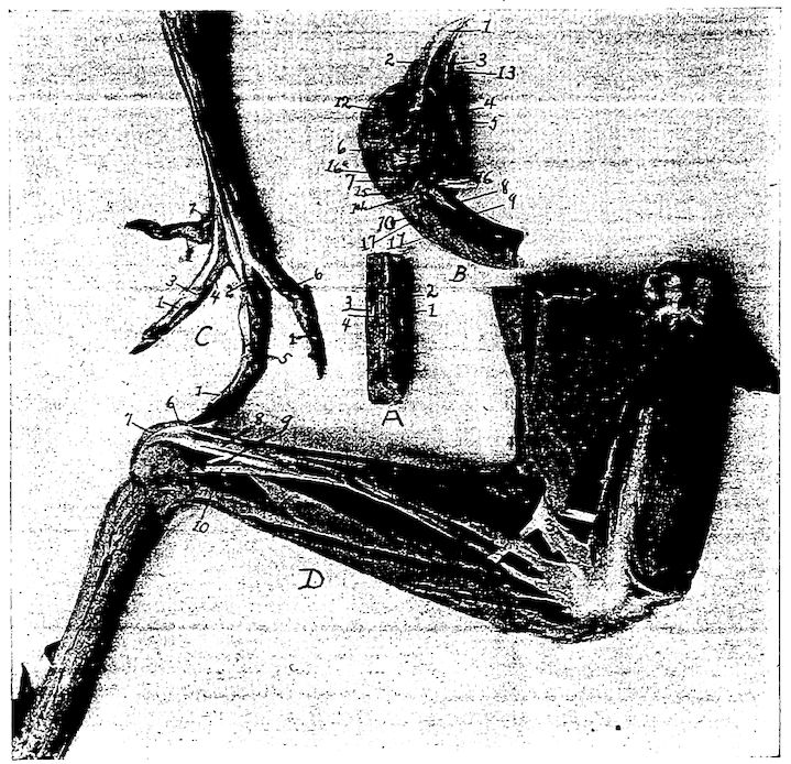
Fig. 17.—Muscles and tendons of the head and posterior extremity. A. Section of neck with superficial muscles removed. 1, Obliquo-transversales. 2, Interspinales. 3, Intertransversales. 4, Interarticulares. B. Head showing muscles. 1, Beak. 2, Nostril. 3, Tongue. 4, Sublingual salivary gland. 5, Pterygoideus. 6, Stylo-hyoideus. 7, Temporal. 8, Flexor capitis inferior. 9, Rectus capitis lateralis. 10, Rectus capitis posticus major. 11, Trachelo-mastoideus. 12, Eye. 13, Mylo-hyoideus. 14, Genio-hyoideus. 15, Biventer maxilla. 16, Digastricus. 17, Rectus capitis anterior minor. C. Foot of hen showing tendons. 1, Flexor perforans digitorum profundus. 2, Flexor perforatus medius primus pedis. 3, Sheath at joint through which tendon passes. 4, Flexor perforatus indicis primus pedis. 5, Flexor perforatus medius secundus pedis. 6, Flexor perforatus annularis primus pedis. 7, Flexor longus hallucis. D. Outside view of leg of hen. 1, Biceps flexor cruris. 2, Semitendinosus. 3, Loop through which biceps flexor cruris passes. 4, Tibialis anticus. 5, Flexor perforatus indicis secundus pedis. 6, Flexor perforatus medius primus pedis. 7, Gastrocnemius. 8, Flexor perforatus annularis primus pedis. 9, Flexor perforatus medius secundus pedis. 10, Extensor longus hallucis.
The ligamentum tibio-fibulare extends from the head of the fibula, or capitulum fibulæ, to the inner surface of the external crest, or crista externa tibiæ, of the tibia (Fig. 25, No. 3).
69The ligamentum interosseum is a delicate ligament located between the tibia and fibula (Fig. 25, No. 4) which very early becomes ossified.
Ligaments of the Tibio-metatarsal Joint (Fig. 16).—The tibio-tarsal joint is made up of the tibia and metatarsus.
The ligamentum capsulare ossis tibio-metatarsi connects the tibia and the metatarsus and surrounds the joint (Fig. 16, Nos. A, 7 and C, 7).
The ligamentum externum, a long strong ligament, originates on the upper and outer surface of the external condyle, or condylus externus of the tibia and is attached to the upper rim of the os metatarsi (Fig. 23, No. 15).
The ligamentum anticum is spread out between the fossa intercondyloidea of the tibia and the tuberculum ossis metatarsi of the proximal end of the metatarsus (Fig. 16, No. C, 3). Just posterior to this ligament, there is an interosseous ligament, the ligamentum interosseum.
A semi-lunar pad of fibrous cartilage, the cartilago semi-lunaris is located between the outer distal extremity of the tibia and upper articular surface of the metatarsus (Fig. 16, No. C, 6). Its concave portion is directed anteriorly and receives an insertion from the ligamentum externum, or external ligament.
The tendon Achillis (Fig. 16, No. A, 1) may become ossified.
Ligaments of the Toes (Fig. 16).—In addition to the capsular ligaments, which all true joints have, we find the following in connection with the toes:
The ligamentum superius and the ligamentum inferius connect the second toe with the great toe, or hallux.
The ligamentum laterale externum and the ligamentum laterale internum unite the bases of the second, third and fourth toes to the lateral faces of the trochleas of the inferior extremity of the metatarsus.
The bases of the toes are held together by the ligamenta transversa. All the rest of the toes are held together by the ligamenta capsularia digitorum pedis and by two lateral ligaments, namely: the ligamentum laterale externum and the ligamentum laterale internum.
70