SPLANCHNOLOGY
Reading edition
The Digestive Apparatus.—This apparatus, apparatus digestorius, consists of the organs directly concerned in the reception of food, in its passage through the body, and in the expulsion of the unabsorbed portion. For convenience, these organs are grouped as follows: the alimentary canal and the accessory organs.
The alimentary canal is a tube which extends from the mouth to the anus. It has a complete lining of mucous membrane, external to which is an almost continuous muscular coat. The abdominal portion of the tube is largely covered with a serous membrane, the visceral peritoneum. The canal consists of the following consecutive segments: mouth, pharynx, first portion of the esophagus, crop, second portion of the esophagus, proventriculus, gizzard, small intestine, large intestine, and cloaca.
The accessory organs are beak, tongue, salivary glands, liver, and pancreas.
The Mouth (Fig. 27).—The distinctive character of the mouth of the fowl consists, in the absence of lips and teeth and instead of jaw bones of other animals, of a beak. The edge of the beak is covered by a horn-like gum. The shape of the beak differs in the various classes of birds. In the chicken the beak is short, strong, thick, and pointed; in palmipeds it is flattened. The upper mandible extends out over the lower mandible. The outer portion of the upper and the lower beaks is covered by a horny sheath. There is no velum, and the mouth cavity is extended rather continuously into the pharynx (Fig. 27, No. 4). The posterior cross bar of the hard palate (Fig. 27, No. 5), which possesses a row of filiform papillæ projecting backward, marks the upper boundary line of the mouth; while the posterior end of the tongue (Fig. 27, No. 6), likewise with a row of filiform papillæ, marks its lower boundary line.
Parts of the Mouth.—The mouth of fowls is divided into an upper and a lower half. The upper half is divided into an anterior and a posterior part.
The anterior part extends from the anterior tip of the beak to the posterior nares. It is further divided into an outer hard part and 136an inner soft part. The outer part is covered by epithelium which may be considered as a continuation from the upper outer portion of the beak. A portion of this is cornified. The inner part is covered by a mucous membrane containing mucous glands, nerves, and blood-vessels.
In the posterior part of the upper half of the mouth, are situated the posterior nares (Fig. 27, No. 10) which appear as a longitudinal slit in the center of the hard palate. There is also found in this part a furrow which contains the opening of the Eustachian tubes, or tubæ auditivæ. There are also two ridges near the same posterior part in which are red and white papillæ, which may appear in rows, and which contain mucous glands.
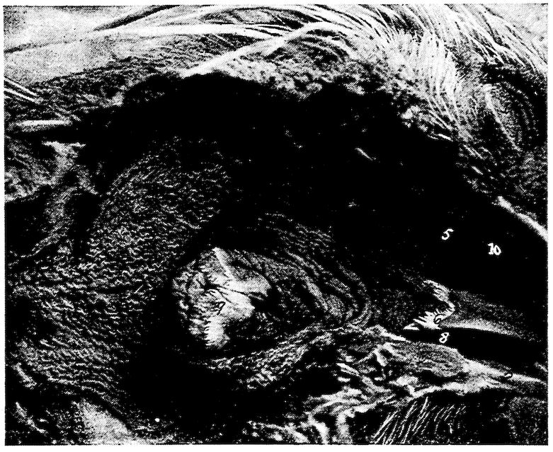
Fig. 27.—Mouth and pharynx of a cock laid open. 1, Eye. 2, Edge of beak covered with a horn-like gum. 3, Tongue. 4, Pharynx. 5, Posterior cross bar of hard palate. 6, Posterior end of tongue. 7, Anterior end of esophagus. 8, Phrenum. 9, Palatal papillaries. 10, Posterior nares.
The lower half of the mouth lies between the lower jaw bones and the walls of the mouth cavity. The tongue is attached here by 137means of the phrenum (Fig. 27, No. 8). This half of the mouth forms a pocket-like structure which aids in taking up the food.
The mouth cavity is lined with stratified squamous epithelium, continuous with that of the pharynx and the esophagus. Taste buds are located in the mucous membrane of the mouth.
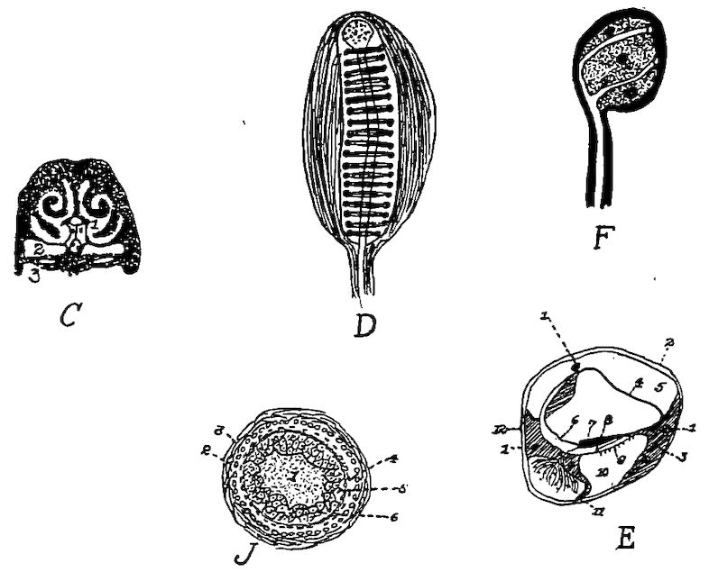
Fig. 28.—Various anatomical parts of the fowl. C. A section through the nasal region of the fowl. 1, Nasal passages showing the turbinated bones dividing the nasal passage into the superior, middle and inferior meati. 2, The infraorbital sinus. 3, The hard palate. D. Herbst’s touch corpuscle from the beak of a quail. E. A vertical section through the ductus cochlearis of a pigeon. 1, A blood-vessel. 2, The periosteum. 3, The bony structure. 4, The vascular integument. 5. The scala vestibuli. 6, The inner hyaline cylindrical cells. 7, The membrana tectoria. 8, The papilla acustica basilaris. 9, The membrana basilaris. 10, The scala tympani. 11, The ganglion of the cochlear nerve in the ramus basilaris. 12, The periosteum (Gadow). F. A corpuscle of the soft papilla of a duck’s tongue. J. A transverse section through a feather papilla. 1, The pulp. 2, The malpighian layer. 3, The corium. 4, The stratum corneum of the papilla. 5, The malpighian cell group of the main shaft. 6, The horny sheath.
In many water fowls, as geese and ducks, the gum edge of the mandible has grooves extending crosswise, in which are numerous terminals of the trigeminus nerve arranged as taste organs (Fig. 29, G and Fig. 28, D and F). In many birds of prey and in water birds, e.g., the goose and the duck, there is found at the base of the beak, a very thin, nervous or sensitive skin, waxy in appearance, called the ceroma.
138
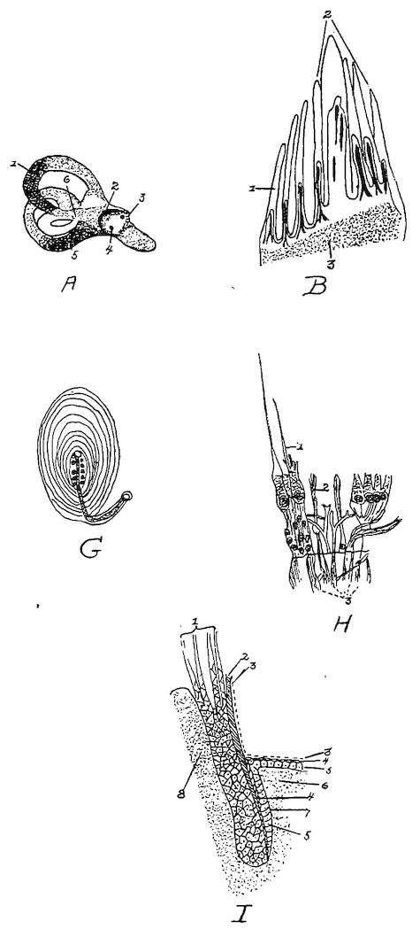
Fig. 29.—Various anatomical parts of the fowl. A. 1, The superior semicircular canal. 2, The vestibular nerve. 3, The meatus auditorius internus—the entrance of the auditory nerve. 4, The entrance of the cochlear nerve. 5, Posterior semicircular canal. 6, The ampulla. B. Side papilla of the tongue. 1 and 2, Papillæ showing a fibrous central core supporting blood-vessels and nerves. 3, The basement membrane consisting of connective tissue. G. Herbst’s corpuscle of the tongue of a duck, showing the capsule, lamella and around the nerve zone, numerous round bodies arranged in two rows. H. From the cristæ acoustica of the ampulla of the dove. A, A vertical section with isolated fiber cells. 1 and 2, The nerve fibers. 3, The hair cells. I. A longitudinal section through a feather papilla showing a young feather. 1, The developing feather. 2, The horny sheath. 3, The epithelium. 4, Stratum corneum. 5, Stratum malpighi. 6, Corium. 7, Malpighian cell group of the follicle.
139The Hard Palate.—The hard palate, palatum durum, forming the roof of the mouth has for its boundaries the beak anteriorly, the mandibular arches laterally, and the pharynx posteriorly. It has four or five transverse bars, projecting posteriorly from each of which is a row of filiform papillæ (Figs. 27 and 29).
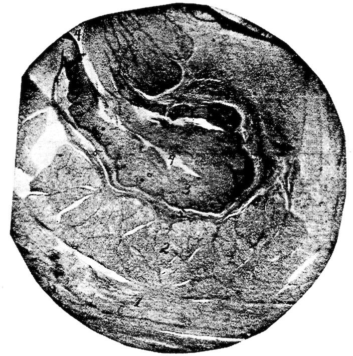
Fig. 30.—A section through the wall of the crop of a hen. 1, The outer muscular layer showing longitudinal fibers. 2, The inner muscular layer showing the sectioned ends of the bundles of muscle fibers. 3, Stratified squamous epithelium. 4, The outer surface or lumen of a fold. 5, The stroma, muscularis mucosa, and submucosa.
The beak (Fig. 4, No. 1) with little if any aid from the tongue, is the prehensile organ.
The Tongue (Fig. 27, No. 3). Location.—The tongue (lingua) of the fowl is situated in the floor of the mouth between the rami of the 140lower mandible, and is slung to the cranium by the cerato-branchial element of the os hyoideum (Fig. 18, No. 2).
Shape.—The tongue, is pointed in front and wide behind, shaped like an Indian arrow-head, and is supported by the bony and cartilaginous hyoid apparatus (Fig. 9, A).
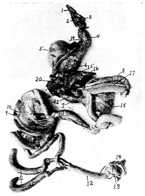
Fig. 31.—The visceral organs of the hen. 1, Tongue. 2, Larynx. 3, Glottis. 4, First portion of the esophagus. 5, Crop. 6, Second portion of the esophagus. 7, Proventriculus. 8, Duodenum. 9, Free or floating portion of the small intestine. 10, Mesentery supporting the free portion of the small intestine. 11, Cæca. 12, Rectum or large intestine. 13, Cloaca. 14, Anus. 15, Liver. 16, Gall-bladder. 17, Pancreas. 18, Gizzard. 19, Trachea. 20, Lungs. 21, Spleen.
Structure.—The free part of the tongue consists of two long tubes which are formed by the rolling in of the ventral sides of its horny coverings. The dorsal plate, which constitutes one-half of the 141horny sheath, does not form a part of the tubes, but ends in the region of the point of the entoglossum.
The top surface is covered by a thick stratum corneum, giving it a rather horny surface. Glands occur in the posterior part of the tongue.
While not so freely movable as in mammals, the tongues of birds are very flexible.
The Pharynx (Fig. 27). Location.—The anterior part of the pharynx joins with the mouth and the posterior part with the upper portion of the esophagus and the superior larynx. The pharynx is bounded anteriorly by the base of the tongue and by the posterior edge of the hard palate. Posteriorly, it is marked by the entrance of the esophagus and by a ridge of filiform papillæ located on the supero-posterior part of the larynx, called the palatal papillaries. A few filiform papillæ stud the roof. The pharynx communicates anteriorly with the posterior nares and the mouth, and, posteriorly, with the esophagus and the larynx.
Shape.—The pharynx is a musculo-membranous sac.
Structure.—The pharynx is lined with a mucous membrane covered by squamous epithelium. The mucous membrane is thrown into irregular folds. The bird has two Eustachian tubes. Pneumatic apertures conduct the air from the Eustachian tubes to the pericranial diploë.
In the posterior pharyngeal roof is situated the infundibular crevice.
Function.—The function of the pharynx is to give passage for the air from the posterior nares to the larynx, and to give passage for the food from the mouth to the esophagus.
Glands Adjacent to the Mouth and to the Pharynx.—The mucous membrane adjacent to the glottis and in the roof of the mouth contains alveolar glands. The angular gland of the mouth, located beneath the zygomatic arch, by some anatomists is considered the rudimentary parotid gland. The sublingual glands, or glandulæ sublinguales, are well developed (Fig. 17, No. B, 4). They form conical masses, with the apex directed anteriorly, and occupy a portion of the intermaxillary space. Several ducts from these glands open into the mouth cavity. In the palatine region there are also located glands called the palatine glands, which open by many stomata upon the surface.
142The Esophagus (Fig. 18, No. 6). Location and Shape.—The esophagus is a musculo-membranous tube, capable of great distension, which extends from the pharynx to the proventriculus (Fig. 35, No. 1, and 3).
The esophagus communicates anteriorly with the pharynx. It extends down the neck, lying supero-laterally with regard to the trachea and toward the right side. It enters the thorax above the trachea, and, passing through between the bronchi, terminates into the proventriculus. At the entrance of the thorax and just to the right of the median line, it expands into the crop which divides it into two portions, designated as the first and the second. The first portion of the esophagus is the longer, the length depending upon the size and kind of fowl.
Structure.—The wall is composed of four coats, the mucous membrane, the submucosa, the muscular coats, and the fibrous sheath or tunic. The mucous membrane is pale and is covered with stratified squamous epithelium.
The mucous membrane is loosely attached to the muscular coat by the submucosa. Except during deglutition it lies in longitudinal folds which obliterate the lumen. Opening from the mucosa are lenticular glands, which may be seen on inflating an esophagus and looking through its transparent walls.
The muscular coat of the esophagus is divided into two layers, an outer longitudinal and an inner circular layer.
The outer fibrous sheath connects the esophagus loosely to the surrounding structures.
Function.—The function of the esophagus is to give passage way for the food from the pharynx to the crop and from the crop to the proventriculus.
The Crop (Fig. 35, No. 2; Figs. 30 and 31). Location and Shape.—The crop, or ingluvies, saccular in shape, is located at the entrance of the thorax and just to the right of the median line. The first portion of the esophagus empties into the crop superiorly and inferiorly the crop opens into the second portion of the esophagus. Like the esophagus the crop is capable of great distension.
Structure.—The wall of the crop is composed of four coats, the mucous membrane, submucosa, muscular and the outer fibrous.
The crop is lined with mucous membrane containing mucous glands which secrete a mucus to keep the surface moist. The surface of the mucous membrane is covered by stratified squamous epithelium.
143The submucosa connects the mucous membrane to the muscular coat.
The wall of the crop is provided with strong muscles. The fibrous coat, or tunic, connects it with the surrounding structures.
Function.—The crop is a storehouse for the food during the hours of feeding, the food when needed by the stomach being gradually discharged from the crop by the contraction of its muscular walls. The subcutaneous cervical muscles which cover this reservoir aid in this discharge.
In the act of deglutition, the food, after being subjected to the fluid supplied in the mouth by the adjacent glands, is poised upon the tongue and swallowed partly by a sudden jerk of the head, and partly by means of the pressure of the tongue against the hard palate, the food then passes down the esophagus and lodges first in the crop, till needed by the stomach, when it is passed through the second portion of the esophagus to the first portion of the stomach, the proventriculus. The time during which food remains in the crop depends upon the nature of the food. Animal food will, in part, be retained about eight hours and vegetable foods may not all be passed on for from sixteen to eighteen hours.
The Stomach (Fig. 31, No. 7; Fig. 32, B).—The stomach, or ventriculus, of fowls is made up of two portions, namely, the pars glandularis, or proventriculus, and the pars muscularis, or gizzard.
The Proventriculus. Location.—The proventriculus lies in the superior part of the groove formed by the two lobes of the liver, is inferior to the aorta, and is directed slightly to the left, communicating anteriorly with the second portion of the esophagus and posteriorly emptying into the pars muscularis, the gizzard.
Shape.—The proventriculus is round transversely and elongated, in fact, nearly fusiform. In the hen of average size it measures about 1.62 inches long and 0.8 inch in diameter.
Structure.—The wall of the proventriculus has four coats, the mucous, submucous, muscular, and serous.
The inner mucous coat which is raised in folds, is lined with columnar epithelial cells. The mucous membrane contains lymphoid tissue. The mucous coat throughout contains simple tubular glands which secrete a highly acid fluid which finds its way to the surface through small cylindrical ducts lying at right angles to the inner surface of the mucous membrane (Fig. 32, No. B, 1).
144
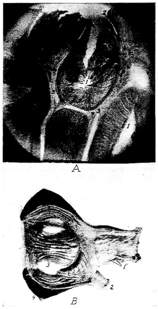
Fig. 32.—Gizzard and stomach of the fowl. A. 1, Alveolar glands. 2, Mucous membrane of the inner surface of the proventriculus showing the tubular glands. 3, Connective tissue between the alveolar glands. 4, An artery. 5, Muscularis mucosa. B. Photograph of the proventriculus and gizzard. 1, Proventriculus showing stomata of glands. 2, End of duodenum. 3, Gizzard showing hornified epithelium formed into grooves. 4, The heavy muscular walls.
145The submucosa, connects the mucous and muscular coats and the muscularis layer throws the mucous membrane into folds.
Outside the muscularis mucosa there is another layer (Fig. 32, A), of simple tubular glands, grouped in lobules, and lined by cuboidal cells and separated from each other by clefts. These tubular glands converge toward the center and open into the same cavity.
The middle muscular coat can be divided into three layers: two thin, longitudinal layers, and a thick, circular layer interposed between the other two.
Function.—The function of the proventriculus appears to be to soak the food with a secretion. The secretion from the proventriculus is similar to that produced by the fundus glands of the stomach of mammals. It contains acid and a ferment-pepsin.
The Gizzard (Fig. 31, No. 18). Location.—The gizzard, or muscular stomach, occupies a portion of the central part of the abdominal cavity (Fig. 33, No. 4). It lies slightly to the left and just behind the liver, the proventriculus, and the spleen, and rests upon a mass of intestines. The gizzard communicates at its anterior portion with the proventriculus and with the duodenum. These openings are close together. The gizzards of a large number of hens of average size averaged in weight as follows: full, 0.215 pound; empty, 0.126 pound. The gizzard stands perpendicularly and somewhat obliquely in the abdominal cavity.
Shape.—In shape the gizzard is roundish, flattened laterally.
Structure.—The walls of the gizzard are very thick and are made up of three coats: mucous, muscular, and serous.
The cavity of the gizzard is covered by a thick skin-like structure possessing a heavy stratified squamous epithelial layer which is thrown somewhat into ridges (Fig. 32, B). This membrane becomes thinnest near the edges.
The mucous membrane, being cornified and readily detachable, is by some anatomists considered as a special membrane (Fig. 37, B).
At the pyloric opening there is a valve formed by a fold of the mucous membrane, which prevents grit and large particles of food from passing out of the gizzard.
146
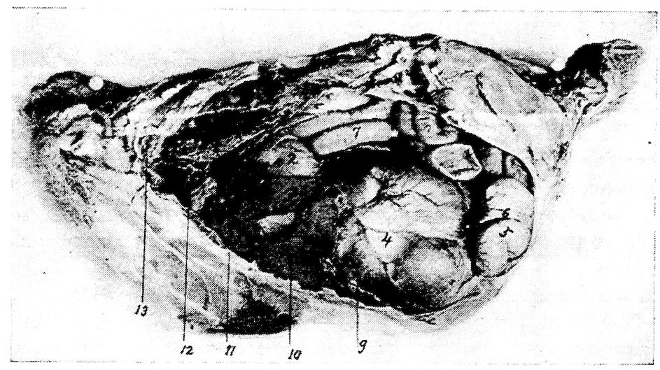
Fig. 33.—View of viscera of the left side of a hen. 1, The base of the heart. 2, Proventriculus. 3, Left lobe of the liver. 4, Gizzard. 5, End of the duodenal loop. 6, Pancreas. 7, Free portion of the small intestine. 8, Lungs. 9, Diaphragm.
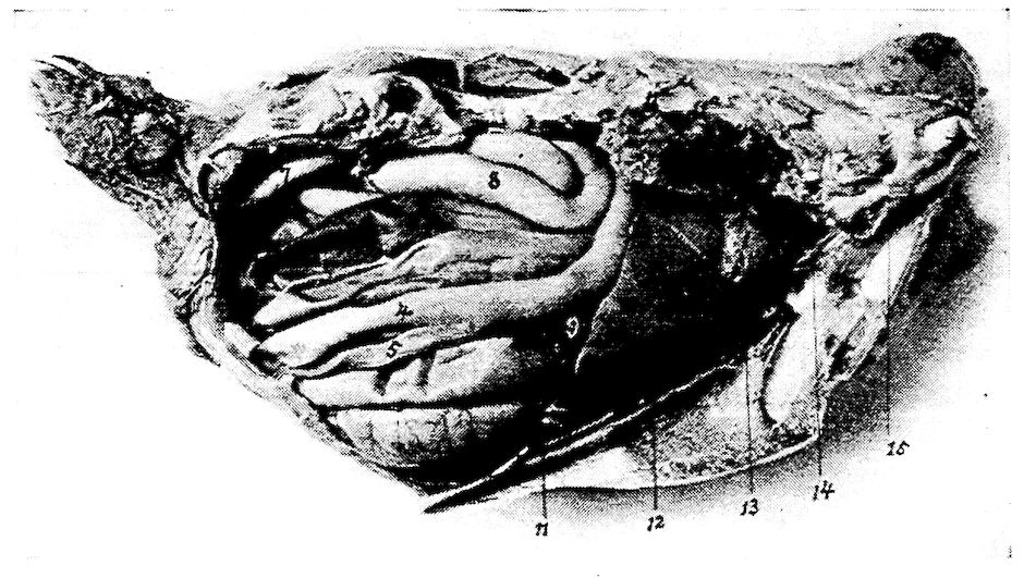
Fig. 34.—View of the viscera of the right side of a hen. 1, Base of the heart. 2, Lungs. 3, Right lobe of the liver. 4, Duodenal loop. 5, Pancreas. 6, Cæcum. 7, Large intestine or rectum. 8, Free portion of the floating small intestine. 9, Gall-bladder. 10, Right kidney. 11, The ribs. Note the lungs pushing up between them.
On each side it has a powerful fleshy muscle, the muscularis lateralis. These musculares laterales are hemispherical, consist of very closely packed fibers extending transversely, and are attached to strong anterior and posterior ligamentous tendons. They are joined at the edge of the organ by a strong aponeurosis. The muscular fibers are red but do not possess cross striations; they are of the smooth type. The muscularis intermedii, thinner and less 147developed than the musculares laterales, occurs on each of the anterior and the posterior parts of the gizzard.
The serous coat covers over the greater part of the external surface of the gizzard and closely adheres to the muscular coat.
Function.—The hard callous pads of the gizzard, operated by the powerful muscles above described, together with grit, act like mill stones and make reduction to fineness very complete.
At the posterior part there is a sacculated portion containing glands of the long tubular type which secrete a fluid ferment similar to that secreted by the glands of the pyloric portion of the stomach of mammals. These glands also exist in a small band near the entrance of the gizzard. Here the food is mixed with strongly acid secretion containing pepsin which makes gastric digestion perfect.
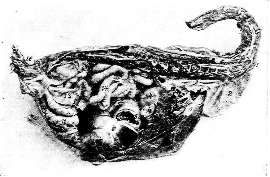
Fig. 35.—A median antero-posterior section through the body of a 1-pound pullet. 1, First portion of the esophagus. 2, Crop. 3, Second portion of the intestine. 4, Proventriculus. 5, Gizzard. 6, Spleen. 7, Liver. 8, Heart. 9, Point where duodenum was severed from gizzard. 10, Point where duodenum was severed. 11, Duodenum. 12, Pancreas. 13, Cæcum. 14, Floating small intestine. 15, Ovary. 16, Oil sac.
The Small Intestine (Fig. 31, No. 8 and 9).—The small intestine, intestinum tenue, is the tube which connects the gizzard with the large intestine. It is divided into two parts, the duodenum and the free portion. Of the three parts as considered in the mammalian 148intestine, only the first, the duodenum, can be distinguished. There is no demarcation between the jejunum and the ileum. The jejunum and the ileum or that part which represents these sections, are in coils suspended from the free border of the mesentery the other border of which is, in turn, attached to the dorsal wall (Fig. 64). The length of the small intestine in a hen of average size is about 61.7 inches.
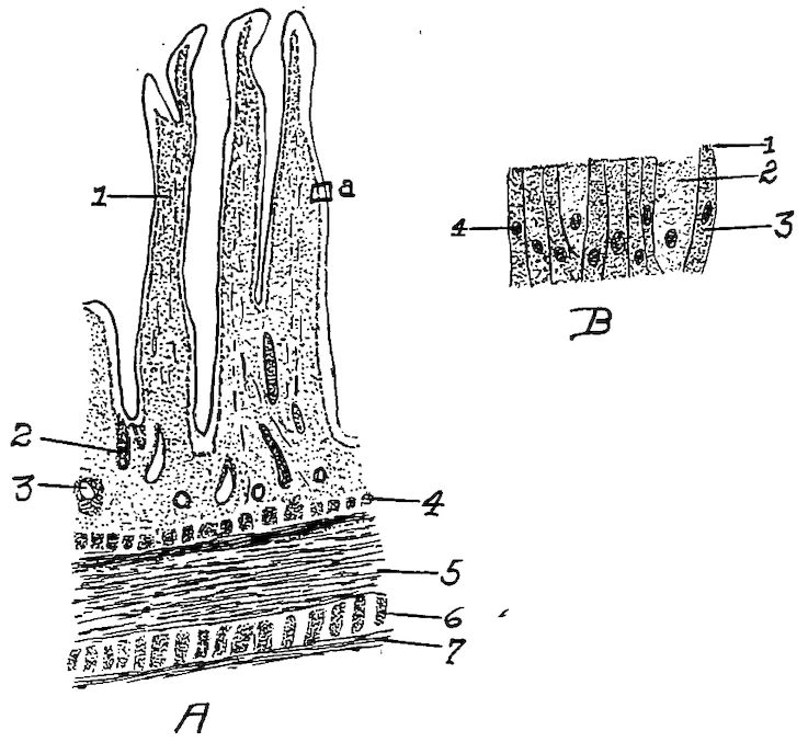
Fig. 36. A. Section of the duodenum of the fowl. 1, Villus. 2, Gland. 3, Mass of lymphoid tissue. 4, Muscularis mucosa. 5, Longitudinal layer. 6, Circular muscular layer. 7, Serous layer. B. A section from A at a. 1, The striated free border of the cells. 2, Goblet cell. 3, Columnar cell. 4, Nucleus of cell.
The wall of the small intestine is provided with four coats, as follows: a mucous, submucous, muscular coat made up of two layers—the outer, longitudinal and the inner circular layer, and an external serous.
149
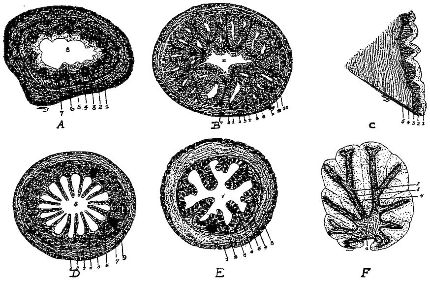
Fig. 37.—Histological studies of various anatomical parts. A. A transverse section of the first portion of the esophagus of a fowl. 1, The outer longitudinal muscular layer. 2, The circular muscular layer. 3, The submucosa. 4, The muscularis mucosa. 5, Stroma. 6, Epithelial layer. 7, The lenticular glands. 8, The lumen. B. A transverse section of the proventriculus. 1, The outer longitudinal muscular layer. 2, The middle muscular layer. 3, The inner longitudinal muscular layer. 4, Stroma. 5, The muscularis mucosa. 6, The submucosa. 7, Stroma. 8, Tubular glands. 9, Tubulo-alveolar glands. 10, A tubulo-alveolar gland with the tubular glands cut transversely, 11, The lumen of the proventriculus. C. A section of the inner wall of the gizzard. 1, The hyaline mucous membrane. 2, Branched tubular glands. 3, Submucosa. 4, Muscle. 5, A connective tissue septum. D. A transverse section of the small intestines. 1, The outer longitudinal muscular layer. 2, The inner circular muscular layer. 3, The muscularis mucosa. 4, The stroma. 5, Brunner’s glands. 6, A villus. 7, Mass of lymphoid tissue. E. A transverse section through the cecum. 1, The outer longitudinal muscular layer. 2, The inner circular muscular layer. 3, The submucosa. 4, The muscularis mucosa. 5, The stroma. 6, Tubular glands. 7, The lumen. F. The cerebellum showing the arbo-vitæ. 1, White, fiber portion. 2, The granular layer. 3, The layer of Purkinje cells. 4, The molecular layer.
The mucous membrane which lines the intestine is thick, soft, and highly vascular. It has a velvety appearance, due to numerous long, thin projecting villi. The villi (Fig. 36, A) are concerned in the absorption of the digested food, absorbing principally the emulsified fats. Each villus is covered with a single layer of high columnar epithelial cells. Some of these, the so-called goblet cells, provide mucin which lubricates the mucous surface. These cells are found in all mucous surfaces and prevent the surface from becoming dry. There are a few goblet cells near the summits of the villi. Openings of simple intestinal tubular glands the duodenal glands, or the glands formerly known as Brunner’s glands, are located between the villi. These glands secrete the succus entericus, or intestinal juice. These openings, or stomata, are lined with granular cells. The 150reaction of the contents of the small intestine is strongly acid, but gradually less so in proportion to the distance down the intestinal tract until the cæca are reached, where the reaction is found to be faintly acid, neutral, or slightly alkaline.
Function.—The function of the small intestine is that of digestion and absorption.
The Duodenum. Location and Shape.—The duodenum, a small tube, originates from the gizzard about ½ inch to the right of the entrance of the proventriculus. Extending from left to right, it passes under and behind the gizzard along the inner side of the right abdominal wall backward to the posterior portion of the wall, and a trifle more than half way toward the left side, where the loop rests. This loop, the duodenal loop, is about 5 inches in length (Fig. 31, No. 8). The two branches of the loop the first and the second portions or the descending and ascending limbs, are loosely held by connective tissue, and have the pancreas lodged between them.
The Free Portion of the Small Intestine. Location.—That section of the small intestine following the duodenum is called the free portion of the small intestine and occupies the space between the abdominal air-sac and the median line of the abdominal cavity. It is disposed in coils and is suspended from the dorsal wall of the abdomen by a thin membrane, the mesentery.
The bile ducts enter the small intestine about 14 inches from its point of origin. The pancreas also pours its contents into the small intestine.
The Large Intestine (Fig. 31, No. 12). Location.—The large intestine extends in a straight line along the inferior border of the vertebral column communicating anteriorly with the small intestine, and the cæca and posteriorly with the cloaca.
Shape.—The diameter of the large intestine is approximately twice that of the small intestine. In fowls of average size its mean length is 4.61 inches. The large intestine has sometimes been spoken of as the rectum, or straight gut.
Structure.—Like the small intestine the large intestine has four coats: an inner mucous, a submucous, a middle muscular and an outer serous. The folds of mucous membrane of the large intestine have tubular glands lined with columnar cells (Fig. 37, A, No. 1).
Function.—The large intestine is similar in function to that of the small intestine, in that digestion and absorption may take place within it.
151The Cæca (Fig. 31, No. 11). Location.—The fowl has two cæca extending forward from their point of origin at the juncture of the small and large intestine.
Shape.—The cæca average 7.61 inches in length. They are large in caliber toward the blind extremity and are constricted near their origin.
Structure.—The parietal coats are continued from the small intestine.
Ebarth has described an elevated body in the cæcum, which is located about 4 millimeters from the opening and is composed entirely of lymphoid tissue.
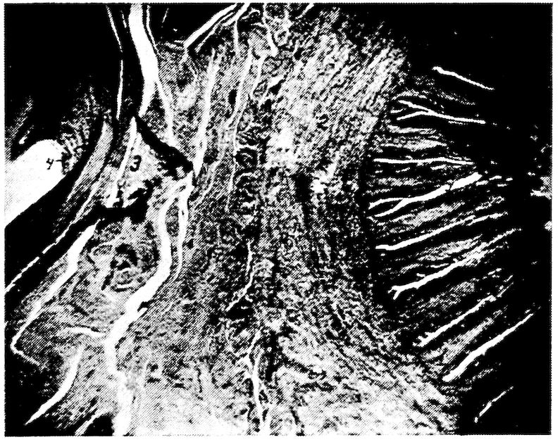
Fig. 37A.—Photomicrograph of a transverse section of large intestine and oviduct just anterior to the cloaca, showing the tubular glands of the large intestines, mucosa of oviduct with intervening structures. 1, The tubular glands of large intestine, or rectum. 2, Muscular wall of intestine. 3, Wall of the oviduct. 4, Mucosa of the oviduct.
The cæca are usually partly filled with a soft pultaceous material of a pasty consistency.
The Cloaca (Fig. 31, No. 13). Location.—The rectum opens by a circular, valvular orifice into the dilated portion just in front of the anus, which dilatation is called the cloaca.
Shape.—The cloaca is saccular in shape.
Structure.—The cloaca is divided into two portions, the coprodeumal and the urodeumal. That portion of the cloaca into which the intestine empties is called the coprodeum; and the ureter and 152oviduct empty into the urodeum. The seminiferous tubules, carrying the semen from the testes in the male empty in teatlike projections on the cloacal mucous membrane into the urodeumal portion. The cloacal walls are similar in structure to the large intestines.
On the dorsal wall of the cloaca between it and the spine, is a small sac, called the bursa of Fabricius, which has a duct communicating with the cloaca. The mucous membrane of this sac is thrown into folds and is studded with glands. The bursa of Fabricius is larger in the young than in the adult bird. It apparently atrophies as the bird becomes older. When the bird is four months old this bursa is best developed, and at this age it may be as large as 2 or 3 centimeters in diameter.
Function.—The function of the cloaca is to give passage way to the feces, the urine, and the egg and to act as an organ of copulation.
Course of the Food.—The food first enters the mouth, after being picked up by aid of the beak. From here it passes through the pharynx and first portion of the esophagus to the crop, without mastication, as the bird is not provided with teeth. The food is passed from the crop by aid of its muscular walls as needed; thence through the second portion of the esophagus to the proventriculus, an expansion in the digestive tube just before it terminates in the gizzard. The glands of the proventriculus produce a secretion in which the food is soaked before passing into the gizzard. The gizzard is provided with strong muscular walls which, by aid of grit, thoroughly reduce the food to fineness. From the gizzard the food passes through the first portion of the small intestine, where it is subjected to the action of the bile from the liver, the pancreatic juice from the pancreas, and of the succus entericus from the glands of the intestinal wall. The food is then passed into the cæca. The indigestible portion of the food passes from the cæca through the large intestine, or rectum, to the cloaca and thence to the external world.
The digestive functions of the bird are very potent and rapid. This compensates for the waste caused by their extensive, frequent, and energetic motions, and is in accordance with the rapidity of their circulation and their high state of irritability.
THE ACCESSORY ORGANS OF DIGESTION
The accessory organs of digestion are the liver, pancreas, and some anatomists include also the spleen. The first two manufacture 153fluids containing ferments which aid in splitting or digesting the food.
The Liver (Figs. 31, 33 and 34). Location.—The liver, hepar, lies ventrally and posteriorly to the heart. It is related anteriorly with the diaphragm, inferiorly with the sternum, posteriorly with the gizzard and intestine, and superiorly with the ovary, oviduct and proventriculus and laterally with the abdominal wall.
Shape.—The liver is a voluminous deep livid brown gland, soft and friable in texture. It is divided into two principal lobes, a right and a left.
The right lobe is larger than the left. In the hen of average size the liver weighs 35 grams. The parietal surface is convex and smooth. The surface which lies against the viscera is irregularly concave. The visceral surface furnishes exit for the bile duct and passage for the nerves and blood-vessels. This part is called the porta.
The left lobe may be cleft from below so deeply as to form two lobes on that side.
Structure.—Each lobe is covered by a double serous membrane, one closely adherent, the other surrounding the structure loosely. These tunics, which are reflections of the peritoneum, are continued from the base of the liver, over both the anterior, and the posterior surface. The loose layer is formed by the air cells surrounding the lobes. The thin border of the liver is usually free.
The two lobes of the liver are connected by a narrow isthmus of liver tissue. Occasionally there is a bird in which there occurs a lobus Spigelii located at the posterior of the liver between the two principal lobes.
The apex of the heart sacculates the diaphragm backward, so that part of this apex lies between the right and the left lobes (Fig. 43, No. 6 and 7). A ligament, the falciform, extends from the apex of the pericardial membrane, and attaching it rather firmly to the central connective tissue, or interlobar ligament. This ligament also has attachments to the inner surface of the sternum. The broad ligament of the liver is formed posteriorly by a fold of the peritoneum.
The interlobar, or principal, ligament of the liver is formed by a large and strong duplicature of the peritoneum, which makes a longitudinal division in the abdominal cavity similar to the lateral division made by the thoracic mediastinum in mammals. It 154is reflected upon the pericardium from the linea alba and the middle line of the sternum, and passes deeply into the interspace of the lobes of the liver. It is attached to these lobes throughout their whole length and connects them below to one side of the gizzard. The lateral and posterior part of the liver attach to the adjacent air cells, and the whole viscus is thus kept fixed in its position during rapid and violent movements of the bird.
The remains of the umbilical veins are traceable within the duplicature of the membranes forming the septum. These remains thus represent the round ligament of mammals.
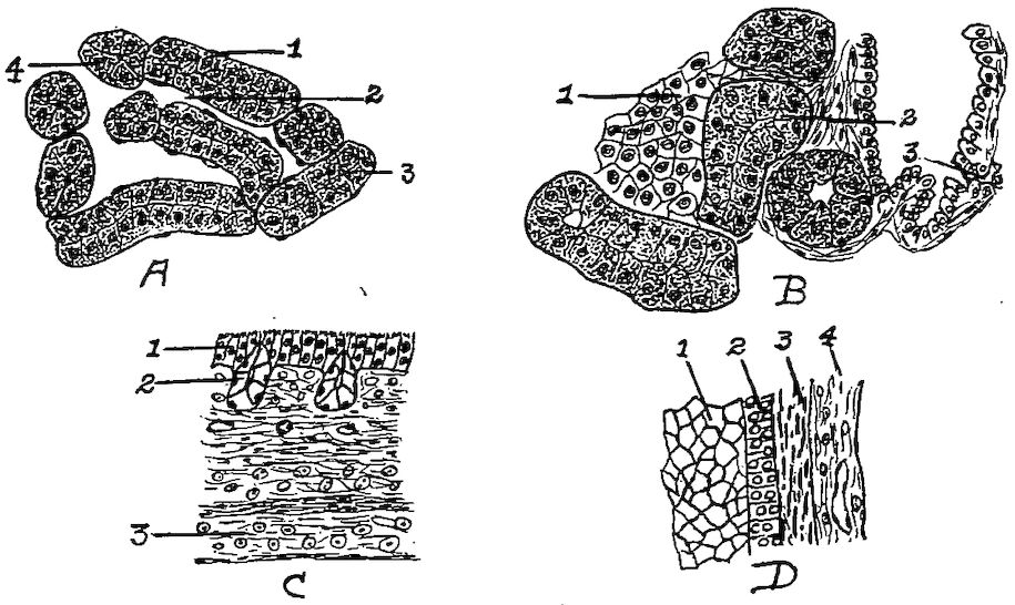
Fig. 38.—Cellular structure of liver, pancreas, and trachea. A. Liver. 1, Liver cells. 2, Sinusoid. 4, Nucleus of cells. B. Pancreas. 1, Island of Langerhans. 2, Alveolar cells. 3, Duct. C. Trachea. 1, Ciliated epithelia. 2, Glands. 3, Hyaline cartilage. D. Section of wall of ovum at Fig. 57 letter d. 1, Yolk. 2, Granular membrane. 3, Theca. 4, Blood-vessel.
A microscopic study of the liver of the fowl shows a compact mass of liver cells polyhedral in shape, with large nuclei (Fig. 38, A). The liver tissue differs from that of mammals in that there is no clearly outlined lobular arrangement; neither is the outline of the individual cell so well marked. The parenchymatous portion is made up of columns of liver tissue. These columns anastomose and show narrow channels between. They are best seen in the young chick.
Function.—One of the functions of the liver is to secrete bile. A gall-bladder, which receives part of the bile secreted by the right lobe of the liver, is located on the posterior face of this lobe. Extending 155from this gall-bladder there is a duct, the cystic duct, which empties into the small intestine toward the extremity of the second branch of the duodenal loop. Another duct called the hepatic duct, proceeds directly from the two lobes of the liver and empties into the intestine just in front of the cystic duct.
The Pancreas (Fig. 31, No. 17). Location and Shape.—The pancreas, an organ lying in the duodenal loop is a yellowish-white, lobulated gland, elongated in shape. Its average length in fowls of average size is 4.96 inches, and the average weight about 0.008 pound. The pancreas is divided into lobes, which in turn are divided into lobules.
Structure.—The pancreas has a supporting connective tissue. The lobules are made up of small alveolar glands, which are lined with columnar epithelial cells. The ducts leading from the alveoli are small; these unite to form larger ducts in which the epithelium is taller. Between the alveoli throughout the pancreas there are clusters of polyhedral cells which form the islands of Langerhans. These islands are said to produce an internal secretion, or hormone, which is absorbed by the blood or lymph capillaries and thus enters the circulation (Fig. 38, B).
Function.—The function of the pancreas is to secrete a fluid containing digestive ferments.
The Spleen (Fig. 31, No. 21). Location.—The spleen, or lien, lies in a triangle formed by the proventriculus, the liver, and the gizzard.
Shape.—It is a reddish-brown body shaped like a buckeye, is small in size, weighing only about 0.005 pound.
Structure.—The outer surface of the spleen is covered by a reflection of the peritoneum. After this covering is removed, there is observed a firm, white, fibrous layer, the cortical portion. This covering sends into the interior small and large trabeculæ, forming a framework and dividing it into acini, or compartments. The spaces are filled with a dark red parenchymatous material called splenic pulp. The framework, in both deep and surface portions, are found elastic fibers and smooth muscular fibers.
The spleen is essentially a lymphatic organ, its peculiar structure depending largely upon the arrangement of the blood-vessels. Compact lymphatic tissue occurs in the spleen in spherical, oval, or cylindrical collections of closely packed lymphoid cells. These masses are known as the Malpighian bodies, or splenic corpuscles. 156They are distributed throughout the splenic pulp. Each splenic corpuscle contains one or more small arteries. These extend near the periphery of the corpuscle and more rarely in the center.
The splenic artery passes in and the splenic veins out at the hilum which is located on the concave, or attached, side of the spleen. The splenic artery, upon entering the organ, at once branches, the trabeculæ forming a support for the vessels.
After the arteries have entered the hilum, as stated above, they divide into many branches which follow the septa of the connective tissue. At first the arteries are accompanied by branches of the splenic veins. Soon, however, the arteries leave the veins and the septa, and penetrate the splenic pulp. In the splenic pulp the adventitia of the smaller arteries assume the character of reticular tissue, and become infiltrated with lymphoid cells. This infiltration forms masses which are called the splenic corpuscles, or Malpighian bodies. The terminal arteries break up into capillaries, which still retain an adventitia, and empty into border spaces, or sinuses, sometimes spoken of as ampullæ. These sinuses in turn empty into cavernous sinuses of the splenic pulp. From these are finally formed the venules; and the collections of venules form the splenic veins through which the blood gains exit from the spleen.
The Abdominal and Pelvic Cavities.—In birds the abdominal cavity is divided into two smaller cavities by a fibrous septum. The anterior cavity representing the abdominal contains the liver, and the other representing the pelvic contains the gizzard, intestines and oviduct.
The Peritoneum and the Mesentery.—The abdominal and pelvic cavities are lined by the peritoneum. Like all serous membranes this is composed of a parietal and a visceral portion, which together form a complete sac, with the organs it covers situated on the outer side. The peritoneum like other serous membranes consists of a mesothelial and a submesothelial portion, the cells of the former being arranged in a single layer. Since a serous membrane is so arranged as to line a closed cavity, and at the same time to cover its contents, it follows that the entire membrane must be a closed sac, the mesothelial layer being on the inside; such a sac is called a serous sac, or cavity. Synovial membranes are also regarded as a variety of serous membranes. The fold, or layer, of the membrane which lines the cavity is called the parietal, that which covers the greater part of the organs contained therein is called the 157visceral portion; the two surfaces contacting, and gliding upon each other, are lubricated by a fluid secretion contained in the sac; hence one use of these membranes is to prevent friction between the walls of cavities and the organs contained therein. Serous membranes line the abdominal cavity, pericardium, cavities of the heart and is continuous throughout the vascular structures.
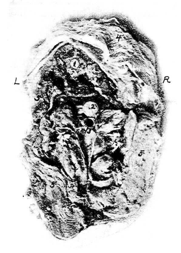
Fig. 39.—A transverse section of body of hen through 15, Fig. 34. R, Right side. L, Left side. 1, Spinal cord. 2, Esophagus. 3, Trachea. 4, Skin. 5, Pectoral muscles.
The serous membrane besides covering the external surface of the viscera, double folds pass from one organ to another, or from an organ to the parietes of the cavity. These double folds of the peritoneum are known as ligaments, or as mesenteries. In ligaments the two folds are strengthened by an interposed layer of fibro-elastic tissue. A mesentery is a broad, double fold of peritoneum, attached to the abdominal parietes above, and containing a portion of the intestine in its free or remote extremity. Between its folds we find blood-vessels, nerves, and lymphatics or lacteals, hence it permits 158vascular and nervous communications with the organ attached to it. The free portion of the small intestine is attached to the free margin of the mesentery.
THE RELATIONS OF THE VISCERAL ORGANS OF THE DOMESTIC FOWL
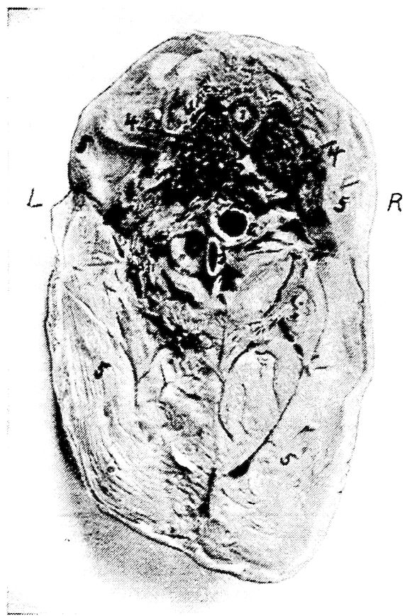
Fig. 40.—Transverse section through the body of a hen through 14, Fig. 34. R, Right side. L, Left side. 1, Spinal cord. 2, Esophagus. 3, Trachea near inferior larynx. 4, Lungs. 5, Pectoral muscles.
Figure 33 shows a fowl with the left abdominal wall and the left thoracic wall removed. No. 1 in this figure shows the base of the heart in front of the left lobe of the liver. Above the liver is the proventriculus; above this, the diaphragm; and above the diaphragm and the base of the heart the left lung occupying the superior part of the thoracic cavity, and that there is no distinct pleural sac, as in mammals but that the lung pushes out between the ribs, thus pressing against the ribs on the inner and the lateral sides. The gizzard is back of the liver, to the left side of the abdominal cavity, 159and beneath and in front of the duodenal loop. The small intestine from this side is above the gizzard. Supero-anterior to the gizzard is the blind end of the cæcum. The pancreas is within the duodenal loop.
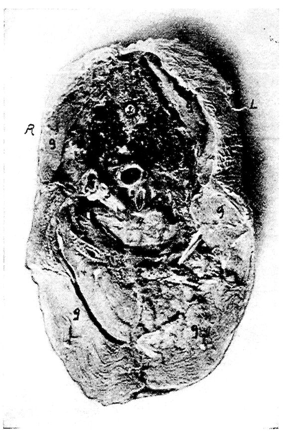
Fig. 41.—Transverse section through the body of a hen at 12, Fig. 33. R, Right side. L, Left side. 1, Spinal cord. 2, Esophagus. 3, Inferior larynx. 4, Base of the heart. 5, Aorta. 6, Vena cava. 7, Lungs. 8, Skin. 9, Pectoral muscles.
Figure 34 shows the viscera from the right side after the removal of the right abdominal and the right thoracic wall. The base of the heart is in front of the right lobe of the liver. Above these the right lung occupies the upper part of the thoracic cavity as in the preceding illustration. Just back of this is the anterior lobe of the kidney. The gall-bladder is observed at No. 9 on the right lobe of the liver. Just inferior to a longitudinal central line is the duodenal loop, between the limbs of which is seen the pancreas. Above this loop the cæca are located. The gizzard is not visible 160from the right side; on this side posterior to the liver is the small intestine. Superior to this at No. 7 the rectum.
The relative position of the visceral organs in the median line is observed in Fig. 35. No. 1 of this figure shows the stump of the first portion of the esophagus, and No. 2, the left wall of the crop. Following this, and located just below the vertebræ is the second portion of the esophagus. The major portion of the crop is located on the right side.
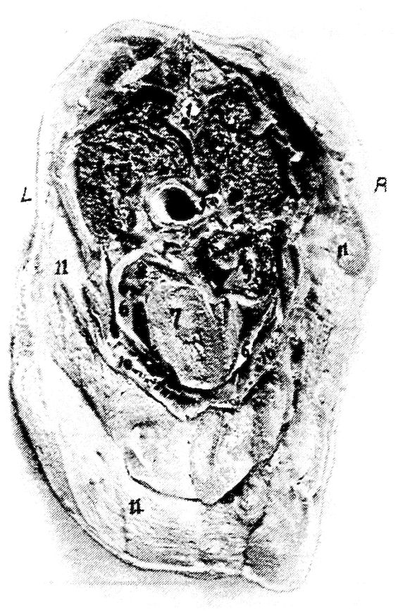
Fig. 42.—Transverse section through the body of a hen at 11, Fig. 33. R, Right side. L, Left side. 1, Spinal cord. 2, Vertebra. 3, Spinous process of vertebra. 4, Lungs. 5, Esophagus. 6, Pericardial sac. 7, Sectioned surface of heart. 8, Auricle of heart. 9, Blood clot in right auricle. 10, Section of sternum. 11, Pectoral muscles.
It will be noted that the second portion of the esophagus passes over the base of the heart and the superior part of the liver, and then terminates in the proventriculus. The proventriculus extends downward and empties into the gizzard. This organ lies antero-laterally to the gizzard, supero-posterior to the liver, and to the left of the spleen. The spleen lies in a triangle formed by the liver, the proventriculus, 161and the gizzard. The heart is noted to lie supero-anterior to the liver, and between the anterior portion of the fissure formed by the right and the left lobe. The ovary is located back of the diaphragm at the anterior end of the kidney and below the inferior surface of the bodies of the vertebræ. The bulk of the floating portion of the small intestine is located above the gizzard.
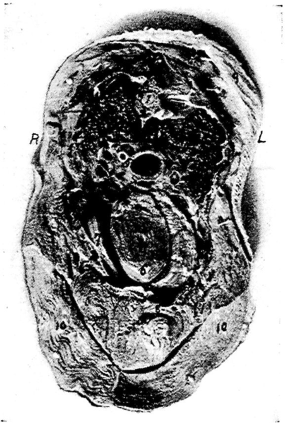
Fig. 43.—Transverse section through the body of a hen at 13, Fig. 34. R, Right side. L, Left side. 1, Spinal cord. 2, Body of vertebra. 3, Superior spinous process of vertebra. 4, Lungs. 5, Esophagus. 6, Heart. 7, Right and left lobes of liver. 8, Sternum. 9, Skin. 10, Pectoral muscles.
A transverse anterior section of a normal laying hen is shown in Fig. 39, the section being made at 15 of Fig. 34. At this point the esophagus lies centrally and above the trachea. Figure 40 shows a transverse anterior section through the thoracic region at 14 of Fig. 34. At this point the esophagus is slightly to the right of and is superior to the trachea. At this level the apex of the lung is sectioned. This is in the region of the cervical air-sac. Figure 41 shows a posterior section at 12 of Fig. 33. The 162esophagus here is above and to the right of the inferior larynx, and directly below and between the lungs. The inferior larynx is above the base of the heart.
Figure 42 shows a transverse anterior section made at 11 of Fig. 33. It shows that at this level the esophagus is centrally located and passes over the base of the heart. The heart occupies the lower portion of the thorax and the lungs the upper. Figure 43 shows a photograph of a transverse anterior section of the body made at 13 of Fig. 34. At this level the apex of the heart lies within the anterior fissure of the liver. The sectioned portion of the heart shows the lower portion of the ventricle.
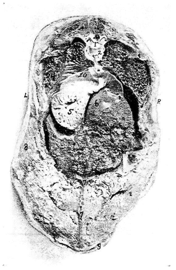
Fig. 44.—Transverse section through the body of a hen at 12, Fig. 34. R, Right side. L, Left side. 1, Spinal cord. 2, Articular surface of vertebral segment. 3, End of rib. 4, Lungs. 5, Proventriculus. 6, Liver showing Glisson’s capsule. 7, Sternum. 8, Pectoral muscles. 9, Skin.
163
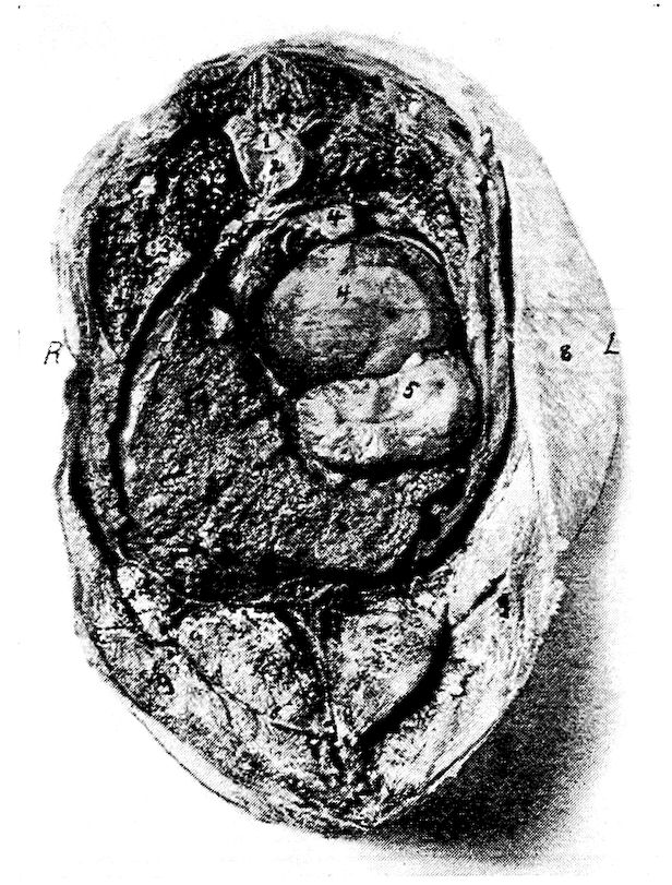
Fig. 45.—Transverse section through the body of a hen at 10, Fig. 33. R, Right side. L, Left side. 1, Spinal cord. 2, Body of vertebra. 3, Lungs. 4, Ova. 5, Proventriculus. 6, Liver. 7, Sternum. 8, Skin. 9, Pectoral muscles.
In this figure the lungs show, on the sectioned surface, the ends of some of the larger bronchi. The esophagus is located above the heart. Showing the relations of the visceral organs back of the heart girdle, photograph number 44, gives an anterior section made at 12, Fig. 33. The lungs are spread out occupying the posterior thoracic region and below at No. 10 is the diaphragm. The diaphragm does not appear to have that rigidity and firmness of position as in mammals. It is rather rudimentary. Below the left lung and above the left lobe of the liver is the proventriculus. Note that the viscus is empty and that the mucous membrane is thrown into folds. At this point the liver occupies much of the abdominal cavity. Figure 45 is a view of an anterior section made at 10, 164Fig. 33. In this section the lungs are decreasing in caliber. The liver occupies much of the space in the lower right abdominal quadrant, and above and to the right is the ovary with many of the ova developing yolks. Below No. 4 which is a developing yolk, is the proventriculus. Figure 46 is a view of an anterior section made at 9, Fig. 33. At this level are shown sectioned surfaces of the kidneys, which lie on either side of the spinal column. Below the spinal column and occupying the left upper quadrant is the ovary containing ova in the process of developing yolks. To the right is the sectioned surface of one of the cæca and below this and on the right side is the sectioned ends of many of the loops of the floating portion of the small intestine. Occupying the left lower quadrant is the sectioned surface of the gizzard and on the abdominal floor and to the right of the gizzard, the posterior end of the right lobe of the liver.
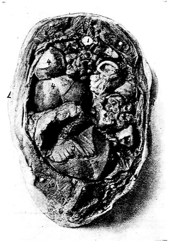
Fig. 46.—A transverse section through the body of a hen at 9, Fig. 33. R, Right side. L, Left side. 1, Spinal cord. 2, Vertebra. 3, Kidneys. 4, Ovary. 5, Cæcum. 6, Small intestine. 7, Gizzard. 8, Right lobe of liver. 9, Skin. 10, Pectoral muscles.
165
THE RELATIONS OF THE VISCERAL ORGANS OF THE BABY CHICK
There is approximately 47 per cent. of the yolk retained in the abdominal yolk sac of the baby chick at hatching. Figure 47 shows a photograph of a longitudinal section through a baby chick. This figure shows all the posterior portion of the abdominal cavity occupied with abdominal yolk. The abdominal viscera are pushed forward, and as the yolk is gradually absorbed the visceral organs gradually occupy their normal position.
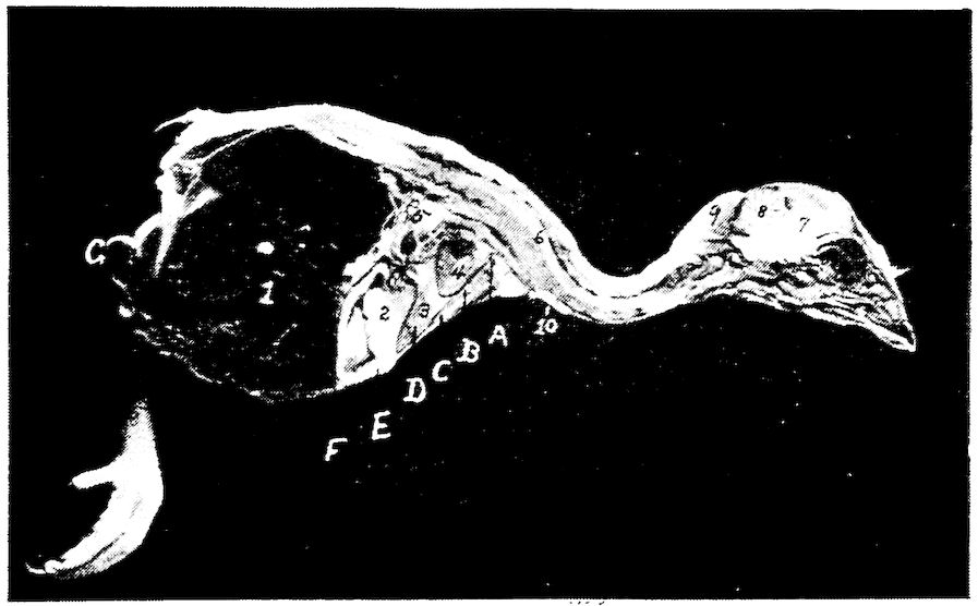
Fig. 47.—An antero-posterior section through the body of a baby chick just hatched. 1, Abdominal yolk sac. 2, Gizzard. 3, Liver. 4, Heart. 5, Intestines. 6, Spinal cord. 7, Cerebrum. 8, Cerebellum. 9, Fat in the post-occipital region. 10, The thymus gland.
166
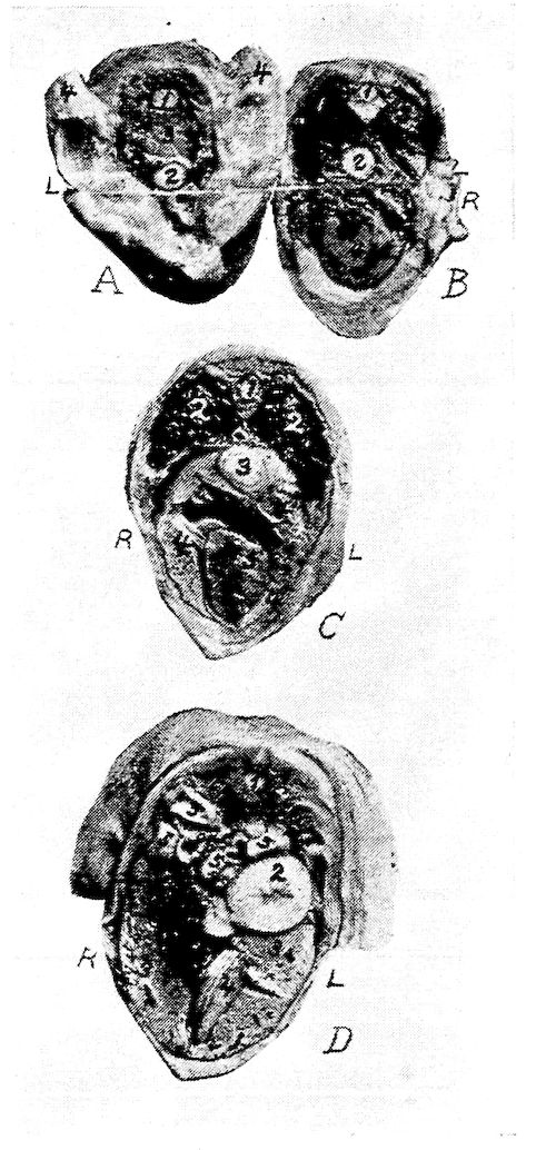
Fig. 48.—Transverse sections of the body of a baby chick at hatching. R, Right side. L, Left side. A. A transverse section at A, Fig. 50. 1, Spinal cord. 2, Esophagus. 3, Entrance to thorax. 4, Stubs of the wings. B. A transverse section at B, Fig. 50. 1, Spinal cord. 2, Esophagus. 3, Lungs. 4, Heart. C. A transverse section through the body at C, Fig. 50. 1, Spinal cord. 2, Lungs. 3, Esophagus. 4, Liver. 5, Heart. D. A transverse section at D, Fig. 50. 1, Spinal cord. 2, Proventriculus. 3, Liver. 4, Gizzard. 5, Intestine. 6, Kidneys. 7, Gall-bladder.
167
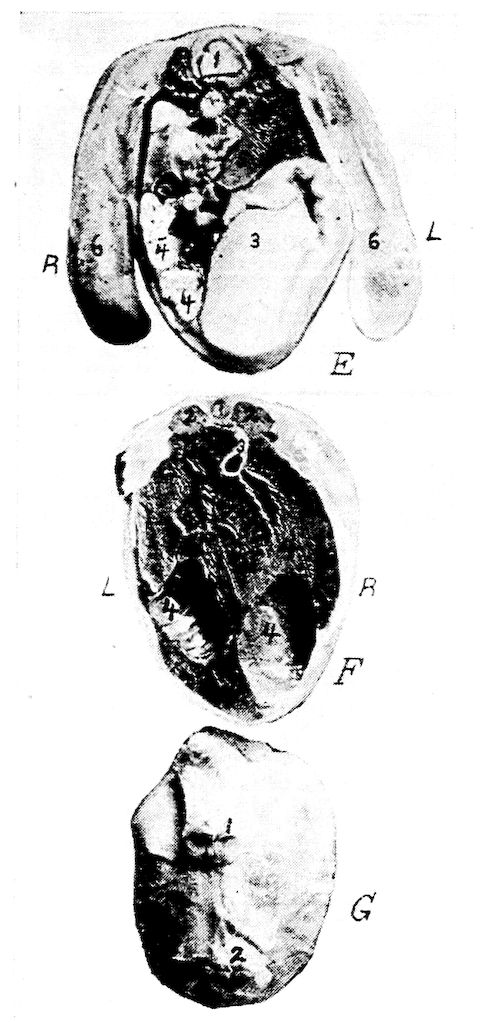
Fig. 49.—Transverse section through the body of a baby chick. E at E, Fig. 50. 1, Spinal cord. 2, Kidneys. 3, Gizzard. 4, Intestines. 5, Unabsorbed yolk. 6, Stubs of legs. F. A section at F, Fig. 50. 1, Spinal cord. 2, Kidneys. 3, Intestine. 4, Unabsorbed yolk. G. A section at G, Fig. 50. 1, Anus. 2, Umbilicus.
A section through this body Fig. 47 at A is shown in Fig. 48, A. At this point the esophagus appears below the vertebral column. Figure 48, B, a section through the body at Fig. 47, B, shows the apex of the lungs. In a median line and below the lungs is the esophagus. Note the mucous membrane thrown into folds. Here the heart is sectioned, showing both auricles and both ventricles. This is a view looking forward. Figure 48, C, is a view of a posterior section made at Fig. 47, C. It shows the same relations of the esophagus and lungs, but shows at this level both the right and the left lobe of the liver. Between these lobes we note the sectioned apex of the heart. Figure 48, D shows a posterior section at Fig. 16847, D. At this level the kidneys are above and infero-laterally to the spinal column. The intestines are below the kidneys; and on the left side, between the kidneys and left lobe of the liver, and near the abdominal wall, is the proventriculus. Occupying the major portion of the inferior abdominal cavity are the right and the left lobe of the liver, and between these the anterior border of the gizzard. Note the gizzard sitting at an angle inclining toward the left side. Figure 49, E shows a section at Fig. 47, E. Here the gizzard occupies the left lower abdominal quadrant. To the right are the intestines; and directly above the gizzard is a small portion of the anterior end of the abdominal yolk sac. Infero-laterally are the kidneys. Figure 49, F, shows the kidneys similarly located as in the preceding; and just below is the rectum suspended by the mesentery. The rest of the cavity is occupied by the abdominal yolk.
169