MYOLOGY
Reading edition
Kinds and Structure of Muscles.—Muscles are highly specialized structures which have the property of contractility when stimulated, and thus produce motion. Muscular tissue is sometimes called flesh. Two kinds of muscles are recognized; muscles of locomotion and visceral muscles.
The muscles of locomotion may be in masses of different shape attached to the skin, the dermal, or to the skin and skeletal structure, the dermo-osseous, or from one bone to another, the skeletal. The visceral muscles form sheets and make up a portion of the wall of many of the hollow organs, such as the intestines, the stomach, the gizzard, the esophagus, and the blood-vessels. A special type of muscle forms the heart.
When classified with reference to structure, muscular tissue is divided into three types, as follows: voluntary-striated, involuntary, and involuntary-striated. The microscopic examination of each of these types of muscle shows it to be made up of fibers, these fibers to be made up of muscle cells, the muscle cell to be inclosed in a delicate tubular sheath, or membrane, called the sarcolemma. This membrane, tough and elastic, isolates each fiber. The bundles of fibers, called fasciculi, are surrounded by a fibrous sheath, which is called the perimysium internum; and the entire muscle has likewise an investing sheath of connective tissue, called the perimysium externum. The muscular cells show a longitudinal striation marking the fibrillæ, and they also show a cross striation. Nuclei are found just beneath the covering, or sarcolemma, in the striated muscle cell (Fig. 77, No. 2). In the involuntary and in the involuntary-striated muscle cell, the nuclei are centrally located.
The voluntary-striated muscles consist of cylindrical fibers and with a few exceptions, are under the control of the will. A muscle of this type, the regular skeletal form, usually has at each extremity a fibrous structure, called a tendon, by means of which it is attached to the bones. The intermediate fleshy portion of the muscle, in case of considerable bulk, is called the belly of the muscle.
The involuntary, non-striated, or smooth muscle, cells are spindle-shaped, long, and pale in color (Fig. 77, No. 1). The cells lie end to end forming fibers. These fibers do not terminate in tendons, but 71are arranged in sheets and aid in forming the walls of the digestive tract, to which they give the power of contraction and expansion.
Involuntary-striated, or heart muscle (Fig. 77, No. 4), occupies an intermediate position between the two muscles just described. It is composed of cells which branch, are somewhat rectangular. They possess both longitudinal and transverse striation. Among the fibers is found a small amount of connective tissue, as in the former types of muscle, which gives support to the blood-vessels and nerves.
Fascia.—The term fascia is applied to membranous expansions, differing materially in strength, texture, and relations. Fascia is composed of loosely arranged white fibrous connective tissue. At least two layers may usually be distinguished, the superficial fascia and the deep fascia.
Below the skin is the superficial fascia, which forms a continuous covering over the whole body and serves to attach the skin to the underlying structure.
The deep fascia more densely constructed, may be attached to the skeleton, ligaments, and tendons.
When the fascia spreads out, becomes denser, and acts as a continuation of a muscle, it is called an aponeurosis.
The Muscular Nomenclature.—In the fowl there are 162 muscles, single or in pairs. These muscles are named from their location, as the lingualis; others from their attachments, as the dermo-temporalis; some from their form, as the rhomboideus; others from their use, as the flexor or extensor; and still others from their direction, as the transversus.
The Dermal Muscles
Birds are provided with a system of delicate muscles divided into numerous fasciculi, which harmoniously act upon the feather quills and collectively agitate the plumage. These are the dermal muscles. This group is divided into two subgroups: the true dermal muscles, that is, those that have their origin and insertion to the under surface of the skin; and the dermo-osseous, those that originate on the surface of some bone and insert to the inner surface of the skin.
TRUE DERMAL MUSCLES
Dermo-frontalis
Dermo-dorsalis
Dermo-tensor patagii
Dermo-humeralis
Dermo-pectoralis
72Dermo-frontalis.[3] Location.—This muscle is somewhat rudimentary and may be entirely absent. It is located in the frontal region and is about 2 or 3 centimeters long and not so wide.
Origin and Insertion.—Closely attached to the skin.
Shape.—Flat and rather rectangular.
Relations.—Superiorly with the skin and inferiorly with the frontal bones.
Action.—By contraction the feathers on the top of the head lie flat. Those above the eyes are elevated.
Dermo-dorsalis. Location.—In the median line of the neck and back.
Origin and Insertion.—Adhering closely to the skin, it generally becomes lost at the occiput; it is most highly developed in the mid-cervical region, and it gradually disappears over the caudal region.
Shape.—Delicate and ribbon-shaped.
Relations.—Superiorly it is attached to the skin. Fat sometimes surrounds the muscle.
Action.—By contraction it raises the feathers along the superior part of the neck and dorsal region.
Dermo-tensor Patagii. Location.—Between the root of the neck and top of the shoulder.
Origin and Insertion.—Attached to the skin in the region of the anterior part of the root of the neck, some fibers passing obliquely upward and blend with fibers of the dermo-temporalis. It blends, by a slender tendon, with that of the tensor patagii longus.
Shape.—A bundle of muscular fibers later becomes thin, delicate, and triangular in shape.
Relations.—Externally to the skin.
Action.—Auxiliary to the tensor patagii longus. A tensor of this region.
The patagii are associated with the wing fold of skin which fills the angle between the arm and forearm. This fold contains elastic tissue and muscle.
Dermo-humeralis. Location.—Lateral thoracic region.
Origin and Insertion.—Fan-like delicate fibers from the skin in the abdominal integument, contracting into a long narrow fasciculus of fibers, again spreading out in fan-shape to be inserted to the tendon of the pectoralis major, just below its insertion.
73Shape.—Triangular, fan-shape.
Relations.—Superiorly with the skin.
Action.—Controls the skin in this region. The anterior end being fixed, the muscles are raised; and the posterior end acting as the fixed point, the feathers are caused to lie close to the body.
Dermo-pectoralis. Location.—On each side of the chest, lying in a longitudinal manner.
Origin and Insertion.—Attached to the skin on either side of the thorax corresponding to the dermo-dorsalis. Anteriorly it disappears over the region of the origin of the cleido-trachealis, and posteriorly just behind the tips of the post-pubic parts of the pelvis.
Shape.—Thin, delicate.
Relations.—Superiorly with the skin.
Action.—The anterior part acting as the fixed point, the feathers of the chest are raised; and the posterior end acting as the fixed point, the feathers are made to lie flat.
THE DERMO-OSSEOUS MUSCLES
Dermo-temporalis
Platysma myoides
Dermo-cleido dorsalis
Cleido-trachealis
Dermo-spinalis
Dermo-iliacus
Dermo-ulnaris
Dermo-temporalis (Fig. 18, No. 1). Location.—From the temporal region down the side of the neck to the anterior part of the thoracic region.
Origin.—By a broad tendinous attachment from a small depression above and anterior to the temporal fossa. From here the fibers pass upward and backward and then downward.
Insertion.—A few fibers blend with those of the cleido-trachealis. The fibers are then lost upon the skin in front and opposite the shoulder-joint. Some of the fibers blend with those of the dermo-tensor patagii.
Shape.—Long, flat, and ribbon-shaped. The lower portion consists of delicate fibers.
Relations.—Superior to the temporal muscle and sphenotic process. Superior to the cleido-trachealis and inferiorly to the shoulder-joint. The superior portion touches the skin throughout its entire length.
74Action.—A tensor of the lateral cervical integument. It is an auxiliary to the tensor patagii.
Platysma Myoides. Location.—The inferior portion of the throat.
Origin.—From the lower margin of the rami of the jaw. Just inferior to the attachment of the masseter.
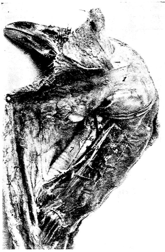
Fig. 18.—Muscles of the upper neck region, 1, Dermo-temporalis. 2, Cornu of the os hyoideum. 3, Carotid artery. 4, Jugular vein. 5, Pneumogastric nerve. 6, Esophagus. 7, Trachea.
Insertion.—From the point of origin it spreads out into a thin fan-like layer and meets its fellow in the median raphe. It is attached to the skin in this region.
Shape.—A very thin fan-shaped muscle.
Relations.—Closely adherent to the skin.
Action.—Assists in supporting the lingual apparatus and superior larynx. Compresses and elevates the part.
75Dermo-cleido Dorsalis. Location.—Anterior shoulder region.
Origin.—From the upper mesial part of the clavicular bone.
Insertion.—Three fasciculi forming a fan-like arrangement attached to the skin in the shoulder-joint and scapular region. The extremities of the fasciculi may meet in the median line of the dorsal region and merge with the dermo-dorsalis.
Shape.—Three fasciculi forming a fan-shaped radiation.
Relations.—Superiorly with the skin. Inferiorly with the shoulder-joint.
Action.—Contracting with the origin as the fixed point, they brace the skin over the forepart of the back. With the integumental attachment as the fixed point, they aid in the act of respiration.
Cleido-trachealis. Location.—Extending from the shoulder to the superior laryngeal region.
Origin.—From a small area on the antero-inner part of the middle of the arm of the furcula.
Insertion.—The fibers pass upward and become flattened and attached to the skin and dermo-temporalis muscle. They touch each other on the anterior border of the superior larynx, the trachea, and the skin over these parts.
Shape.—A slender bundle of muscles.
Relations.—Superiorly the skin, and internally the larynx and the trachea.
Action.—Controls the skin region of this part of the neck.
Dermo-spinalis. Location.—In the region of the shoulder.
Origin.—In an attenuated fascia from the crest of the neural spines of the first, the second, and the third dorsal vertebræ.
Insertion.—To the skin in a broad pale stratum, over the scapular region.
Shape.—Thin, pale, and very delicate.
Relations.—Superiorly with the skin, and inferiorly with the shoulder.
Action.—Controls the skin of the region.
Dermo-iliacus. Location.—Along the back on either side of the spine.
Origin.—From the inner angle of the marginal portion of the antero-dorsal border of the ilium.
Insertion.—Passing forward as a delicate band it spreads out and becomes lost in the skin of the shoulder region.
Shape.—Thin, delicate, and ribbon-shaped.
76Relations.—Superiorly with the skin.
Action.—If the posterior end is fixed, it will cause the feathers to lie close to the skin. Its action would then be opposite to the dermo-dorsalis.
Dermo-ulnaris. Location.—Outer surface of the anterior upper costal and the posterior humeral region.
Origin.—By a thin fascia from the outer part of the third and the fourth true ribs at the base of their epineural appendages. Also from the fascia between them.
Insertion.—The fibers pass forward and upward as a thick muscular bundle and is loosely attached to the skin at a point just back of the humerus. The tendon later becomes spread out and attached to the fascia as far as the elbow-joint. It covers the olecranon of the ulna.
Shape.—At first rather thick, fascicular-like; later becomes tendinous; and at its attachment it becomes spread out over the olecranon.
Relation.—Inferiorly with the ribs and humerus, and externally with the skin.
Action.—A depressor of the humeral region.
The Skeletal Muscles
THE MUSCLES OF THE HEAD
Temporal
Masseter
Biventer maxillæ
Entotympanicus
Pterygoideus internus
Pterygoideus externus
Digastricus
Temporal (Fig. 19, No. 4). Location.—Occupies the temporal fossa.
Origin.—From the mesian line of the sphenotic process, and the adjacent wall of the orbit.
Insertion.—The fibers passing downward and forward blend with the fibers of the masseter. Inferiorly it inserts by a tendon to the coranoid process upon the superior ramal margin of the mandible.
Shape.—Fan-shaped with broad portion uppermost.
Relations.—It occupies the temporal fossa. It is related superiorly with the dermo-temporalis and skin. Inferiorly, with the biventer maxillæ and the masseter.
77Action.—It aids in closing the jaw.
Masseter (Fig. 19, No. 1). Location.—Occupies the supero-lateral portion of the surface of the lower jaw.
Origin.—In two portions: the first, by a broad and thin tendon from the entire length of the bony ridge above the external auditory meatus, from the squamosal process, and from the outer portion of the quadrate bone; the second, from the side and under border of the zygoma.
Insertion.—The first portion: The fibers pass downward and forward beneath the zygoma; a few of the fibers blend with those of the temporal; one tendon inserts to a small tubercle on the upper border of the jaw behind the coronoid process, and by fleshy insertion to the outer side of the ramus of the lower jaw, quite as far forward as the horny portion of the beak. The second portion: by a small tendon to the mandible on its upper border immediately in front of the articular portion.
Shape.—Flat, and elongate. Somewhat fleshy.
Relations.—Superiorly with the temporal, and inferiorly with the biventer maxillæ and stylo-hyoideus. Externally with the skin, and internally with the jaw bone.
Action.—Aids in closing the jaw.
Biventer Maxillæ (Fig. 19, No. 3). Location.—Covers the outer posterior portion of the mandible.
Origin.—It arises in two portions: The first, from a ridge bounding the posterior part of the auditory canal; the second, from a depression of the mesial side. These two parts blend and extend downward and forward.
Insertion.—To the posterior part of the articular end of the mandible.
Shape.—A curved fleshy mass.
Relations.—Superiorly with the temporal and stylo-hyoideus. Posteriorly with the digastricus and genio-hyoideus. Internally with the jaw bone, and externally with the skin.
Action.—It aids in opening the jaw.
Entotympanic. Location.—Posterior to the pterygoid.
Origin.—From the side of the basi-sphenoid and from the base of the rostrum immediately beyond it.
Insertion.—The fibers pass downward and forward, and insert by a double tendinous slip. One slip inserts to a spine-like process on 78the upper side of the pterygoid, and the other to the quadrato-pterygoidean articulation.
Shape.—Spindle-shaped.
Relations.—Anteriorly with the pterygoid. Internally and posteriorly with the basi-sphenoid. Externally with the pterygoideus internus.
Action.—Aids in raising the upper mandible by pulling forward the quadratus and pterygoidean against the palatines.
Pterygoideus Internus (Fig. 17, No. B, 5). (Synonym.—Pterygoideus medialis.[4])
Location.—A muscular mass at the roof of the mouth.
Origin.—From the major part of the surface of the palatine bone and the distal head and shaft of the pterygoid and the sphenoidal projection.
Insertion.—By a tendon to the antero-internal part of the articular part of the mandible.
Shape.—Fusiform with a thick fleshy belly.
Relations.—Superiorly with the palatine, the pterygoid, and the sphenoid bones. Inferiorly with the skin.
Action.—It aids in closing the jaw.
Pterygoideus Externus. (Synonym.—Pterygoideus lateralis.)
Location.—Supero-external to the pterygoideus internus.
Origin.—From the outer part of the extremity of the orbital process of the quadrate.
Insertion.—The fibers pass downward, outward and forward and are inserted to the inner part of the mandibular ramus.
Shape.—A small round bundle.
Relations.—Inferiorly with the pterygoideus internus. Externally with the inferior maxilla.
Action.—Aids in closing the jaw.
Digastricus (Fig. 17, No. B, 16). Location.—Extends from the basi-temporal to the side of the neck.
Origin.—From the external lateral angle of the basi-temporal.
Insertion.—Opposite the angle of the jaw the fibers spread out in fan-like arrangement. The muscle meets its fellow of the opposite side; extends longitudinally over the superior larynx, for some distance down the neck.
Shape.—Thin and ribbon-shaped.
Relations.—Externally to the platysma myoides. The anterior 79fasciculi blend with those of the mylo-hyoideus; internally with the larynx at the upper part.
Action.—To raise the trachea and hyoid apparatus against the pharynx.
THE MUSCLES OF THE TONGUE
Mylo-hyoideus
Stylo-hyoideus
Genio-hyoideus
Cerato-hyoideus
Sterno-hyoideus
Depressor-glossus
Cerato-glossus
Hyoideus transversus
Mylo-hyoideus. Location.—The forepart of the inter-ramal space.
Origin.—From the inner side of the lower jaw at a point just above the lower border and the inturned edge of the horny sheath of the beak.
Insertion.—By aponeurosis to the under side of the hyoid, on the median line between the first and the second basi-branchial.
Shape.—Thin, flat, and delicate.
Relations.—Inferiorly with the skin. Thin and rather broad, it meets its fellow of the opposite side. Laterally with the rami of the jaw. Superiorly with the hyoid apparatus.
Action.—Lifts the tongue upward against the roof of the mouth.
Stylo-hyoideus (Fig. 17, No. B, 6). Location.—Supero-posterior to the hyoid apparatus.
Origin.—From the outer portion of the articular enlargement of the lower jaw.
Insertion.—By a tendon to the basi-branchial of the thyro-hyal.
Shape.—A long transversely flattened fasciculus.
Relations.—Supero-posterior part of the hyoid apparatus along side the genio-hyoideus and the cerato-hyoideus.
Action.—Singly, pulls the tongue to one side; acting with its fellow, pulls the tongue upward.
Genio-hyoideus (Fig. 9, No. 7). Location.—Supero-posterior to the hyoid apparatus.
Origin.—From the anterior portion of the inner side of the lower jaw.
Insertion.—To the middle of the outer side of the basi-branchial of the corner of the os hyoides which cornu it completely envelops.
Shape.—A long, rather thick fasciculus.
Relations.—With the stylo-hyoideus and cerato-hyoideus.
80Action.—Protrudes the tongue from the mouth.
Cerato-hyoideus (Fig. 9, No. 8). (Synonym.—Kerato-hyoideus.)
Location.—Inferior to the hyoid apparatus.
Origin.—By a delicate tendinous slip from the under side of the shaft of the basi-hyal element of the hyoid apparatus.
Insertion.—To a small elevation on the under side of the anterior end of the glosso-hyal.
Shape.—A small round muscular fasciculus becoming tendinous in its anterior half.
Relations.—Superiorly with the hyoid apparatus.
Action.—Singly, pulls the tongue to one side; acting with its fellow, depresses the tongue.
Sterno-hyoideus. Location.—Inferior to the hyoid apparatus.
Origin.—From the outer anterior surface of the thyroid bone of the superior larynx.
Insertion.—To the anterior part of the basi-hyal.
Shape.—At first broad and rather fleshy, becomes contracted and somewhat tendinous.
Relations.—Inferior to the hyoid apparatus and superior larynx.
Action.—When the larynx is fixed, acting alone deflects the tongue laterally; acting with its fellow, depresses the tongue. If the base of the tongue is fixed the two pull the larynx forward. Hence muscles of deglutition.
Depressor-glossus. Location.—Superior to the basi-hyal.
Origin.—From the under portion of the basi-hyal.
Insertion.—To under part of the glosso-hyal.
Shape.—Small, short.
Relations.—Superiorly with the basi-hyal and glosso-hyal bones.
Action.—Depresses the tip of the tongue and elevates the base.
Cerato-glossus. Location.—On the upper portion of the cornu of the hyoid.
Origin.—One-half of the surface of the first basi-branchial.
Insertion.—To the upper side of the shaft of the cerato-branchial element of the hyoid apparatus.
Shape.—A small muscular fasciculus.
Relations.—Supero-laterally to the cerato-branchial element.
Action.—Elevates the cornua of the hyoid arches and presses them against the skull.
Hyoideus Transversus (Fig. 9, No. 9 and 10). Location.—Envelops the spur process of the euro-hyal.
81Origin and Insertion.—It extends from the inner side of one cornu to the spur process.
Shape.—Thin, flat.
Relations.—Posterior to the euro-hyal, which it envelops, and between the cornua of the os hyoides.
Action.—Approximates the cornua of the os hyoides.
THE CERVICAL MUSCLES
Complexus
Rectus capitis anticus minor
Flexor capitis inferior
Rectus capitis posticus major
Biventer cervicis
Longus colli posticus
Obliquus colli
Longus colli anterior
Rectus capitis lateralis
Trachelo-mastoideus
Interspinales
Interarticulares
Obliquo-transversales
Intertransversales
Scalenus medius
Complexus (Fig. 19, No. 5). Location.—Supero-lateral portion of the neck.
Origin.—By three tendinous slips from the antero-lateral portion of the transverse process of the fourth, the fifth, and the sixth cervical vertebræ.
Insertion.—The muscular fibers pass around the neck and meet their fellows in the median line in a fascia formation. The thin tendinous sheet inserts into the occiput just above the occipital ridge.
Shape.—From point of origin this muscle expands into a broad sheet.
Relations.—It overlies the muscles of the occipital region.
Action.—It extends the head.
Rectus Capitis Anticus Minor (Fig. 19, No. 8). (Synonym.—Rectus capitis ventralis minor.)
Location.—Infero-lateral portion of the neck.
Origin.—From the apices of the hyapophyses of the second, the third, and the fourth cervical vertebræ.
Insertion.—To the occiput just below the complexus.
Shape.—A long and rather thick fasciculus.
Relations.—Externally with the skin. The tendinous slips of origin pass between the flexor capitis inferior and longus colli anterior.
82Action.—Extends the head.
Flexor Capitis Inferior (Fig. 19, No. 7). Location.—Antero-inferior part of the neck.
Origin.—From the apices of the hyapophyses of the second, the third, and the fourth cervical vertebræ.
Insertion.—To the triangular area on the basi-temporal of the base of the cranium.
Shape.—At first tendinous, becomes somewhat fleshy as it passes forward.
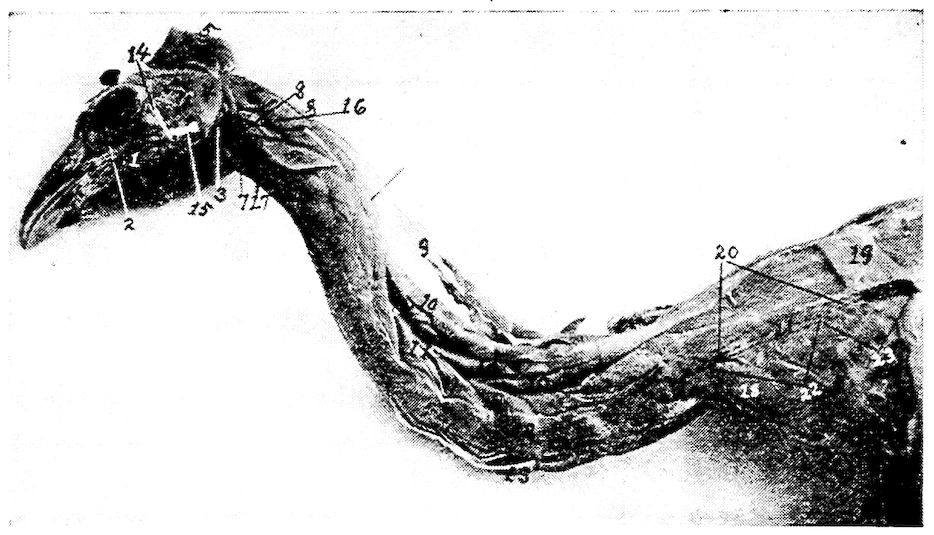
Fig. 19.—Muscles of the head and neck. 1, Masseter muscle. 2, Infraorbital sinus. 3, Biventer maxilla. 4, Temporalis. 5, Complexus. 6, Rectus capitis posticus major. 7, Flexor capitis inferior. 8, Rectus capitis anticus minor. 9, Biventer cervicis. 10, Longus colli posticus. 11, Obliquus colli. 12, Intertransversales. 13, Longus colli anterior. 14, Lateral temporo-maxillary ligament. 15, Lateral ligament of the jaw. 16, Trachelo-mastoideus. 17, Rectus capitis lateralis. 18, Scalenus medius. 19, Rhomboideus. 20, Trapezius. 21, Location of scapula. 22, Supraspinatus. 23, Teres et Infraspinatus.
Relations.—Antero-inferior portion of the neck close to the rectus capitis anticus minor.
Action.—Flexes the head upon the neck.
Rectus Capitis Posticus Major (Fig. 19, No. 6). (Synonym.—Rectus capitis dorsalis major.)
Location.—Supero-lateral area on the anterior portion of the neck.
Origin.—From the median anterior aspect of the second cervical vertebra, and the supero-anterior border of the neural canal.
83Insertion.—To the crescent-shaped area at the back of the skull.
Shape.—Short, thick, fleshy bundle.
Relations.—Superiorly with the complexus; inferiorly with the trachelo-mastoideus and the vertebræ.
Action.—Singly, pulls the head to one side; acting with its fellow, extends the head.
Biventer Cervicis (Fig. 19, No. 9). Location.—Occupies the superior part of the neck.
Origin.—From the side of the neural spine of the first dorsal vertebra, and from the adjacent tendon of the longus colli posticus.
Insertion.—To the occiput.
Shape.—Tendinous in the middle with a more or less spindle-shaped belly at each end.
Relations.—Superior to the longus colli posticus. It lies next to the skin.
Action.—Extends the head on the neck and elevates the neck.
Longus Colli Posticus (Fig. 19, No. 10). (Synonym.—Longus colli dorsalis.)
Location.—Occupies the superior part of the neck.
Origin.—From the marginal edges of the summits of the neural spines of the first two dorsal vertebræ.
Insertion.—To the transverse process of the axis, and superior part of the cervical vertebræ.
Shape.—A long, somewhat narrow, flattened muscle, the inferior portion of which is divided into five or six fasciculi.
Relations.—Superiorly with the biventer cervicis and inferiorly with the vertebræ.
Action.—Raises the neck upward.
Obliquus Colli (Fig. 19, No. 11). Location.—On the lateral side of the neck.
Origin and Insertion.—The first of the seven fasciculi originates from the diapophysis of the eleventh cervical vertebra, winds obliquely over the tenth, and is inserted into the posterior margin of the postzygapophysis of the ninth vertebra. In its passage it receives the slip from the longus colli posticus. The next fasciculus originates from the transverse process of the tenth cervical vertebra, winds obliquely over the ninth vertebra, and is inserted to the postzygapophysis of the eighth vertebra. In its passage it also receives a fasciculus from the longus colli posticus. The next three fasciculi originate in a similar manner being attached to similar postzygapophyses 84and have slips extending forward which slips are inserted to the neural spines of the alternate vertebræ. The sixth fasciculus originates from the transverse process of the sixth vertebra, passing obliquely up the neck is inserted to the extremity of the diapophysis of the fourth vertebra, the outer extremity of the transverse process of the third vertebra. The seventh, or anterior, fasciculus originates from the transverse process of the fifth vertebra, and is inserted to the extremity of the diapophysis of the third vertebra.
Shape.—Short, thick fasciculi.
Relations.—Laterally with the vertebræ. Inferiorly with the intertransversales. Superiorly with the longus colli posticus, and externally with the skin.
Action.—Flexes one vertebra on the other laterally.
Longus Colli Anterior (Fig. 19, No. 13). (Synonym.—Longus colli ventralis.)
Location.—Occupies the anterior portion of the neck.
Origin and Insertion.—This muscle is divided into a vertical portion, and a superior and an inferior oblique portion. The vertical portion originates from the hypophyses of the tenth to the fifteenth vertebra and is inserted by a tendon to the tubercle on the inferior portion of the atlas. Small, slender tendons are given off and insert to the apices of the parapophyses of the fourth to the tenth cervical vertebræ. There is more or less attachment to the bodies of the cervical vertebræ mentioned. The superior oblique portion originates from the diapophyses of the third, the fourth, the fifth cervical vertebræ and becoming tendinous is inserted to the tubercle on the inferior portion of the body of the atlas. The inferior oblique portion originates from the transverse processes of the fourth, the fifth, and the sixth cervical vertebræ. It is inserted by a slender tendon to the apex of the parapophysis of the third cervical vertebra.
Shape.—A long fleshy muscle extending the entire length of the neck.
Relations.—Inferiorly with the skin and superiorly with the cervical vertebræ.
Action.—Pulls the neck downward.
Rectus Capitis Lateralis (Fig. 19, No. 17). Location.—The infero-lateral anterior portion of the neck.
Origin.—Originates tendinous from the diapophysis of the third, the fourth, and the fifth cervical vertebræ.
85Insertion.—Passes obliquely upward in front of the spinal column to the inner tubercle on the basal ridge of the basi-temporal bone, by a subcompressed tendon.
Shape.—A somewhat thick fasciculus.
Relations.—Superiorly with the trachelo-mastoideus, inferiorly with the longus colli anterior, and externally with the skin.
Action.—Singly, pulls the head down and to one side; acting with its fellow, pulls the head downward.
Trachelo-mastoideus (Fig. 19, No. 16). (Synonym.—Longissimus capitis et atlantis.)
Location.—Laterally on the anterior portion of the neck.
Origin.—Semitendinous from the diapophyses of the second, the third, the fourth, and the fifth cervical vertebræ.
Insertion.—By subcompressed tendon to the base of the cranium, at the outer tubercle of the basal ridge of the basi-temporal.
Shape.—Flattened from side to side; broad at the posterior portion, becomes angular at the anterior portion.
Action.—Singly, pulls the head down and to one side; acting with its fellow, pulls the head downward.
Interspinales (Fig. 17, A, No. 2). Location.—Superior to the vertebra.
Origin and Insertion.—A series of muscles connecting the superior neural spines of the cervical vertebræ.
Shape.—Thin, flat.
Relations.—Anteriorly the posterior border of the vertebræ in front and posteriorly the anterior border of the succeeding vertebræ, inferiorly by the vertebræ.
Action.—To approximate the spinous portion of the vertebræ.
Interarticulares (Fig. 17, No. A, 4). Location.—Supero-laterally to the vertebræ. Between the postzygapophysis.
Origin and Insertion.—From the posterior margin of the ring of the atlas to the postzygapophysis of the axis. Then in the succeeding vertebræ from the postzygapophysis of the vertebra to the same of the succeeding vertebra.
Shape.—Muscular bundle.
Relations.—Inferiorly with the vertebræ. Supero-laterally with the obliquo-transversales.
Action.—Aids in approximating the vertebræ in a supero-lateral direction.
86Obliquo-transversales (Fig. 17, A, No. 1). Location.—Supero-lateral to the vertebræ.
Origin and Insertion.—Passes obliquely from the transverse process of one vertebra to the postzygapophysis of the vertebra beyond.
Shape.—A thin fasciculus.
Relations.—Inferiorly with the interarticulares and laterally with the intertransversales.
Action.—Aids in flexing the vertebræ supero-laterally.
Intertransversales (Fig. 19, No. 12). Location.—Laterally to the vertebræ.
Origin and Insertion.—Extend between the transverse processes beginning at the third cervical vertebra.
Shape.—Short, thick.
Relations.—Laterally the vertebræ. Superiorly the obliquo-transversales.
Action.—Aids in flexing the vertebræ laterally.
Scalenus Medius (Fig. 19, No. 18). (Synonym.—Scalenus.)
Location.—Supero-laterally to the entrance of the thorax.
Origin.—From the diapophysis and pleurapophysis of the eleventh cervical vertebra.
Insertion.—To the entire border of the first rib. A few fibers pass over to the anterior free margin of the middle third of the second rib.
Shape.—Rather pyramidal in shape with base uppermost.
Relations.—Posteriorly with the levatores costarum. Internally with the longus colli.
Action.—When the first rib is fixed singly, turns the neck to one side; acting with its fellow, extends the neck. When the neck is fixed, by drawing the first rib forward, it acts as an inspiratory muscle.
THE MUSCLES OF THE AIR PASSAGES
The Superior Larynx
Constrictor glottidis
Thyreo-arytenoideus
The Inferior Larynx
Tracheo-lateralis
Broncho-trachealis posticus
Broncho-trachealis anticus
Broncho-trachealis brevis
Bronchialis posticus
Bronchialis anticus
Sterno-trachealis
87Constrictor Glottidis. Location.—Supero-anterior portion of the superior larynx.
Origin.—From the superior and longitudinal line of the thyroid plate.
Insertion.—All along the inner margin of the arytenoid bone, and to the apex of the mid-cricoidal segment.
Shape.—Thin, flat sheet extending outward and upward, and then inward.
Relation.—Interiorly with the larynx.
Action.—The two muscles of this kind acting together, close the glottis by drawing the apices of the arytenoids to the median line.
Thyreo-arytenoideus. Location.—Supero-lateral portion of the superior larynx.
Origin.—From the entire outer margin of the thyroid plate and the outer margin of the cricoid bone.
Insertion.—All along the outer margin of the arytenoid bone and the outer border of the central cricoid piece.
Shape.—A thin, flat sheet.
Relations.—Infero-laterally with the superior larynx.
Action.—The two muscles of this kind acting together, open the glottis by pulling the arytenoid bone outward.
Tracheo-lateralis (Fig. 20, No. 2). Location.—Along the lateral side of the trachea.
Origin.—By the union of the bronchio-trachealis posticus and bronchio-trachealis anticus, on the lateral side of the trachea, about 1½ centimeters from the bifurcation of the trachea.
Insertion.—In delicate fan-like structure to the side of the trachea, near the superior larynx.
Shape.—Thin, ribbon-shaped, closely attached to the trachea.
Relations.—Internally with the trachea, and externally with the skin and other integument.
Action.—Acts as a brace to the sides of the trachea, and contracting, approximates the tracheal rings and thus shortens the trachea.
Broncho-trachealis Posticus (Fig. 21, No. 3). Location.—Postero-superior portion of the trachea. The fibers pass downward and backward.
Origin.—From the inferior end of the tracheo-lateralis.
Insertion.—To the end of the third half of the bronchial ring of the same side.
Shape.—A small fasciculus.
88Relations.—Internally with the trachea, and externally with the skin and other integument.
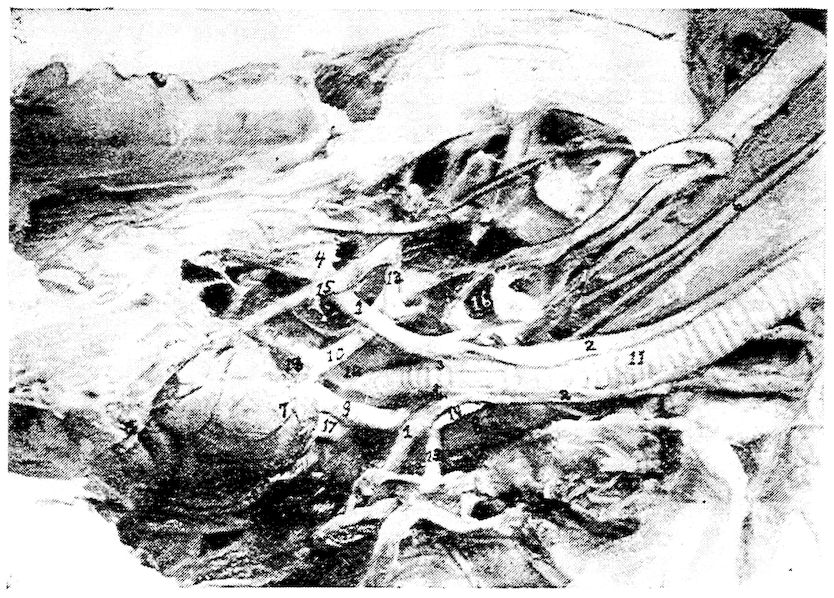
Fig. 20.—The anterior pectoral region. 1, Sterno-trachealis. 2, Tracheo-lateralis. 3, Broncho-trachealis brevis. 4, Costal process of sternum. 5, Right and left carotid converging and occupying same sheath at 6. 7, The heart showing branches of coronary artery. 8, Cervical air-sac. 9, Left brachio-cephalic artery. 10, Right brachio-cephalic artery. 11, Trachea. 12, Inferior larynx. 13. Subclavian artery. 14, Carotid trunk. 15, Internal thoracic artery. 16 Thyroid gland. 17, Anterior vena cava. 18, Auricular appendage.
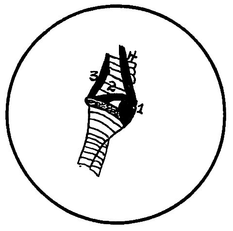
Fig. 21.—The inferior tracheal region. 1, Bronchialis anticus. 2, Bronchialis posticus. 3, Broncho-trachealis posticus. 4, Broncho-trachealis anticus.
Action.—A tensor of the true or inferior larynx.
Broncho-trachealis Anticus (Fig. 21, No. 4). Location.—Anterior portion of the inferior extremity of the trachea.
89Origin.—The anterior branch of the bifurcated tracheo-lateralis.
Insertion.—To the anterior extremity of the third half ring of the bronchus of the same side.
Shape.—A small fasciculus.
Relations.—Internally with the trachea, and externally with the skin and other integument.
Action.—A tensor of the inferior larynx.
Broncho-trachealis Brevis (Fig. 20, No. 3). Location.—Posterior part of the inferior extremity of the trachea.
Origin.—From just beneath the broncho-trachealis anticus muscle.
Insertion.—Extends obliquely across the inferior larynx and is inserted to the posterior end of the second bronchial ring.
Shape.—Rather short, strong, straight, subcylindrical.
Relations.—Internally with the trachea, and laterally with the broncho-trachealis posticus.
Action.—A tensor of the inferior larynx.
Bronchialis Posticus (Fig. 21, No. 2). Location.—Supero-lateral side of the anterior end of the bronchi.
Origin.—From the lateral inferior margin of the last tracheal ring.
Insertion.—Passing obliquely across the larynx it inserts to the posterior extremity of the second half ring of the bronchus.
Shape.—Small, thick, spindle-shaped.
Relations.—Internally with the trachea, inferiorly with the bronchialis anticus, and superiorly with the broncho-trachealis brevis.
Action.—A tensor of the larynx.
Bronchialis Anticus (Fig. 21, No. 1). Location.—Inferior to the former muscle.
Origin.—From the last ring of the trachea.
Insertion.—The fibers pass obliquely forward and become inserted to the rim of the arytenoid cartilage of the inferior larynx, and the anterior extremities of the first and second half rings of the bronchus.
Shape.—Thick, spindle-shaped; about twice the size of the bronchialis posticus.
Relations.—It is crossed at its origin by the broncho-trachealis anticus and is related internally with the bronchus and end of the trachea.
Action.—A tensor of the larynx.
Sterno-trachealis (Fig. 20, No. 1). Location.—The inferior region of the trachea above the above-named muscles.
90Origin.—From the side of the trachea beneath the posterior border of the broncho-trachealis anticus.
Insertion.—Passes downward and backward across the cavity of the chest and is inserted to the inner part of the costal process of the sternum.
Shape.—A delicate cord of fibers.
Relations.—Internally with the broncho-trachealis muscles.
Action.—A relaxor of the larynx, and hence, of the tympanic membrane.
THE STERNAL GROUP
Triangularis Sterni. Location.—In the floor of the thoracic cavity.
Origin.—From the entire superior margin of the summits of the costal processes.
Insertion.—The fibers extend upward and backward, dividing into four digitations which cover the inner surface of the three principal sternal ribs, and are inserted to the first four as high as their articulations with the vertebral ribs.
Shape.—Flat and somewhat triangular.
Relations.—Interiorly with the floor of the thoracic cavity.
Action.—A powerful muscle of expiration. In contracting, lessens the cubic content of the thorax.
The Diaphragm (Fig. 47, No. 10; Fig. 33, No. 9). Description.—The diaphragm in the domestic fowl is rudimentary. It consists of a thin semi-transparent membrane, situated between and separating the thoracic and the abdominal cavities. It readily conforms to the various organs pressing upon it from each side. On each side are three rudimentary muscles which extend from the vertebral heads of the second, the third, and the fourth sternal ribs. The fibers of these muscles spread out in fan-like upon the diaphragm, and are seen just above the digitations of the triangularis sterni.
THE ABDOMINAL MUSCLES
Obliquus abdominis externus
Obliquus abdominis internus
Rectus abdominis
Transversalis abdominis
Obliquus Abdominis Externus (Fig. 22, No. 1). Location.—The outermost muscle of the lateral abdominal wall.
91Attachments.—By a delicate aponeurotic membrane, from the sides of all the true dorsal ribs, from the posterior border of the last vertebral rib and the adjoining margins of the pelvis, and from the entire posterior surface of the inferior border of the post-pubic element of the pelvis; by aponeurosis, blending with the fascia toward the root of the tail and lower part of the abdomen; and by aponeurosis to the sides of the sternum, to the under part of the pectoralis major muscle, and to the zyphoid prolongation. Also by aponeurosis it meets its fellow from the opposite side, at the linea alba.
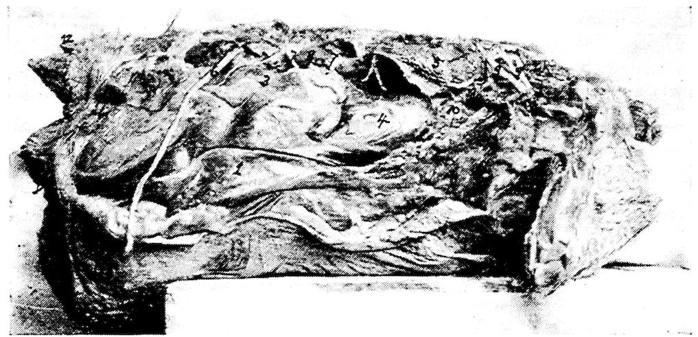
Fig. 22.—The abdominal muscles of a hen. Right side. 1, Obliquus abdominis externus. 2, Obliquus abdominis internus. 3, Peritoneum covering intestines. 4, Peritoneum covering the gizzard. 5, The lungs (note how small they are from a relative standpoint). 6, The great sciatic or ischiadic nerve. 7, Lumbar nerves. 8, Kidney. 9, Brachial plexus. 10, Pericardium of the base of the heart. 11, Anterior vena cava. 12, Oil gland. 13, Showing a sacculation of the abdominal muscles. There is no tunica abdominalis in the fowl.
Obliquus Abdominis Internus (Fig. 22, No. 2). Location.—Just internal with regard to the external oblique.
Attachments.—By aponeurosis, from the posterior third of the post-pubic element of the pelvis; by muscular fibers, from the balance of the bone; and by a few fibers, from the iliac border posterior to the acetabulum. Anteriorly, these fibers are inserted to the entire posterior margin of the last vertebral rib, and into the pleurapophysial head of the last costal rib.
Rectus Abdominis (Fig. 13, No. 10 and Fig. 23, No. 1). Location.—The inferior median abdominal wall.
92Attachments.—By aponeurosis, from the distal extremity of the post-pubic element of the pelvis and from the semitendinous ligament which stretches from one post-pubic tip to the other. Attaches anteriorly to the zyphoid margin of the sternum, and passes, by a broad aponeurotic membrane, over the outer surface of the thoracic wall beneath the external oblique.
Transversalis Abdominis (Fig. 25, No. H, 1). Location.—Infero-lateral portion of the abdominal wall.
Attachments.—By a thin, tendinous attachment from the entire post-pubic and iliac margins of the pelvis and from the interpubic ligament. Its fibers cross the abdomen between the peritoneum, the rectum, and the internal oblique. It is inserted over the entire pleural part of the last two vertebral ribs, the intercostal muscles between them, and the same surface of the hemapophyses connected below. Inserts into the linea alba.
Action.—The abdominal muscles give support to the abdominal organs, aid in flexing the spine, draw the last rib backward, thus aiding in respiration, and compress the abdominal organs to aid in defecation and expiration.
THE DORSO-LUMBAR REGION
Sacro-lumbalis
Longissimus dorsi
Sacro-lumbalis. Location.—The lateral lumbo-sacral region.
Origin.—Tendinous from the anterior margin of the ilium, from the angles of the last two vertebral ribs, and by tendinous slips, from the outer ends of the transverse processes of the last three dorsal vertebræ.
Insertion.—By a few fleshy fibers into the angle of the first dorsal rib and to the corresponding points upon the free cervical ribs, and by a strong semitendinous insertion into the outer extremity of the diapophysis of the twelfth cervical vertebra.
Shape.—A close fitting, tendo-muscular sheet extending between the anterior margin of the ilium and the root of the neck.
Relations.—Intimately blended with the longissimus dorsi externally.
Action.—Assists the longissimus dorsi.
Longissimus Dorsi (Fig. 24, No. 4). Location.—The superior dorso-lumbar region.
93Origin.—From the inner portion of the anterior margin of the ilium, and from the various surfaces afforded by its walls and the walls of the ilio-neural canal; by a series of short and distinct tendons alternately from the anterior and posterior extremities of the summits of the neural spines of all the dorsal vertebræ. From the diapophyses of the dorsal vertebræ, from the crests of the neural spines of the last three dorsal vertebræ, from the bodies of the dorsal vertebræ, and from the fascia between them and the sacro-lumbalis; and also by a tendinous sheet continuous with the origin of the longus colli posticus.
Insertion.—By four fasciculi into the free posterior margins of the oblique processes of the eleventh, the twelfth, the thirteenth, and the fourteenth cervical vertebræ.
Shape.—A large flat sheet.
Relations.—Laterally, with the superior spinous processes of the vertebræ, and with the superior surface of the ribs.
Action.—Singly, flexes the back laterally; with its fellow, aids in elevating the body upward.
THE COCCYGEAL MUSCLES
Levator coccygis
Levator caudæ
Transversus peronei
Depressor caudæ
Depressor coccygis
Lateralis caudæ
Lateralis coccygis
Infracoccygis
Levator Coccygis (Fig. 23, No. 2). Location.—The superior part of the caudal apparatus.
Origin.—From a limited area of the ilium just beyond and to the side of the anterior free caudal vertebræ.
Insertion.—Into the tuberosity of the anterior margin of the pygostyle.
Shape.—Short, fleshy.
Relations.—Superior portion of the coccygeal vertebræ.
Action.—Elevates the tail.
Levator Caudæ (Fig. 23, No. 4). Location.—Supero-lateral side of the tail.
Origin.—From the posterior surface of the post-acetabular area of the pelvis, and from the superior surface of all the coccygeal vertebræ except the pygostyle.
94Insertion.—To the four inner quill butts of the main tail feathers.
Shape.—Long, spindle-shaped.
Relations.—Internally, with the levator coccygis and externally, with the lateralis caudæ.
Action.—Powerfully elevates the four inner main tail feathers.
Transversus Peronei (Fig. 23, No. 3). Location.—The posterior abdominal region.
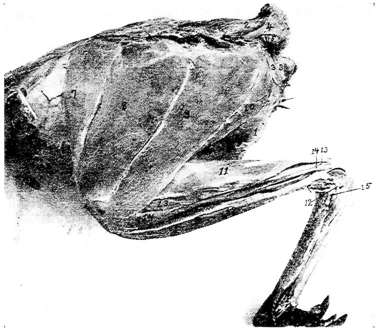
Fig. 23.—Outer layer of muscles of the tail and thigh, 1, Rectus abdominis. 2, Levator coccygis. 3, Transversalis peronie. 4, Levator caudæ. 5, Lateralis caudæ. 6, Depressor caudæ. 7, Sartorius. 8 and 9, Gluteus primus. 10, Semitendinosus. 11, Gastrocnemius. 12, Peroneus longus. 13, Flexor perforatus indicis secundus pedis. 14, Flexor perforatus medius secundus pedis. 15, Lateral ligament of the hock.
Origin.—From the entire posterior margin of the ischium, and from the posterior margin of the post-pubis extending beyond it.
Insertion.—Becoming aponeurotic, it passes toward the coccyx and is attached to the entire posterior margin of the ilium. Passes to the median line, meeting its fellow in front of the anus.
Shape.—Thin, sheet-like.
95Relations.—Infero-anteriorly with the anus. Posteriorly with the edges of the ilium and of the ischium. Internally with the depressor caudæ.
Action.—Gives support to the viscera in the post-anal region, and aids in attaching the anus to the structures above.
Depressor Caudæ (Fig. 23, No. 6). Location.—The infero-lateral side of the tail.
Origin.—From the lower half of the posterior border of the ischium, and from the entire posterior border of the post-pubis beyond.
Insertion.—To the quill butts of the three or four outer main tail feathers.
Shape.—Strong, conical.
Relations.—Externally, with the depressor coccygis.
Action.—Singly, pulls the tail downward and outward; with its fellow, pulls the tail downward.
Depressor Coccygis. Location.—The outermost of the infero-lateral muscles of the tail.
Origin.—From the lower half of the posterior margin of the ischium and the anterior three-fourths of the posterior margin of the post-pubic element of the pelvis beyond it.
Insertion.—To the thickened rim of the inferior and expanded portion of the pygostyle.
Shape.—Flat, triangular.
Relations.—Internally with the depressor caudæ.
Action.—Singly, pulls the tail downward and to one side; with its fellow, pulls the tail downward.
Lateralis Caudæ (Fig. 23, No. 5). Location.—The lateral side of the tail.
Origin.—From the tip of the transverse process of the first free caudal vertebræ.
Insertion.—To the outer three quill butts of the main tail feathers.
Shape.—Four fasciculi forming a fleshy belly.
Relations.—Inferiorly, with the levator caudæ and superiorly with the depressor caudæ.
Action.—Singly, pulls the tail downward and outward; with its fellow, pulls the tail downward. The outermost fasciculus contracting, spreads the tail feathers.
Lateralis Coccygis (Fig. 25, No. E, 2). Location.—The infero-lateral side of the caudal vertebræ.
Origin.—From the surface of the posterior end of the ilium and by 96tendons from the under side of the ends of the first three or four caudal vertebræ.
Insertion.—To the side of the posterior margin of the expanded portion of the pygostyle.
Shape.—Subcompressed mass.
Relations.—With the transverse processes of the caudal vertebra, inferiorly.
Action.—Controls the lateral movements of the tail and its feathers, and the oblique downward movement.
Infracoccygis (Fig. 25, No. E, 1). Location.—The extreme inferior portion of the caudal vertebræ.
Origin.—From the inferior surface of the diapophysis of the last vertebra which anchyloses with the pelvic sacrum, and from all the free caudal vertebræ.
Insertion.—Into the lower side of the pygostyle.
Shape.—Flat, somewhat triangular.
Relations.—Superiorly, with the vertebræ, laterally, with the lateralis coccygis, and mesially, with its fellow of the opposite side.
Action.—Depresses the tail.
THE COSTAL REGION
Latissimus dorsi
Trapezius
Rhomboideus
Serratus magnus anticus
Serratus parvus anticus
Teres et infraspinatus
Intercostales
Levatores costarum
Appendico-costales
Latissimus Dorsi (Fig. 24, No. 7). Location.—Supero-lateral portion of the dorsal region.
Origin.—From two portions, the anterior slip from the outer edge of the superior margins of the neural spines of the second and the third dorsal vertebræ, and the second portion from a similar point on all the succeeding dorsal vertebræ. The second portion is fascia-like in its attachments.
Insertion.—The fibers converge toward the humerus and enter between the deltoid the scapular head of the triceps and the remaining heads of this muscle and is inserted to the anconal part of the humerus, just within in the radial crest.
97Shape.—Thin, triangular.
Relations.—Superiorly, with the skin. The most superficial of the dorsal muscles.
Action.—To elevate the humerus and thus flex the shoulder-joint. An expiratory muscle when the wing is fixed.
Trapezius (Fig. 19, No. 20). Location.—In the shoulder region, just below the longissimus dorsi.
Origin.—From the neural spines of the second, the third, the fourth, and the fifth cervical vertebræ just below the latissimus dorsi.
Insertion.—To the mesian upper border of the scapula.
Shape.—A flat oblong layer of fibers.
Relations.—The posterior part of the trapezius overlies the anterior part of the rhomboideus. The posterior two-thirds is covered by the latissimus dorsi. The anterior third is superficial.
Action.—Draws the scapula forward.
Rhomboideus (Fig. 19, No. 19). Location.—Supero-posterior scapular region.
Origin.—From the neural spines of the first four dorsal vertebræ beneath the latissimus dorsi and trapezius muscles.
Insertion.—The fibers passing outward and backward attach to the posterior third of the mesial or upper border of the scapula.
Shape.—Thin, flat, delicate.
Relations.—Superiorly, by the latissimus dorsi and the trapezius.
Action.—To draw the free end of the scapula upward and forward.
Serratus Magnus Anticus (Fig. 13, No. 16). Location.—The supero-lateral side of the thorax.
Origin.—By three strong digitations: first, from the outer part of the second true rib just above the base of the epineural appendage; the other two, from similar locations on the two succeeding ribs, just above the origin of the dermo-ulnaris.
Insertions.—The first by tendinous attachment to the interpleurapophysial membrane. The rest of the fibers pass upward and slightly forward, and are inserted to the inferior surface of the apex of the scapula.
Shape.—Three flat, strong digitations.
Relations.—Internally, with the ribs.
Action.—If the scapula is fixed, it is a muscle of inspiration.
Serratus Parvus Anticus. Location.—The outer and upper surface of the anterior part of the thorax.
98Origin.—By three digitations from the outer surface of the first three ribs above the origin of the serratus magnus anticus and the thoraco-scapularis.
Insertion.—The thin sheet of fibers passes upward and backward, and is inserted to the inferior margin of the scapula.
Shape.—Thin, flat.
Relations.—Internally, with the ribs.
Action.—If the scapula is fixed, it is a muscle of respiration.
Teres et Infraspinatus (Fig. 13, No. 11). Location.—Scapular region.
Origin.—From the superior surface and outer margin of the posterior two-thirds of the scapula.
Insertion.—The fibers pass forward and outward, and insert to the humerus at the middle of the ulnar margin of the pneumatic fossa, between the forks of the triceps.
Shape. —Flat, triangular-shaped.
Relations.—Anteriorly, with the supraspinatus. Internally, with the ribs.
Action.—Aids in keeping the humerus in its socket. Assists in closing the wing by drawing the humerus to the side of the body.
Intercostales (Fig. 24, No. 5). Location.—Between the ribs of the upper thoracic region.
Origin and Insertion.—From the anterior border of one rib, the fibers, passing obliquely upward and forward, and the lower portion downward and forward, are inserted to the posterior border of the rib just in front.
Shape.—They are best developed in the anterior portion of the thoracic region and extend down as far as the sternal ribs.
Relations.—Internally the pleura, and anteriorly and posteriorly the ribs.
Action.—The first rib being rendered fixed by the scalenus medius, the muscles draw the ribs forward, thus aiding inspiration.
Levatores Costarum (Fig. 24, No. 2). Location.—Supero-lateral portion of the thorax.
Origin and Insertion.—A series of muscles, the first of which extends from the extremity of the transverse process of the twelfth vertebra, the fibers passing downward and backward to the anterior free margin of the upper third of the long posterior rib, and to the external surface close to its margin. The others arise and insert in a similar manner from the ends of the diapophyses of all 99the dorsal vertebræ and attach to each succeeding rib. Those in front are best developed.
Shape.—Thin and triangular.
Relations.—External to the intercostales.
Action.—Aids in respiration.
Appendico-costales (Fig. 24, No. 1). Location.—The lateral side of the thorax.
Origin and Insertion.—From the posterior edge of an uncinate process or epineural appendage, the fibers extending downward and backward to the outer surface of the succeeding rib.
Shape.—A series of thin triangular-shaped muscles.
Relations.—Internally, with the intercostales.
Action.—Aids in respiration.
THE ANTERIOR PECTORAL GROUP
Tensor patagii longus
Tensor patagii brevis
Tensor Patagii Longus (Fig. 15C, No. 1; Fig. 14, No. 2). Location.—Anterior shoulder region, in the triangular patagium of the wing.
Origin.—By a flat tendon common to it and the tensor patagii brevis from the supero-mesial line of the head of the clavicle.
Insertion.—By a long slender tendon extending in the duplicature of the patagium, with which the fibers blend. Passing to the side of the extensor metacarpi radialis longus, it extends over the end of the radius and is inserted to the os carpi radiale, and to the fascia which binds down the other tendons on the anterior part of the wrist-joint.
Shape.—Cone-shaped belly terminating in a long tendon.
Relations.—Externally, with the skin; by its tendon, with the border of the extensor metacarpi radialis longior.
Action.—Tenses the soft part in the fold of the wing, and aids in flexing the forearm.
Tensor Patagii Brevis (Fig. 14, No. 3). Location.—In the triangular patagium of the wing.
Origin.—From the head of the clavicle in common with the preceding.
Insertion.—The tendon bifurcates, one branch blending with the fascia of the extensor metacarpi radialis longior, the other inserting just below the tubercle on the external condyle of the humerus.
100Shape.—Fleshy; somewhat broader and longer than the preceding.
Relations.—Interiorly, with the preceding.
Action.—Assists the tensor patagii longus in flexing the forearm.
THE PECTORAL MUSCLES
Pectoralis major
Pectoralis secundus
Pectoralis tertius
Pectoralis Major (Fig. 13, No. 5). Location.—On the lateral side of the sternum.
Origin.—From the posterior portion of the lateral wing of the sternal body, from the outer marginal third of the keel of the sternum, and from the entire outer side of the limb of the furcula or clavicle.
Insertion.—The fibers converging form toward the proximal third of the humerus a broad tendon which by its insertion covers the entire palmar part of the pectoral crest. A few of the tendinous fibers pass over the shoulder-joint and blend with the long head of the biceps. Near this point it receives the insertion of the dermo-humeralis.
Relations.—Interiorly, with the skin; superiorly, in the sternal region, with the pectoralis secundus and the pectoralis tertius.
Shape.—Fleshy; largest of the pectorales.
Action.—Powerfully depresses the humerus. The chief muscle of flight.
Pectoralis Secundus (Fig. 13, No. 9; Fig. 15, No. A, 6). Location.—The entire lateral side of the sternum.
Origin.—From the anterior sternal extremity of the lower third of the coracoid, from the keel and sternal wing, and by a tendon from a membraneous expansion between the coracoid and the clavicular bones and from the lower third of the coracoid.
Insertion.—The fibers converge into a tendon, which passes upward around the coracoid to its posterior through a canal formed by the scapula, the coracoid, and the clavicle, the foramen triosseum (Fig. 15, No. A, 7). This tendon then passes outward and downward, and becoming flat, inserts to the humerus just anterior to the radial crest and nearer to the humeral head than does the pectoralis major.
Relations.—Internally, with the breast-bone; externally, with the pectoralis major; and superiorly, with the pectoralis tertius.
101Shape.—Long, fusiform, fleshy.
Action.—Raises the wing.
Pectoralis Tertius (Fig. 13, No. 6; Fig. 15, No. A, 5). Location.—On the antero-lateral side of the sternum.
Origin.—From the anterior half of the exterior of the body of the sternum, from the fascia of the subclavius on the outer border of the costal process, and from the outer, lower third of the coracoid process.
Insertion.—The fibers pass upward to the outer side of the coracoid, and becoming tendinous as they reach the humerus, by a strong flattened tendon insert to the ulnar crest of the humerus on the proximal margin at about a middle point of the pneumatic fossa.
Relations.—Externally, with the pectoralis major; interiorly, with the pectoralis secundus; internally, with the fascia of the subclavius and the sternum.
Shape.—Fleshy, fusiform.
Action.—Assists the pectoralis secundus in elevating the humerus.
MUSCLES OF THE SCAPULAR REGION
Coraco-humeralis
Scapulo-humeralis
Supraspinatus
Subclavius
Coraco-brachialis
Teres minor
Levator scapulæ
Thoraco-scapularis
Subscapularis
Coraco-humeralis. Location.—The scapulo-humeral region.
Origin.—From the outer side of the head of the coracoid, supero-laterally to the long head of the biceps.
Insertion.—To the palmar part of the head of the humerus just inside of the insertion of the pectoralis major.
Shape.—A delicate subcylindrical muscle.
Relations.—Superior to the head of the humerus.
Action.—Aids in extending the humerus.
Scapulo-humeralis (Fig. 14, No. 10). Location.—The scapulo-humeral region.
Origin.—From the inner side of the neck of the scapula just within the head of the deltoid.
Insertion.—Passing over the top of the shoulder-joint it is inserted to the palmar part of the humeral head between the insertion of the pectoralis major and the pectoralis secundus.
102Shape.—Narrow, flat ribbon.
Relations.—Along the upper margin of the larger portion of the deltoid.
Action.—Aids in extending the humerus.
Supraspinatus (Fig. 19, No. 22). Location.—Scapular region.
Origin.—From the superior surface and outer third of the scapula.
Insertion.—To the lower border of the pneumatic fossa of the humerus.
Shape.—Thin, flat, triangular.
Relations.—Externally, with the trapezius and posteriorly, with the teres et infraspinatus.
Action.—If the scapula is the fixed point it will pull the humerus upward and backward.
Subclavius. Location.—Anterior sternal region.
Origin.—From the entire outer surface of the sternal process of the sternum and the adjacent outer surfaces of three or four of the hemapophyses.
Insertion.—To the inferior margin of the coracoid bone, the longer fibers passing over to the fossa in the lower third of the posterior part of the coracoid.
Shape.—Rather small, fleshy.
Relations.—Overlapped by the pectoralis tertius.
Action.—Pulls the coracoid outward. Also aids in keeping the coracoid in place.
Coraco-brachialis (Fig. 13, No. 15). Location.—Along the coracoid shaft.
Origin.—By a delicate tendon from a small circular point on the postero-mesial part of the shaft of the coracoid immediately above the attachment of the subclavius and the fossa at that point.
Insertion.—To the top of the ulnar tuberosity of the humerus. This subcircular space is common to the teres minor and to this muscle.
Shape.—Long, fusiform.
Relations.—With the shaft of the coracoid between the teres minor and subscapularis.
Action.—To depress the wing.
Teres Minor (Fig. 13, No. 14). Location.—The coraco-scapular region.
Origin.—From under the side of the anterior tip of the scapula.
Insertion.—Passing outward behind the coracoid head and beneath 103the neck of the scapula, it is attached by a small tendon to the top of the ulnar tuberosity of the humerus in common with the coraco-brachialis, with which tendon it fuses.
Shape.—Small, chunky.
Relations.—Anteriorly, with the tip of the scapula and behind the coracoid head. Posteriorly, with the coraco-brachialis.
Action.—Aids in the downward stroke of the wing.
Levator Scapulæ. Location.—Scapular region.
Origin.—By two strong digitations, one from each of the lateral processes of the first cervical vertebra.
Insertion.—To the middle third of the inferior part of the blade of the scapula.
Shape.—Small and flat.
Relations.—Inferior to the scapula.
Action.—Pulls the scapula forward, and with it the entire shoulder girdle, which articulates like a hinge-joint at the costo-sternal juncture.
Thoraco-scapularis. Location.—Lateral side of the chest.
Origin.—From the outer part of the lower half of the first free rib, the outer side of the next succeeding rib and its epineural appendage, and from a similar surface on the next rib.
Insertion.—By a broad, flat tendon which passes between the two divisions of the subscapularis and inserts at a point at the juncture of the anterior and middle third of the outer margin of the scapula.
Shape.—Broad, flat.
Relations.—Internally, with the ribs and the scapula.
Action.—When the scapula is fixed it is a muscle of inspiration. When the ribs are fixed, it draws the scapula downward.
Subscapularis. Location.—Scapulo-humeral region.
Origin.—From the anterior and outer half of the scapula.
Insertion.—By a strong and subcylindrical tendon to the top of the ulnar tuberosity of the humerus close to the combined tendons of the coraco-brachialis and teres minor.
Shape.—A large and powerful muscle.
Relations.—The flat tendon of the thoraco-scapularis divides the posterior portion of the belly into two portions.
Action.—A powerful rotary muscle of the head of the humerus; aids in keeping the humeral head in the glenoid cavity.
104
THE MUSCLES OF THE BRACHIAL REGION
Biceps
Triceps
Deltoid
Brachialis anticus
Biceps (Fig. 13, No. 8). (Synonym.—Biceps brachii.)
Location.—The anterior brachial region.
Origin.—By a broad tendon it covers the top of the shoulder-joint, dividing into two heads, giving rise to the long and the short heads. The long is inserted into the outer part of the head of the coracoid just beyond the glenoid cavity; the short head to the distal angle of the ulnar tuberosity of the humerus.
Insertion.—To the ulna just in front of the articular cavity for the trochlear surface of the distal extremity of the humerus.
Shape.—Large, subfusiform.
Relations.—On one side by the triceps and the other the deltoid and anteriorly partially covered by the patagii muscles.
Action.—A powerful flexor of the forearm.
Deltoid (Fig. 13, No. 12; Fig. 15, No. 8). Location.—Occupying the lateral side of the humerus; the brachial region.
Origin.—Divided into two portions, the long narrow head extends from the clavicular process of the scapula and from the adjacent surface of the same bone. These fibers then extend around the back of the shoulder-joint, and are joined by the fibers that arise from the entire outer surface of the large os humero-scapulare. These latter fibers are inserted upon an extensive area upon the anconal part of the bone beyond the humeral articular head, and to almost the entire shaft below it.
Insertion.—By a subcylindrical tendon to the proximal side of the tubercle of the external condyle, and above the insertion of the extensor metacarpi radialis longus.
Shape.—Large, fleshy, with tendinous attachment.
Relations.—On one side by the biceps and the other by the triceps also the patagii.
Action.—To extend the arm.
Triceps (Fig. 15, No. 7). (Synonym.—Triceps brachii.)
Location.—The posterior humeral region.
Origin.—By three portions: by the internal and external heads, and by the long scapular head. The internal and the external heads are blended except at their proximal extremities. Each head arises from the anconal surface of the shaft of the humerus. The 105internal head being located toward the deltoid attachment and the external head into the pneumatic fossa. The long head extends from a circumscribed area just posterior to the glenoid cavity of the scapula. The fibers pass around the shoulder-joint, beneath the deltoid.
Insertion.—The broad tendon passes over the elbow-joint and is inserted to the entire under surface of the olecranon of the ulna.
Shape.—Long, large, fleshy.
Relations.—The supraspinatus passes between the internal and the external heads. It is bounded by the biceps and the deltoid.
Action.—An extensor of the antibrachial region directly antagonizing the biceps.
Brachialis Anticus (Fig. 15, No. 9; Fig. 14, No. 5). Location.—In the flexure of the humerus and the ulna.
Origin.—It arises fleshy from a circumscribed area on the inner side of the anconal part of the distal extremity of the humerus.
Insertion.—The fibers passing directly over the elbow-joint become inserted to the lateral surface of the proximal end of the ulna close to the margin of the sigmoid cavity.
Shape.—Small, fleshy.
Relations.—With the joint.
Action.—Assists in flexing the forearm upon the arm. Protects the structures in the anterior part of the elbow-joint.
THE MUSCLES OF THE FOREARM AND THE HAND
Extensor metacarpi radialis longior
Supinator brevis
Extensor digitorum communis
Pronator brevis
Flexor metacarpi radialis
Extensor ossis metacarpi pollicis
Pronator longus
Extensor indicis longus
Flexor digitorum sublimis
Flexor digitorum profundus
Flexor carpi ulnaris
Flexor carpi ulnaris brevior
Extensor Metacarpi Radialis Longior (Fig. 13, No. 17). Location.—Superior to the radius.
Origin.—By two strong tendinous heads; the outer from the tubercle of the external condyle of the humerus, just above the origin of the tendon of the tensor patagii brevis; the inner and stronger 106portion from the tubercle found above the oblique trochlear facet of the distal end of the radius.
Insertion.—Becoming a flat, broad, and strong tendon at about the middle of the forearm, finally becomes inserted to the apex of the anchylosed first metacarpal of the carpo-metacarpus.
Shape.—A thick, fusiform belly, tendinous at both extremities.
Relations.—By the side of the pronator brevis.
Action.—Raises the hand, and draws it forward toward the radial margin of the forearm and retains it on the same side.
Extensor Digitorum Communis (Fig. 15, No. 2). Location.—The upper metacarpal region.
Origin.—Immediately below the tubercle of the external condyle of the humerus.
Insertion.—By a small tendon to the outer side of the base of the pollex, and at a mid-point upon the anterior rim of the proximal phalanx of the middle finger.
Shape.—Small spindle-shaped, becoming tendinous at about the middle of the forearm.
Relations.—Occupies the middle of the group of muscles on the outer side of the forearm.
Action.—An extensor of the digit.
Supinator Brevis (Fig. 14, No. 7). Location.—The superior part of the radius.
Origin.—From the external condyle of the humerus below the origin of the tendon of the extensor digitorum communis.
Insertion.—To the outer side of the shaft of the radius, for nearly one-third of its length.
Shape.—Thin, slender.
Relations.—Inferiorly, with the bone.
Action.—A supinator of the radial region, and antagonistic to the pronators.
Flexor Metacarpi Radialis (Fig. 15, No. 10). Location.—The lowest of the group of three muscles on the outer part of the forearm.
Origin.—By two tendinous heads, the longer from the external condyle of the humerus, and the other just beyond the base of the olecranon.
Insertion.—Becomes tendinous at about the middle of the shaft of the ulna and inserts to a prominent process on the proximal third of the posterior part of the shaft of the mid-metacarpal.
Shape.—Fusiform.
107Relations.—Inferiorly, the anconeus and superiorly, the extensor digitorum communis.
Action.—A powerful flexor of the hand.
Pronator Brevis (Fig. 13, No. 18). Location.—Supero-lateral side of the forearm.
Origin.—From just above the internal condyle of the humerus.
Insertion.—The tendon passing obliquely across the interosseous space and is inserted to the ulnar side of the shaft of the radius, just beyond the juncture of the proximal and the middle thirds.
Shape.—Fusiform.
Relations.—Superior to the pronator longus.
Action.—Pronates the forearm, and flexes the forearm upon the arm.
Pronator Longus (Fig. 14, No. 6). Location.—Lateral side of the radio-ulnar region.
Origin.—From the middle of the internal condyle of the humerus.
Insertion.—To the shaft of the radius just beneath the pronator brevis.
Shape.—Massive, ellipsoidal.
Relations.—Between the pronator brevis and flexor digitorum profundus.
Action.—A pronator.
Extensor Ossis Metacarpi Pollicis (Fig. 13, No. 23). Location.—Slightly interposed between the ulna and radius.
Origin.—Immediately in front of the greater sigmoid cavity of the ulna.
Insertion.—To the palmar side of the base of the first metacarpal, in common with the extensor metacarpi radialis longior.
Shape.—Delicate, straight.
Relations.—Superiorly, the extensor indicis longus, and inferiorly, the anconeus and the flexor digitorum profundus.
Action.—Extends the hand upon the forearm.
Anconeus (Fig. 15, No. 4). Location.—Between the ulna and the radius.
Origin.—By a short, strong, subcylindrical tendon from the lower posterior of the external condyle of the humerus.
Insertion.—To the latero-radial side of the ulna somewhat beyond its middle.
Shape.—Fusiform.
108Relations.—Superiorly, the extensor indicis longus, and inferiorly, the ulna.
Action.—A flexor of the forearm.
Extensor Indicis Longus (Fig. 13, No. 22; Fig. 15, No. 13). Location.—Infero-lateral to the radius.
Origin.—From about one-half the surface of the proximal portion of the radius.
Insertion.—Possesses a long tendon which passing over a groove at the distal end of the ulna and receiving muscular fibers at the base of the metacarpus, extends down the anterior part of the hand and is inclosed in a sheath in front of the superior part of the first phalanx of the index-finger. This tendon is inserted into the anterior upper rim of the distal phalanx.
Shape.—Small, thin.
Relations.—Supero-laterally with the radius.
Action.—Extensor of the digit.
Flexor Digitorum Sublimis. Location.—The forearm.
Origin.—From the internal condyle of the humerus.
Insertion.—Passing over the wrist, it is inserted to the middle phalanx of the hand.
Shape.—A musculo-tendinous band, rather delicate and rudimentary.
Relations.—Closely adhering to the integument that stretches from the internal condyle of the humerus to the wrist.
Action.—Assists these muscles. A flexor.
Flexor Digitorum Profundus (Fig. 13, No. 21). Location.—Lateral side of the ulna.
Origin.—By two heads from the proximal extremity of the ulna, between which the brachialis anticus passes.
Insertion.—The two bellies uniting are attached to the under side of the shaft of the ulna. It becomes tendinous about the middle of the shaft, and inserts to the ulnar side of the base of the distal joint of the index-finger.
Shape.—A fleshy belly with long tendon.
Relations.—Inferiorly, with the flexor carpi ulnaris, superiorly, with the pronator brevis.
Action.—A flexor of the digit.
Flexor Carpi Ulnaris (Fig. 13, No. 19). Location.—Inferior part of the forearm.
Origin.—By two strong tendons; one from the side and posterior 109of the internal condyle of the humerus, passing through the humero-ulnar pulley at the side of the base of the olecranon process; the other, to the posterior of the internal condyle of the humerus. It does not pass through a pulley.
Insertion.—A subcylindrical tendon extends back of the ulnare ossicle of the carpus giving off a tendinous slip to the flexor digitorum profundus, and becomes inserted to the anterior rim of the proximal phalanx of the index digit. It has an attachment at the carpus.
Shape.—A strong fleshy belly terminating in a long tendon.
Relations.—The outermost inferior muscle of the forearm.
Action.—A powerful flexor of the hand upon the forearm.
Flexor Carpi Ulnaris Brevior (Fig. 13, No. 20). Location.—Inferior part of the antibrachial region.
Origin.—From a broad area on the middle third of the upper side of the shaft of the ulna.
Insertion.—Near the top of the outer edge of the anchylosed os magnum of the carpo-metacarpus.
Shape.—Thin, somewhat flat.
Relations.—To the inside of the flexor digitorum profundus.
Action.—Flexes the hand upon the forearm and rotates the hand toward the body.
OTHER MUSCLES OF THE DIGITS
Extensor proprius pollicis
Flexor minimi digiti
Abductor minimi digiti
Interosseous palmaris
Flexor brevis pollicis
Flexor minimi digiti brevis
Interosseous dorsalis
Flexor metacarpi brevis
Extensor Proprius Pollicis (Fig. 13, No. 29a). Location.—Anterior to the radio-carpal joint.
Origin.—From the ulnar side of the tendon of the extensor metacarpi radialis longior.
Insertion.—To the antero-ulnar side of the pollex.
Shape.—Small, spindle-shaped; tendinous at both attachments.
Relations.—Inferiorly, with the proximal and anterior part of the metacarpus.
Action.—An extensor of the pollex.
Flexor Brevis Pollicis (Fig. 13, No. 28a). Location.—Anterior to the pollex.
110Origin.—From the shaft of the mid-metacarpal bone, just below the pollex.
Insertion.—Distal apex of the pollex.
Shape.—Short, small, fleshy.
Relations.—Superiorly, with the inferior part of the pollex.
Action.—Flexes the pollex.
Flexor Minimi Digiti. Location.—Inferior side of the metacarpus.
Origin.—From the posterior part of the median metacarpal close to the os carpi ulnare. A few fibers extend to the ulna.
Insertion.—Into the base of the median phalanx.
Shape.—Small, slender, short.
Relations.—Superiorly, with the metacarpal bone.
Action.—A flexor of the digit.
Flexor Minimi Digiti Brevis (Fig. 13, No. 30). Location.—Inferior to the third, or small finger.
Origin.—From the lower and posterior end of the median metacarpal and from the adjacent tendon of the preceding.
Insertion.—Inserted to the apex of the small finger.
Shape.—Small and rather rudimentary.
Relations.—Inferior to the small finger.
Action.—Acts as a posterior ligament of this joint.
Abductor Minimi Digiti. Location.—Supero-lateral side of the small finger.
Origin.—From the proximal extremity of the anterior aspect of the third finger.
Insertion.—To the posterior border of the proximal phalanx of the second, or index-finger at a point just above the tip of the third finger.
Shape.—A few muscular fibers with some tendinous material.
Relations.—At the base of the median segment of the index-finger and to the side of the third.
Action.—Antagonizes the powerful flexors on the back of the joint.
Interosseus Dorsalis (Fig. 13, No. 31). Location.—By the side of the metacarpus.
Origin.—From the margin of the shaft of the median metacarpal.
Insertion.—To the anterior part of the base of the distal phalanx of the index digit.
Shape.—Short, delicate, with long tendon.
Relations.—Internally, the metacarpal bone.
111Action.—Extends the last bone of the middle finger.
Interosseus Palmaris (Fig. 13, No. 32). Location.—On the opposite side of the bone from the interosseous dorsalis.
Origin.—From the shaft of the median metacarpal under the preceding.
Insertion.—To the apex of the last joint of the second, or middle finger.
Shape.—Small, delicate, with long slender tendon.
Relations.—Beneath the preceding and along the metacarpal bone.
Action.—Flexes the terminal phalanx.
Flexor Metacarpi Brevis. Location.—To the lateral side of the metacarpal bone.
Origin.—From the outer side of the distal extremity of the ulna.
Insertion.—To the base, in front, and on the ulnar side of the proximal phalanx of the index digit.
Shape.—Delicate, with long slender tendon.
Relations.—On the lateral side of the ulna and the metacarpal bone.
Action.—Flexes the metacarpus.
MUSCLES OF THE POSTERIOR LIMB
Sartorius
Gluteus medius
Extensor femoris
Biceps flexor cruris
Semitendinosus accessorius
Femoro-caudal
Obturator internus
Abductor longus
Gluteus primus
Gluteus minimus
Vastus internus
Semitendinosus
Semimembranosus
Obturator externus
Gemellus
Adductor magnus
Sartorius (Fig. 23, No. 7). Location.—Extreme anterior portion of the thigh.
Origin.—From the outer two-thirds of the superior surface of the raised emarginations of the anterior border of the ilium, and by fascia from the neural spines of the fourth dorsal vertebra.
Insertion.—Obliquely by a semitendinous fascia to the inner edge of the ligamentum patella, and to the inner and adjacent border of the anterior half of the summit of the tibia.
Shape.—Large, fleshy.
112Relations.—Posteriorly, with the gluteus primus and the vasti.
Action.—Extends the leg, flexes and adducts the thigh.
Gluteus Primus (Fig. 23, No. 8 and 9). Location.—The outer flat massive muscle of the thigh.
Origin.—By a thin fascia from nearly the entire length of the supero-internal margin of the pre-acetabular portion of the ilium, above the antitrochanter, and from the entire length of the post-acetabular ridge.
Insertion.—Near the patella it joins the tendon of the extensor femoris, and by aponeurosis spreads over the knee and is inserted to the crest of the upper border of the tibia, the patella being incorporated in its aponeurotic ligament.
Shape.—Triangular in shape, it is aponeurotic anteriorly and more fleshy posteriorly.
Relations.—Anteriorly, covers over the posterior portion of the sartorius and the body of the gluteus medius; posteriorly, bordered by the semitendinosus and the biceps.
Action.—Abducts the thigh.
Gluteus Medius (Fig. 24, No. 11). Location.—Supero-anterior part of the ilium under the gluteus primus.
Origin.—From the entire supero-internal margin of the pre-acetabular surface of the ilium and the concave surface on the adjacent bone.
Insertion.—By a strong tendon which passes over a bursa on the anterior rim of the trochanter and is inserted obliquely to this trochanter.
Shape.—A fleshy muscle filling the concavity of the pre-acetabular division of the ilium.
Relations.—Superiorly, with the gluteus primus; inferiorly, with the gluteus minimus.
Action.—Abducts and pulls the femur forward.
Gluteus Minimus (Fig. 24, No. 12). (Synonym.—Gluteus profundus.)
Location.—Beneath the medius and the smaller of the two.
Origin.—To the anterior margin of the outer border of the ilium, and from the supero-external surface of the last rib.
Insertion.—The fibers extending downward and backward and outward become tendinous and are inserted below the trochanter of the outer part of the upper third of the femur.
Shape.—Small, fleshy.
113Relations.—Inferiorly, with the bone and superiorly, with the gluteus medius.
Action.—Abducts and pulls the thigh forward.
Extensor Femoris (Fig. 24, No. 15). Location.—Anterior femoral region. It is divided into two parts: the vastus externus and the crureus.
Origin.—The vastus extemus (Fig. 24, No. 14) arises tendinous from the base of the trochanter of the outer part of the femur and from this point down the bone approximately to the condyle. The crureus (Fig. 24, No. 13) originates by a tendon from the anterior upper prominent rim of the trochanter, from this point extending down the antero-external part of the shaft of the femur.
Insertion.—Merges with the gluteus primus, and, by a fascia-like arrangement, spreads over the front of the knee-joint inserts to the patella and is also inserted into the crest of the tibia.
Shape.—Large, fleshy.
Relations.—Posteriorly, with the femur and anteriorly, with the sartorius.
Action.—Extends strongly the leg upon the thigh.
Vastus Internus (Fig. 16, No. 3). Location.—Internal and lateral side of the femur.
Origin.—From the postero-internal part of the shaft of the femur just below the head, and in a straight line extending down the femur.
Insertion.—By a broad tendon along the thickened inner border of the summit of the tibia.
Shape.—Thick, long.
Relations.—Anteriorly, with the bone; surrounded by the other crural muscles.
Action.—Extends the leg upon the knee; a powerful assistant of the extensor femoris.
Biceps Flexor Cruris (Fig. 17, No. D, 1). Location.—Posterior tibial region.
Origin.—By a tendinous fascia from the post-acetabular ridge, extending between the antitrochanter and the anterior point of the insertion of the semitendinosus.
Insertion.—To the tuberosity on the external part of the shaft of the fibula a short distance below the head.
Shape.—Large, rather cone-shaped with base directed upward.
Relations.—Anteriorly, with the vastus externus; posteriorly, with 114the semitendinosus. The inferior tendinous portion passes through a tendinous sling or pulley called the biceps band.
Action.—Flexes the leg upon the thigh.
Semitendinosus (Fig. 23, No. 10). Location.—Posterior femoral region.
Origin.—By a tough, strong fascia from the surface of the caudal muscles and from the posterior third of the post-acetabular ridge.
Insertion.—The fibers pass downward and forward, and insert to the tendinous raphe along the posterior margin of the semitendinosus accessorius; lower down it merges with the median fascia of the inner head of the gastrocnemius.
Shape.—Fleshy, broad, flat and long.
Relations.—Internally, with the semimembranosus and anteriorly, with the biceps flexor cruris.
Action.—Flexes the leg.
Semitendinosus Accessorius. Location.—Posterior femoral region.
Origin.—From an oblique line just above the condyle, on the posterior of the shaft of the femur.
Insertion.—Its fibers, passing upward and backward, attach themselves to the tendinous raphe common to this muscle and the semitendinosus, and are finally inserted to the inner side of the shaft of the tibia.
Shape.—Flat, oblong.
Relations.—Posteriorly to the shaft of the femur, and externally to the long adductors of the thigh.
Action.—Aids the preceding in flexing the leg.
Semimembranosus (Fig. 24, No. 17). Location.—Postero-internal to the semitendinosus.
Origin.—From the outer surface of the ischium, beginning at the lower margin of its notch on the posterior pelvic border, extends on a curved line on the adjacent surface beyond.
Insertion.—The fibers, passing downward and forward, insert by a broad, thin tendon to the shaft of the tibia a short distance below the head and on a line parallel to the long axis of the tibia.
Shape.—Long, narrow, ribbon-shaped.
Relations.—Lies adjacent to the semitendinosus and in the same plane.
Action.—Directly flexes the leg.
Femoro-caudal (Fig. 24, No. 8). Location.—Infero-lateral to the caudal and post-femoral region.
115Origin.—By a delicate tendon from the base of the pygostyle.
Insertion.—By a thin, flat tendon upon the outer part of the shaft of the femur below the trochanter and at about the juncture of the upper and middle third of the bone.
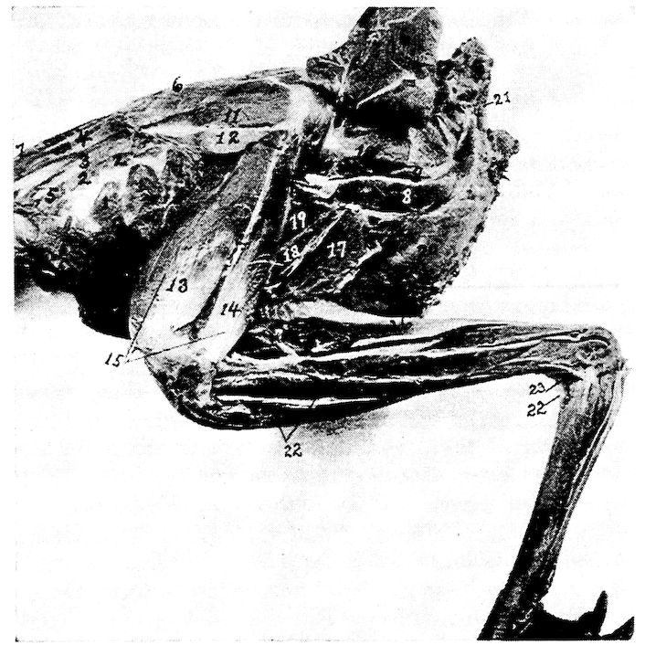
Fig. 24.—Second layer of muscles of the thigh of a cock. Outside view. 1, Appendico-costales. 2, Levatores costarum. 3, Sacro-lumbalis. 4, Longissimus dorsi. 5, Intercostales. 6, Longissimus dorsi. 7, Latissimus dorsi. 8, Femoro-caudal. 9, Edge of obturator externus. 11, Gluteus medius. 12, Gluteus minimus. 13, Crureus. 14, Vastus externus. 15, Extensor femoris. 16, Biceps flexor cruris. 17, Semimembranosus. 18, Adductor magnus. 19, Adductor longus. 20, External abdominal oblique. 21, Depressor coccygis. 22, Tibialis anticus. 23, Tibialis posticus. 24, Flexor perforans digitorum profundus. 25, Flexor perforatus medius secundus pedis. 26, Flexor longus hallucis.
Shape.—Long, narrow, spindle-shaped, flattened from side to side.
Relations.—Interiorly to the obturator externus, and along the upper border of the long adductors of the thigh and the semimembranosus.
Action.—Pulls the tail down and to one side.
116Obturator Externus (Fig. 24, No. 9). Location.—External posterolateral side of the pelvis.
Origin.—From the posterior half of the periphery of the ischiatic foramen and the concavity found on the external surface of the lateral part of the pelvis.
Insertion.—The fibers pass across to the femur and are inserted by a broad, flat tendon to the shaft of the femur just below the trochanter.
Shape.—Thick, fleshy.
Relations.—Lies just above the femoro-caudal muscle, the sciatic nerve and femoral artery pass over and external to its tendon.
Action.—Pulls the head of the femur backward.
Obturator Internus (Fig. 16, No. E, 1). Location.—Occupies the space between the ischium and the pubis.
Origin.—From the ventral surface of the ischium, including the posterior border, from the inner line of the corresponding post-pubis, and from the membrane covering the space between these two bones.
Insertion.—Becoming dense, subcylindrical, and strong, it passes through the obturator foramen and is inserted to the outer part of the trochanter of the femur, in common with the gemellus and opposite to the insertion of the gluteus medius.
Shape.—Subtriangular and flat with a central tendon; bipenniform.
Relations.—Outside the pelvis the tendon lies upon the gemellus and is external to it. Fills the obturator foramen and the space between the ischium and the pubis.
Action.—Acts as a posterior stay to the head of the femur.
Gemellus (Fig. 25, No. 5). Location.—Posterior to the head of the femur.
Origin.—From the fossa between the acetabulum and the obturator foramen, and on the outer side of the pelvis.
Insertion.—Its fibers are attached by fascia to the tendon of the obturator internus, and pass directly to the trochanter of the femur where they insert in common with that of the obturator internus.
Shape.—Strong, thick, chunky.
Relations.—Postero-external to the head of the femur.
Action.—Like the two preceding, when the head of the femur is fixed, it pulls the pelvis forward and steadies it on the head of the femur.
117Adductor Longus (Fig. 24, No. 19). Location.—In the posterior femoral region.
Origin.—From a line on the lateral part of the pelvis.
Insertion.—The fibers, passing downward and forward, are inserted on a longitudinal line along the posterior part of the shaft of the femur.
Shape.—Broad, flat, fleshy.
Relations.—Posteriorly to the femur, and anteriorly to the adductor magnus.
Action.—Adducts the thigh.
Adductor Magnus (Fig. 24, No. 18). Location.—Posterior femoral region.
Origin.—From the fine constituting the lower boundary of the ischiatic fossa on the outer lateral part of the pelvis.
Insertion.—The fibers pass downward and are inserted to the superior curve of the internal femoral condyle. The internal head of the gastrocnemius blends with this muscle just above its attachment.
Shape.—Long, narrow.
Relations.—Posteriorly to the adductor magnus and anteriorly to the semimembranosus.
Action.—Strongly adducts the thigh.
THE TIBIAL GROUP
Gastrocnemius
Peroneus longus
Extensor longus digitorum
Tibialis posticus
Flexor longus hallucis
Flexor perforatus annularis primus pedis
Flexor perforatus medius secundus pedis
Flexor perforans digitorum profundus
Soleus
Tibialis anticus
Extensor hallucis brevis
Flexor perforatus indicis secundus pedis
Flexor perforatus medius primus pedis
Flexor perforatus indicis primus pedis
Extensor brevis digitorum
Flexor hallucis brevis
Extensor annularis brevis
Gastrocnemius (Fig. 23, No. 11). Location.—Posterior tibial region.
118Origin.—There are three heads: the internal, the external, and the tibial. The external head extends by a short, flattened, strong tendon from the postero-external part of the external condyle of the femur; the internal head extends from the outer surface of the inner condyle of the femur; and the tibial head extends from the entire inner rim of the tibial summit and from the free edge of the adjacent crest.
Insertion.—At the lower fourth of the tibial shaft, terminates in a broad, flat tendon which passes over the shallow, longitudinal groove of the tibial cartilage, and, crossing the tibio-tarsal joint, is inserted to the posterior surface of the hypotarsus of the metatarsal bone, and finally, below this point, merges into the podothecal sheath confining the flexor tendons.
Shape.—Large, fleshy, somewhat cone-shaped with the base upward.
Relations.—The posterior fleshy muscle of the post-tibial region located anterior to the soleus.
Action.—Extends the metatarsus on the tibia.
Soleus (Fig. 25, No. G, 1). Location.—The posterior tibial region.
Origin.—From the posterior part of the head of the tibia.
Insertion.—By a long slender tendon to the proximal end and toward the inner angle of the tibial cartilage, some of the fibers passing to the tendon of the gastrocnemius.
Shape.—Small, flattened.
Relations.—Anteriorly, with the flexor perforans digitorum profundus and posteriorly, with the gastrocnemius.
Action.—Similar to that of the gastrocnemius.
Peroneus Longus (Fig. 23, No. 12). Location.—Antero-lateral tibial region.
Origin.—From the raised crest in front of the head of the tibia, and from the fascia that covers the outer side of the knee-joint.
Insertion.—The fibers, passing downward, then downward and outward, terminate in a small tendon at the lower third of the tibia. This tendon bifurcates just above the tibial condyles at the outer part of the limb, the shorter and stronger attaches to the fibrous fascia covering the tibial cartilage, the other merging with the tendon of the flexor perforatus medius primus pedis about 1 centimeter below the hypotarsus of the tarso-metatarsus.
Relations.—Covers over all the muscles of the anterior tibial region.
119Shape.—A broad muscular sheet.
Action.—Assists the flexor perforatus medius primus pedis.
Tibialis Anticus (Fig. 24, No. 22). Location.—In the anterior tibial region.
Origin.—The muscular belly may be easily divided into two parts. The inner head extends from the head of the tibia immediately beneath the peroneus longus. The outer head arises by a strong tendon from a depression on the antero-inferior ridge of the outer condyle of the femur.
Insertion.—The tendon passing through the fibrous ligamentous loop just above the tibial condyles, inserts to a tubercle on the shaft just below the head of the tarso-metatarsus.
Shape.—Large, fusiform.
Relations.—Beneath the peroneus longus.
Action.—Flexes the metatarsus upon the tibia.
Extensor Longus Digitorum (Fig. 25, No. G, 6). Location.—Anterior tibial region.
Origin.—From the inferior portion of the crest and a portion of the shaft of the fibula.
Insertion.—It becomes tendinous at the lower third of the bone, passing under the bony ridge just above the condyle in front, and over the ankle-joint. It is bound down by firm fascia, and at the trochlea of the basal toe joints, divides into three small tendinous slips. These pass over the superior part of the second, the third, and the fourth toes, bifurcating in their course, and are inserted to the distal ones.
Shape.—Long, penniform, with a long, slender tendon.
Relations.—Immediately below the tibialis anticus.
Action.—Extends the digits as their long extensor.
Extensor Hallucis Brevis (Fig. 25, No. G, 4). Location.—The anterior metatarsal region, along the antero-internal edge.
Origin.—From just below the summit of the antero-internal part of the head of the tarso-metatarsus, and from the adjacent shaft below, and from the tendon of the tibialis anticus.
Insertion.—Into the process at the superior part of the bone of the bony claw of the hallux.
Shape.—Slender, thin in diameter, with a long, slender tendon.
Relations.—Superiorly, with the antero-internal edge of the metatarsus and interiorly, its tendon with the top of the hallux.
Action.—Extends the hallux.
120Extensor Brevis Digitorum. Location.—On the anterior surface of the metatarsus.
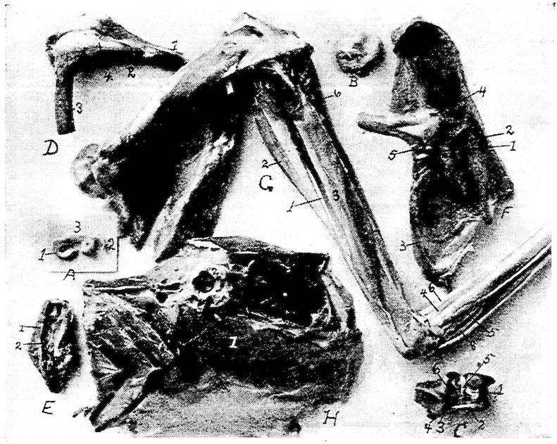
Fig. 25.—Inner layer of muscles of the thigh of a hen. Outside view. A. Pad of fibrocartilage or meniscus of femoro-tibial articulation. 1, Inner semi-lunar fibrocartilage. 2, External semi-lunar cartilage. 3, Anterior border. B. Tarsal meniscus or pad of fibrocartilage. C. Articular surface of the tarsal joint. 1, Posterior ligamentum cruciatum genu. 2, Femoral tendinous insertion of tibialis anticus. 3, Ligamentum tibio-fibulare. 4, Ligamentum interosseum. 5, Ligamentum transversum of the meniscus. 6, Anterior ligamentum cruciatum genu. D. 1, Tibia. 2, Fibula. 3, Femur. 4, External ligamentum laterale genu. E. Inferior surface of the tail of a hen. 1, Infracoccygis. 2, Lateralis coccygis. F. Outside pelvic view. 1, Obturator ligament. 2, Tendon of the obturator internus. 3, Ligamentum oblongum. 4, Ligamentum capsulare. 5, Gemellus. G. Outside view of leg of a cock. 1, Soleus. 2, Flexor longus hallucis. 3, Flexor perforans digitorum profundus. 4, Extensor brevis hallucis. 5, Extensor brevis annularis. 6, Extensor longus digitorum. 7, Tendon of the tibialis posticus. H. 1, Transversalis abdominalis. 2, Cotyloid cavity.
Origin.—By a small fleshy belly to the anterior part of the proximal end of the metatarsus.
Insertion.—It becomes tendinous about the middle third of the metatarsus. Extending down the anterior face of the metatarsus it is 121inserted to the inner tubercle of the base of the first phalanx of the outer or fourth toe.
Shape.—Long, slender.
Relations.—Posteriorly with the metatarsus.
Action.—Pulls the fourth toe upward and inward.
Flexor Hallucis Brevis. Location.—Postero-internal side of the metatarsus.
Origin.—Side and lower margin of the inner aspect of the hypotarsus of the tarso-metatarsus and from the shaft of this bone immediately below it.
Insertion.—Winds round inner side of basal joint of the hallux and is inserted on its under side.
Shape.—Small flat muscle gradually tapering into a tendon.
Relations.—Posteriorly with the metatarsus.
Action.—To flex the hallux.
Extensor Annularis Brevis. Location.—On postero-external side of metatarsus.
Origin.—External aspect of the hypotarsus and the shaft below.
Insertion.—Supero-external part of the basal phalanx of the fourth toe.
Shape.—Small flat muscle.
Relations.—Posteriorly with the metatarsus.
Action.—Extensor of the fourth toe.
Tibialis Posticus (Fig. 24, No. 23). Location.—In the posterior tibial region.
Origin.—From the whole shaft of the fibula below the insertion of the biceps flexor cruris, and from the shaft of the tibia a short distance below this insertion. Also from the interosseous membrane, and the adjacent surface of the tibial shaft.
Insertion.—Passing in front of the external malleolus, crossing the ankle-joint, finally inserted into the outer edge of the summit of the tarso-metatarsal bone.
Shape.—Long, subcylindrical.
Relations.—Medially, with the flexor perforans digitorum pedis; superiorly, with the postero-external portion of the tibia.
Action.—Extends the metatarsus upon the tibia.
Flexor Perforatus Indicis Secundus Pedis (Fig. 17, No. 5). Location.—The posterior tibial region.
Origin.—From the external surface of the outer condyle of the femur, just below the head of the gastrocnemius.
122Insertion.—Passing in an oblique manner along posterior to the tibia, extends through the superficial part of the tibial cartilage to the outer side of the leg; then crossing the tibio-tarsal joint, it passes through a special canal of the hypotarsus, down the back of the tarso-metatarsus, under the annular ligament of the sole of the foot and a cartilaginous structure below this ligament, over the trochlea, and is inserted to the middle shaft of the second phalanx of the second toe.
Shape.—Thin, flat, broadly spindle-shaped.
Relations.—Posteriorly, with the flexor perforatus medius secundus pedis, and anteriorly, with the flexor perforatus annularis primus pedis.
Action.—Flexes the toes.
Flexor Longus Hallucis (Fig. 17, No. 7; Fig. 25, No. 2). Location.—The posterior tibial region.
Origin.—By two tendinous heads: one from the posterior part of the femur, just below the two condyles; the other from the outer part of the external condyle of the femur. The deep flexor passes between these two heads.
Insertion.—Becoming tendinous at the lower third of the leg, passes beneath the superficial flexors in a special canal on the outer side of the tibial cartilage, and extends through the large canal of the hypotarsus (Fig. 12, No. F, 5) next to the shaft, then down to the apex of the accessory metacarpal bone. It is inserted to the tubercle on the inferior proximal end of the ungual phalanx.
Shape.—Large, fusiform.
Relations.—With the exterior of the deep flexors.
Action.—Flexes the hallux as its long flexor.
Flexor Perforatus Annularis Primus Pedis (Fig. 17, Nos. D, 8 and C, 6). Location.—The posterior tibial region.
Origin.—From the inner side of the belly of the flexor longus hallucis.
Insertion.—Becoming tendinous at the lower third of the tibia, passes a little to the outer side and through the tibial cartilage, then over the ankle-joint and through the hypotarsal canal (Fig. 12, No. 5) to the under side of the outer toe, where it forms a sheath through which the deep flexors extend. It gives off on either side a tendinous slip which attaches to the basal phalanx.
Shape.—Long, slender, compressed laterally.
123Relations.—Internally, with the flexor perforatus indicis secundus pedis, and externally, with the flexor longus hallucis.
Action.—Flexes the toes as their long flexor. A second flexor of the outer toe.
Flexor Perforatus Medius Secundus Pedis (Fig. 17, No. 9). Location.—The posterior tibial region.
Origin.—From a strong fascia that merges with the knee-joint, and by a tendon from the external condyle of the femur in common with the tendon of the flexor perforatus indicis secundus pedis.
Insertion.—Continued as a flattened tendon extending from the lower third of the leg, passing obliquely through the tibial cartilage and then through the interno-posterior canal of the hypotarsus, (Fig. 12, No. F, 5) inserted to the second joint of the middle toe.
Shape.—Long, fusiform.
Relations.—With the shaft of the fibula, which it covers.
Action.—Flexes the middle toe.
Flexor Perforatus Medius Primus Pedis (Fig. 17, No. 2). Location.—Posterior tibial region.
Origin.—From the inner side of the muscular portion of the flexor perforatus annularis primus pedis.
Insertion.—Terminating in a flattened tendon, passes through the tibial cartilage and hypotarsus, and then extends along the shaft of the basal joint of the third toe, and, dividing into two slips, inserts to either side of its shaft.
Shape.—Long, rather small.
Relations.—With the flexor perforatus annularis primus pedis on the inner side.
Action.—Flexes the third toe.
Flexor Perforatus Indicis Primus Pedis (Fig. 17, No. 4). Location.—The posterior tibial region.
Origin.—From a thin, broad tendon, on the external condyle of the femur, in common with the flexor perforatus medius secundus pedis.
Insertion.—Becoming tendinous, passes through the tibial cartilage and the hypotarsus of the metatarsus (Fig. 16, A) and inserts to the sides of the basal joint of the second toe.
Shape.—Long, slender, fusiform; somewhat flattened laterally.
Relations.—With the flexor perforatus medius secundis pedis, posteriorly.
Action.—A flexor of the toes.
124Flexor Perforans Digitorum Profundus (Fig. 24, No. 24 and Fig. 25, No. G, 3). Location.—The posterior tibial region.
Origin.—From the posterior part of the external condyle of the femur, from the posterior part of the tibia just below the summit, and from a point near the origin of the soleus.
Insertion.—Its heavy flattened tendon passes through the tibial cartilage, this tendon dividing, the branches pass along the under side of the toes, perforating the outer tendons and become inserted to the proximal tubercle at the base of the under side of the ungual phalanxes of the second, third, and fourth toes.
Shape.—Long, fleshy, with a long tendon.
Relations.—Closely with the tibial bone; the deepest of all the flexors.
Action.—Flexes the digits.
THE MUSCLES OF THE EYE
Orbicularis palpebrarum
Depressor palpebræ inferioris
Pyramidalis nictitans
Obliquus inferior
Rectus inferior
Levator palpebræ superioris
Quadratus nictitans
Obliquus superior
Rectus superior
Rectus externus
Rectus internus
Orbicularis Palpebrarum (Fig. 7, No. 30). (Synonym.—Orbicularis oculi.)
Location.—Upon the lacrimal and maxillary bones.
Origin.—From the lacrimal and maxillary bones and the ciliary margin.
Insertion.—To the lower free edge of the tarsal cartilage.
Shape.—Thin, delicate layer of fibers.
Relations.—Externally, with the skin; internally, with the lacrimal and the maxillary bones.
Action.—Aids in closing the eye.
Levator Palpebræ Superioris (Fig. 7, No. 24). Location.—Along the superior roof of the orbit.
Origin.—Along a longitudinal line found near the middle of the roof of the orbit.
125Insertion.—To the ciliary margin of the upper lid, near the outer canthus.
Shape.—Thin, delicate layer.
Relations.—Superiorly, with the bony wall, and inferiorly, with the eyeball.
Action.—Raises the superior eyelid.
Depressor Palpebræ Inferioris (Fig. 7, No. 29). (Synonym.—Malaris.)
Location.—Along the inferior border of the eye.
Origin.—From the inferior border of the interorbital foramen.
Insertion.—To the lower lid.
Shape.—Thin, flat, delicate.
Relations.—Superiorly, with the eyeball; inferiorly, with the inferior orbital wall.
Action.—Pulls the lower lid down.
Quadratus Nictitans (Fig. 26, No. B, 7). Location.—Above the eyeball along the inferior border of the upper wall of the orbit.
Origin.—From the sclerotic from the upper part of the ball.
Insertion.—To the upper part of the sheath of the optic nerve near the optic foramen.
Shape.—Broad, thin, quadrate.
Relations.—Inferiorly, with the eyeball; superiorly, with the orbital wall, with the superior oblique, and with the superior rectus.
Action.—Aids in pulling the nictitating membrane obliquely outward and downward over the forepart of the eyeball.
Pyramidalis Nictitans (Fig. 26, No. B, 8). Location.—Inferoposteriorly to the eyeball.
Origin.—From the lower nasal side of the eyeball.
Insertion.—The fibers converge toward the upper part of the optic nerve, into a tendon which passes through a pulley at the free margin of the quadratus. Inserts to the lower part of the margin of the third eyelid, the nictitans.
Shape.—Pyramidal; of thin layers of fibers.
Relations.—Internally, with the bony orbital wall; externally, with the eyeball.
Action.—Aids the quadratus nictitans.
Obliquus Superior (Fig. 26, No. B, 5). (Synonym.—Obliquus dorsalis.)
Location.—Superior to the eyeball.
126
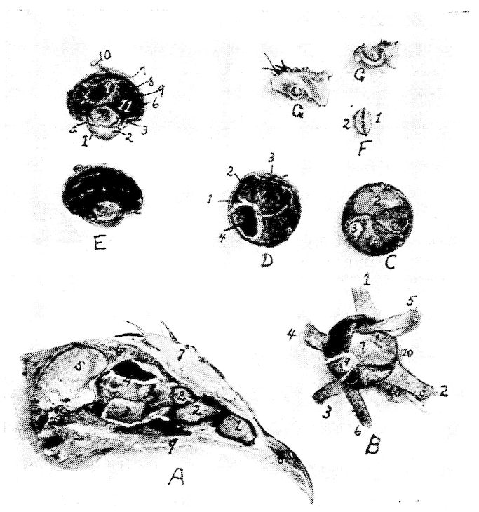
Fig. 26.—Structures of the eye and nasal passage. A. 1, The anterior turbinated bone. 2, The posterior turbinated bones. 3, Orbital gland. 4, Ophthalmic division of the fifth pair of cranial nerves. 5, Section through the cerebrum. 6, Section through the skull showing the air spaces. 7, Section through the comb. 8, Anterior portion of the upper beak. 9, Edge of the hard palate. B. The intrinsic muscles of the eyeball. 1, Superior rectus. 2, Internal rectus. 3, Inferior rectus. 4, External rectus. 5, Superior oblique. 6, Inferior oblique. 7, Quadratus. 8, Pyramidalis. 9, Optic nerve. 10, Edge of membrana nictitans. C. Posterior part of the eye showing at the inferior portion of the ball the pyramidalis muscle at 1, and at 2, the quadratus. 3, Optic nerve. D. Inner view of the posterior part of the vitreous chamber. 1, Sclerotic coat. 2, Choroid coat. 3, Retina. 4, Pecten. E. Longitudinal section through the eye of a hen. 1, Cornea. 2, Anterior chamber. 3, Scleral ring. 4, Crystalline lense. 5, Iris. 6, Retina. 7, Sclerotic. 8, Choroid. 9, Pecten. 10, Optic nerve. 11, Vitreous chamber. F. An edge view of the crystalline lense of a hen showing it to be asymmetrical biconvex. 1, The anterior side. 2, The posterior side. G. The upper section through the anterior portion of the anterior turbinated bone showing one complete circle and the lower a section through the middle turbinated showing one and one-half turns.
127Origin.—From the orbital septum just back of the inner canthus, the fibers passing backward and forward.
Insertion.—Into the sclera, anterior to the sclerotic plates.
Shape.—Small, thin, fan-shaped.
Relations.—With the eyeball, superiorly; at its extremities with the superior rectus, interiorly.
Action.—Rotates the eyeball.
Obliquus Inferior (Fig. 26, No. B, 6). (Synonym.—Obliquus ventralis.)
Location.—Inferior to the eyeball.
Origin.—From the interorbital septum near the inner canthus of the eye.
Insertion.—By a broad expansion to the inferior portion of the eyeball.
Shape.—Tendinous at its insertions; fan-shaped as it passes downward and outward.
Relations.—Internally, with the eyeball; and externally, with the bony orbital wall, with the inferior rectus muscle, inferiorly.
Action.—Aids in rotating the eye; opposes the obliquus superior.
Rectus Superior (Fig. 26, No. B, 1). (Synonym.—Rectus dorsalis.)
Location.—Superior to the eyeball.
Origin.—Along the supero-posterior border of the optic foramen.
Insertion.—To the sclera just within the margin of the bony circle of the eye.
Shape.—Flat, thin.
Relations.—Internally, with the obliquus superior; superiorly, with the bony wall of the orbit.
Action.—To pull the eyeball upward.
Rectus Inferior (Fig. 26, No. B, 3). (Synonym.—Rectus ventralis.)
Location.—Inferior to the eyeball.
Origin.—From the inferior border of the optic foramen.
Insertion.—To the lower portion of the eyeball within the margin of the bony circle of the eye.
Shape.—Thin, fan-shaped.
Relations.—Superiorly, with the eyeball; inferiorly, with the bony wall of the orbit.
Action.—Pulls the eyeball downward.
Rectus Externus (Fig. 26, No. B, 4). (Synonym.—Rectus lateralis.)
Location.—External to the eyeball.
128Origin.—By two heads infero-laterally to the optic foramen.
Insertion.—To the postero-external side of the eyeball.
Shape.—Short, fan-shaped.
Relations.—Internally, with the eyeball; externally, with the bony wall of the orbit.
Action.—Pulls the eyeball outward.
Rectus Internus (Fig. 26, No. B, 2). (Synonym.—Rectus medialis.)
Location.—Inner side of the eyeball.
Origin.—From the supero-anterior border of the optic foramen.
Insertion.—To the sclera on the anterior portion of the eyeball.
Shape.—Thin, fan-shaped.
Relations.—Internally, with the eyeball and with the pyramidalis; externally, with the bony wall of the orbit.
Action.—Pulls the eyeball inward.
THE MUSCLES OF THE EAR
Circumconcha
Tensor tympani
Circumconcha (Fig. 7, No. 32). Location.—Surrounds the external ear.
Origin and Insertion.—The circumconcha, surrounding the periphery of the ear, is adherent to the skull and loosely to the skin, and is attached to the outer terminus of the supra-occipital crest.
Relations.—Internally, with the skull; externally, with the skin.
Action.—Relaxes the tympanum.
Tensor Tympani (Fig. 7, No. 31). Location.—External to the quadrate and to the external auditory meatus.
Origin and Insertion.—From the surface of the quadrate and the inner end of the quadrato-jugal to the inner surface of the tympanum.
Shape.—A few fibers.
Relations.—Internally, with the quadrate and the quadrato-jugal; externally, the integumental duplicature.
Action.—Tenses the tympanum.
FUNCTIONS OF MUSCLES
To cause the feathers on the top of the head to lie flat:
Dermo-frontalis.
To raise the feathers along the superior part of the neck and along the dorsal region:
Dermo-dorsalis.
129To tense the patagial region:
Dermo-tensor patagii,
Dermo-temporalis.
To cause the feathers to lie close to the body:
Dermo-humeralis,
Dermo-pectoralis.
To tense the lateral cervical integument:
Dermo-temporalis.
To support the lingual apparatus and superior larynx:
Platysma myoides.
To manipulate the feathers and skin of the inferior part of the neck:
Cleido-trachealis.
To control the skin in the shoulder region:
Dermo-spinalis.
To cause the feathers of the back to lie close to the skin:
Dermo-iliacus.
To depress the humeral region:
Dermo-ulnaris.
To close the jaw:
Temporalis,
Pterygoideus internus,
Pterygoideus externus,
Masseter.
To open the jaw:
Biventer maxilla,
Entotympanic.
To raise the trachea and hyoid apparatus:
Digastricus.
To elevate the tongue:
Mylo-hyoideus.
To pull the tongue to one side:
Singly[5]—
Cerato-hyoideus,
Stylo-hyoideus.
Alone when the larynx is fixed—
Sterno-hyoideus.
Those which protrude the tongue from the mouth:
Together—
Cerato-hyoideus,
Genio-hyoideus.
To aid in deglutition:
When the base of the tongue is fixed—
Sterno-hyoideus.
To depress the tongue:
Together—
Sterno-hyoideus.
130To depress the tip of the tongue and elevate the base:
Depressor glottis.
To elevate the hyoid arches:
Cerato-glossus.