OUTLINE FOR LABORATORY STUDY OF THE CHICK
Reading edition
The objects of laboratory study of the embryos are as follows:
1. To study the living embryo.
2. To study the entire embryo:
With the dissecting microscope, as an opaque object.
With the compound microscope after killing, hardening, clarifying, and mounting.
3. To study embryos by dissection, in later stages.
4. To study the serial sections with the compound microscope.
THE LIVING EMBRYO
The egg is opened under warm physiological salt solution: 0.8 per cent. sodium chlorid in distilled water heated to a temperature of 38°C.
Gradually pick away the shell at the large end. Note that there are two membranes and an air cell. Strip off the membranes. When sufficient shell and membranes have been removed from the large end, invert the open end of the egg in the salt solution and allow the contents to flow out. Care must be taken not to break the yolk. The embryo, or blastoderm, lies upon the surface of the yolk, which is usually turned with this body uppermost. Separate the blastoderm by cutting around the outside of the area vasculosa. In doing this a small pair of slightly curved scissors is needed. After the embryo, or blastoderm, has been separated, gently float it into a watch crystal with the flat bottom submerged in the salt solution. The watch crystal with its contents may now be gently lifted out. Next remove the vitelline membrane. The vitelline membrane is the delicate transparent membrane covering the blastoderm, The embryo is now ready for study.
THE PREPARATION FOR STUDY OF ENTIRE EMBRYOS AND SECTIONS
The following processes may be used in killing embryos up to four days, or ninety-six hours, of age. After removing the embryo as described above, spread the blastoderm out in the watch crystal and pipette off the salt solution. Allow it to stand till the edge of 331the tissue begin slightly to adhere. Then slowly add the killing fluid by aid of a pipette, dropping it on the center of the embryo. The pipette must be held low or the mechanical interference will dislocate the parts.
Older embryos are submerged with their membranes intact into the killing fluid. The quantity of fluid should be several times the bulk of the specimen. Kleinenberg’s picrosulphuric acid may be used as a killing fluid. This fluid is a saturated solution of picric acid plus 2 per cent. sulphuric acid, to which is added twice its volume of water.
Chick embryos from one to two days old should be left in this fluid from one and one-half to six hours. Embryos from two to four days old two and one-half to six hours. Remove the specimen from the killing fluid, and place it in 70 per cent. alcohol. Change the alcohol every twenty-four hours until the color ceases to come out of the embryo. Preserve in 80 per cent. alcohol.
If the specimen is to be mounted whole, transfer it from 80 per cent., then to 50 per cent., then to 35 per cent., and, finally to water. Small embryos should remain in each fluid thirty minutes and large ones sixty minutes.
The following method may be used for staining embryos: Dilute Delafield’s hematoxylin with four times its volume of water. To every 6 cubic centimeters of this diluted hematoxylin, add one drop of Kleinenberg’s undiluted picrosulphuric acid, and leave specimen in the fluid thus prepared until it is stained through. This will require from one to three hours. Now pass up through the series of alcohols to 70 per cent. Next extract the excessive stain with 1 per cent. hydrochloric acid in 70 per cent. alcohol. Wash repeatedly with 70 per cent. alcohol to free from the acid; then transfer the specimen to 80 per cent. alcohol and leave in this for several hours for complete removal of the acid; then transfer to 95 per cent. alcohol for thirty minutes. Allow the specimen to remain in absolute alcohol for one hour. Introduce a layer of oil of cloves or xylol beneath the alcohol. This may be done by gradually allowing the fluid to run down the side of the bottle. After the embryos have sunk into the oil and begun to appear transparent, remove the fluid and add fresh oil. After the specimen is sufficiently transparent, mount in balsam, supporting the cover slip so that it will not rest on the embryo.
In staining for section, place the embryo in borax carmine from 332the 50 per cent. alcohol and leave twelve hours. Then wash in 50 per cent. alcohol; after which transfer to 70 per cent. alcohol for six hours. Clarify in oil of cedar or oil of cloves, and place in melted paraffin for two hours. Imbed in paraffin, and section.
Embryos may be hardened, imbedded, and sectioned after the usual methods, using either paraffin or celloidin, and the sections stained with hematoxylin and eosin after sectioning.
POINTS TO BE OBSERVED IN THE STUDY
1. The embryo twenty-nine to thirty-four hours old with from ten to fourteen somites.
A study of the egg.
In opening the egg, observe that the shell membranes are double.
Observe that the shell is porous.
Note the air cell at the large end of the egg and note that the space or cell lies between the outer and the inner shell membrane.
After the egg contents have dropped into the salt solution, note extending from the ends of the yolk the twisted denser cords of albumen. These are the chalazæ, which act as stays to the yolk. Note that the yolk is surrounded by a delicate membrane. This is the vitelline membrane. The yolk is the true ovum and serves as food for the developing embryo.
A study of the living embryo.
Note the amount of yolk that is covered by the blastoderm. Note the slipper-shaped, transparent center of the blastoderm. This is the area pellucida. In the center of this there is a narrow white streak. The area opaca is the area lying external to the area pellucida. In this there is the area vasculosa.
A study of the embryo entire, including the vascular area.
The blood islands show as irregular deeply stained masses in the vascular area. At this stage they are inclosed in wide anastomosing tubes, the extra-embryonic blood-vessels, which open peripherally into the bounding sinus terminalis.
The following structures may be identified:
The neural tube forming the axis of the embryo. In the anterior region may be noted the forebrain, the optic vesicles, the midbrain. The hindbrain is subdivided into the neuromeres. The cord, or myelon, of the neural tube back of the hindbrain is closed in front but is open behind.
333The head projects above the blastoderm. The fold which unites the ventral surface of the head with the blastoderm is called the head fold.
The diverging folds of the myelon encloses the primitive streak.
On each side of the neural tube are the mesoblastic somites. The series is continued behind by the undivided segmental plate.
The heart is beneath the hindbrain. The portion of the body cavity in which it lies, is bounded in front by the head fold and behind by the diverging limbs of the splanchno-pleure. Its posterior or venous end receives the vitelline veins from the vascular area. The anterior or arterial end is prolonged into the ventral aorta.
Note that the axis is somewhat bent.
The head fold of the amnion is noted to extend over the anterior end of the head.
A study of the transverse sections.
The sections about 20 micro-millimeters thick should be cut serially, and so mounted. Knowing how many micro-millimeters the embryo is in length will enable one to make a diagram of the fetal structure in the study of the series of transverse sections. The sections should be drawn in the order studied so as to obtain relative structural ideas.
With the microscope study the following regions:
1. Optic vesicle.
2. Midbrain.
3. Posterior half of the heart.
4. Myelon in the closed region.
5. Myelon in the open region.
6. Through the primitive streak.
2. The embryo twenty to twenty-four hours old with from two to six somites.
Compare the parts in this embryo with those of the embryos from twenty-nine to thirty-four hours old. Note and describe the relations of the embryo, the area pellucida, and the area opaca. Note how much of the yolk the blastoderm covers.
In studying the entire embryos, note the condition of the medullary plate. Observe if the tube is formed in any part; how far back the head plate extends, if the heart can be seen; and if the primitive streak is longer or shorter than in 1.
Study relations of structures and make drawings through the point 334of divergence of the walls of the fore gut; also through the somatic region, and through the primitive streak.
Study all parts, as the head fold, the heart, and the fore gut.
3. The embryo forty-four to forty-eight hours old with twenty-four to twenty-nine somites.
Remove the embryo with the entire area, and preserve it.
Note and carefully describe the changes visible to the naked eye since the thirty-fourth hour.
In making a study of the entire embryo we note there has been a rapid growth of the dorsal surface of the head, which has become more bent. This bend, in the region of the midbrain is called the cephalic flexure. The forebrain and part of the midbrain form almost a right angle with the rest of the head. The head is compressed laterally and free from the blastoderm. The dorsal side of the trunk is turned up, and there is a twisting of the axis of the embryo just back of the heart. The tail fold begins at about this time and may or may not be visible. The optic vesicles are relatively smaller in relation to the brain than in 1. Note the part of the forebrain to which they are attached. Observe the inner and outer layers of the retina, the lens, the choroid fissure, and the cavity of the vitreous humor.
Note the auditory vesicles and whether or not they are closed sacs.
Note that the heart has grown in length and has become doubled on itself. The two ends are fixed. Note the relation of the heart to the afferent and efferent blood-vessels. Note that two, and possibly three, visceral pounches are visible. Note whether or not they are ventral to the midbrain. The first, the hyomandibular pouch, is bounded in front by the first visceral, or mandibular, and behind by the second visceral, arch. The second pouch is bound in front by the hyoid, and behind by the third visceral arch. The third pouch is bounded in front by the third visceral arch, and behind by the fourth. Note the number of mesoblastic somites and the condition of the mesoblastic segmental plates.
Note how far back the foregut is closed. Locate the head fold of the amnion, and note how far back it is closed. Note changes that have taken place in the vascular area. In studying the sections it will be found that a section cut transversely to the trunk will pass horizontally through the forebrain and through the midbrain.
Study a section through the trunk a short distance behind the heart. Observe the elevation of the axis of the body. Note the 335way in which the lateral folds, or the lateral limiting sulci, in the somatopleure, delimit the embryonic from the extra-embryonic area.
Note the appearance of the mesenchyme, the approximation of the two dorsal aortæ, the appearance of the amniotic folds. Observe in the mesoderm the posterior cardiac veins, and the myotomes, or muscle plates. The sclerotome is made up of the mass of mesenchyme between the myotome, on the one hand, and the neural tube and the notochord, on the other. Note the folding of the splanchno-pleure, and note if there is present the Wolffian duct, or the nephrotome.
Study sections through the optic vesicles. Note if there is the beginning of the lens. Do you note the diverticula of the pharynx? Can you identify the closed amnion and the chorion?
Study sections through the auditory pit. Note fusion of the gill pouches with the ectoderm. Note the blood-vessels. Study sections through the region of the heart, through the roots of the vitelline veins, and through the primitive streak, if it is still present.
For this study it will appear that the anterior end has developed in advance of the posterior end. The tail fold has probably just begun.
Write a description of the pharynx, and of the circulation at this stage.
4. The embryo sixty-eight to seventy-two hours old with cervical flexures formed.
In a study of the living embryo note the changes visible to the naked eye since forty-eight hours old. Note the difference in the blood-vessels of the vascular area. Name the arteries and veins. Note the beating of the heart.
In a study of the entire mount note that a second, the cervical flexure, has appeared in the head. Note that the tail fold is well formed. Note the position of the embryo on the blastoderm. Determine if the amnion is completely closed. Note the olfactory pits on the ventral surface of the head, a short distance in front of the optic stalks. Note the telencephalon, a rudiment of the cerebral hemispheres and an extension of the primary forebrain. It is bilobed anteriorly. The optic stalks are attached to the floor near the anterior end of the thalamencephalon. Note the infundibular region, which is the depressed region behind the optic stalks. In the roof of the thalamencephalon there is a short diverticulum, the epiphysis. The mesencephalon, or midbrain, forms the apex of 336the cranial flexure, and is united to the hindbrain by a narrow isthmus. The metencephalon, or rudimentary cerebellum, appears as a thick portion on the most anterior division of the hindbrain. The rest of the hindbrain is provided with a transparent roof and constitutes the myelencephalon, or the rudimentary medulla oblongata. Observe the inner and the outer wall of the optic cup, the lens, the choroid fissure, and the posterior, or vitreous, chamber.
Note the form of the otocyst, or auditory sac. Note above which visceral arch it lies. Note the number of visceral clefts. The visceral arches are formed by the thickening of the walls of the bounding clefts. The visceral arches are as follows: the first is the mandibular, or hyomandibular, arch, which is in front of the first cleft. From this there is developed the lower jaw. Note if there is a maxillary process arising from the dorsal angle of the arch. The second arch is the hyoid arch, which is located behind the first cleft. Then follow in order the third, the fourth, and the fifth visceral arches. Note above the mandibular arch the rudimentary trigeminal ganglion, and above the hyoid arch the rudimentary acoustico-facialis. The latter is in contact with the anterior walls of the auditory sac. The rudimentary glosso-pharyngeal ganglion is noted above the third visceral arch. The vagus, or pneumogastric, ganglion is located above the fourth and the fifth. Note the form and the position of the heart. Note the anterior and the posterior limb rudiment in the trunk.
In a study of the sections it is found that cuts transverse to the trunk pass about horizontally through the forebrain. At this age the following sections of the embryo should be studied:
First, through the hindbrain, at which level will be noted the auditory sacs, the neuromeres, the trigeminal, the acoustico-facialis, and the glosso-pharyngeal and the vagus ganglion.
Second, through the upper part of the pharynx, at which level will be observed the midbrain, the hindbrain, the visceral pouches, the nerves, and the blood-vessels.
Third, through the choroid fissure of the optic cups. Note the parts of the eye, and, on the other side of the section, the heart.
Fourth, a study of a section through the olfactory pits.
Fifth, a study of a section through the pancreatic and the hepatic diverticula.
Sixth, a study at the beginning of the allantois through the hind-gut.
337At this age it is of interest to study the systems of organs. Observe the manner in which the splanchno-pleure folds to form the walls of the intestine. Note the commencement of the mesentery. Note that the foremost part of the alimentary tract is formed from the stomodeal invagination of the ectoderm. The hypophysis is formed from a dorsal outgrowth of this. Note its relation to the brain.
The following structures are formed from outgrowths of the ectoderm at this stage:
First, the visceral pouches.
Second, the median rudiment of the thyroid. This is an outgrowth from the pharynx between the two hyoid arches.
Third, the rudimentary lungs, which develop in a pair from a median ventral diverticulum of the alimentary tract, just behind the last visceral pouch. The esophagus is just posterior to this. The esophagus, very short at this stage, is continuous with a slightly wider part that develops into the stomach.
Fourth, the first liver diverticulum, and, at a short distance posterior to this, the second liver diverticulum.
Fifth, the pancreas is at a point where the intestine opens ventrally. It first appears as a slight thickening of the dorsal angle of the intestine.
Sixth, the ventral wall of the hind-gut forms a wide evagination.
Seventh, the beginning of the allantois.
After the whole series of transverse sections have been studied and drawn, construct a longitudinal section of the fetus, including a reconstruction of the alimentary tract.
At this stage the heart is a simple tube. The following divisions are distinctly visible: the auricular portion, the ventricular portion, the sinus venosus, and the bulbus arteriosus. The union of the two ductus Cuvieri and the ductus venosus form the sinus venosus. The two ductus Cuvieri are formed by the union of the anterior and the posterior cardinal veins. The ductus venosus is formed by the union of the small right and the large left vitelline vein. These latter veins return the blood from the yolk sac. The sinus venosus empties into the single auricle above which it is located. The single auricle is later divided into two chambers, the right and the left auricle. At this stage it is widest in the lateral direction. The auricle empties directly into the ventricle. The ventricle lies ventrally and behind the auricle. This location is due to the bending of the heart at this stage of development. Its hindmost portion 338forms the future apex of the heart. If the series of sections be studied from the posterior forward, the ventricle will be first to appear in the sections. Just beneath the auricular portion of the heart there is the bulbus arteriosus. The bulbus arteriosus soon divides into a number of aortic arches. There is an ascending pair in each of the visceral arches. The dorsal aorta is formed by the union of the aortic arches above the visceral arches. The aortic arches are first continued a short distance forward as the carotid arteries. The dorsal, or posterior, aorta passes backward under the notochord. The dorsal aorta divides into two parallel aortæ which give off on each side the vitelline arteries. Note other branches of this aorta.
The veins at this stage consist of the anterior and posterior cardinal, the ductus venosus, the ductus Cuvieri, and the vitelline veins.
Make drawing of the circulatory system after a completion of the study of the series of sections.
Make a study of the nervous system according to hints already given.
The dorsal and the ventral roots of the spinal nerves are given off separately, and secondarily unite. From the neuroblasts of the cord there are at regular intervals outgrowths representing the ventral roots. From the neural crest there develop segmental collections of neuroblasts which form the spinal ganglia, from which the dorsal spinal nerve roots develop. In fact this developmental stage can be observed in embryos only forty-eight hours old, first appearing as a line of cells springing on each side from the angle between the neural canal tube and the external epiblast. In the section from the embryo seventy-two hours old there are observed the rudiments of the development of these spinal nerves. Four primary ganglia develop in the neural crest of the head. These ganglia are as follows: the acoustico-facialis ganglia, which is located over the hyoid arch; the ganglia of the trigeminus, which is located over the mandibular arch; the ganglia of the glosso-pharyngeus, which is located over the third visceral arch; and the ganglia of the vagus, which is located over the third and the fourth visceral cleft.
In this stage of development the trigeminal and the acoustico-facialis are clearly visible.
On each side and dorsal to the aorta is noted the Wolffian body, or mesonephros. The Wolffian body (Fig. 82, E) consists of a series of tubules imbedded in the mesenchyme. The openings into the Wolffian duct lie just beneath the cardinal vein.
339In a study of the Wolffian duct determine just how far anteriorly and how far posteriorly it extends. Note whether it empties into the cloaca.
Each tube beginning in a blind extremity is later dilated. It has a thin wall, and is situated near the median portion of the Wolffian body. The tubule proper passing transversely, opens into the duct. The upper wall of the thin walled part is invaginated by a mass of mesenchyme that receives a small vessel from the dorsal aorta. The Malpighian corpuscle, consisting of a glomerule and Bowman’s capsule, is thus established. In the study of the four days old chick note the further development of these parts.
In a study of a chick four days, or ninety-six hours old, note to what extent the yolk is covered by the blastoderm. It will be noted that the embryo lies in the extra-embryonic cavity. This cavity is bounded above by the chorion and below by the splanchno-pleure. In removing the amnion from the embryo, note the relation to the somatic umbilicus. Note the relation of the splanchnic umbilicus to the splanchno-pleure. What relation has the allantois to the above?
In examining the head, locate the cerebral hemispheres, and note their development. Locate the pineal gland. Note changes in the olfactory pit and the eye. Locate the lens and the choroid fissure. Note that the maxillary process of the mandibular arch lies beneath the eye and behind the olfactory pits. Note the otocyst and the relations of the other arches to the above structures.
In a study of the trunk, note the tail, the allantois, Wolffian ridges, the heart, and the condition and the position of the rudimentary limbs.
Make drawing of embryo from the side view. Carefully cut off the head immediately behind the last visceral arch, and study and draw the structures observed on the ventral side. Note the maxillary processes, the mandibular and the hyoid arch, the nasal pits, and the fronto-nasal process, which is just beginning its development.
In a study of transverse sections observe from your drawing at what level the section is made. Study and draw a section made through the region of the anterior limbs. Note the spinal ganglion, the muscle plate, or myotome, the condensation of mesenchyme around the notochord, the pancreas, the liver, and the intestine; and note the distribution of the mesenchyme. Note the ventral roots of the spinal nerves, and the neuroblasts in the spinal cord.
340In sectioning the embryo from before backward, the first sections will pass horizontally through the hindbrain and the midbrain region. Note the parts, including the auditory vesicle. In the first series of sections also locate the ganglia of the pneumogastric, or vagus, the acoustico-facialis, the trigeminus, and the glosso-pharyngeal nerves. Note the notochord and the cardinal veins. After the disappearance of the ear, we observe the midbrain, which, is located at one end of the section, and the cord at the other end. The region between lies just above the pharynx. In the following series study the visceral arches. Note the third pair of cranial, or motor ocular, nerves. This latter nerve springs from the floor of the midbrain. From the ventral prolongation of the floor of the thalamencephalon there arises the infundibulum. The hypophysis is located just beneath the infundibulum. At this stage the hypophysis appears as a tube to empty into the mouth. This is an ingrowth of the oral epithelium.
In a study of a section through the center of the eye we should observe the lens and optic stalk. This is in the region of the optic chiasm. Note the choroid fissure and the pineal gland, the latter appearing just beyond the eyes. Just forward lies the telencephalon, or rudiments of the cerebral hemispheres.
In a study of the alimentary tract we note that the mouth is bounded by the mandibular arches. Note the maxillary processes, and the ventral surface of the head. A finger-like diverticulum, extending from the roof of the ruptured double membrane, formerly separating the pharynx from the arches, forms the hypophysis. The great development of the visceral pouches makes the pharynx rather complex. In studying sections horizontally through the pharyngeal region note the various visceral arches and pouches. Note the arteries of the thyroid diverticulum.
In the series note the changed development of the lung rudiments, the glottis, the esophagus, the trachea, and the bronchi. The bronchi appear in pairs. Note that the liver has assumed proportions, and that it surrounds the common trunk of the vitelline veins, which it divides into two parts. The sinus venosus lies close to the heart. The ductus venosus is also surrounded by the liver. Above the tip of the ventricle we note a dilatation which represents the stomach. The hepatic, or bile duct is located immediately behind the stomach. This duct is formed by the fusion of the right and the left duct. Locate, draw, and describe the pancreas; trace the intestine; 341locate the splanchnic umbilicus, or yolk stalk; locate the allantois stalk, and trace its connection with the hind-gut.
In the series, locate and study the Wolffian ducts, the beginning of the Müllerian duct, the embryonic kidney, or mesonephros, the permanent kidney, or metanephros. The urino-genital ridge is made up of all the above except the last named. The urino-genital ridge forms a rounded projection on each side of the mesentery into the dorsal angles of the body cavity.
The Wolffian ducts empty into the cloaca. There are two ducts which may be traced far forward, and which are found to extend backward along the lateral margin of the ridge to the cloaca. Along the greater part of their length we note tubules emptying into them.
Beginning near the anterior end of the urino-genital ridge, we note that the Müllerian ducts arise from a thickened line of epithelium. The greater part of the ridge is formed by the mesonephros. This is made up of a series of tubules in each of which we may distinguish two parts as follows: a tuft of capillaries from the aorta forming the glomerules, surrounded by a thin walled invaginated capsule, making up the Malpighian corpuscle; and the tubules proper. The tubules lead from the corpuscles, or glomerules, to the Wolffian duct.
The germinal epithelium constitutes the essential portions of the gonad, or ovary, or testis. The germinal epithelium arises from a thickening of the peritoneum of the median wall of the ridge. The gonad is found near the anterior end of the ridge. At about this age of the embryo there should appear the primitive ovary or testis. Near the posterior termination of the Wolffian duct and from the dorsal diverticulum there arises the ureter, or metanephros duct.
In these series there should be studied the heart and circulation, which will be found similar to the sections from the embryo seventy-two hours old.
Work out a summary of the relations of the allantois and the yolk stalk to the intestines; the relations of the Wolffian ducts to the intestines; the origin of the ureters from the Wolffian ducts; the relations of the lungs, the liver, and the thyroid gland; the relations of the blood-vessels; a study of the muscle plates; the relations of epiphysis, hypophysis, infundibulum, mouth, and pharynx; the relations of the vagus, or pneumogastric, trifacial, acoustico-facialis, and glosso-pharyngeal nerves to the visceral arches.
In addition to the study of embryos at the end of each of the first four days of incubation, a study should be made of preserved 342museum specimens each of which represents the development of a day up to and including the twenty-first day.
THE DERIVATIVES OF THE GERM-LAYERS
From the three primary germ-layers are developed the various tissues and organs of the body by metamorphoses which may be referred to the two fundamental processes of specialization, or the adaptation of structure to function, and of unequal growth, which latter results in the formation of folds, ridges, and constrictions.
From the ectoderm are produced:
The epidermis and its appendages, including the nails, the epithelium in connection to the feathers and the feathers.
The infoldings of the epidermis, including the epithelium of the mouth, epithelium of the salivary glands and the anterior lobe of the pituitary body, or hypophysis.
The epithelium of the nasal tract with its glands and communicating cavities.
The epithelium lining the external auditory canal, including the outer stratum of the membrana tympani.
The lining of the anus.
The epithelium of the conjunctiva and of the anterior part of the cornea, the crystalline lens.
The spinal cord, the brain with its outgrowths, including the optic nerve, the retina, and the posterior lobe of the pituitary body.
The epithelium of the inner ear.
From the entoderm are produced:
The epithelium of the respiratory tract.
The epithelium of the digestive tract, from the back part of the pharynx to the anus, including its associated glands, the liver, and the pancreas.
The epithelial parts of the middle ear and of the eustachian tube.
The epithelium of the thymus and the thyroid bodies.
From the mesoderm are developed:
Connective tissue in all its modified forms, such as bone, cartilage, lymph, blood, fibrous and areolar tissue.
Muscle tissue.
All endothelial cells, as of joint-cavities, bursal sacs, lymph sacs, blood-vessels, pericardium, and endocardium, pleura, and peritoneum.
The spleen.
343The kidneys and ureters.
The testicles and the system of excretory ducts.
The ovary and oviduct.
PREPARATION OF STRUCTURES FOR STUDY
It is hoped that the following suggestions will be helpful in the laboratory work.
Directions for Dissecting Muscles.—The muscles that are brought into great play in movements of the bird’s limbs are dark carmine in color, while those which are not brought greatly into use are pale or white in color.
The tendons are made up of white fibrous connective tissue, are very dense, and pearly white in color.
In securing a bird for dissection of muscles it is best to select one in rather poor flesh, as the fat is annoying. The bird may be chloroformed or killed in a bell jar by aid of illuminating gas. After the bird is dead pluck all the feathers and immerse it in a 10 per cent. solution of formaldehyde or of 80 per cent. alcohol. It is best to puncture the abdominal wall so that the fluid may at once fill the abdominal cavity and more readily gain access to the chest cavity; and to puncture the skin at various points so that the liquid may more quickly become disseminated among the muscular structures. Post-mortem changes quickly take place if these precautions are not taken. The liquid surrounding the carcass should be at least twice the quantity of the bulk of the carcass.
The first dissection should be to lay bare the dermal muscles. The dermal muscles are of two kinds, true dermal and dermo-osseous. The dermal muscles have their origin and insertion in the skin, and control the movements of the different groups of feathers. The dermo-osseous have their origin on some part of the skeleton, and insert to the integuments.
The dermal muscles vary with the characteristics of the bird, we do not find all the known dermal muscles in any one specimen. A cock of the Cornish breed will show these muscles best developed. Birds possess an enormous system of minute muscles divided up into an infinite number of fasciculi, to act harmoniously upon the feather quills and to agitate collectively the plumage. By the aid of a low-power lens the action of the feather muscles in the large quill butts of the wing or the tail may be studied.
The muscles may be studied in groups as outlined in the text. 344Make an incision through the skin down to the bone on the superior part of the head, parallel and close to the base of the upper mandible, and extending completely across. From the outer end of this make an incision backward and down to the skull and posteriorly. The muscles of the upper part of the neck will then be exposed. The straight incision should extend to about a half inch on the inside of the upper eyelid of the same side. Reflect the flap of skin from the top of the skull, and carefully examine the under side of it in the median line, where it overlies the frontal region. The dermo-frontalis will be observed if it be present. In many birds, especially in females, it may not be discernible, and may be considered absent. To expose the circumconcha make an incision completely around the ear; then carefully dissect to the ear base. A dermal circular muscle should be observed. To expose the dermo-temporalis extend the longitudinal incision down the back of the neck to a point between the clavicular heads, carrying it just through the skin and about one-fourth of an inch to the side of the median line. Remove the skin from the throat and the anterior portion of the chest. Lay open the alar and parapatagial duplicatures of the skin. This exposes a number of dermal muscles. The dermo-temporalis is now observed to extend from a small depression just above and anterior to the temporal fossa. It makes slight attachments to the temporal muscle, which it covers, and extends backward as a thin ribbon-shaped muscle, the fibers blending with those of the cleido-trachealis, and becomes lost upon the skin in front and opposite the shoulder-joint. At times its fibers blend with those of the dermo-tensor patagii.
As the musculature of the fowl is loosely arranged, the rest of the dissection is easily done if care be exercised.
The ligaments may be dissected after the completion of the study of muscles, using the same subject or, a two pound broiler be prepared by killing in the gas chamber, plucking the feathers and parboiling just till the flesh becomes tender and is easily removed, it will be observed that all structures can be removed from the points exposing the ligaments distinctly. The ligaments appear swollen and more easily observed for study.
Directions for the Study of the Viscera.—Carefully remove the right and the left abdominal and thoracic walls, allowing a strip of tissue to remain in the median line to hold the organs in their normal position. To open these cavities it is necessary to use the 345bone saw and the scalpel. The organs may now be studied from each side. To make a longitudinal section through the median line, select a small bird, one weighing not more than 2 pounds, kill, and preserve in a 10 per cent. solution of formaldehyde for three days. Then with a sharp, thin, long-bladed knife make an incision at one sweep through the median line of the body down to the back bone, and with the bone saw section through the vertebræ. If it is difficult to cut through the breast-bone, saw through before making the incision. In small birds the entire cut may be made without the aid of the saw.
Directions for the Study of Arteries.—Arteries should be injected. Veins are usually more or less filled with blood so that the tracing of these is not so difficult as the tracing of uninjected arteries. Nerves are white and no difficulty is usually encountered in tracing them.
The courses of arteries, veins, and nerves are side by side, and many of them, as in mammals, are arranged in the order of veins, arteries, nerves, the veins being in front.
The injection apparatus consists of the following parts: air-compression chamber, to which is attached a pressure pump. A manometer made of glass tubing 6 millimeters in diameter, inside measurement. This tube is partly filled with mercury, and a scale in centimeters is made from the top of the right-hand tube (Fig. 84, No. 8). This ruling, or gauge, should be about 15 centimeters long. Extending from the right extremity of the U-tube is a small rubber tubing which is attached to the chamber containing the injection fluid; and extending from the inferior part of this chamber is another tubing which has the injection needle attached to the free end.
The injection should be done under 120 millimeters pressure. The stop cock of the pressure chamber is released sufficiently to raise the mercury in the U-tube six centimeters, which multiplied by 2, the amount of work required to raise two columns, makes 120 millimeters pressure. If this pressure be maintained, all vessels should be injected without rupture. The same process may be used in injecting the air cells through the trachea.
The injecting material may consist of one part finely sifted plaster of Paris, four parts water, and sufficient gentian violet to make a violet color. For the coloring, red aniline may be used in preparing this material. The dye should be dissolved in the water to be used in making the injection liquid. Caution must be used and the work rapidly done, as the plaster soon sets, or becomes solid, in the 346needle or in the tubing. A small cannula should be used, since the endothelial lining of the arteries are easily injured and difficulty may thus be created.
Select for arterial dissection an old cock, as in a bird of this kind the arteries are larger and the difficulties are reduced. Select for bleeding and injection the ischiadic artery in the thigh region. With the sharp point of the thin blade of a knife make an incision lengthwise of the artery being careful not to strip back the endothelial lining of the artery. Allow as much of the blood as will escape before injecting; in fact, the arteries should be thoroughly emptied, so that there is no longer danger of a clot’s plugging some vessel and thus preventing its filling. Since the blood of most birds coagulates in about thirty seconds, this work must be done rapidly, care being exercised to keep the flow running as long as possible. After bleeding is completed, insert the cannula and tie the vessel tightly around the cannula to prevent the escape of the injecting fluid. See that all connections are sufficiently tight to prevent the escape of liquid under pressure. After the injection is completed, remove the cannula and tie the artery with a small twine, preferably cotton. Quickly remove all injecting fluid from the needle, the tubing, and the injecting chamber.
During the operation of bleeding the cock may be chloroformed, care being taken not to administer chloroform to kill him; for it is necessary to maintain life as long as possible so that the heart may be kept beating and all blood possible drained from the arteries. After the injection is completed the bird may be plucked and immersed in the preservative fluid in the same manner as in the preparation for dissection for muscles.
The arteries, the veins, and the nerves may now be dissected and studied in relation to one another and in relation to the muscles, the bones, and other structures. The skin should not be removed from the shanks till it is desired to dissect these parts, as the tissues quickly dry out. In fact, the tendons of the shanks and toes can best be dissected while the specimen is fresh.
A Study of the Structure of Bones.—Longitudinal and transverse sections of old bone may be made by making thin longitudinal and transverse sections with the bone saw, and then by making them very thin with a fine three-cornered file. Examining under the low power microscope, we note the lacunæ, the canaliculi, and the Haversian system.
347Similar sections in green bone may be studied if prepared as follows. Secure specimen of bone just removed from a fowl and place it for three days in a 10 per cent. aqueous solution of hydrochloric acid. Test by puncturing with a needle, and, if all the mineral salts are removed, place in a water bath and wash for four hours. Pass it through the fluids usually employed in preparing specimens for sectioning with the microtome. Stain as sections of other tissue for microscopic study. See the description below. If the ends of the bone be included, it will enable the student to study not only compact bone but also cancellated bone and articular, or hyaline cartilage, and in some of the bones, as the femur, the red marrow.
Special Technic for the Dissection of Cranial and Spinal Nerves.—It is rather difficult to dissect the cranial and spinal nerves of the fowl, owing to the fact that the structures are very small. The bone is rather hard and the nerve tissue so delicate that great skill must be attained to achieve any degree of success.
A simple technic has been developed as follows: Place the head and neck, or other structures of the spinal column in a 10 per cent. aqueous solution of hydrochloric acid for three or more days, the time depending on the size of the specimen and the amount of soft structures surrounding it. This solution removes all the calcium salts from the bone and makes the removal of the bony structures a less difficult task.
Directions for the Study of Soft Structures.—Secure a specimen of the tissue to be studied—lung, muscle, intestine, liver, pancreas—from a normal fowl just killed. The specimen should be not more than ½ inch square. After first hardening three days in 10 per cent. formaldehyde. Pass through the following fluids:
1. Alcohol, 95 per cent. 24 hours
2. Alcohol, absolute 24 hours
3. Alcohol and ether, equal parts 24 hours
4. 1 per cent. celloidin 24 hours
5. 2 per cent. celloidin 24 hours
6. 4 per cent. celloidin 24 hours
7. 6 per cent. celloidin 24 hours
8. 10 per cent. celloidin 24 hours
9. Place on block, and as soon as solid, place in 80 per cent. alcohol until ready to section. Histoloid or parlodion will take the place of celloidin.
348In placing specimen on the block be careful that the specimen lies conveniently for cutting the sections in the right direction. As soon as the surface has hardened a little, add a few drops of thick celloidin, and repeat until there is a good body of celloidin. Allow to stand until the tissues are quite firmly fastened to the block, but not long enough to permit shrinking. Then place in 80 per cent. alcohol until the specimen is perfectly firm, 12 or more hours, before cutting.
All tissues, cut sections, and mounted blocks are to be placed in 80 per cent. alcohol. As containers for this purpose shell vials will be most handy.
Cut the sections with the microtome as thin as possible, the thinner the better. The following process of staining will make the nucleus blue and the cytoplasm reddish.
1.
Float section in a tumbler of tap water.
2.
Place section on slide, and immerse in hematoxylon for five to ten minutes.
3.
Immerse in acid alcohol from two to five seconds.
5.
Place on slide, and immerse in eosin from one-half to three minutes.
6.
Wash thoroughly in alcohol.
7.
Clarify in oil of cloves, oil of cedar, or beechwood creosote, ten minutes.
8.
Mount in balsam.
9.
Label and study.
Delafield’s hematoxylon is prepared as follows:
Hematoxylon crystals 4 grams
Alcohol, 95 per cent 25 c.c.
Saturated aqueous solution of ammonia alum 400 c.c.
Add the hematoxylon dissolved in the alcohol to the alum solution, and expose in an unstoppered bottle to the light and air for three or four days. Filter and add:
Glycerin 100 c.c.
Alcohol, 95 per cent. 100 c.c.
Allow the solution to stand in the light until the color is sufficiently dark; then filter, and keep in a tightly stoppered bottle. The solution keeps well and is extremely powerful. So long as it is good the solution has a purplish tinge. If time permits, it would be wise to combine the alum, the hematoxylin, and the water, and to ripen the solution for two or three weeks before adding the other ingredients, which have a tendency to prevent oxidation.
349The acid alcohol is made as follows:
Absolute alcohol 70 c.c.
Distilled water 30 c.c.
Mix.
Above solution 99 c.c.
Hydrochloric acid 1 c.c.
Eosin is sold in two forms, that soluble in water and that soluble in alcohol. The eosin soluble in water is preferred, because with it a greater degree of diffusion in stain can be obtained.
Keep on hand a saturated aqueous solution and dilute with water as needed. The strength of the solution to be used varies somewhat with the tissue and the reagent in which it is to be fixed; but usually the strength should be between ⅒ and ½ per cent. when eosin is used after hematoxylin. The diluted solutions should contain 25 per cent. of alcohol, otherwise they will not keep well. When eosin is used before an aniline dye, such as methylene blue, a 5 per cent. or even a saturated solution should be used.
To Stain Sections of Liver for the Study of Kupffer Cells.—To bring out this reaction Keys suggests the following technic:
Fix small blocks of the fresh tissue of spleen or liver for eighteen to twenty-four hours in Müller’s fluid plus 5 per cent. mercuric sublimate. Imbed in paraffin and section to 4 microns. Fix sections to slide, and stain twenty to forty minutes with acid carmine. Wash, and transfer to equal parts of a 2 per cent. aqueous solution of potassium ferrocyanid and of a 2 per cent. aqueous solution of hydrochloric acid. Remove after three to ten minutes, wash in distilled water, and pass quickly through a 0.5 per cent. aqueous erythrosin solution. Dehydrate in alcohol, clarify in xylol, and mount in Canada balsam.
To Prepare Anatomical Specimens for a Museum.—The Keiserling method gives the best results, since by this method the tissues retain their normal color. The three steps are as follows:
1. Place the specimen in the following solution and leave from one to seven days, the length of time depending upon the size of the specimen.
Formalin 200 c.c.
Potassium acetate 30 grams
Potassium nitrate 15 grams
Water 1000 c.c.
3502. Pass the specimen through each of the following solutions, leaving it in each twenty-four hours or until the normal color is obtained. The specimen should be removed from alcohol as soon as color is attained. If it is left in the alcohol too long it will again lose some of its color.
Alcohol 40 per cent.
Alcohol 60 per cent.
Alcohol 80 per cent.
Alcohol full strength
3. Place the specimen in the following permanent solution, label, and place in museum.
Glycerin 40 c.c.
Potassium acetate 40 grams
Distilled water 400 c.c.
A small piece of thymol must be placed on the top of the liquid in each jar, or mold will develop and spoil the specimen.
To Make Specimens Transparent.—Specimens may be rendered transparent by the method of Spalteholz.
The steps are essentially as follows:
I. Preparation of the fresh tissue. If any parts are to be made conspicuous, as blood-vessels, or the lymphatic system, they must be injected with an insoluble, unbleachable substance. Spalteholz recommended carmine or methylene blue, and carbon. I have found Higgin’s ordinary black carbon ink excellent for this purpose. The system or systems are injected with this substance while the tissue is perfectly fresh.
II. Fixation. The tissues are fixed preferably in 10 per cent. formalin. The length of time for fixation depends on the size of the tissue. In formalin it requires from eight hours upward to fix completely.
III. Rinsing. Running tap water for five or ten minutes accomplishes the rinsing. If it is impracticable to pass directly into the bleaching fluid, the tissues may be kept temporarily in 60 per cent. or 70 per cent. alcohol. They should then be rinsed again thoroughly in water when ready to bleach.
IV. Bleaching. The bleaching fluid used is hydrogen peroxid to which ammonia is added until a white precipitate forms. The proportion is approximately two parts of peroxid and one of ammonia. The material is bleached until all the color is removed 351and the tissue looks perfectly clear or slightly opaque. It may be quite transparent as the protein coagulate is somewhat white, but all yellow or reddish color should be bleached away.
V. Washing. Rinse thoroughly in running tap water or through several changes of water until the odor of ammonia is quite gone.
VI. Dehydration. Pass gradually through alcohol solutions, 30 per cent., 50 per cent., 60 per cent., 70 per cent., 80 per cent., 95 per cent., and last through absolute alcohol. The time required for dehydration will vary with the size of the material, but in either 95 per cent. solution or in absolute alcohol the tissue should remain until well hardened, twenty-four hours or more. Dehydration is completed by passing from absolute alcohol, to a fluid one-half absolute alcohol and one-half benzol; thence for a few days to pure benzol.
VII. Clarifying. From benzol the material is finally clarified in winter-green oil or any other standard clearer, and put up in its final position in the museum jar.
In commenting on this method Dr. A. F. Contant says: “I have found this to work very successfully in the injection of the lymphatics of the human skin, which, as you know, is a rather delicate injection and has been difficult of demonstration by other methods. With this method, however, I have prepared whole portions, as the side of the face, the leg, etc., of small animals and embryos which show clearly the whole course of the lymphatic vessels and nodes, in situ, and their relations to surrounding parts.”
EQUIPMENT FOR THE DISSECTION LABORATORY
Figure 84 illustrates some essential equipment for the dissection laboratory. The equipment should consist of a pump, No. 1, which forces air through the rubber tube, No. 2, into the pressure tank, No. 4, which is guarded by the valve at No. 3. A dial, No. 5, indicates the pressure of the air within the tank. The air as needed is released through an outlet valve. The air now passes through the rubber tube, No. 6, into the left arm of the manometer at No. 7. The manometer is simply a glass tubing filled with mercury to the point indicated at No. 7. By the side of the opposite arm there is made a scale graduated in centimeters. The tube at No. 9 being attached to the Y-tube, conveys air under the same pressure into the injection chamber as that supporting the column of mercury 352of the manometer. It is necessary to have a pinch cock, as indicated at No. 11, to control the liquid within the tube. No. 12 illustrates the tube in the end of which, No. 13, there is inserted a trocar which is introduced into the artery and tightly tied with a cord.
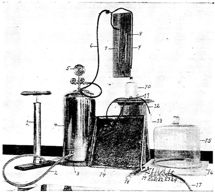
Fig. 84.—Photograph showing injection apparatus, dissection instruments, dissection pan and death chamber. 1, Pump. 2, Rubber tube leading from pump to compression chamber. 3, Stop cock. 4, Compression air tank. 5, Pressure indicator. 6, Rubber tube leading from pressure tank to manometer. 7, Top of mercury in manometer tube. 8, Open end of glass tube showing scale in centimeters. 9, Tube leading from 4 to injection chamber. 10, Injection chamber containing injection fluid. 11, Pinch cock on rubber tube connecting injection chamber with injection canula. 12, The tube. 13, Injection canula. 14, A metal dissection tray. 15, Death chamber. 16, Base of same. 17, Rubber tube connecting gas cock with death chamber. 18, Bone saw. 19, Bone cutter. 20, Scapula. 21, Tenaculum. 22, Forceps. 23, Straight scissors. 24, Small pair of curved scissors.
A convenient dissection tray may be made from galvanized iron. It should be 16 inches square and about 1 inch deep (Fig. 84, No. 14).
353A death chamber is made by using a bell jar 12 inches in diameter at the bottom. This is used on a perfectly smooth board 16 inches square. Through the center of this board is inserted a glass tube ⅜ inch in diameter. To this glass tube is attached a rubber tube the other end of which is attached to a gas jet.
The instruments needed in dissecting are illustrated in Fig. 84. No. 18 is a bone saw; No. 20 is a scalpel; No. 21 is a tenaculum; No. 22 is a pair of forceps; No. 23 is a pair of straight scissors; and No. 24 is a small pair of curved scissors.