ANGIOLOGY (continued 2)
Reading edition
The vena umbilicalis originates in the umbilical region and empties into the vena hepatica magna sinistra at a point where it comes out of the liver (Fig. 70, No. 29). This is the remains of an embryonal vein which collected all the blood of the yolk sac, passed on the left side of the large intestine to the body, took up the vena mesenterica and ended as the vena umphalo-mesenterica.
The Lymphatic System
The peculiarity of the lymph vessels is that they are associated with organs in which lymph cells are formed.
The lymphatic system consists of the lymph vessels and the cell-forming organs. In some instances the cell-producing organs are lymph follicles and in others lymph glands. For the most part the glands are replaced by plexuses which in many places surround the blood-vessels.
The lymph of birds is similar to that of mammals. The larger lymph vessels are similar to the veins, although the walls are always thinner. Its tunica intima is rich in elastic fibers and has a layer of endothelial cells on the inner side. The tunica media is formed of rings of smooth muscle fibers. The adventitia is composed of loose connective tissue.
The lymph vessels frequently form plexuses. The large lymph trunks follow the course of the larger blood-vessels, and frequently surround the arteries. All the lymph vessels of the body, exclusive of the lymph of the caudal region, form into a large trunk which originates on both sides of the celiaca communis and passes upward 255along the side of the abdominal aorta, reaching a point anterior to the celiaca. By receiving many vessels in this region it forms a plexus around the aorta, and finally divides into two vessels, the right and the left, ducti thoracici.
The lymph vessels of the left side of the head, the neck, and the lung, and the left wing, and also lymph vessels of the proventriculus and the throat enter into the left ductus thoracicus. They accompany the jugular vein and are closely associated with the thyroid gland.
The right thoracic duct receives the lymph veins from the right cervical lymph vein, and the right side of the head, the neck, the lung, and from the right wing.
After the right cervical lymph vein has passed through the right thyroid gland, it divides into two branches, one branch emptying into the right thoracic duct and the other into the vena cava dextra.
The lymph vessels of the liver, the stomach, the pancreas, and the duodenum enter near the root of the arteria celiaca into the large lymph trunk. The lymph vessels of the remainder of the intestines, of the kidney, and of the generative organs empty farther caudally.
The lymph vessels of the intestines take up the emulsified fat. This emulsion in birds is colorless. The vessels pass upward along the mesenteric arteries. There are no mesenteric glands. These vessels form a plexus around the arteria celiaca. The lymph vessels of the posterior extremities accompany the artery, especially the anterior iliaca externa, and empty into the thoracic duct at the point of the anterior iliaco-communis.
The lymph vessels in birds are numerous. The lymph glands are few. They are only visibly found in the anterior breast and the neck region, and sometimes in the wings. Lymph follicles are numerous in the intestines.
The thin walls of the lacteals, of the lymph vessels, and of the thoracic duct are made up of two tunics, the inner being the thinner and weaker.
The lymphatics of the foot unite to form the vessels along the sides of each toe. In palmipedes there are anastomosing branches which pass from the lateral vessels of one toe to those of the adjoining toe, forming arches in the uniting web of the foot. These branches form a small plexus at the anterior part of the digito-metatarsal joint, from which pass three or four lymph vessels. The anterior and 256internal branches accompany and form a network around the blood-vessels. The posterior and external branches receive the lymphatic vessels from the sole of the foot. They then ascend along the metatarsus and form, at its proximal articulation, a close network from which vessels pass along the tibial region, forming a plexus around it as far as the middle of the leg. From this there arises two branches. The smaller passes along the anterior part of the depression between the tibia and the fibula, as far as the knee-joint, where it joins the other branch, which accompanies the blood-vessel. The trunk formed by the union of these two vessels accompanies the femoral vessels. Forming plexuses in its course, it receives tributary vessels from the adjacent muscles. The iliac trunk accompanies the femoral vein into the abdominal cavity, entering just in front of the anterior end of the pubis. At this point it receives branches from the lateral parts of the pelvis and then separates into two branches. The posterior vessel receives some lymph from the anterior lobes of the kidney, and from the ovary, or testis, and communicates anteriorly with a branch formed by the lymph vessels adjacent to the anterior mesenteric artery, and posteriorly with a large vesicular plexus surrounding the aorta and its branches. This plexus receives the lymph from the renal plexus and from those accompanying the arteria media.
There are two sacral or pelvic vesicles which are situated at the angle between the tail and the thigh in the posterior part of the abdominal cavity. Each vesicle is a trifle more than a half inch long and a quarter inch wide, and is shaped somewhat like a kidney bean. They have muscular coats with striated fibers. These sacs are called “lymph hearts.”
The anterior division of the femoral lymphatic trunk accompanies the aorta, on which it forms a plexus with the branches of the opposite side, and with the intestinal lymph vessels. These vessels commence from a continuous plexiform network located between the mucous and the muscular coat of the intestine. They are larger at this point than where they leave the intestine to pass through the mesentery. They accompany the trunk of the anterior mesenteric artery and form a plexus around it.
Before reaching the region of the aorta, the intestinal lymphatic vessels communicate with the posterior division of the femoral trunk and with the lymph vessels of the ovary or of the testis. After passing to the region of the aorta they receive vessels from 257the pancreas and the duodenum, and terminate around the celiac axis with the lymphatics of the liver, the proventriculus, the gizzard, and the spleen, forming a rather voluminous plexus (Lauth).
The aortic plexus represents the receptaculum chyli and gives origin to two thoracic ducts, mentioned above, which passing on each side of the bodies of the vertebræ, pass one right and one left, over the lungs, from which they receive lymph vessels, and terminate after receiving the lymph vessels of the wing, into the jugular vein of their respective sides. The left thoracic duct, before emptying into the vein, receives the trunk of the lymphatics of the left side of the neck, and the right duct that of the right side of the neck, each tributary collecting lymph from all the structures of its side.
The lymphatics of the wing follow the course of the brachial artery, forming a plexus around it. These vessels are well developed in the elbow region. The principal trunk, following the humerus, receives collateral branches in its upper third. This vessel, when nearing the chest, receives two or three large lymph vessels from the pectoral muscles, and a branch which accompanies the brachial plexus.
The lymph vessels of the head accompany the branches of the jugular vein, collecting the lymph from the structures of the head and the neck.
The lymphatic vessels communicate at the anterior and posterior oblique anastomosing vessels. At the lower part of the neck each trunk receives a vessel, which accompanies the carotid arteries. Further on they are provided with a lymph gland which rests on the jugular vein.
The Blood and Its Functions
The special function of the blood is to nourish all the tissues of the body, and in this way to aid growth and repair. It furnishes material for the purpose of the elaboration of body secretions; it supplies the organism with oxygen; and it carries away carbon dioxid and other effete material. Blood is constantly in circulation.
Blood is red, opaque, and is, in the fowl, quite viscid. The exact tint of the blood depends on whether it is drawn from an artery or from a vein. Blood from a vein has a purplish tinge while that from an artery is a bright scarlet. The color of blood is largely due to pigment in the erythrocyte, called hemoglobin.
The reaction of the blood is alkaline, due to the phosphate and the 258bicarbonate of soda. The alkalinity of the blood is reduced by work. This is due to the formation of sarcolactic acid in the muscle. The odor of blood is due to volatile fatty acids. Each kind of fowl has its own peculiar odor. The taste of the blood is saltish, due to a small amount of sodium chlorid it contains.
The blood consists of the following substances:
First, the unorganized part, or fluid, the liquor sanguinis or plasma. It contains in solution proteids, extractives, mineral matter, and gases. The gases are held in loose chemical union.
The liquor sanguinis constitutes fully 66 per cent. of the volume of the blood. It is albuminous in nature and contains a small amount of coloring matter of a fatty nature. It holds in solution three proteids—fibrinogen, serum globulin, and serum albumin.
Second, the organized parts, or the cellular structure (Fig. 74, Nos. 1 to 21). The cells float in the plasma and consist of three groups: the erythrocytes, or red blood cells, the leucocytes, or white blood cells, and the thrombocytes.
Erythrocytes.—The average number of red cells (Fig. 74, No. 21) of the domestic fowl range between 3,000,000 and 4,000,000 per cubic millimeter. The red blood cells are flattened and elliptical in shape, and possess an oval elliptical nucleus. The average length is ¹⁄₂₁₀₀ inch and the diameter ¹⁄₃₈₀₀ inch, or 7 to 8 micra in diameter and 12 to 13 micra in length. However the diameters vary in different kinds of birds. The cytoplasm is yellow and glassy, and the nucleus takes basic stains and appears somewhat picnotic.
Thrombocytes.—The thrombocyte (Fig. 74, No. 19) is of about the same length as the erythrocyte but somewhat narrower. The nucleus is round, stains purple with the Wright’s stain, and the chromatin material is somewhat diffused. The diameter of the nucleus is nearly equal to that of the cell. The cytoplasm is pale and may show vacuoles near the nucleus. They may contain small circumscribed red structures. They vary somewhat in size and shape. There are in the domestic fowl between 45,000 and 55,000 per cubic millimeter.
Leucocytes.—There are, in the blood of the hen, 28,000 to 35,000 leucocytes per cubic millimeter. The leucocytes may be divided into five distinct types. These are as follows:
Polymorphonuclear leucocytes with eosinophilic rods (Fig. 74, No. 2) are round and have a diameter about equal to the length of the erythrocyte. The nucleus is polymorphous; that is, it has two or more lobes. The nucleus stains a pale blue, and the chromatin is diffused. The cytoplasm is colorless with bright red staining spindle-shaped rods. There are 28 to 32 per cent. of this type of cell in the blood of the hen.
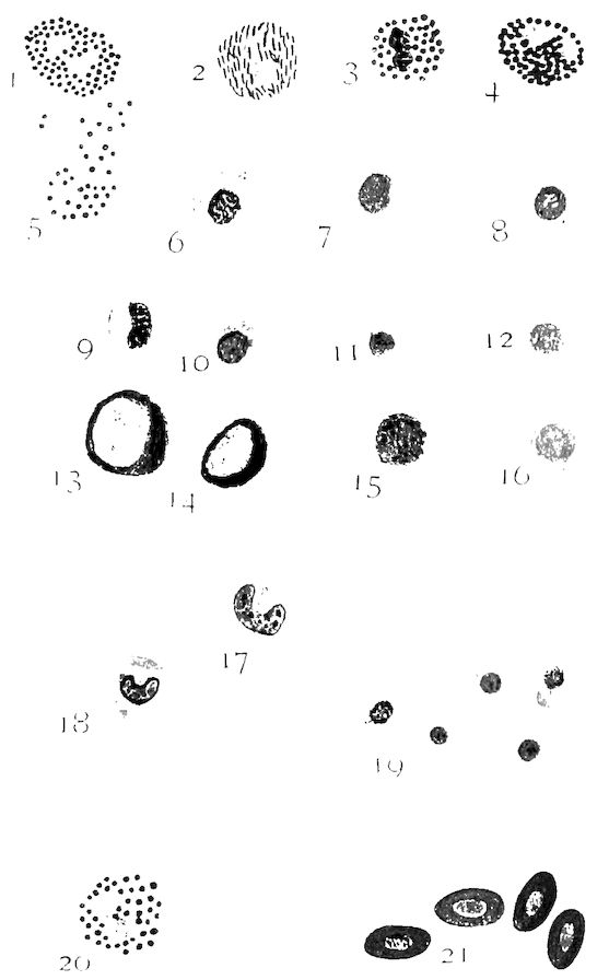
Fig. 74.—Blood cells of the fowl. Wright’s stain. From a S. C. Rhode Island Red cockerel. 1, Basophile (mast cell) with lilac staining spherical granules. 2, Eosinophile with rod-shaped acidophile bodies. 3 and 4, Eosinophiles with acidophile staining round granules. 5, Eosinophile ruptured. 6, Mononuclear leucocyte with vacuoles in the cytoplasm. 7 and 9, Transitional leucocytes (first stage). 8, Mononuclear leucocyte. 10, 11, and 12, Small lymphocytes. 13 and 14, Large lymphocytes. 15, A lymphocyte. 16, Lymphocyte (nucleus centrally located). 17 and 18, Transitional leucocyte. 19, Thrombocyte. 20, Neutrophile (polymorphonuclear). 21, Erythrocytes.
259Polymorphonuclear leucocytes with eosinophilic granules (Fig. 74, No. 3 and 4) are of about the same shape and size as the preceding. The nucleus is similar to the former except that it may appear slightly picnotic. The cytoplasm stains not at all or faintly blue; it contains round or spherical granules which stain a dull red. There is from 4 to 6 per cent. of this type of eosinophiles found in the domestic fowl.
Lymphocytes are round in shape and of about the diameter of the width of a thrombocyte (Fig. 74, No. 11). The nucleus is round, staining somewhat purple, and contains a diffused chromatin material. The cytoplasm exists in only small amounts; it lies to the side of the nucleus and stains a pale blue. This is the small lymphocyte. A similar cell but much larger also exists. This is the large lymphocyte (Fig. 74, No. 13). There is from 40 to 44 per cent. of the lymphocytes in the blood of the fowl. The small lymphocytes are most abundant.
Large mononuclear cells (Fig. 74, No. 6) either round or oval, in shape, whose diameter may be about that of an erythrocyte and at times much larger. The nucleus may be round, oval, or irregular, and at times rather crescent or U-shaped (Fig. 74, No. 17). The cytoplasm is abundant and completely surrounds the nucleus. The cytoplasm stains a paler blue than the nucleus. Both taking the basic stain as do the lymphocytes. These constitute 18 to 20 per cent. of the cells of the blood.
Mast cells or basophiles (Fig. 74, No. 1) are of about the same size and shape as the eosinophiles. The nucleus is round or oval, and stains a very pale blue. The cytoplasm is colorless, mostly to one side of the nucleus, and contains round or spherical purple staining granules. This type of cell constitutes from 2 to 4 per cent. of the white cells of the blood.
Structure of the Red Blood Cell.—The red blood cell is composed of a spongy stroma holding in its meshes the red coloring matter. The stroma, or framework, of the erythrocyte consists principally of nucleo-albumin; it contains lecithin, cholesterin, and salts. The red matter consists of an albuminous crystalline substance called hemoglobin, which forms about 90 per cent. of the total solid matter 260of the dried corpuscle. Each red cell offers a certain absorbing surface for oxygen. As the blood circulates through the delicate walls of the lungs and the air-sacs, it takes up oxygen; the blood at the same time delivers to the air carbon dioxid which has been brought from the tissues where active cell metabolism has been going on. This oxygen taken up by the erythrocyte forms a loose chemical union and is known as oxy-hemoglobin. In this form it is carried to the tissues of the body where it is given up by the erythrocyte to the tissues where oxidation is going on.
Hemoglobin is a crystallizable proteid substance containing carbon, hydrogen, oxygen, nitrogen, sulphur, and iron.
Formation of the Cells of the Blood.—The red blood cells are formed in the red marrow of the bone.
Polymorphonuclear leucocytes are formed in the red marrow of the bones, and the lymphocytes in the lymph glands and lymph follicles.
The bird carries a normal body temperature of 105° to 107° F. The average temperature of 50 mature hens and cocks was 106.8° F. The blood is of a deep red color.
Composition of the Blood.—The average composition of the blood of the domestic fowl as given by Owen, is as follows:
Whole shed blood:
Water 780 parts
Clot 157 parts
Albumin and salts 63 parts
1000 parts
Moist blood cells:
Average total weight 456.69
Water 342.52
Solid matter 97.50
Plasma:
Total weight 543.30 parts
Water 495.72 parts
Solid matter 30.72 parts
Blood when drawn and allowed to stand soon coagulates. In the blood of birds this process is very rapid, the blood coagulating, in most instances, in about one-half minute. Blood coagulates only in the presence of calcium salts.
During life, the liquor sanguinis is termed plasma; but after it has been shed from the body and coagulation has taken place, the 261liquid residue is called serum. Serum is plasma with its modifications as the result of coagulation, and as this latter process is brought about by the production of fibrin, we may say that serum is plasma minus fibrin-forming elements.
The proteids of the serum are serum globulin, serum albumin, and a ferment produced as the result of coagulation. As fibrinogen is used up in the process of coagulation, it is not found in the serum, but there is in the serum a proteid known as fibrino-globulin. This is produced from fibrinogen during the process of fibrin formation. The following tabulation gives a clear idea of the difference between the proteids of plasma and of those of serum:
Proteids of Plasma
Fibrinogen
Serum globulin
Serum albumin
Proteids of Serum
Serum globulin
Serum albumin
Fibrin ferment (nucleoproteid)
Fibrino-globulin
Fibrinogen is the precursor of fibrin.
The fibrin of the blood clot of the bird is soft and very lacerable. The serum is usually yellow.
THE FATE OF THE ERYTHROCYTE OF THE FOWL
The power of vascular endothelium to ingest red blood corpuscles has been studied by Keys.
When bacteria or other minute foreign bodies are injected into the blood stream of pigeons, they are rapidly withdrawn from the circulation into the tissues of the liver and of the spleen. The foreign bodies are noted to be contained within cells of a distinct type, which is found in both liver and spleen. This type of cell contains, in addition to the foreign substances injected, much yellow pigment, and when tested for iron by Pearl’s method gives a positive Prussian-blue reaction.
In such specimens there is a display of contrast to other tissues. There is an extensive content of cells possessing the distinct tone of Prussian-blue iron reaction. These cells are distributed rather evenly throughout both the spleen and the liver, but more numerously in the liver.
In the liver, under low-power magnification, these cells appear as blue patches, sharply differentiated from the red-stained parenchyma. These cells are larger in their greater diameter than the liver 262cells. They vary much in size and form. They bear a constant relationship to the venous capillaries, and often appear to occupy the lumen of the vessels. Under higher magnification it is noted, however, that each cell is an integral part of the endothelial intima lining of the capillaries. They are therefore fixed tissue cells, engaged by one of its surfaces upon the reticulum of the vessel wall, with a free surface bulging a greater or less degree into the lumen of the vessel. The attached surface of the cell follows exactly the line of the vessel wall. These cells are similar to those described for mammals by Kupffer and are called Kupffer cells or stellate cells. In the fowl Keys proposes the name hemophages. The nucleus of the hemophage stains a deep garnet with the carmine used in the above-given technic, and contains two or three very distinct and intensely stained nucleoli. In the hemophages, which are more nearly flat, the nucleus appears like those of the typical endothelial cells; whereas in the protruding hemophages of greater bulk, the nucleus is more vesicular and is irregularly pyramidal in form. Rarely two nuclei are found in one cell. Within this cell may be seen vacuoles of the cytoplasm which contain red blood corpuscles. These blood corpuscles have been phagocyted from the circulating blood stream. Approximately one-third of the intimal cells are hemophages. Each hemophage displays evidence that it contains, or has recently contained, one or more red blood cells. The cell body of the hemophage has no fixed morphology, but changes from time to time according to its phase of phagocytic activity. In a stage which the hemophage has recently ingested a red blood cell, the cell body bulges out into the lumen of the vessel and the nucleus is crowded to one side. At this time the red blood cell appears as those in the blood stream and possesses the characteristic staining reactions. The nucleus of the red blood cell stains deep reddish brown and the cytoplasm an even yellow bronze. In the next stage the cytoplasm of the hemophage gives a diffuse Prussian-blue reaction. Then in hemophages which represent later stages there are various stages of disintegration and digestion of the red blood cell. The first changes of the phagocyted red blood cell is hemolysis, the hemoglobin escaping into vacuoles of the cytoplasm of the phagocytic cell, leaving the nucleus-containing stroma distinctly outlined. The stroma may retain the original ovoid form or may become spherical; the nucleus in such instances remains ovoid. Gradually, both the stroma and nucleus lose their staining reaction until finally the 263vacuoles contract about a small indistinct remnant of the nucleus, which in its turn ultimately disappears. During this latter process the size of the hemophage gradually decreases. The hemoglobin, which has escaped into the cytoplasm of the hemophage, is seen to undergo a series of changes. At first the greater part of the pigment does not give the iron reaction but retains its yellow-bronze tone with erythrosin and occupies vacuoles of various sizes. Later the contents of the vacuoles give the iron reaction and with increasing intensity. Later there is a gradual decrease in the staining reaction indicating that the iron gradually disappears from the cells which extracted it from the red blood cells it digests. As a summary we find, that these cells take care of the worn out red blood cells. They devour them; hemolyze them, destroying the stroma and nucleus; split the hemoglobin and free the iron; and then finally return to their normal form.
The spleen contains the same type cells, but they are fewer in number. For the most part they are confined to the pulp cords and have no such evident relation to the vessel wall, or lumen, as in the liver.
The function of the cells of the spleen are essentially the same as those in the liver.
Iron freed from the worn out red blood cells is not retained by the cells freeing it, nor is it found in the bile. It does not occur elsewhere in the tissues of the liver and spleen. It is possibly discharged into the blood stream, and transported to the hemapoietic tissues. Cells which hemolyze red blood cells and liberate the iron are to be seriously thought of in connection with bile formation since bilirubin is approximately, if not identical with, iron-free hematoidon.
264