Volume 5 — DISEASES OF THE NERVOUS SYSTEM. (continued 37)
Reading edition
TUMORS OF THE SPINAL CORD AND ITS ENVELOPES.
BY CHARLES K. MILLS, A.M., M.D., AND JAMES HENDRIE LLOYD A.M., M.D.
DEFINITION.—Under Spinal Tumors will be included the growths or adventitious products which arise in the substance of the spinal cord or spring from its envelopes, membranous or bony, in such manner as to directly or by pressure involve the spinal cord. Tumors strictly confined to the cord are extremely rare. First in order of frequency are the new growths which develop from the spinal membranes, either the dura mater or pia mater, most frequently the former. Tumors originating in the bony spine, like those of the substance of the cord, are comparatively rare.1
1 A “Table of Fifty Cases of Spinal Tumor” (which will be frequently referred to) is appended to this article.
ETIOLOGY.—Under the predisposing causes of spinal as of intracranial growths are such diatheses or constitutional affections as cancer, tuberculosis, and syphilis. Under Pathology a table will be given from which it appears that of 50 tabulated cases, 3 were cancerous, 5 syphilitic, and 4 tubercular.
Traumatisms, such as a fall from a height, a blow on the back, a wrench or twist of the spine, or a sudden concussion as in a railway accident, sometimes serve as exciting causes of spinal tumors. Even when a diathetic or infectious predisposition exists, the patient might frequently escape from the special intraspinal localization of the disease were it not for the accidental infliction of direct injury to the axis. When no special predisposition is present, an injury is more likely to produce an osteoma, fibroma, or sarcoma than some of the other forms which will be mentioned, such as a glioma, myxoma, neuroma, or psammoma.
Spinal tumors are said by most authors to occur much more frequently in the male than in the female sex. Our tabulated cases, however, gave 22 cases among males, 21 among females, and 7 in which the sex was not given.
Fifty cases of spinal tumor gave the following result as to age:
Under 10 years 4 cases.
From 10 to 20 years 3 cases.
From 20 to 30 years 7 cases.
From 30 to 40 years 12 cases.
From 40 to 50 years 10 cases.
From 50 to 60 years 6 cases.
From 60 to 70 years 1 case.
Age not given 7 cases.
50 cases.
SYMPTOMATOLOGY.—Can tumors of the spinal membrane be separated by a study of symptoms from intramedullary tumors? While this may be theoretically possible, in practice it will be found difficult, and of little value even when it can be done. The spinal canal is of such narrow calibre that a growth of any size either in the membranes or the cord itself will soon directly or indirectly involve both. On this subject Erb2 speaks as follows: “The attempt has thus far been made in vain to secure, from amongst the individual symptoms, at least a few fixed points on which to base the diagnosis of intramedullary tumors. The following have been claimed as such: A somewhat long antecedent history of active local manifestations of irritation, belt-like pains, eccentric pains, definite paræsthesiæ, local paralysis, as in meningeal tumors; early and well-marked atrophy, which points to a larger involvement of the gray substance; striking fluctuations in the course of the disease, spontaneous improvement, and equally spontaneous growing worse again (Schueppel); and, finally, Schueppel has also tried to connect the occurrence of scoliosis (curvature of the spinal column to the side on which the tumor is situated) with the presence of a tumor. In this he is doubtless wrong, as this manifestation merely depends on unilateral paralysis of the muscles of the back, which may depend on all sorts of causes.”
2 Ziemssen's Cycl. Prac. Med., Am. trans., p. 754.
In the discussion of symptomatology and diagnosis which follows therefore no effort will be made to separate the phenomena of meningeal and intramedullary growth. In most cases the symptoms indicate involvement early of the membranes, and later compression of the cord.
The symptomatology will differ according to the stage of the affection. The symptoms can be arranged into those of a first or early, a middle, and a late stage. As a rule, but not invariably, the symptoms of the early stage are those of beginning irritation—such sensory phenomena, for instance, as pain in the parts supplied by certain nerves in the neck, arms, hands, abdomen, legs, or feet. This pain is sometimes associated at an early period with more or less stiffness, which later may become well-marked contracture. Pain in the back is occasionally an early symptom. Paræsthesiæ, as numbness, formication, constriction or girdle sensations, coldness or heat or alternations of hot and cold feelings, are sometimes early symptoms, but occur in a more positive manner in the middle stages of the progress of the spinal growth. Hyperæsthesia is more likely to be pronounced in the middle stage of the disease. A slight paresis, which may vary a little from day to day for a time, slight twitchings or spasms, usually localized to certain muscles or groups of muscles, are also present, in some cases as an incipient manifestation. The presence of these motor symptoms will depend largely upon the location of the incipient growth with reference to the columns of the cord.
One general point of differentiation between intramedullary and membranous tumors is the fact that irritative phenomena, such as pain in the back and along nerve-tracts, spasmodic twitchings, etc., are not likely to appear as early in the intramedullary cases as in the meningeal or mixed forms.
In the middle period of the progress of a spinal tumor the irritative phenomena, such as pain, paræsthesiæ, hyperæsthesia, twitchings, cramps, stiffness, and paresis, will be found to persist and increase, and in addition other manifestations will appear, chiefly those of compression of the cord. Anæsthesia frequently develops, and, when the posterior segment of the cord is the one chiefly implicated, soon becomes profound; it is, however, often variable in distribution for a time, and sometimes shows peculiar areas. One leg or one arm may be partially involved, or both lower or both upper extremities, or certain portions of any one of the limbs. Anæsthesia dolorosa—that is, absence of sensation to objective tests, although the patient suffers pain or distress in the affected part—is common. Hyperæsthesia, best observed in this middle period, is sometimes cutaneous, but in other cases follows certain nerve-trunks, probably indicating a neuritis descending from the seat of the spinal growth. In this and in the last stage the paresis advances to more or less complete paralysis, which is also variable in distribution according to the localization of the growth. Contractures and rigidity come on and changes in the reflexes now become important symptoms. These may be increase of knee-jerk with ankle-clonus, or diminished or abolished knee-jerk, according to the position and the extent of the lesion.
The late symptoms of spinal tumors, especially of those which are slowly developed, are—usually profound anæsthesia and paralysis of the limbs, with rigidity and contractures, atrophy, marked changes in the spinal reflexes, bed-sores, paralysis of bowels or bladders or their sphincters, impotence, œdema, dyspnœa, vomiting, cardiac palpitation, cystitis, and pyelitis, and, when the lesion is near the medulla oblongata, dysphagia and certain intracranial symptoms, as amblyopia, diplopia, deafness, contracted pupils, affections of speech, etc.
The size of the growth and the rapidity of its development will of course influence the character and the time of appearance of certain symptoms. According to the position of the growth, either as to its level in the spinal axis or as to its relative position to the various longitudinal segments of the spinal cord, the symptoms will also differ at special stages. These differences will appear as symptomatology and diagnosis are now further considered.
Pain in the back occurs, but is not as frequent a symptom as eccentric pain. Unlike headache in intracranial tumors, it is not a constant symptom. The headache of brain tumors is due in part at least to the conditions of tension which are produced by the growth interfering with the balance of pressure within the skull. Headache is also, as has been pointed out in the article on Brain Tumors, frequently due to the irritation of the membranes; but in this case the one great nerve through which pain expresses itself is the trigeminal, which has its distribution both within the skull and outside of it to all parts of the head. In spinal tumors the pains are more likely to be eccentric, because of the limited character of the lesion and the almost exclusively peripheral distribution of the nerves. Twisting the trunk or jarring the spinal column by blows on the head will sometimes cause pain along the spine, most frequently when the bone is involved. It sometimes cannot be elicited.
Leyden3 pointed out the fact that the movement of the spinal column is often difficult and painful in a certain direction, because this motion brings a greater pressure upon the tumor.
3 Quoted by Erb.
Pain on percussion over the spinal column might be expected from the character of the affection, but has not been frequently reported. Like spinal hypersensitiveness and inflexibility with muscular rigidity, it is much more to be expected in those cases in which the vertebral bones and cartilages are implicated.
Constriction or girdle sensations are of comparatively frequently occurrence. When the cervical cord is involved, choking sensations or a sense of constriction about the neck are common. Tumors located in the lower cervical and dorsal region give girdle sensations most frequently in the chest or abdomen. So far as the assistance afforded by such sensations toward localizing the exact level of the growth is concerned, however, it must not be forgotten that curious and unexpected conditions sometimes occur. Thus, in one case (Case 16) a myxoma at the level of the sixth and seventh cervical vertebræ caused constriction sense about the legs and abdomen, and in another (Case 44), a glioma of the filum terminale, constriction of the chest.
The paralytic phenomena of spinal tumors have certain peculiarities which are not exhibited by any other spinal or by cerebral affections. A glance at the clinical history of a number of cases shows that many of them began with paresis of a single limb or part of a limb, in addition to the irritative phenomena. This paresis deepens after a time into complete paralysis, or before this occurs one or more of the other extremities become paretic. The progress toward bilateral paralysis may be comparatively rapid. The appearance and progress of the paresis or paralysis vary somewhat according to the level of the cord at which the tumor is located. In tumors of the cervical cord the paresis usually, but by no means invariably, first attacks the upper extremity. The fact that the arms are first the seat of irritative phenomena and paresis is in a case of spinal tumor indicative of a cervical location or a location in the upper dorsal region; but, on the other hand, not a few cases are recorded in which in tumors in these locations the loss of power first exhibited itself in one or both of the lower extremities. These cases are to be explained by the manner in which the descending motor tracts are affected directly or by pressure. In mid-dorsal tumors and those below this level the paresis shows itself first in the legs, and generally becomes before long a complete paraplegia.
To Brown-Séquard, more than to any other observer, we owe our accurate practical knowledge of unilateral lesions of the spinal cord, both in the cervical and other regions. When the lesion is localized in one lateral half of the cord and is situated in the cervical region, we have the affection known as spinal hemiplegia. The main symptoms of this affection are motor paralysis of the arm and leg on the side of the lesion and anæsthesia of the opposite limbs. Sensory fibres decussate in the cord soon after entering it, while the motor tracts cross at the anterior pyramids of the medulla oblongata; in which physiological facts we have a simple explanation of the peculiar motor and sensory phenomena presented by such a case. When the lesion is below the cervical portion of the cord, instead of spinal hemiplegia we have the affection known as spinal hemiparaplegia, in which the paralysis and hyperæsthesia in one lower extremity stand out in strong contrast to the anæsthesia and retained muscular power in the other. With a lesion so strictly localized as a spinal tumor it might be expected that these crossed phenomena would present themselves in some cases. They are recorded, more or less distinctly, in Cases 4, 16, and 38, and it is probable that they would have been more frequently observed if they had been anticipated and looked for carefully.
Ataxia does not seem to have been a frequent symptom in reported cases. It has probably been sometimes overlooked or confounded with paresis. In a case of myxoma of the dura mater in the left dorsal region ataxia of both leg and arms was present; but in this case, however, the dura mater of the brain contained fluid and lymph. In the light of the commonly accepted views as to the physiology of the spinal cord regarding the posterior columns, as related in function both to co-ordination and sensation, ataxic manifestations might be frequently expected. Owing, however, to the narrowness of the spinal canal, compression of the entire cord takes place so early as to make paretic symptoms displace those of ataxia.
Atrophy which varies in distribution according to the extent of the destructive involvement of the cord is frequently present. In a few instances the atrophy will be of certain muscles or muscular groups. When true atrophy is present the anterior horn will be involved directly or indirectly, and accompanying changes in the electrical reactions will also be found.
The electrical condition of the atrophy of the paralyzed parts will vary with the extent of the trouble produced by the tumor. In a case of tumor of the cervical enlargement, for instance, producing more or less paralysis both of the upper and lower extremities, reactions of degeneration will be present only in the muscles supplied by the nerves which spring directly from the seat of lesion.
Spontaneous twitchings or spasms due to irritation of motor-centres or tracts are comparatively frequent. More or less permanent contracture in a limb or part is of frequent occurrence, particularly after the growth has advanced. Forms of torticollis or retraction of the head, strong flexures of the arms or legs, and, late in the history of many cases, complete and extremely painful drawing up of the limbs upon the body, may occur. Persistent subsultus was observed in one case, a glio-myxoma involving the gray columns from the medulla oblongata to the cauda equina. Fibrillary twitching is rare, and in our tabulated cases was observed only in one instance, a glioma of the filum terminale. General convulsions with unconsciousness are exceedingly uncommon. In one case, however, a round-celled sarcoma at the level of the seventh, eighth, ninth, and tenth cervical vertebræ, the patient is recorded as having died in a fit. Nystagmus was observed once, but probably had no significance so far as the spinal affection was concerned, as the cerebral dura mater exhibited evidences of inflammation.
The condition of the reflexes, both cutaneous and tendinous, is often peculiar and almost diagnostic, but varies greatly according to the position and extent of the lesion. Increase of reflex action is sometimes a marked symptom. The slightest irritation of the soles of the feet, the calves of the legs, palms of the hands, or other special regions will often produce decided contractions, and sometimes that symptom which has been designated by Brown-Séquard as spinal epilepsy, in which both lower extremities are thrown into violent clonic spasms, which may last for many seconds or even minutes. In tumors of the dorsal region causing marked compression and preventing cerebral inhibition, clonus and knee-jerk are also markedly exaggerated. The so-called diplegic contractions—that is, contractions in one extremity from irritation of the other—are observed, especially when a transverse area of the cord, large or small, is involved in an irritative or inflammatory process. They probably result from the abnormal facility of transmitting impressions which has been acquired by the cord. When the cervical or the lumbar enlargement of the cord is completely compressed or destroyed by a tumor, reflex activity is diminished or abolished in the region supplied by nerves originating at the seat of lesion.
Alterations in the body-temperatures have been frequently noted in lesions of the spinal cord, such as fractures of the vertebræ and inflammatory changes in the cord and its membranes. Some of the observations are almost incredible, as that of J. W. Teale,4 who records an axillary temperature of 122° F. The subject has been somewhat obscured by physiological speculations. Thus, it has been asserted that paralysis of motor centres and strands causes an increase of temperature, while paralysis of sensory tracts produces a diminution. The accepted clinical facts apparently are as follows: after crushing the cervical cord a uniform rise in temperature occurs if peripheral cooling is prevented. This rise is caused by a paralysis of the vaso-motor nerves, which permits a paralysis of the vessels and floods the parts with blood. This assumes, of course, that mere increase of blood in a part means increase of heat. After a variable period this increase is followed by a decrease which is permanent. Hutchinson records5 a case of fracture of the cervical spine at the fifth vertebra in which the patient's body felt almost as cold as a corpse and the rectal temperature was only 95° F. In Cases 4 and 16 the affected parts are recorded as cold, just as in atrophic and other lesions of the cord. The tabulated cases do not show many exact thermometric observations, but in Case 8 the average temperature for two weeks before death is given as slightly below normal, while Case 7 shows a sudden rise on the day preceding death. It is probable that the permanent decrease following crushing and compressing lesions would also be found in cases of spinal tumors. The following exact observations, made by one of us6 upon a case of injury to the cervical cord, are directly illustrative of this subject. The case was of several months' standing. The patient could walk imperfectly, and the right arm was more paretic than the left. The observations were made at 10 o'clock A.M. on six successive days:
Right Axilla. Left Axilla.
First observation 98.2° 97.3°
Second observation 99.8° 98.2°
Third observation 96.4° 96.2°
Fourth observation 95.6° 96.4°
Fifth observation 97.2° 96.8°
Sixth observation 95.2° 93.8°
It will be seen that the temperatures range higher in this series on the more paretic side; but this did not hold in a series taken a few weeks later. The fact is to be noted that all these temperatures, with one exception, are below the normal.
4 Lancet, March 6, 1875.
5 Ibid., August, 1875.
6 Hospital Gazette, Nov. 7, 1879.
Cystitis, pyelitis, and pyelo-nephritis are usually secondary symptoms, due to retention of urine, distension of the bladder, etc. in consequence of paralysis of this viscus. Bed-sores result in two ways: in the first place, they may be due to emaciation and immobility and the uncleanliness which it is almost impossible to prevent; or, in the second, trophic eschars may arise because of the involvement of nutritive regions of the cord. The so-called sacro-ischiatic eschars are of this character. Febrile phenomena, such as chills, increased temperature, increased respiratory action, are frequently secondary phenomena due to bed-sores, cystitis, continued pain, exhaustion, or septic infection.
Priapism was recorded in two of the fifty cases, both tumors of the cervical cord. Impotence was only recorded once, a dorsal myxoma. It is probable that both priapism and impotence, particularly the latter, were present, but overlooked in other reports.
Headache was present in three of fifty cases. One of these was a fibro-sarcoma at the level of the fourth cervical nerve; the other two were low down in the spinal axis, one in the lumbar enlargement, and the third, strange to say, in the filum terminale. Vertigo is a rare symptom in the spinal tumors. In one case in which the growth was located at the position of the third cervical vertebra its presence was recorded.
Out of five cases in which vomiting was present, two were in the cervical regions, one in the dorsal, one in the lumbar, one in the filum terminale.
Tumors involving the upper cervical and bulbar region of course will give rise to special symptoms indicating more or less involvement of cranial nerves.
Unfortunately, very few ophthalmoscopic observations have been made in cases of spinal tumor. Optic neuritis has been found in a few cervico-dorsal cases, and in other cases located in the same region no ophthalmoscopic alterations have been discoverable.
Mental disturbances were only especially recorded in four of the fifty cases. Such symptoms were certainly not of frequent occurrence, except those emotional manifestations which were due to the great suffering which the unfortunate patient was called upon to endure. These emotional disturbances, as in all forms of painful disease, varied according to the mental stamina of the patient. In one case the mental symptoms, in association with other phenomena and a probability of dog-bite, led to the suspicion of hydrophobia.
In one case (31) an interesting observation was made of an anal sphincter reflex, with frequent stools. The tumor in this case is reported at the level of the tenth dorsal vertebra, which would be at the level of the eleventh dorsal segment. The tumor was tubercular, and therefore probably meningeal, so that the irritation to the anal centre, which is in the lumbo-sacral segment, may have been caused by extension of inflammation along the meninges.
PATHOLOGY.—We present in tabulated form the various kinds of tumors as found in the fifty cases which have been collected:
Aneurism 1 Myxoma 2
Cancer (?) 2 Neuroma 1
Carcinoma 1 Organized blood-clot 1
Cysticercus and hydatids 3 Osteoma 1
Cyst (Dermoid?) 1 Phlegmon 1
Fibroma 5 Psammoma 2
Glioma 5 Sarcoma 7
Gumma 5 Tubercle 4
Myo-lipoma 2 Unclassified 6
It will be observed that the predominance in this list is decidedly in favor of the sarcomata and structures which are likely to be associated with or to graduate into them, such as the gliomata, myxomata, and the psammoma. In one instance the resemblance (Case 15) to psammoma is referred to by the reporter. The comparatively large number of unclassified, and the two cases referred to vaguely as cancer, would probably, on more exact report, have added several more to the group of the sarcomata. The table shows that next in frequency come the fibromata and gummata, while the carcinomata have but a single representative in the group. Tubercular tumors occurred with comparative frequency, no less than 8 per cent. being recorded. Although the exact origin of only somewhat more than one-half (29) of all the tumors is given, it is stated of this fraction that 17 sprang from the membranes, while of the remainder 8 were located in the cord itself and 4 in the vertebræ. Of the 3 oases of parasitic invasion, it is recorded of one (Case 43) that a hydatid cyst was also found in the liver; and it is probable that in any given case the spinal cord would not be the only part to suffer. Cobbold's work refers to one case of hydatid of the spinal cord. Erb refers to 13 cases, all but 2 external to the dura mater. In Case 43 of the table pains in the back and hip, simulating rheumatism, were present early.
The dimensions of the spinal tumors of whatever character are never very great, for the reason that they have but little space in which to enlarge, and that their presence soon causes such grave changes as to be incompatible with life. They rarely exceed an inch in their longest diameter, and not unfrequently are smaller than this. There is usually about them a more or less marked meningitis and an area of vascular fulness. Œdema of the membranes is sometimes noted. The substance of the cord beneath is compressed, atrophied, or softened, and this softening sometimes extends for a considerable distance both above and below the neoplasm. These changes were reported in Case 8, in which there was the addition of an abscess. Secondary degenerations would probably be found in all cases, unless very recent; and these changes, following the Wallerian law, would ascend the posterior and descend the lateral columns. Such degenerations are reported in some of the cases. The spinal nerves are sometimes compressed and atrophied. Old or recent hemorrhages are found, as in Case 18. Among the changes which occur, probably at a late stage, are the formation of cysts, either large or small, either in the substance of the cord or consisting simply of a dilatation of the central canal of the cord. This condition is known as syringo-myelia, and is of exclusive pathological interest. In Case 1 is recorded, apparently, a well-marked dilatation of the central canal (hydromyelus), and cyst-formation is recorded with gliomata, sarcomata, and gummata in other cases. Caries of the vertebræ is recorded in a number of cases, and occurred both in cases of gumma (Case 8) and carcinoma (Case 14). These cases were, however, exceptions to the general rule that the bony envelope of the cord does not furnish external evidence of the location of the tumor. It is worthy of note that the one instance of phlegmon or inflammatory exudate (Case 20) also presented infiltration of the tissues of the throat and mediastinal space. A case of organized blood-clot (Case 39) has been included in the list, although, properly, a spinal hemorrhage, because it became and acted as a tumor. The location of the single case of aneurism (Case 48) is not given. The symptoms were those of tumor in the dorsal spine.
As sequelæ of tumors of the cord may be mentioned especially bed-sores, which sometimes commit frightful ravages, as in a case (37) in which the spinal canal was laid open. The bronzing of the skin and diseased condition of the suprarenal capsules, as recorded in Case 41, were mere coincidences, and not probably at all connected in pathological sequence with the spinal lesion. Cystitis, pyelitis, and pyonephritis are not uncommon in cases of tumor of the cord, just as they are observed in other compressing and destructive lesions of that organ. In those cases in which the tumor is the result of a general taint, as in gummy and tubercular growths, the evidence of this taint is not usually wanting in other organs; thus in Case 37, of tubercle of the cord, tubercles were also found in the lungs, bowels, and uterus. In gummata of the cord it would not be likely to escape careful inquiry that the patient's history or his body presented evidence of the disease.
In a case (47) of congenital sacral neuroma amyilinicum the infant was also hydrocephalic and had a bifid spine—conditions of faulty development with which the patient cannot long survive.
In one case of psammoma (Case 5) a resemblance to endothelioma is noted, while in another (Case 15), already referred to, a sarcoma is said to have resembled a psammoma.
Vascular changes are usually notable. In addition to the congestion already spoken of, it is recorded in one case (No. 14) that the right vertebral artery was obliterated. This was a carcinoma which had partly destroyed one vertebra.
Virchow's case (No. 50) of a stillborn child with a large tumor of the size of the head of a child of two years, and containing bone, has some analogies in three cases, referred to by that author, in which both hair and bone were found.
In several cases a brown or yellow exudate (plastic lymph?) is mentioned as extending along the cord far beyond the immediate neighborhood of the tumor.
The histology of tumors of the whole cerebro-spinal axis will be found described in the article on Tumors of the Brain.
DIAGNOSIS.—The diagnosis of tumors of the spinal cord presents itself naturally under two heads—the differential, or general, and the local diagnosis. The conclusions reached in this paper are based on a careful study of the cases appended, two of which were personal observations, and the remainder were collected from American, English, French, and German literature. It cannot be denied that much obscurity rests upon the diagnosis of tumors of the spinal cord, and that the doubts expressed by Erb and other writers have much to support them. It is hoped that the systematized study presented in this table will do something to dispel this obscurity.
(1) General Diagnosis.—The differential diagnosis has regard first to certain general phenomena which are broadly indicative of a spinal disorder as distinct from a cerebral or peripheral one. Thus, mental symptoms are absent, or if present are an accompaniment of tumors high up in the spinal axis, are the results of suffering, or appear very late in the disease because of progressive weakness. Briefly stated, the phenomena which point with comparative certainty to the existence of spinal tumors are symptoms of meningeal irritation gradually increasing, and symptoms of slow compression of the cord. These have been sketched at the beginning of Symptomatology. As to duration, the data in the cases studied were somewhat meagre. The usual duration is from six months to three years.
The differential diagnosis of spinal tumors will be considered in reference to the following affections: congestion, hemorrhage, meningitis (simple and specific), caries, traumatisms, sclerosis, aneurisms, neuritis, metallic and infectious disorders, and hysteria. Spinal tumors, it will be recalled, are from constitutional or special causes, as syphilis, cancer, and tuberculosis. The onset is gradual and irregular. The duration is comparatively long. The progress is by irregular advances toward a fatal termination. The symptoms are inclined to be at first unilateral or local; later, bilateral. Special symptoms, as paralysis, spasm, sensory and visceral disorders, occur irregularly as to time. Decubitus and trophic changes are common late in the history. Reactions of degeneration are often present. Gowers refers to the fact that two morbid processes often occur, one consecutive upon the other, as a secondary degeneration or a hemorrhage, after the establishment of the morbid growth, with characteristic increase of symptoms.
In spinal congestion a constitutional cause is not likely to be present. The onset is usually sudden and after exposure. The duration is shorter than in tumors, and is from a few days to four months. The disease is stationary for a while; then retrogression of symptoms toward recovery occurs. The symptoms are more uniformly bilateral, and motor and other symptoms develop about the same time. Decubitus is rare. Reactions of degeneration are rare (?). It is desirable that cases of so-called spinal congestion should be differentiated from the forms of peripheral neuritis above referred to, the most characteristic symptom of which appears to be tenderness of nerve-trunks.
In spinal hemorrhage there is no special history, or a history and signs of cardiac and vascular degeneration may be present. The onset is quite sudden and the progress of the case regular. The first symptoms persist, and secondary degenerations follow, and differ according to the extent and location of the lesion, but are most likely to be uniformly bilateral.
In meningitis the symptoms of localized compression are absent. The girdle symptom is absent. The affection is sometimes curable, and especially so if it has been of syphilitic origin. The reactions of degeneration are not marked.
In caries of the spinal vertebræ deformity is rarely absent, especially if the case has continued a few months. Rigidity of the muscles of the back is an important symptom, which, however, is occasionally found with tumor. Jarring of the spinal column by tapping upon the head or jumping from a chair or stool is more likely to elicit pain in caries than in tumors. Strumous symptoms and evidence of tubercles in the lungs or other organs are often present.
In traumatisms usually a history of the injury can be obtained. The symptoms are those of caries, myelitis, meningitis, or of combinations of these, according to the character of the case.
In sclerosis the symptoms are usually those of progressive systemic affections, with absence of compression symptoms. The duration is longer. The progress is gradual and more regular.
Aneurisms are only to be distinguished when extra-spinal, causing erosion and compression.
In neuritis there is the soreness of the nerve-trunk already referred to, while compression symptoms and visceral disorders are absent. The motor and sensory symptoms are confined to the area of distribution of the affected nerve. It is amenable to treatment. In advanced stages the reactions of degeneration are marked. In the form of general peripheral neuritis, the existence of which, as a distinct disease, is being at present claimed, the characteristic symptoms are as yet not sufficiently determined or the pathology demonstrated by post-mortem research to admit of much discussion.
In metallic and infectious disorders a history of definite causation is present. Metallic disorders may present special distinctive signs, such as lead-line, wrist-drop, etc.
In hysteria a precedent characteristic history is usual. The onset is often sudden and an emotional element is present. The symptoms are bilateral and protean. Trophic changes are absent. No reactions of degeneration are present.
(2) Local Diagnosis.—It may be said of spinal tumors in a modified sense, as it can be said of brain tumors, that they are not good pathological experiments for illustrating the functions of the exact areas which they occupy. The spinal canal has such narrow limits, the tumor itself soon attains such a relatively large size and causes such wide vascular engorgement, and the different tracts and systems of the cord are so closely packed together, that the tumor does not often invade only one functional area and escape another. Hence the regional diagnosis presents special and greater difficulties than the diagnosis of the level of the cord at which the tumor presents itself. A tumor which destroys the trophic centres for the arm in the anterior cornua might exert sufficient backward pressure to paralyze the motor tracts running to the leg; or a cervical tumor, as in Case 5, might produce symptoms which are almost wholly observed in the legs. It will be seen, however, by reference to the table, that in Case 3, reported by Wilks, we have a tumor whose exact anatomical seat could have been predicted, and which seems to have reproduced almost the upper-arm paralysis of Remak. When we compare these two cases, in which the pathological conditions are so similar, it will be observed that the paralyzed arm is much wasted, which indicates a lesion of its trophic centre, whereas the affected legs in the other case are irritated by pressure and by isolation, but are not wasted, because their trophic centres are far below the point of lesion. The invasion of the trophic centres, and the accompanying wasting of particular groups of muscles, especially when this occurs early in the case, with the consequent reactions of degeneration in these muscles, would furnish very valuable indications both as to the region and the level of the cord involved (Case 29). Unfortunately, the exact observations are wanting in most of the cases as reported.
M. Allen Starr, in a recent paper,7 has devoted much labor to the elucidation of the functions of different segments and regions of the cord. He demonstrates the existence of groups of cells in the gray matter, especially in the anterior horns, each of which he believes constitutes a physiological unit. He affirms that these cell-groups preside over certain associated movements or combinations of certain muscles, and, quoting from Spitzka, says that “the nearer a muscle is to the ventral aspect of an animal the nearer will its nucleus be to the median line of the cord; and the nearer the muscle is to the dorsal aspect of the animal the nearer will its nucleus be to the lateral cornua of the cord. Flexor nuclei are therefore in internal, extensor nuclei in external and posterior, cell-groups.” The only light that such a theory throws upon the subject of diagnosis is by affording a possible explanation of the fact that spastic flexion is much more common than spastic extension, and may be due to the fact that the cell-groups for flexion lie deeper and are more protected than those for extension; and the additional fact above referred to, that a paralysis of associated muscles or groups of muscles, with degeneration, as in the types of Remak, would indicate with great clearness the destruction of the cell-group which presides over them. Starr, in his article, also tabulates the various reflexes and their seats in the cord. As this subject is of much importance in any exact study of spinal-cord diseases, we will state here some of the facts as given in that article: The neck-pupil reflex (dilatation of the pupil on irritation of the neck) has its seat from the fourth to the seventh cervical segment; the elbow-tendon reflex in the fifth and sixth cervical; the wrist tendons from the sixth to the eighth cervical; the palmar in the seventh and eighth cervical; the epigastric and abdominal skin reflexes in the fourth to the eleventh dorsal segments; the cremasteric reflex in the first to the third lumbar; the patellar tendon in the second to the fourth lumbar, and bladder and sexual centres in same; the rectal centre in the fourth lumbar to the third sacral; the foot-clonus and Achilles-tendon reflex in the first sacral. A destructive lesion, such as a tumor, at any one of these points would cause abolition of that particular reflex, and this would probably occur early in the case. Our table of cases does not present any such observation, whereas exaggerated reflexes, such as occur from a compressing lesion above the seat of the excited centre, are recorded in abundance. Many of these deductions are of course only possible early in the history of the case, as at a late stage the secondary degenerations have caused too widespread havoc to admit of any exact localization. The distinction must also be sought for between a destructive lesion and the symptoms of irritation which it may project to distant parts. Fürstner's cases of syringo-myelia,8 in which were marked vaso-motor changes, such as pallor, flushings, copious sweat, and trophic disorders in the integument and its appendages, seem to show that a lesion in the gray matter just posterior and external to the central canal is necessary for such phenomena. Similar vaso-motor changes may be observed in some of the tabulated cases, as in No. 4, in which there were islands of heat and cold in the leg, with a hydromyelia in the cord. Sensory symptoms are very common in cases of spinal tumor, but they furnish indications rather of the exact level of the lesion than of its region.
7 “Localization of the Functions of Spinal Cord,” Am. Journ. Neur. and Psych., 2-3, p. 443.
8 Quoted by Starr.
FIG. 45.
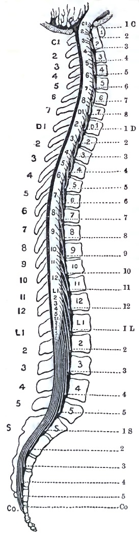
Diagram of Spinal Column, Cord, and Nerve-exits (after Gowers).
FIG. 46.
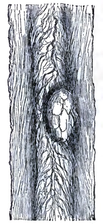
Sarcoma compressing Cervical Cord, Case 17 of Table (E. Long Fox).
Before considering briefly the indications which point to the various levels of the cord as a possible seat of spinal tumor, it will be necessary also to make plain a few anatomical facts. It must be borne in mind, first, that the nerve-origins in the cord are never at the same level as their exits from the spinal canal, or, in other words, that the spinal segments do not correspond with the bodies of the same numerical vertebræ, and that there is, in fact, one more cervical segment than there are cervical vertebræ. The tendency is for the nerve-trunks to run downward before passing out of the canal, so that in every instance, without exception, from the medulla oblongata to the filum terminale the segments of the cord are above the corresponding vertebral body. This discrepancy increases as we descend the cord; whereas it is approximately correct to say of the cervical and dorsal regions that every segment is opposite the vertebral body which is numerically just above it, this difference becomes much greater in the lumbar and sacral regions. The cord itself terminates in the lumbar enlargement which ends opposite the interval between the first and second lumbar vertebræ. All the remainder of the canal is occupied by the descending trunks of the lumbar, sacral, and coccygeal nerves as they pass to their respective foramina, constituting the cauda equina. It must be recalled, however, that the vertebral bodies, lying very deep, cannot serve as guides, but that we are dependent upon the spinous processes as landmarks in diagnosis. These again differ in their levels from their respective vertebral bodies, as they are deflected at somewhat different angles at different regions of the spine. Gowers has illustrated these facts by a very graphic wood-cut9 (Fig. 45), from which the general rule may be drawn that each vertebral spine is about opposite the spinal segment which is numerically two places below it; thus the eighth dorsal spine is opposite the tenth dorsal segment, etc. The indications afforded by this exact anatomical knowledge have reference largely to the existence of pain on pressure and to any deformity of the bony structures. The cases as reported do not indicate that this method of research has been utilized, and it may possibly be of only theoretical importance; but it has been considered worthy of reference as an indication in diagnosis.
9 Diagnosis of Diseases of Spinal Cord, p. 6.
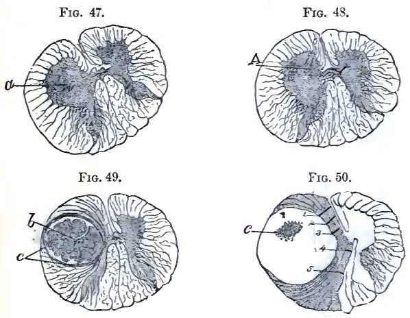
Sarcoma of Lower Cervical Cord, Case 13 of Table (Adamkiewicz).
It will be seen by reference to the table that usually certain general features in the symptomatology indicate the seat of the lesion. Thus in tumors of the cervical region pain and stiffness of the neck occur, while the first appearances of paresis and sensory disturbances are usually observed in the arms and about the chest. The centres for the forearm and hand lie in the lower portion of the cervical enlargement; that for the upper arm, including the supinator longus, in the upper portion. Mental symptoms are more marked, and in Case 4 several of the cranial nerves were implicated. In the lower cervical and upper dorsal region there are symptoms of dyspnœa, fixation of the chest (Nos. 20, 22, 24), and cough. The girdle symptom is an important indication at any level, as it is due to irritation of the nerves at the lowest level of healthy cord just above the transverse lesion. It has already been discussed under Symptomatology. In many of the dorsal cases (Nos. 32, 37, 38, 39, and 45) the symptoms are almost entirely confined to the legs and lower trunk, the arms escaping entirely. The condition of the bladder is usually given in the table as one of paralysis; this does not indicate whether automatic evacuation existed at the beginning of the case; which condition would indicate that the centre for micturition was below the lesion, and intact. It is probable that later in these cases the bladder is actually paralyzed by destruction of its centre in the cord, and this even when the tumor has been situated some distance above.
FIG. 51.
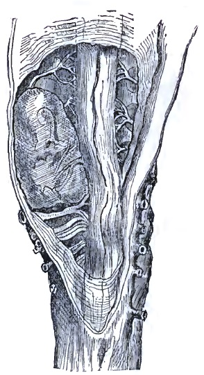
Fibroma of Lower Dorsal Cord, Case 32 of Table (W. Cayley).
FIG. 52.
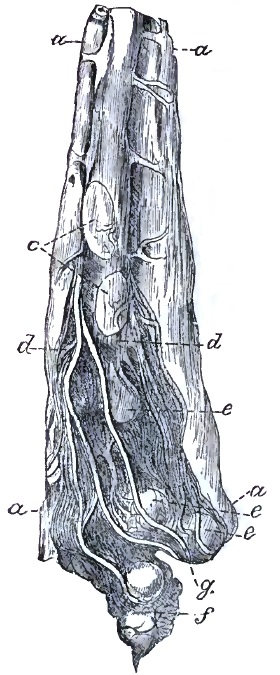
Tumor of Cauda Equina, Case 45 of Table (W. W. Fisher).
With reference to tumors of the cauda equina, Erb10 says that they have in every respect a great resemblance to those which are situated higher and affect the cord proper. “They are hard to distinguish from the latter, but may be in many cases perhaps, if it is borne in mind that tumors of the cauda produce exclusively nerve-root symptoms, and that the signs of compression of the cord, of secondary myelitis, etc. are absent. The higher the tumor, the nearer it approaches the lumbar portion of the cord, the harder will it be to draw the distinction. In respect to tumors seated lower the following points may be attended to: the seat of the pains (which in such cases often attain enormous violence) is strictly localized in certain nerve-districts; all nerves leaving the spinal canal above the tumor are free; thus in myxo-sarcoma telangiectodes of the cauda I observed the pain strictly limited to the district of the sciatica, while the crural and the dorsal nerves were perfectly free; constant violent pain in the sacrum. If palsy occurs the reflex actions necessarily cease at once. Spasms are seldom observed, more frequently contractures. Atrophy of the muscles occurs rather frequently. The palsy and anæsthesia by their localization often give us the opportunity of fixing the upper limit of the lesion. Increase of the reflex acts and marked tendinous reflexions do not occur. Paraplegia, palsy of the bladder, bed-sores, etc. may develop exactly as in tumors occupying a higher seat, but the symptoms of paralysis do not seem to belong necessarily to the disease, as is shown in my case (just mentioned), which terminated fatally before paralysis or anæsthesia occurred.”
10 Op. cit.
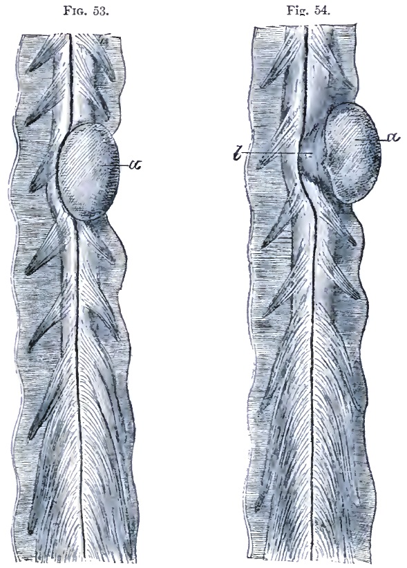
Psammoma of Dorsal Cord, 38 of Table (after Charcot).
PROGNOSIS.—The prognosis of spinal tumors is generally very unfavorable. Syphilitic cases are of course the most hopeful, but even in these cases it is only when they are recognized early that much can be expected. A gumma that has grown to any dimensions will have so compressed the cord that even when the tumor is melted away by specific treatment its effects will remain.
DURATION AND TERMINATION.—Most cases of spinal tumor last from about six months to three years. Occasionally death may result, as from a rapidly-developing sarcoma, in less than six months, and somewhat more frequently in slowly-developing tumors, or in those which are held more or less in abeyance by treatment the sufferings of the patient are prolonged to four or five years or more. Hemorrhages into or around the growths sometimes take place, and are the cause of death, or more frequently of a sudden aggravation and multiplication of severe symptoms. Death sometimes takes place from the complete exhaustion which results from the disease and its accompanying secondary disorders, such as bed-sores, pyelitis, etc. Occasionally death results from intercurrent diseases, such as pneumonia, infectious fevers, etc., whose violence the weakened patient cannot well withstand. Sometimes the symptoms of a rapidly-ascending paralysis appear, probably due to an ascending myelitis or meningo-myelitis.
COMPLICATIONS AND SEQUELÆ.—Spinal tumors are sometimes complicated with other similar growths in the brain or the evidences of the same constitutional infection in other parts of the body. In one case of cysticercus of the cord sclerosis of the posterior columns was also present.
TREATMENT.—The treatment of spinal tumors can be compressed into very small compass. In cases with syphilitic history, or when such history is suspected, although not admitted, antisyphilitic remedies should be applied with great vigor. It should be borne in mind, however, that even in syphilitic cases after destruction of the cord by compression or softening specific remedies will be of no avail. In tubercular cases and in those in which the system is much run down tonics and nutritives are indicated. Bramwell11 advises an operation in any case in which the symptoms are urgent, in which the diagnosis clearly indicates the presence of a tumor, when there is no evidence of malignant disease, when the exact position of the growth can be determined, and when a vigorous antisyphilitic treatment has failed to produce beneficial results. As some meningitis, meningo-myelitis, or myelitis is usually present in cases of spinal tumor, treatment for the complication will assist in relieving the torments of the patient. Anodynes, particularly opium and its preparations, should be used freely in the later stages of the affection. Bromides and chloral are of little value except in association with opiates. Operation offers even less hope than in brain tumor, but in very rare cases should be taken into consideration.
11 Diseases of the Spinal Cord, Edinburgh, 1884.
TABLE OF FIFTY CASES OF SPINAL TUMOR.
No. Sex and Age. Clinical History. Path. Anat. and Location. Remarks.
1 M. 33. Paresis of forearms, left worse. Paraplegia, then paralysis of all limbs; paralysis of intercostals. Contractures of hands, then of feet. Pain and stiffness of neck on motion. Wasting of interossei. Diplegic contractions of legs. Only partial paralysis of sphincters. Sensation perfect. Bed-sores. Duration, thirteen months. Glioma; syringo-myelus. Dilated lymphatics. Entire length of cord, and involving medulla oblongata. Upper four inches of cord greatly enlarged. T. Whipham, Trans. Path. Soc. London, 1881, xxxii. 8-12.
2 F. —. Constricting pains about abdomen. Paresis of legs. Persistent subsultus. Temporary improvement after labor. General paralysis. Scoliosis. Glio-myxoma. In gray columns from medulla oblongata to cauda equina. Schueppel, Arch. d. Heilk., viii. Bd., 1867 (quoted by Rosenthal).
3 M. 15. Paresis of left arm. Pain back of neck. Later, paralysis of left arm, and wasting of arm, shoulder, and neck muscles. Slight paresis of right arm. Prolonged vomiting. Constriction of neck; dysphagia; paralysis of chest. Gelatinous tumor left side of cord, and involving in some parts the gray matter. From medulla to sixth cervical vertebra. S. Wilks, Lectures on Dis. of Nervous System, p. 266.
4 M. 18. Paresis of left leg, increasing; some atrophy. Weakness in left arm. Later, numbness in both legs. Contracture of fingers. Some mental confusion. Left hand and leg livid and cold. Hyperæsthesia of left leg; anæsthesia of right leg, perineum, penis, scrotum, rectum, and inguinal region, and of left arm. Right arm normal. Islands of heat and cold in leg, and of cold in arms. Left ankle clonus. Left pupil contracted. Vomiting. Dysphagia. Occipito-cervical pain and contracture of cervical muscles. Leg contractures and tremor. Later, hyperæsthesia disappeared. Incontinence of urine. Patellar and skin reflexes increased. Facial spasm. Amblyopia, optic neuritis, diplopia, deafness, paralysis of left abducens; pupils contracted. Sacral bed-sores. Thick speech. Round-celled sarcoma or glio-sarcoma, growing from ependyma of central canal, causing hydromyelia, softening, and secondary degeneration. Dura mater thickened. Brown exudate in cord and base of brain. From medulla oblongata to dorsal cord. Schultze (F.), Arch. f. Psychiat., Berlin, 1878, viii. 367-393, 1 pl.
5 F. 48. Pain in abdomen and down legs, worse on left side. Tonic spasm in flexors and adductors of thighs. No anæsthesia. Two months before death paralysis of sphincters. Great emaciation. Tumor (psammoma), growing from dura mater on right side in cervical region. Upper part of cervical region. J. Hutchinson, Jr., Tr. Path. Soc. Lond., 1881-82, xxxiii. 23, 24.
6 —— Pain in arms. Contracture of fingers of right, then left side. Numbness in right foot, then upward, then left foot. Girdle feeling. Priapism and dysuria. Complete anæsthesia, later, up to third rib, with paralysis of legs and paresis of fingers. Respiration diaphragmatic. Legs very jerky. Later, arms paralyzed. Sarcoma of left post. aspect of cord; adjacent cord compressed and soft. Belt of yellow substance enveloped cord to cauda equina. Between cervical bulb and second cervical vertebra. E. Long Fox, Bris. Med.-Chir. Journ., 1883, i. 100-106, 2 pl.
7 F. 31. Pain, stiffness in neck; pain radiating, aggravated by jarring. Sudden paralysis of both arms; next day paralysis of legs, incomplete. Partial anæsthesia. Marked skin reflexes in legs. Patellar reflexes retained, weaker on right than left. Dyspnœa. Profuse perspiration. Cardiac irregularity. Day before death temperature in right axilla 100°; left, 102.2°. Gumma of dura mater two inches long, with intercurrent hemorrhage; flattening and softening of cord, with secondary sclerosis. From first to fifth cervical vertebra. Charles K. Mills, Philada. Med. Times, Nov. 8, 1879, p. 58.
8 M. 34. Pain in back of neck, with stiffness and torticollis. Paresis of arms; later, of legs. Anæsthesia of arms, then of legs; also paræsthesia of legs. Late symptoms: shortening and great rigidity of neck, with choking sensation (girdle sensation at neck). Dimness of vision. Atrophy of arms and less of legs. Complete paralysis of arms, almost complete of legs. Electro-contractility preserved. Violent skin reflexes in legs. Involuntary evacuations and incomplete priapism. Severe pains in knees and ankles. No acute bed-sores. Paroxysms of dyspnœa. Average temp. for two weeks before death, M. 97.9°, E. 98.3°. Gumma of dura mater; caries, probably syphilitic, of vertebræ. Abscess. Total (almost) transverse sclerosis of cord. Secondary degeneration. Some softening above and below tumor. Cervical nerves compressed and atrophied. From second to fifth cervical vertebra; most in front. Charles K. Mills, Philada. Med. Times, Nov. 8, 1879, p. 58.
9 M. 43. Pain between shoulders. Numbness in right hand and arm, with weakness and swelling. Numbness in left arm, which spread over chest and abdomen. Unable at first to lie down. Felt as though encased in armor. Pain in back of neck. Tongue protruded to right. Exaggerated reflexes in legs. Right arm and leg weaker than left. Vertigo. Dysphagia. Sense of constriction about neck. Breathing impaired. At third cervical vertebra, to right of front of cord. Destruction of opposite vertebra. E. H. Clark, Bost. Med. and Surg. Journal, 1859-60, lxi. 209-212.
10 —— No record of symptoms especially referable to the cysticercus. Symptoms of tabes dorsalis. Cysticercus in substance of cord. Lesions of tabes dorsalis. On level with third cervical nerve. Geo. L. Walton, ibid., vol. cv. p. 511.
11 M. 25. Pain in back of neck; stiffness. Numbness of left hand. Gradual loss of power of left arm. Jerking of arm. Paresis of left leg. Constriction of upper chest. Right limbs involved, and eventual complete paralysis of trunk and extremities. Severe headache. Last three days absolute anæsthesia of arms and legs. No ophthalmoscopic changes. Constipation and dysuria. Fibro-sarcoma at level of fourth cervical nerves. Cord compressed. H. A. Lediard, Tr. Path. Soc. Lond., 1881-82, xxxiii. 25-27.
12 F. 25. Œdema of ankles; pain in legs; afterward numbness, formication, and stiffness of legs. Painful contractures in upper extremities. Slight left scoliosis. Abdominal pains. Paresis of arms. Fingers flexed. Fever. Respiration became involved, and bowels and bladder paralyzed. Mind clear. Died in attack of suffocation. Duration, two years and three months. Fibroma, size hazelnut, under pia mater. Between fourth and fifth cervical vertebræ. Bernhuber, Deutsch. Klin., Berlin, 1853, v. 406.
13 M. 16. Restlessness. Cramps in pharynx on swallowing. Excitability. Delirium. Hallucination. Pain in the neck. On touching neck general cramps. Grimaces. Salivation. In three days complete paraplegia. No fever. Sudden change. Pulse 120. Pupils alternating. Blepharospasm. Irregular respiration. Pulmonary œdema. Suspicion of hydrophobia, because patient had been with hydrophobic dog; when offered coffee had symptoms simulating rabies. Sarcoma. Extending from fifth to seventh cervical nerve on antero-lateral face of cord, compressing left half and penetrating into right half, so that anterior longitudinal fissure described arc of circle around it. Adamkiewicz, Arch. de Neurol., Paris, 1882, iv. 323-336, 1 pl.
14 F. Paresis and partial anæsthesia in all limbs for many months, most marked on left side. Brain and special senses unaffected. Had a tumor at bottom of right side of neck. Extensive bed-sore. Carcinoma. Tumor caused partial absorption of sixth cervical vertebra. Cord compressed and twisted. Right lateral aspect especially affected. Cord atrophied. At level of sixth cervical vertebra. J. W. Ogle, Tr. Path. Soc. Lond., 1885, 6, vii. 40, 41.
15 F. 34. Pain in right foot, and paresis increasing to paraplegia. Paresis of arms. Contractures of legs. Hyperæsthesia in both legs up to crest of ilia. Later, great pain; paralysis of sphincters. Bed-sores. Sarcoma, growing from dura mater; nerves passing through and over tumor. Cord congested and pushed to one side. Thin, but not softened. Growth resembled psammoma. Between sixth and seventh cervical nerves of left side. T. Whipham, Tr. Path. Soc. Lond., 1873, xxiv. 15-19.
16 M. 57. Pain in right arm. Numbness in hand, and paresis. Paresis and coldness of left leg. Some anæsthesia and wasting of right leg. Later, paraplegia. Diminished reflexes. Contractures. Constriction sense about legs and abdomen. Triceps, deltoid, and serratus magnus of right side paralyzed. Incontinence of urine, difficult defecation, decubitus, fever. Abdominal muscles paralyzed. Later, other muscles of arms paralyzed. Complete anæsthesia of legs. Dyspnœa, œdema of lungs. Myxoma from arachnoid. Cord compressed and softened on right postero-lateral side. Secondary degeneration. Some œdema of brain. At sixth and seventh cervical vertebra on postero-lateral surface of cord. Pel (P. K.), Berlin. Klin. Wochensch., 1876, xiii. 461-463.
17 F. 35. First, pain in right arm, weakness in right hand. Then paralysis almost complete in arms, and impaired sensation. In legs paralysis complete, sensation impaired. Alternate incontinence and dysuria. Ankle clonus and increased knee-jerks and plantar reflex. Tapping biceps causes reflex in little and ring fingers. No atrophy or bed-sores. Cold on one side, hot on other. Pain and little swelling over sixth cervical vertebra. No eye symptoms. Brain clear. Inability to turn head. Before death respiratory paralysis and bed-sores. Duration, fifteen months. Spindle-cell sarcoma, springing from arachnoid and destroying cord by pressure, except posterior columns. Cord below tumor soft. At sixth cervical vertebra. E. Long Fox, Bris. Med.-Chir. Journ., 1883, i. 100-106, 2 pl.
18 M. 50. Paresis in right arm. Stiffness in neck and back. Paralysis of all extremities gradually developed. Glioma in right half of cord. Old hemorrhages in adjacent parts and in medulla oblongata. A more recent hemorrhage in dorsal cord. In lower cervical region. Schueppel, Arch. d. Heilk., viii. Bd., 1867 (quoted by Rosenthal).
19 —— Coldness, numbness, violent pains, first in left arm, later in both legs. Paralysis of all limbs and muscles of trunk. Atrophy. Reactions of degeneration. Violent leg reflexes. Tubercle, large as hazelnut. Consecutive myelitis of adjacent parts and left anterior horn. In lower cervical region. Chvostek, Med. Press, 33-39, 1873 (quoted by Rosenthal).
20 M. 45. Interscapular pain. Chest-pressure and dyspnœa. Paræsthesia and pain in legs. Spastic paralysis. Difficulty in stools; bloody urine and dysuria. Œdema of legs. Bed-sores. Kypho-scoliosis. Pain on pressure over spine. Paralysis of left leg, paresis of right, some anæsthesia of both. Broncho-pneumonia, fever. Phlegmon of dura mater, compressing cord. Some infiltration of tissues of throat and mediastinal space. From seventh cervical to second dorsal vertebra. Mankopff (E.), Berl. Klin. Wochensch., 1864, i. 33-46, 58, 65, 78.
21 M. 22. Pain in back and side of neck and in limbs. Marked pain in sternal region on coughing. Pressure and jarring cause pain. Rapid loss of power in both arms. Feeble and slow movements of thighs, legs, and feet. Right deltoid and flexors of fingers much wasted. No paralysis of face. Knee-jerks exaggerated. Later, complete paralysis, including bladder and rectum. Tumor of membrane. Cord beneath compressed and degenerated. Lower cervical and upper dorsal region. H. C. Wood, “Proceedings of Philadelphia Neurological Society,” Medical News, vol. xlviii. No. 9, Feb. 27, 1886.
22 F. 50. Pain in neck, shoulders, and chest. Stiffness of neck, back, and arms. Chest fixed; breathing diaphragmatic. No paralysis or altered sensation. Secondary cancer of vertebræ. Cervical region. Gull, by Wilks, in Lect. on Dis. of Nerv. System.
23 F. 40. Severe pain in back. At height complete paralysis in legs, some paresis in arms. Variable anæsthesia. Girdle sensation and mammary pain. Lively and distressing reflexes. Contractures in legs. Bed-sores and paralysis of sphincter of bladder. Toward close rigors (pyæmia?). Fibro-cyst on right side, between cord and dura, and between anterior and posterior nerves. Top of dorsal region. Risdon Bennett, Tr. Path. Soc. Lond., 1855-56, vii. 41-45.
24 M. 30. Cough, dyspnœa, wasting, simulating phthisis. Pain in back of neck and shoulders. Pain in joints; paresis of legs and bladder. Pain in chest. Paresis of arms. Later, increased paralysis, bed-sores, sweating. Tumor, size of hazelnut, inner anterior surface of dura mater. Flattening and softening of cord. Top of dorsal region. Gull, in Guy's Hosp. Rep., (quoted by Wilks in Lectures on Dis. Nerv. Syst., p. 264).
25 F. 43. Pain in shoulders, chest, and sides. Contractures of legs; heels to nates. No anæsthesia. Later, retention of urine and bed-sores. Incessant pain in back and abdomen. Fibro-nucleated tumor from inner surface of dura mater. Opposite third dorsal vertebra. Gull, by Wilks, ibid.
26 F. 43. Pain in chest and shoulder, then in legs. Paresis of legs. Contractures and jerking of legs. Spasm of abdominal muscles. No anæsthesia. Paresis of bladder and rectum. Wasting and bed-sores. Finally, paresis increased, but never complete paralysis. Duration, nine months. Fibro-nucleated tumor, size of a bean, from dura mater. Cord compressed backward, and softened. Opposite third dorsal vertebra. Wilks, Trans. Path. Soc. Lond., 1855-56, vii. 37-40.
27 M. 24. Paraplegia. Depressed reflexes; girdle symptom. Partial anæsthesia. Dysuria. Vomiting. Pulse weak and intermittent. Partial recovery from paralysis, and anæsthesia in left leg, and reflexes in right foot regained. Later, complete paraplegia, anæsthesia, and bed-sores. Duration, five months. Probable gumma. Middle dorsal region. B. G. McDowell, M.D., Dubl. Q. J. Med. Sci., 1861, xxxii. 299-303.
28 F. 44. Paresis in legs. Spine hypersensitive and inflexible; least attempt at bending causes great cervico-brachial pain. Paræsthesia; sense of falling out of abdominal viscera through abdominal walls. Pains in extremities increasing, and involving right shoulder, intercostals on both sides, and lumbar region. Paralysis of right arm (first); complete paralysis of leg. Excessive spinal tenderness. Loss of sensation (partial) in legs, body, and right arm. Later, dyspnœa, then dysuria, then complete inability to empty bowels or bladder. Great tympanites. Girdle sense above umbilicus, and finally complete paralysis and anæsthesia below this band. Sense of twisting of legs and feet, so that latter seemed close to face. Œdema. Later, paresis of left arm. One small bed-sore. Alveolar sarcoma. Eighth and ninth dorsal vertebræ. G. W. H. Kemper, Journ. Nerv. and Ment. Dis., xii. No. 1, Jan., 1885.
29 F. 42. Projection of seventh, eighth, ninth, tenth, and eleventh dorsal vertebræ. Numbness below ankles, and early girdle sensation. Peronei and anterior tibial muscles first involved; then all leg-muscles, then sphincters, then arms. Died in a fit. Round-celled sarcoma. The anterior columns soft opposite tumor. Bodies of seventh, eighth, ninth, and tenth vertebræ soft. Opposite seventh, eighth, ninth, and tenth dorsal vertebræ. E. Long Fox, Brit. Med. Journ., 1871, p. 566.
30 F. —. Ill-defined hemiplegia; later, paraplegia, with contractures and rigidity. Gumma and syringo-myelus. Small cavities in anterior cornua. At ninth dorsal vertebra anterior aspect. Taylor, Lancet, 1883, p. 685.
31 M. 7. Paraplegia, except adductors and rotators of thigh. Reflex contractures; most intense from irritation of penis and scrotum. Rigidity of legs. Complete anæsthesia of lower half of body. Later, anuria, incontinence of feces. Anal sphincter reflex; figured stools. Cystitis. Pain on percussion in dorsal region. Pain in back. Complete paraplegia. Very late, brain symptoms. Duration, nine months. Tubercle (?). Cord soft for two inches. Tenth dorsal vertebra. Geoghegan, Dublin Med. Press, 1848, xix. 148-151.
32 F. 46. Fixed pain in left iliac region. Paresis in left leg, increasing to paraplegia. Formication. Girdle sensation. Incomplete, increasing to complete, anæsthesia of legs. Spontaneous twitchings. Bladder and sphincter ani paralyzed. Bed-sores. Duration, one year. Fibroma (?) from inner surface of dura. Cord hollowed out and softened. Interval between tenth and eleventh dorsal vertebræ. William Cayley, Tr. Path. Soc. Lond., 1864-65, vol. xvi. 21-23.
33 M. 30. Hyperæsthesia; later, anæsthesia in legs; then complete paraplegia. Tubercle size of pea. Adjacent myelitis. In lower dorsal region. Chvostek, Med. Presse, 33-39, 1873 (quoted by Rosenthal).
34 M. 31. Ataxia; stiffness of legs and cramps in abdomen and legs. Slight nystagmus. Difficulty in forming words. Ataxia of arms. Slight wasting of legs, especially of left. Lumbar pains; abdominal cramps. Dysuria. Impotence. Later, increased spastic state of legs. Mind depressed and emotional; attempts at suicide. Anuria. Bed-sores. Urine albuminous. Duration, one year. Myxoma of dura mater 3 inches long. Dura mater of brain contained fluid and lymph. Dorsal region, left side. Shearman, Lond. Lancet, vol. ii. 1877, p. 161.
35 F. 50. Pains in limbs (thought to be rheumatic). Paresis in legs. Hyperæsthesia in right leg; burning pains alternating with sense or coldness. Cancer of vertebræ (sarcoma?). Dorsal region. Gull, by Wilks, Dis. Nerv. Syst.
36 F. 35. Paresis of left leg; soon of right leg. Pain in back and left side. Tonic spasms of legs. Darting pains in knees. Partial anæsthesia. Exalted plantar reflexes. Dysuria. Later, complete paraplegia and anæsthesia; violent reflexes; severe pain in back. Bed-sores. Duration, seven and a half years. Tumor, osseous or fibrous, three-fourths of an inch long, growing from dura mater. Cord flattened, and softened below tumor. Lower part of dorsal cord. H. Ewen, Tr. Path. Soc. Lond., 1848-50, i. 179.
37 F. 28. Weakness in legs. Aching and shooting pains in legs. Numbness and formication. Slight spasm in legs. “Felt as if ground was some distance below feet.” Œdema of ankles. Later, numbness extended to abdomen. Paralysis of bladder. Hyperæsthesia in right leg. Obstinate constipation. Bed-sores. Some paralysis of respiratory muscles. Duration, fourteen months. Tubercle the size of cherry, which had almost obliterated cord. Tubercles in lungs, bowels, and uterus. Bed-sore had opened spinal canal. Lower part of dorsal cord. S. O. Habershon, M.D., Guy's Hosp. Rep., London, 1872, 3d S., xvii. 428-436.
38 M. 63. Progressive paresis of left leg for five years. Right leg then paretic. Paralysis then in left leg. Rigidity on extension of right leg. Paroxysms of clonic spasms in right leg. Joint pains, sciatic pains. In left leg, hyperæsthesia, in right leg, anæsthesia. Plantar reflex retained; other reflexes exaggerated. Diplegic contractions in right leg from irritation in left. Late symptoms: purulent urine, with retention; chest and lumbar pains like bone pain; extension changed to flexion; swelling of legs and ecchymosis; sacral and other eschars. Psammoma adherent to dura mater. Cord softened. Ascending degeneration in posterior columns, and descending degeneration of lateral columns. In dorsal region just above lumbar enlargement, anterior left side. Charcot, Arch. de Physiol., Paris, 1869, ii. 291-296.
39 M. 20. Paralysis of lower extremities; tremor; exaggerated reflexes, hyperæsthesia of trunk; bed-sores. Œdema of feet. Fever. Pus in urine. Organized blood-clot exterior to dura mater. Cord compressed and softened. Opposite lower dorsal and upper lumbar. C. B. Nancrede, Am. Journ. Med. Sci., O. S., lxi. 156.
40 F. 38. Pain around abdomen, in back, and legs. Paraplegia. Anæsthesia and tingling of feet and legs. Paralysis of bladder. Hydatid cysts of vertebræ (?) and spin. canal. Lower part of spinal canal (probably lumbar region). S. Wilks, Dis. Nerv. Syst., p. 265.
41 F. 23. Bronzing of skin for two years; then headache, giddiness, fever. Choreic movements in left arm, then in leg, then general. Bronzing increased. Vomiting after meals. Duration, two years and two months. Tumor, consisting of granular matter, with a few nerve-fibres and cells, springing from centre of cord backward to posterior fissure. Cord slightly widened. Suprarenal capsules large and nodulated. Lumbar enlargement. W. H. Broadbent, Trans. Path. Soc. Lond., 1861-62, viii. 246.
42 10 ms. Twitching and convulsive movements of right leg. After removal of exterior tumor the movements ceased. Child died of peritonitis. Tumor outside of sacrum, and also protruding through sacral opening. Reported to have been behind and pressing upon cord (?). Fatty growth within membranes. Arthur Johnson, ibid., 1856-57, viii. 28, 29.
43 F. 54. Paresis, first of left arm and leg; then paralysis of these and of right arm and leg. Pain in back and hips early; then, suddenly, darting pains and incontinence of urine. Paræsthesia of left arm and leg; no anæsthesia. Coma. Hydatid cyst. Cyst also in liver. Fluid beneath membranes of cord and brain. At first and second left sacral foramen, opposite last lumbar and upper three sacral vertebræ. H. S. Wood, Australian Med. Journ., 1879, N. S. i. 222.
44 M. 46. Fibrillary twitching. Increased patellar reflexes. Paræsthesia and hyperæsthesia in legs, disappearing. Constriction of chest (?). Headache. Dysuria for two years. Straining at stool. Indigestion. Bloody vomiting. Cardiac palpitation; intracardial murmurs; slow pulse. Swollen inguinal glands. Variations in temperature. Bed-sores. Glioma. At filum terminale, upper part. Lachman, Arch. f. Psychiat., Berl., 1882, xiii. 50-62, 1 pl.
45 M. 38. Pain in legs. Œdema. After two years could not lie down: rested on hands and knees. Paralyzed in legs; pain in seat. Anæsthesia in legs, not complete in right. Paræsthesia in left. Dysuria and constipation. Before death had incontinence with hæmaturia, and was able to lie down. A lobulated tumor from pia mater at lower end of spinal canal, surrounded by nerves of cauda equina. Structure not made out. At cauda equina. W. W. Fisher, Tr. Prov. M. and S. Ass., 1882, x. 203-208.
46 —— This case had symptoms of posterior spinal sclerosis, which possibly had no relation to growth, according to reporter. Myo-lipoma attached to conus medullaris. Crescentic, clasping cord from anterior to posterior fissure. Nerve-roots of cauda equina imbedded in it. Contained striated muscular fibres. W. R. Gowers, Tr. Path. Soc. Lond., 1875-76, xxvii. 19-22.
47 Und. 1 yr. Spina bifida (?); hydrocephalus; convulsions, bloody stools; partial paraplegia. (Above symptoms came on after closing of sacral opening by surgical operation.) Congenital sacral neuroma amyilinicum. W. F. Jenks, M.D., Trans. Path. Soc. Philada. (1871-73), 1874, iv. 190-192.
48 M. 30. Pain in back; abdominal girdle sensation. Pain in legs; paraplegia; nearly complete anæsthesia; paralysis of bladder; bed-sores. Aneurism, eroding vert. and compressing cord. Location not given. Wilks, Dis. Nerv. Syst.
49 M. 54. Paralysis of both legs, of sphincter ani, and of bladder; urine alkaline, with pus and blood. Partial anæsthesia. Pyonephritis. Gumma from inner layer of dura mater and involving pia mater. Location not given. Delafield, N. Y. Med. Rec., 1875, x. 131.
50 Still- born. —— Tumor, size of head of child two years old, projected between legs from spinal column. Nerves of cauda equina over anterior part. Some bone in tumor (dermoid cyst?). Virchow, Monatschr. f. Geburtsk., Berl., 1857, ix. 259-262.
INFANTILE SPINAL PARALYSIS.
BY MARY P. JACOBI, M.D.
SYNONYMS.—Essential paralysis of childhood (Rilliet and Barthez); Myogenic paralysis (Bouchut); Acute fatty atrophic paralysis (Duchenne); Atrophic paralysis (Ferrier); Acute anterior poliomyelitis (Kussmaul, Erb, Seguin); Regressive paralysis (Barlow); Tephromyelitis (Charcot).
DEFINITION.—Of all the titles which have been given to the disease it is our purpose to describe, two alone may be considered irreproachable. In the present state of our knowledge it is unnecessary to argue that this disease is not essential—i.e. destitute of characteristic anatomical lesions. Neither can the theory of its myogenic origin be maintained; nor even is fatty degeneration invariably present in the paralyzed muscles. Finally, the disease cannot longer be regarded as peculiar to childhood,1 since cases in adults have been in these last years quite numerously reported2—four with autopsies demonstrating the identity of the lesion. But there are two definitions in our list of synonyms which embrace between them the most striking characteristics of the disease, yet contain no error of fact. Atrophic paralysis describes at once the two most salient symptoms; acute anterior poliomyelitis defines at once the seat and nature of the lesion, classes it with the systematic diseases3 of the spinal cord, and notes the peculiarity in the mode of invasion by which it is so remarkably distinguished from nearly all the organic diseases of this centre.
1 W. H. Barlow, On Regressive Paralysis, 1828. See Brain, April, 1879.
2 In Dec., 1873, I quoted 14 cases of adult spinal paralysis, as follows: Duchenne, 4 cases; Charcot and Petitfils, 3; Moritz Meyer, 2; Bernhardt (Archiv Psych., 1873), 1; Cumming (Dublin Quart. Journ., 1869), 1; Lucas Championnière (by Hallopeau, Archives gén., 1861), autopsy, 1; Gombault (Archives de Psych., 1873), 1; personal, 1.
In 1874, Seguin published a summary of all the foregoing cases except the last, and added 6 personal observations, also 3 from Duchenne and 1 from Hammond. In the enlarged edition of his essay in 1877, Séguin increased the list to 45—by new personal cases, 3; cases related by Frey (Berlin. Wochens., 1874), 4; cases by Erb (Arch. f. Psych. u. Nervenkrank., v.), 4; case by Cornil and Lépine (Gaz. méd., 1875), autopsy, 1; case by Soulier (Lyon méd., 1875), 1; case by D. H. Lincoln (Boston Med. and Surg. Journ., 1875), 1; case by Lemoine (Lyon méd., 1875), 1; case by George M. Beard, 1; case by Leyden (Klinik Ruckenmarks Krankheiten) Bd. iv. 1; case by Hammond (6th ed. Treatise), 4; case by Courty (Gaz. méd., 1876), 1; case by Dejerine (Arch. de Phys., 1876), 1.
To these may be added—case by Goltdammer (Berl. klin. Wochen., 1876), 1; case by Webber (Trans. Amer. Neurol. Ass. for 1875, vol. i.), autopsy, 1; case by Klose (Diss. Breslau, 1876), 1; case by Schultze (Virchow's Archiv, Bd. lxviii.; also Bd. lxxiii.), autopsy, 1; case by Bernhardt (Archiv für Psych., Bd. ix., 1879); case by Sinkler (Amer. Journ. Med. Sci., Oct., 1878), 5; case by Althaus (ibid., April, 1878), 2; case by Ross (Dis. Nerv. Syst., vol. ii. p. 139), 1—total, 57 cases.
Morton (St. Bartholomew's Hospital Reports).
Others have doubtless been published since this date, but, as they do not immediately concern our subject, need no further citation.
3 Vulpian, Leçons sur les Myelitis, 1880.
SUMMARY OF CLINICAL HISTORY.—The clinical features of an acute attack of infantile paralysis are well known. The children affected are usually between eighteen months and four years of age (Henoch). The attack is more likely to occur in summer than in winter, as Sinkler4 found that 47 out of 57 cases began between May and September, and Barlow noted 27 out of 53 in July and August.5
4 Amer. Journ. Med. Sci., April, 1875.
5 Loc. cit., p. 75. Among Sinkler's 57 cases, only 6 furnish autopsies, thus:
Case by Cornil and Lepine and case by Webber (quoted and accepted by Erb in Ziemssen's Handbuch, Bd. xi.); case by Gombault (rejected by Erb and Westphal); case by Schultze; cases by Dejerine and Lucas Championnière (quoted by Hallopeau).
The influence of heat is perhaps shown in the case related by Dyce-Duckworth in the Lancet of 1877: a child two and a half years, after exposure to great heat on a steamboat-landing, became paralyzed in all four limbs, but the paralysis was subsequently confined to the lower extremities. Coincidently, the patient became delirious; suffered from anæsthesia and temporary paralysis of the sphincters. The paralyzed muscles wasted rapidly and lost faradic contractility. Treatment by faradization was begun in a month from the date of the attack, and recovery was complete three months later.
The onset of the paralysis is either really sudden, occurring in the daytime, while the child is under competent observation, or apparently sudden, being discovered in the morning after a quiet night, the child having gone to bed in health (West); or is preceded by some hours or days of fever or of nervous symptoms, especially convulsions, or both. The paralysis is almost always at its maximum of extent and intensity when first discovered, and from this maximum begins, within a few hours or days, to retrocede. The improvement may, however, be delayed much longer. A variable number of muscles remain permanently paralyzed, and in these, within a week (thirty-six hours, according to some observers), faradic contractility is first diminished, then abolished; galvanic reaction is exaggerated, ultimately is characterized by the degeneration signs (entartungs reaction). The temperature of the paralyzed limbs falls; the muscles waste; the atrophy may rapidly become extreme. The paralysis and loss of faradic contractility are complete, however, while the atrophy is only incipient and progressing. The absence of lesions of sensibility, of visceral disturbance, of trophic lesions of the skin, or of sphincter paralysis is as characteristic of the disease as are the positive symptoms above enumerated.
In the third or chronic stage the paralyzed limbs often become contracted and deformed. At other times, and with more complete paralysis, the growth of the bones is arrested, the muscles remain flaccid, the entire limb shrivels, and dangles so loosely from its articulations that it may be dislocated by slight effort (membre de Polichinelle). The general health of the patient remains remarkably good, the intelligence clear, the disposition lively. The duration of life seems to be in nowise shortened by the paralysis. Thus, suddenness of development, intimate association of trophic, motor, and electrical disturbance, absence of cerebral or sensory lesion, peculiar localization and grouping of the permanent paralyses,—such are the salient characteristics of this remarkable disease.
SYMPTOMS IN DETAIL.—Three well-defined stages exist—the initial, the paralytic, and the chronic.
Initial Stage.—Seeligmüller6 signalizes three principal varieties in this stage. The first is characterized by fever; the second by nervous symptoms, principally convulsions, sometimes delirium or coma; in the third no symptoms either precede or accompany the local disease—"la paralysie est toute la maladie.”7
6 Gerhardt's Handbuch der Kinderkrankheiten, 1880 (separat Abdruck).
7 Rilliet et Barthez, Traité des Maladies des Enfants, ii. p. 551.
Mode of Invasion.—I have elsewhere8 described eight different modes of invasion: absolutely sudden, coming on in the daytime; morning paralysis (West), discovered after a quiet night, preceded by fever or by vomiting alone (?), or by another typical disease, especially one of the exanthemata, or, finally, by a traumatism, generally slight. An interval of time almost always elapses between the occurrence of the traumatism and the development of the paralysis—a fact which already indicates that a definitely-evolved morbid process must intervene between the two occurrences. An exception is related by Duchenne fils;9 and some apparent exceptions, in which recovery occurred rapidly, seem to belong to the temporary paralysis of Kennedy,10 more recently described again by Frey.11
8 Am. Journ. Obstet., May, 1874.
9 Archives gén., 1864. A father pulled his child from a table by the right arm, and set it rather roughly on the ground. Immediate pain, almost immediate paralysis of arm, which persisted, and was followed by atrophy of its muscles.
10 Dublin Quarterly, 1850.
11 Berlin. klin. Wochens., 1874. Frey considers these cases to be identical in nature with, though differing in severity from, anterior poliomyelitis.
PRODROMATA.—There is rarely any lengthened period of prodromata. Seeligmüller has noticed in some cases an indisposition on the part of the child to stand or walk during several weeks before the occurrence of the paralysis. He does not say whether such children were rachitical. In marked contrast with cases of cerebral paralysis is the habitual absence of generalized nervous symptoms. Thus in only 1 case of Seeligmüller's (total of 75) did the child suffer, and that during six months preceding the paralysis, from intermittent muscular contractions, and also from attacks of laryngismus stridulus.
The fever is usually of moderate severity (Seeligmüller), but sometimes extremely high (Erb)12—as much as 41° C. (Henoch),13 or 104° F. (Barlow).14 Duchenne fils observed 7 cases alleged to be entirely without fever, and Laborde counts 10 cases out of 50 as apyretic.15
12 Ziemssen's Handbuch, Bd. xi. Abh. 12.
13 Vorlesung. über Kinderkrank., 2d Aufl., 1881. Seeligmüller (Jahrb. für Kinderheilk., 1878, p. 345) quotes another case from Henoch's clinic where the fever lasted thirty-six hours, the temperature on the first evening being 39.2°, the following morning 39.0°, the second evening 39.5°; the second morning, at which date the paralysis was discovered, it was normal. The author states this to be the only case known to him in which thermometric measurements were taken.
14 Loc. cit.
15 De la Paralysie de l'Enfance.
The duration of the fever usually varies from a single night to forty-eight hours; much more rarely does it last six, eight, twelve, or fourteen days, or even, but quite exceptionally, three or four weeks. According to Duchenne, its intensity and duration increase with the age of the child, perhaps indicating greater resistance on the part of the nerve-tissues which are the seat of the morbid process of which it is symptomatic. Rarely does it last after the paralysis has once occurred, but ceases then with an abruptness which recalls the defervescence of pneumonia when the exudation process is once completed.16
16 See p. 1144 for pathogenic inferences to be drawn from this fact. Seguin (New York Med. Record, Jan. 15, 1874) seems to throw some doubt on the existence of apyretic cases; but, as Seeligmüller remarks, there is too much testimony to this possibility to render it really doubtful.
There is no proportion between the intensity of the fever and the extent of the subsequent paralysis; nor is there any marked contrast between the fever in children and that in adults in those rare cases in which the disease, instead of being subacute, is sudden as in children.
Erb considers the fever to be purely symptomatic of an inflammatory process in the spinal cord.17 But Vogt regards it rather as an essential factor in the development of a spinal lesion, and thus explains the occurrence of this in the course of febrile diseases which at the outset have no special relation to the cord.
17 Loc. cit., p. 279.
Convulsions, usually accompanied by fever, were observed in 11 of Seeligmüller's cases out of 67; Duchenne had 13 out of 70; Heine, 9 out of 86;18 thus a total of 33 cases of convulsions in 223 cases of infantile paralysis—nearly 15 per cent. The paralysis may set in after a single brief convulsion, or this may be repeated several times at variable intervals before the paralysis is definitely declared (Ross).19 The convulsive movements are apt to be particularly intense in the limbs destined to become paralyzed (Vogt).
18 Die Spinale Kinderlahmung.
19 Loc. cit., p. 107. The author is quoting Laborde.
The convulsion may be very slight—an isolated spasm of a limb or even a single group of muscles. Whether, on the other hand, it can ever be so intense that the child succumbs to it before the development of paralysis, is a question which could only be decided by repeatedly examining the cord in the cases of convulsion which have terminated fatally. In a case of Seeligmüller's the child was affected for eight days preceding the paralysis by tremblings generalized through all his muscles.
The convulsion is usually followed by a soporous or even comatose condition, or this may replace the convulsion. Delirium may take the place of either.
Special interest attaches to those cases where the paralysis develops in the course of an acute specific disease; for then becomes most plausible the suggestion of Vogt, that a fever excited by some cause remote from the spinal cord may itself become a cause of lesion in this centre. In Roger's first and most celebrated case, paraplegia developed suddenly during the course of a fatal scarlatina in a child already suffering from paralysis of the left deltoid of two months' standing.20 The scarlatina was hemorrhagic, and, as will be shown farther on, the autopsy showed traces of a hemorrhagic extravasation in the cord. Thus a double influence was presumably exerted by the scarlatina, while, moreover, the previous and recent occurrence of a deltoid paralysis indicated a morbid predisposition in the spinal cord. Of Seeligmüller's 75 cases, 1 occurred during scarlet fever, 1 with measles, 1 in the course of an erysipelas, and 1 of pneumonia.
20 Gaz. méd., 1871.
Apyretic diseases, especially of the gastro-intestinal tract (Brown-Séquard), also seem to have an influence on the development of infantile paralysis. Two of my own cases occurred during an attack of cholera infantum; another in a child who had been for several weeks in bed with a purulent conjunctivitis. Study of these varied antecedents is of interest in connection with the obscure question of the etiology of infantile paralysis. In this latter connection we will refer to them again.
Vomiting, or even the entire symptom-complex of gastric fever, not infrequently ushers in the paralysis. Fever is then usually present, but I have recorded one case of vomiting where, according to the mother's assurance, no fever at all existed.
At the moment that the symptoms of the invasion subside, and the child seems to enter upon convalescence, the terrifying discovery is made that an arm or a leg or all four limbs, or even they and the muscles of the trunk, are paralyzed.
In the severest form the child lies motionless, unable to stir hand or foot, or even a finger or toe. Yet, singularly enough, this extensive paralysis is sometimes overlooked, especially in very young children, as the immobility of the patient is attributed merely to weakness caused by previous illness. General paralysis, during at least the first few hours of the paralytic stage, is probably more common than appears from our present statistics. Not only, as has just been noted, may this condition be overlooked, but it may exist during the hours of sleep which precede the cases of morning paralysis. Seguin21 speaks as if the paralysis were at first always generalized, but this statement seems to me somewhat exaggerated. Referring merely to the statements of the parents, a considerable number of paralyses would be found limited from the beginning. Heine's third table of partial paralysis is entirely composed of cases so limited. In 16 out of the 19 cases of hemiplegia (monoplegia) the original limitation of the paralysis is also specified; similarly with 7 out of the 20 cases of paraplegia contained in the first table.
21 Loc. cit.
Paralysis of one or both lower extremities is often first detected when the child gets out of bed and attempts to walk; or in children too young to walk the flaccid immobility of the limb attracts attention as soon as they are again carried on the mother's arm. Paralysis of the upper extremities is discovered early in proportion to the liveliness recovered by the child, leading him to occupy himself with his toys as usual. In unilateral paralysis of the trunk the child will fall over to one side when placed in the sitting position; in bilateral paralysis it cannot be made to sit up at all.
From lack of competent observation during the initial stage it is really not quite certain whether any degree of paresis precedes the paralysis; but from the testimony at present accumulated the paralysis is nearly always complete when first observed. This is in striking contrast with adult spinal paralysis. In some few cases the paralysis has been observed to creep on slowly, and not reach its maximum for several days (Ross). Laborde relates a case where recovery from a first attack of paralysis was followed by two relapses in the same limbs at intervals, each ushered in by fever. After the second relapse the paralysis remained permanent.
Significance of Original Extent of Paralysis.—The question of the original distribution of the paralysis is of special interest in connection with that of the original distribution of the morbid process in the spinal cord. The real effect of the latter cannot be adequately measured by the permanent paralyses; for, as will be seen, it is not unusual to find traces of an extensively diffused process in the cord in cases of quite partial paralysis.
It is interesting to notice that certain muscles are always exempt from paralysis. With the exception of a single case of paralysis of one temporal muscle, cited by Seguin,22 the muscles of the head, eyeballs, ears, larynx, and pharynx are always exempt, as are also the diaphragm and intercostals. The arrest of the spinal lesion below the medulla explains the immunity of muscles supplied by the vagus and spinal accessory nerves. But since the cervical plexus is often involved, the constant escape of the diaphragm, innervated by the phrenic nerve which comes from this plexus, is remarkable. Still more so the immunity of the intercostal muscles, whose nerves arise in the dorsal region—a position of the cord frequently affected. This fact tends to confirm Ross's hypothesis, that the nuclei of the intercostal nerves lie in the vesicular columns of Clarke—columns confined to the dorsal region of the cord, and which are invariably found intact at autopsies of atrophic paralysis.
22 Loc. cit.
The immunity of these respiratory nerves explains the absence of the dyspnœa which is so marked in Landry's ascending paralysis. In the adult case described by Schultze and Erb23 dyspnœa was present for a short time. The disease terminated fatally twenty months from the time of invasion. In this case traces of myelitis were found extending through the dorsal region of the cord, and including not only the anterior nerves, but, to a less degree, the columns of Clarke.
23 Arch. Virch., Bd. lxviii.
The facial nerve (itself a respiratory nerve) shares the immunity of the phrenic and intercostals. In the cases in which facial paralysis has been noted the limb paralysis has been hemiplegic, as in Seeligmüller's twentieth case. A cerebral origin is then always to be at least suspected.
Barlow24 has seen 6 cases of paralysis of the facial, but the histories render a cerebral paralysis more probable in 4 out of those 6. Henoch25 gives a case of paralysis of left arm, accompanied by paralysis of corresponding facial nerve. The latter rapidly recovered, but the paralysis of the arm persisted and was followed by atrophy. Ross26 implies that the sides of the neck, face, and tongue are always at first implicated in spinal hemiplegic paralysis, but do not remain permanently affected.
24 Loc. cit.
25 Loc. cit., p. 205.
26 Loc. cit., p. 108.
That the facial should be affected while the other medullary nerves escape probably depends on the more anterior position of its nucleus.
The regression of the original paralysis is characteristic, indeed almost pathognomonic, of the disease. It is on this account that Barlow has proposed the name regressive paralysis.27 This author quotes the case of a boy who at five months was affected with a universal paralysis, even affecting the neck, but entirely recovered except in the extensor longus digitorum of the foot. This improvement constitutes a second apparent convalescence, as deceptive as that which immediately succeeds the pyrexia. Only in rare cases do all the muscles at first paralyzed remain so permanently (Seeligmüller); nor, on the other hand, do all entirely recover (temporary paralysis of Kennedy and Frey). Even when an entire limb appears to be paralyzed, careful examination will usually detect certain muscles that retain their faradic contractility. Thus the order of frequency of paralysis in the different limbs must be distinguished from that observed for different muscles.
27 Brit. Med. Journ., 1882.
Duchenne fils28 and Seeligmüller29 have tabulated, for lists of 62 and 75 cases respectively, the general locality of the permanent paralyses in their order of frequency. The cases of monoplegia are by far the most numerous. Thus in the table quoted below there are 97; in Heine's tables (86 cases), 47; in Sinkler's tables (86 cases), 29—total, 173 from a total of 309, or nearly one-half:
Duchenne. Seeligmüller. Total.
Left lower extremity 7 27 34
Right lower extremity 25 15 40
Right upper extremity Left upper extremity 10 13 9 4 23
All four extremities 5 2 7
Both upper extremities 2 1 3
Both lower extremities 9 14 23
Left over and under extremity 1 1 2
Right over and under extremity 0 1 1
Right over and left under extremity 2 1 3
Muscles of trunk and abdomen 1 0 1
62 75 137
This limitation is all the more noteworthy when compared with the frequency of general paralysis at the outset.
28 Archives gén., 1864.
29 Jahrbuch der Kinderheilkunde, N. H. xii. pp. 338-343.
The next peculiarity is the great preponderance of paralysis of the lower over that of the upper extremities. This is noticeable even in the monoplegias. In Sinkler's cases only two of these affected an arm. But in bilateral paralysis the predilection is still more remarkable, paraplegia of the lower extremities being among the most frequent, paraplegia cervicalis the rarest, form of paralysis. This is not because a lesion situated in the cervical spinal cord must interfere with the motor tract going toward the lumbar, and hence nearly always paralyze all four extremities, if any. Because when this does happen the upper extremities alone exhibit the atrophic changes characteristic of anterior poliomyelitis: the lower, though paralyzed, do not atrophy and retain their faradic contractility. Heine denied the existence of paraplegia cervicalis. But in the table of Duchenne-Seeligmüller 3 cases are recorded; Rosenthal30 relates 1; Lockhart Clarke, 1.31 This is the somewhat famous case, described by Clarke as a progressive muscular atrophy, which contributed one of the earlier autopsies.
30 Klinik der Nervenkrankheiten, 2 Aufl. p. 413.
31 Med.-Chir. Trans., li. p. 219.
Seeligmüller's case32 is remarkable in several respects. The paralysis, occurring after a brief fever in a child seventeen months old, exclusively attacked the two arms at the moment of invasion, and never retreated from them, thus offering a double exception to the usual rule. At four rears of age the arms were much atrophied, and faradic contractility was lost in their muscles, the legs being sturdily developed. Nevertheless, the child constantly fell in walking, because, observes Seeligmüller, he was unable to balance himself with his arms, as is habitual with little children learning to walk.
32 Jahrbuch, loc. cit., p. 349.
The hemiplegic variety of paralysis is again very rare. The Duchenne-Seeligmüller table contains (out of 137) 3 cases; Sinkler records (out of 86) 4; West, 5; Heine (out of 86), 1; Leyden, 1; Duchenne, 1. West's 5 cases all present certain peculiarities, at least unusual in spinal paralysis. In 2 the paralysis came out gradually; in 1 succeeded to remittent fever (pigmentary embolism?); in 1 was preceded by heaviness of the head for several days; and in 1 the leg was paralyzed fourteen days after the arm.33 It is probable that in almost all, if not in all cases, hemiplegic spinal paralysis is the residue of a paralysis originally generalized to all four limbs, if only for a few hours.34
33 In three cases of hemiplegia observed by myself, and previously diagnosed as spinal paralysis by other physicians, I doubted the diagnosis from the coincidence of unusual cerebral symptoms. In the first case the hemiplegia appeared after coma, during cerebro-spinal meningitis; in the second, after a violent convulsion the face was drawn to the opposite side, and the patient, a child of seven, remained for a month in a state of intense maniacal excitement. In a third case, developed during convalescence from scarlet fever, the hemiplegia was preceded during two days by hemiparesis, and accompanied for a year by complete aphasia. Finally, in these cases faradic contractility persisted in the paralyzed limbs (Am. Journ. Obstet., May, 1874).
34 Seeligmüller relates one case where hemiplegia, including the facial nerve, was observed in two days from the beginning of the fever.
The question of hemiplegia is closely connected with that of paralysis of the facial nerve, inasmuch as the existence of the latter often serves to suggest a cerebral paralysis—a suggestion confirmed later by the absence of atrophy and of characteristic electrical reactions. However, in some cases of undoubted spinal paralysis the facial does really seem to have become involved. Thus in the case just quoted from Seeligmüller (Case 20 of his table) Henoch35 relates a case of paralysis of the left facial coinciding with paralysis of the left arm. Rapid recovery from facial paralysis: arm atrophied. Barlow36 records temporary facial paralysis in six cases, but only two of these seem to be really spinal. Such temporary paralysis is not altogether infrequent in the poliomyelitis anterior of adults (Sinkler, Seguin). Ross37 implies that the sides of the neck, face, and tongue are always implicated at first in hemiplegic spinal paralysis, but do not remain so.
35 Loc. cit., p. 203.
36 Loc. cit., p. 76.
37 Loc. cit., p. 108.
Crossed paralysis is extremely rare. There are 3 cases in the Duchenne-Seeligmüller table; Leyden38 has one. But paraplegia of the lower extremities, coinciding with paralysis of one upper extremity, is by no means so rare, especially as a residual paralysis.