Volume 5 — DISEASES OF THE NERVOUS SYSTEM. (continued 31)
Reading edition
There is another fact of personal experience. I have examined many cases of claimed irreparable or serious injury to the spine in private, both for plaintiff and defendant, in impending suits, but I cannot remember a single application of a patient for admission to the hospital to be treated for the after-effects of concussion of the spine, the original injury having been received in a railway collision.
As all sorts of people ride on railways, it is strange that the numerous recipients of concussions of the spine are pecuniarily independent of hospitals. One old fraud I do remember who fell from a street-car and claimed lasting injury to the upper part of the spine and the head, and adequate compensation for it in court. I was not called as a witness at the trial, and the plaintiff recovered very heavy damages. These were afterward reduced to a much smaller amount when it was discovered what I knew about the case.
Other structures of the spine besides the medulla are much more subjected to injury than it is, and their consequences often mislead both patient and doctor, especially the former.
The ligaments and muscles are exposed to contusions, strains, ruptures, and twists which are wrongly attributed to concussion. From these injuries and from so-called concussions the patient recovers rapidly or slowly according to their extent. If damages are looked for from a corporation, he is in a state of what may be called expectant pecuniosity, and shows no amendment until the question is settled. Otherwise, he gets well, as those do who are injured but have no expectations.
There is a striking want of confirmation by post-mortem examination of the terrible effects which are said to follow concussion of the spine.
In fact, the records of such examinations are so few, notwithstanding the immense number of those who have claimed to have the injury that the sceptics are somewhat justified in attributing the few cases which have shown great pathological changes in the cord and its membranes to the coincidence of disease, as myelitis or syphilis, or to much graver injuries than concussion.
I have reported a case in full in the Medical News and Abstract (Philada., Nov., 1881) which illustrates how coincidence might easily play its part in a supposed concussion. This feature of it is not alluded to in that paper. A gentleman began to complain of pain posteriorly at the root of the neck. Paralytic symptoms gradually developed. It is unnecessary to repeat the details here, but the history was a most dreadful one, and precisely that of the few serious ones described in the works on concussion. Within a year the patient died. The autopsy revealed a meningitis and softening and destruction of the cord to the extent of two and a half inches of its brachial enlargement. There was no other disease. Now, this patient frequently travelled on railways, and if he had been subjected to the slightest accidental shock it would have been received on all sides as the cause of the disease. There was, however, no such history, nor was anything ever known to account for the fact that a man in otherwise perfect health should have two and a half inches of his spinal cord as it were spontaneously destroyed.
I will state in passing that this case did not confirm the views of Johnson and others as to there not being any severe pain on pressure in myelitis. It showed also that clinical observation is not always in accord with plausible anatomical facts or reasoning.
Thus, Johnson says: “It is a mistake to suppose that meningitis or myelitis is accompanied by pain on pressure: the spinal cord is surrounded by a bony wall thicker than the bones of the skull, and you might as well press on the head to see if the brain is diseased.”
Now, in this case the pain was simply atrocious and greatly increased by pressure. To relieve both it and the disease the actual cautery was applied on both sides of the spinous processes; and some estimate may be made of the sufferings of the patient, who would not take ether, when he exclaimed as the hot irons were burning through his tissues, “Oh, that is better than the pain.”
To sum up, then, I think I have shown that concussion of the spinal cord proper occurs. I also believe it may occur in a railway collision just as it occurred in the man who fell twenty feet. Why should not a traveller sitting in a peculiar position—with his feet, for example, firmly against a partition or wall of the car—suffer it in a collision.
On the other hand, the great majority of those who after accidents claim injury to the spinal cord as the cause of their disabilities are wholly mistaken. The question is of great importance, for upon it depends the testimony as to whether the patient has sustained temporary or permanent injury.
Each case must be studied on its own merits. There is no class of injuries so full of opportunity for the exaggerator or malingerer. The history of many of them is by no means complimentary to human honesty. Those interested can study the special works on the subject: space is not given to detail them in this paper.
The SYMPTOMS and PROGNOSIS of concussion of the spine may be almost inferred from what has been written above. There are tinglings, pain, and sometimes cramps in the limbs; there may be partial or complete paraplegia which is transient in character. Complete paraplegia is very rare, and when it exists it almost always indicates a more serious injury than concussion. The case I have cited is an exceptional one.
The bladder is almost always affected; there is either suppression or incontinence. The bowels are sluggish for a time. The pulse is mostly quickened; the temperature does not vary much from the normal. Priapism, which is so frequently present in wounds involving the spinal column and cord, is not present in concussion.
I have in the Medical News (Nov., 1881) given my reasons for believing this symptom to be due to a coincident impression or laceration of the sympathetic nerve when there is a fracture or other injury of the vertebra. This view I have been able to sustain by a case reported in the Medical News (Philada., Feb. 25, 1882).
The PROGNOSIS of concussion of the spine is generally favorable. The recovery is slow in pronounced cases. Where such terrible consequences follow as are described in some of the cases which have been caused by railway collision, there is reason to believe that the original injury was either too severe to come under the head of concussion, or that some coincident deterioration was present at the time of the accident.
The DIAGNOSIS from fractures and dislocations is mostly easy. In these cases the local and general symptoms are nearly always so definite as to give no trouble in coming to a conclusion as to their nature. In obscure cases time will develop the truth.
In the TREATMENT of concussion of the spine the great remedy is rest. Under this alone the slight cases will rapidly recover. The more severe ones will require other aids, such as cupping, both wet and dry, to the spine over and about the chief seat of complaint. Sinapisms, blisters, and iodine are useful in the order named. Opium will be borne much better for the relief of pain and restlessness than in like injuries of the head. The natural functions must be looked after. Both the catheter and enemata may be required. Great care must be taken to provide against chafing of the skin and bed-sores. For this I know of nothing better than repeated sponging with alcohol, and drying the surface at once by a good rubbing. The points of pressure should be frequently changed by shifting the patient.
As soon as the acute symptoms pass away the patient should be encouraged to rise and use moderate exertion. This, if well borne, should be increased day by day, for it will be soon found whether the efforts are injurious or not. All the requirements are present in these cases to produce chronic hysterical invalids, both male and female. It is therefore incumbent upon the medical attendant to protect his patient from discouraging surroundings of any kind. It is also his duty to so act that while he will be careful to work no injustice, he will at the same time be on the watch for malingering, for this will often be practised, especially by those who are among the expectants already mentioned in this article.
INTRACRANIAL HEMORRHAGE AND OCCLUSION OF THE CEREBRAL VESSELS, APOPLEXY, SOFTENING OF THE BRAIN, CEREBRAL PARALYSIS.
BY ROBERT T. EDES, M.D.
The various subjects embraced in this article are so closely united to each other, both in a clinical and in a pathological point of view, that they must be considered to a certain extent in common. It is of course more systematic to group them entirely according to the obvious and final lesion, as hemorrhage, thrombosis, or embolism; but when it is considered how very closely the symptoms of one affection may counterfeit those of another—so closely, in fact, that a diagnosis with absolute certainty is not only difficult, but often impossible—and also that similar conditions of the vessels may give rise either to rupture or occlusion, so that not infrequently two sets of lesions may be found in the same brain, and, finally, that the basis of prognosis and of the later treatment is not unlike in different lesions,—we are surely justified in bringing them, at least in the beginning, under a common head.
Intracranial hemorrhage, and especially cerebral hemorrhage, is the lesion which more frequently than any other gives rise to the group of symptoms known as apoplexy, and from this fact has arisen the frequent incorrect application of the word apoplexy, in a pathological as well as a clinical sense, to indicate an extravasation of blood, as in the so-called pulmonary apoplexy, where the anatomical lesion, being an extensive effusion of blood into the tissues of the lung, bears an apparent resemblance to the state of the brain often found in apoplexy primarily and properly so called, the symptoms, however, being entirely different. This error receives additional support from the fact that in some injuries to the brain, especially to the base, pulmonary hemorrhage may secondarily take place. Apoplexy, however, is not always the result of hemorrhage, but occurs with many cases of embolism and of thrombosis, and is sometimes, so far as we can tell, dependent upon neither of these conditions, recovery in one set of cases taking place so rapidly as to preclude the supposition of a considerable organic lesion, and in others, which are fatal, nothing being found beyond an excess of serum or of vascularity, and sometimes not even that. The first of these conditions has been called simple apoplexy, but with our present knowledge its simplicity seems to border closely on ignorance, or at any rate is not of a character to satisfy the inquiring mind. It is therefore better to retain the term apoplexy strictly as a convenient term for a certain set of symptoms, but, whenever possible to substitute for it an anatomical description of the lesion found post-mortem or diagnosticated with reasonable probability during life.
The practitioner may very properly, and without laying himself open to criticism of his diagnostic accuracy, return the cause of death in a case of sudden death, or where his opportunity for observation has been limited, or where no post-mortem examination has been held, as being apoplexy; but in others, where the symptoms were decisive or a post-mortem has disclosed the exact lesion, the condition of the cranial contents should be stated. It is also a not uncommon mistake—or rather piece of carelessness—to speak of small hemorrhages in the brain as small apoplexies. A small extravasation may give rise to slight symptoms or next to none, but a real apoplexy can hardly be small, although it may be short.
The root from which the word apoplexy is derived seems to have been used by the classic writers in something like its present clinical signification (Απορληκτος, seized with (apoplexy or) stupor—Aristophanes; mad—Demosthenes; Αποπλησσομαι, to be struck with amazement—Sophocles). Morbus attonitus, another of its names, expresses a somewhat similar idea.
Morgagni was familiar with cerebral hemorrhage, and Bonetus in the Sepulchretum gives several cases. The allusions of Galen and Hippocrates supposed to refer to this lesion are not unequivocal, although the Father of medicine could hardly have helped being familiar with the symptoms of so striking a form of disease.
Cerebral softening has been recognized since the early part of the present century, and in some of the cases thirty years ago an efficient cause, in the form of arterial disease, assigned to it; but the complete theory of its causation forms a part of the general doctrine of embolism and thrombosis which was so largely developed and systematized by Virchow. Andral and Durand-Fardel had apparently no idea of the exact mechanism of its origin, the latter supposing it to depend upon inflammation, while Todd mentions a case where softening giving rise to paralysis depended upon a dissecting aneurism of the carotid. He seems to have generalized so far as to say that white softening is atrophic, but the precise way in which this localized atrophy was usually brought about evidently escaped him. According to him, the suddenness of the attack was owing to a gradual disorganization of the brain-substance with few or no symptoms, and then a sudden rupture of diseased fibres by some accidental cause or by their having reached the extreme limit of cohesion.
Intracranial hemorrhage may be situated outside of the dura mater, separating this membrane from the bones of the skull and producing more or less compression of the brain. It is usually the result of a blow, but not necessarily of a fracture of the skull. When a fracture is present, blood may pass through it from the interior and give rise to an external extravasation in addition to that which is likely to be the direct result of the blow upon the skin and subjacent soft parts. The middle meningeal artery is a frequent source of this hemorrhage. Hemorrhage in this position will naturally give rise to symptoms of compression, and, if the fact of the blow be not known or the fracture manifest, may be mistaken for some of the deeper-seated forms.
Blood may be effused upon the surface of the brain in the so-called cavity of the arachnoid—that is, outside of the pia mater—or in the meshes of this membrane, following its course along the sulci. This also is not infrequently the result of violence either with or without fracture of the bone. Its source is likely to be found in the veins which empty into the longitudinal sinus from the surface of the brain. Rupture of a lateral sinus from a not very severe blow has been the source of large and fatal hemorrhage.1 Blows upon the head, with or without fracture of the cranial bones, are likely to cause rupture of the cerebral substance with hemorrhage, and this may find its way to the outside and cover more or less of the surface. Such injuries to the brain, it is important to note, do not necessarily correspond immediately to the place of the blow or to the external ecchymoses. Meningeal hemorrhage in this region may, however, be observed when no injury has been received, or at least when there is neither history nor external traces of any.
1 Cincinnati Clinic, p. 135, 1874.
The conditions under which it occurs may not vary greatly from those of the more ordinary intracerebral effusion. In two instances under the observation of the writer the source of hemorrhage has been a vessel of small, but not the smallest, calibre (artery), situated near the fissure of Sylvius, in the lower parietal or temporo-occipital lobe. In children meningeal hemorrhage is, with only a few exceptions, the usual lesion of apoplexy. The blood is usually dark and coagulated in recent cases. Blood found under the membranes where no fracture has taken place is, however, more likely to have been derived from the brain-substance and to form part of a cerebral hemorrhage.
Hemorrhagic pachymeningitis, indicated by a layer of fibrin included between the dura on the one hand and a false membrane on the other, is met with in connection with meningeal and cerebral hemorrhages. It is supposed to depend on a small and thin hemorrhage upon the surface of the brain, which forms, by its irritant action, a false membrane about itself. It is found usually over the vertex.
Hemorrhage into the ventricles is nearly always the consequence of a hemorrhage in the brain-substance breaking through, although it may in rare cases originate in the vessels of the choroid plexus, velum interpositum, or meninges. Its source, however, is sometimes so near the surface as to cause but little laceration of the cerebral tissue. The blood breaking into any one of the ventricles may be found in one or all of them (except the fifth), and form quite an accurate cast of their shape.
The most common form of intracranial hemorrhage, however, which most nearly concerns us here, and which is generally meant when sanguineous apoplexy is spoken of, has its principal seat in the brain itself, which is, of course, more or less lacerated. Such hemorrhages may vary greatly in size, from a mere red point (punctate or capillary), of which many may be present at once, to one of many ounces, filling a large cavity of nearly the length of one hemisphere, and pushing the torn and compressed brain-substance before it in every direction. The amount of laceration produced of course varies greatly; sometimes it seems as if nothing more than a pushing aside of fibres without rupture had taken place, while at others large masses of tissue are torn away and mixed up with the blood into a pulp.
In a recent hemorrhage the clot itself, speaking of those of a size above the capillary, is usually homogeneous, the brain-substance surrounding it ragged, œdematous, yellowish or red, and frequently containing many minute secondary hemorrhages. The rest of the brain is frequently found anæmic from pressure, the convolutions flattened, the surface dry, and the section exhibiting a diminished quantity of blood. In older cases, however, and probably also in some where atrophy, senile or otherwise, has preceded the hemorrhage, this condition is not found, and we may have the convolutions shrunken and the meshes of the pia containing an excess of serum.
If death does not take place speedily, the clot undergoes degenerative changes. Its color becomes somewhat lighter, chocolate color, reddish-yellow, or yellowish-red. A portion is absorbed, and after a time the cerebral substance in the neighborhood forms about it a wall of some density, so that finally nothing is left but a cyst with fluid or semifluid contents, and often remains of connective tissue. Sometimes the absorption of a clot of moderate size is so complete that only a firm mass of a reddish or yellowish-brown color marks the seat of an old hemorrhage. The brain-substance in the neighborhood may be more or less atrophied, and a distinct depression may be noted over the position. The microscope shows in a fresh hemorrhage only broken-down nerve-tissue and blood and vessels more or less degenerated. In an older one the blood-corpuscles have disappeared, but masses of pigment of a dark yellow or a brownish-red remain to show the former presence of blood. This pigment occurs in the form of rounded granules or of small rhombic crystals, and has received the name of hæmatoidin. The light-yellow masses often found along the course of the cerebral vessels are not evidence of hemorrhage, but of congestion merely. The so-called inflammation or granulation corpuscles, which are simply the fattily degenerated cells of the organ in which they are found, and which usually possess no distinctive form, being simply round masses of fat-drops, are often met with in the brain in hemorrhage or softening. It is sometimes perfectly evident, however, from their form, triangular or pyramidal, that they are degenerated nerve-cells. The blood-vessels, those just above the size of capillaries, are usually in a condition of fatty degeneration, masses of dark granules occupying more or less densely the line of their walls. A mere deposit of fatty granules inside the perivascular sheath, but outside of and not involving the walls of the arteries, may present the appearance of a degeneration of the walls themselves. This condition may be a consequence of any lesion involving degeneration of brain-tissue, and in no way a cause.
The intermediate stages of transformation in a hemorrhage are less frequently found than the recent or old ones, since the patient, if he does not die within a few days, is likely to live for some weeks or months.
The changes taking place in the clot itself within the first few days are not very marked, but the walls of the cavity may become softer and more deeply colored, at first red and afterward yellow. Blood-crystals have been detected on the seventeenth day (Virchow). The following descriptions have been given of clots of different ages: Eleven days—reddish-yellow softening clot, with brain-tissue stained for half an inch in depth, and brain rather hard in vicinity. Eighteen days—cavity with its edges anteriorly and superiorly sharply defined, with the edges posteriorly ragged and yellowish, filled with a tolerably firm brick-red mass adherent to surrounding brain, and showing in the centre a softened clot about the size of a pea. Twenty-eight to thirty-five days—soft, brownish, and semifluid. Forty-one days—spot of softening filled with brownish material. One hundred days—somewhat darker, and a little more distinctly marked from surrounding tissues; by microscope granular corpuscles, groups of fatty granules along the swollen vessels, granules of pigment. Eighty-three years—old hemorrhagic focus in right optic thalamus, color yellowish, and areolar structure.
The thirtieth day is given as about the time at which the walls of the cyst become more firm and distinct. The following statement is prepared from a considerable number of cases given by Durand-Fardel: From four to six weeks, the clot is dark-colored, from black to ochrey or reddish-yellow. It varies in firmness. The capsule is tolerably firm. From two to four months, it seems to be generally softer, pultaceous, grumous, or the clot still remaining swimming in serum; in some cases lighter in color. In six months it has lost more in color, and the cavity may be smaller. In a year there is still considerable color left. In a few cases after some years the blood has been found in the form of a dried mass, not changed or darker in color.
Hemorrhages of several months' standing may be indicated either by a brownish-red patch somewhat firmer in texture than the surrounding brain-substance, or by a cavity with firm walls, which often has strings and septa of connective tissue running across it, so as to convert it into a kind of spongy mass filled with brownish fluid.
The most important changes which are found in most if not all cases of ordinary hemorrhages (i.e. such as do not depend upon violence or cachexia) are those of the blood-vessels. They are not, however, visible in the ordinary inspection of the brain at an autopsy, but require to be carefully sought for, either with the microscope or a somewhat tedious process of washing. For this reason there are no trustworthy statistics of large numbers to determine in how large a proportion of all the cases alterations in the blood-vessels are to be found, and in what form. There can be but little doubt, however, that those cases in which no form of arterial disease is present (if, with the reservation just noted as to violence or cachexia, such exist) are to be looked upon as rare exceptions.
The presence of miliary aneurisms in the brain had been noted in some cases, and even in cerebral hemorrhage, without the great importance of the observation having been perceived; but the extended and careful observations of Charcot and Bouchard first showed how extremely common their presence is, while in many instances they were found actually ruptured. These aneurisms are present in the largest numbers in the regions of the brain where hemorrhage is most frequent, and at the age when death from apoplexy is most likely to occur. They have been found at the age of twenty, but very rarely at early ages, while after forty they are not uncommon. It is of course not always that they have gone on to rupture, but may occasionally be found where no hemorrhages have taken place. It is not going far, however, to infer that in such cases the hemorrhage was not very distant.
These aneurisms are dependent upon a periarteritis which is diffused more or less widely over the cerebral arteries, but not over those of the rest of the body. The larger arteries get a thin and shining appearance, compared by Charcot and Bouchard to the skin of an onion, while the smaller ones present besides distinct aneurisms, bulgings and irregularities of outline. This condition may be—and, as would be at once suggested from the age at which both lesions are met with, is—very likely to be associated with atheroma, but it is not the same thing, since the development of the aneurisms depends upon a periarteritis, and that of atheroma upon an endarteritis. It may be remarked, also, that atheroma usually does not affect the very small arteries which bear the aneurisms and give rise to the hemorrhage.
These aneurisms are visible to the naked eye, being from two-tenths to one millimeter, or a little more, in diameter, while the artery to which they are attached can be seen by the naked eye or with a lens magnifying two or three diameters. They may be strung along a small artery like beads or be found in groups like a bunch of grapes. Charcot and Bouchard found them in every case of cerebral hemorrhage which they examined, and, although the possibility of other sources was admitted, concluded that in by far the greater number of cases, excluding those dependent upon traumatism or hemorrhagic diseases, the blood effused in the brain has its origin in one of these aneurisms. The presence of these aneurisms has been abundantly established by other observers, and the fact that they are not usually demonstrated proves nothing except the amount of care and time necessary to find them. One does, however, find occasionally noted that they were sought for and not found. The statements of Charcot and Bouchard relate, so far as their own observations are concerned, and as they themselves remark, chiefly to aged persons, but in their first series of 66 cases there is found 1 of twenty and 1 of forty years of age. The patient of twenty was a semi-imbecile and a drunkard. Extensive cerebral hemorrhage, with atheroma, in the circle of Willis has been found in an apparently healthy and well-nourished boy of fifteen.2
2 Baker, Annals of Anat. and Surg. Soc. Brooklyn, 1879, p. 40.
Larger aneurisms, often multiple, are not very infrequent upon the arteries at the base of the brain. They may before their rupture give rise to symptoms of pressure like any other tumor, and may also be the source of hemorrhage, which is usually meningeal. They are not infrequently symmetrical, and a place of election is one of the early bifurcations of the middle cerebral.
Coats3 states that aneurism of the larger arteries is the most frequent source of hemorrhage in persons under fifty. They may be due to embolism, producing, when the occlusion is not complete, mechanical injury to the walls of the vessels by the constant hammering upon them of the embolus under the impulse of the blood. If this etiology is a common one, it accounts for the frequent situation of these aneurisms in the middle cerebral arteries.
3 Glasgow Med. Journ., 1882, xvii. 109.
Small vessels in a condition of fatty degeneration are often found in the neighborhood of a cerebral hemorrhage. In some cases, undoubtedly, the degeneration is a consequence of the injury to which the cerebral substance has been subjected, but they have also been found too soon after the hemorrhage for this explanation to hold; and in cases where no aneurisms are present the older supposition, that hemorrhage results from this kind of degeneration, seems to have a certain foundation, even if only in a minimum of cases.
In several cases of cerebral hemorrhage in purpura, where the general character of the disease was shown by hemorrhages in other organs, fatty degeneration of the cerebral vessels has been found, together with extensive steatosis of the liver, kidneys, muscles, and heart.4 In a cerebral hemorrhage found in the brain of a girl of eleven the walls of the vessels were dotted with fat-globules and dark granules, and several of them studded with round and oval nuclei closely resembling the nuclei (small cells?) commonly found in tubercle. There was no trace of tubercle in any part of the body.5
4 Gazette hébdomadaire, May 12, 1876, p. 288.
5 Trans. Path. Soc., Cayley.
There is no possible means of determining in which way any given bleeding has arisen, except a very minute search, and this may fail to show the actual point of rupture. It seems highly probable, from the connection of some cases of hemorrhage with valvular disease of the heart, that embolism may give rise to effusions of blood, especially capillary and multiple ones. In such cases the emboli may be deposited in arteries far too small to be obvious in the ordinary process of dissection. (See Capillary Embolism.)
Hemorrhage arises in some rare cases from the backing up of blood in the veins when they are obstructed by thrombosis. A case has been described where meningeal and ventricular hemorrhage resulted from a rupture of the straight sinus at its juncture with the torcular Herophili.6
6 Mullar, Lancet, 1849, i. 607.
In many diseases like purpura, idiopathic anæmia, and leucocythæmia many hemorrhages may take place in the brain as well as elsewhere throughout the body. Their importance under these circumstances is usually not great.
The usual localities of cerebral hemorrhage are stated with much minuteness in the following table from Durand-Fardel, which, although not very recent (1854), is not the less accurate on that account. No subsequent statistics have essentially altered its most important conclusions. In 139 cases the hemorrhage was situated in the hemispheres 119 times; in the protuberance (pons), 21; in the cerebellum, 13; total, 153.
I have placed beside these a small number of cases from the records of the Boston City Hospital and my own practice, and, to avoid the multiplication of headings, have entered some multiple hemorrhages under two or more heads, so that from the whole number of cases (46) there are 81 entries:
Durand-Fardel. B. C. H.
Corpus striatum and optic thalamus, together 22 4
Corpus striatum 13 10
Optic thalamus 5 3
Corpus striatum, optic thalamus, and middle lobe 2 1
Corpus striatum, optic thalamus, and a considerable portion, not well defined, of the hemisphere 12 2
Corpus striatum, optic thalamus, with posterior lobe 1
Corpus striatum and middle lobe 3 3
Corpus striatum, with a considerable portion of hemisphere 5 1
Corpus striatum, with an extended portion of base of ventricle 1
Optic thalamus with middle lobe 2 1
Optic thalamus with posterior lobe 1
Middle (parietal) lobe 19 1
Sphenoidal horn of middle lobe (temporal) 2 4
Anterior (frontal) lobe 11 3
Posterior (occipital) lobe 11 5
A not well-determined extent of one hemisphere 4 3
The superficies of the convolutions (once with corpus callosum) 3 1
Insula — 2
Cortex (not further defined) — 1
Small multiple — 3
Meninges (secondarily) 31 6
Ventricles 66 8
In the cerebellum, right lobe In the cerebellum, left lobe In the cerebellum, middle lobe 6 5 2 4
In the protuberance (pons) 13 4
Protuberance and brain 8
Protuberance, crus cerebri, crus cerebelli, external capsule, fourth ventricle — 1
It would not have been very difficult to increase these figures from the large number of recorded cases, but there is nothing in later statistics to invalidate the statement that the corpus striatum, including both its nuclei, but especially the nucleus lentiformis, the optic thalamus, and the white substance in their neighborhood, are the portions of the brain by far the most frequently affected by hemorrhage, and especially by hemorrhages of considerable size.
There is no essential difference in the frequency of hemorrhage on the two sides. It may occur on both sides at once. Hughlings-Jackson says that he saw a patient who escaped with life from the effects of a clot which had paralyzed both sides of the face as well as all four limbs. Charcot and Bouchard give the following localities as containing in decreasing frequency the miliary aneurisms: optic thalami, corpora striata, the convolutions, the protuberance, the cerebellum, the centrum ovale, the middle peduncles of the cerebellum, the cerebral peduncles, and the bulb. The close correspondence of this list with the table of Durand-Fardel is in itself a strong argument in favor of the importance of the miliary aneurisms as the principal factors in determining cerebral hemorrhage.
The arteries supplying the nucleus lenticularis and external capsule are small branches arising chiefly from the middle cerebral a short distance from its origin, with some assistance from the anterior and posterior cerebral. One of the larger of them runs along the outer side of the nucleus lenticularis where it is covered by the external capsule—a disposition which may have something to do with the occurrence of the larger hemorrhages so likely to take place just outside this nucleus and into the substance of the hemispheres.
The arteries of the optic thalamus arise from the posterior communicating or the posterior cerebral. Why these two groups should furnish, as they do, so large a part of cerebral hemorrhages it is impossible to state, unless it be that from their origin so near to the larger trunks before their division they are exposed to more pressure, and hence a greater tendency to form aneurisms. The functional activity of these regions is another possible reason. The largest hemorrhages also seem to spring from these sources, and if a table of large effusions were compiled it would probably show a greater predilection for this locality than even the general one given above, which includes those of all sizes.7
7 A very careful study of the form and size of foci of bleeding arising from the various nutrient arteries of the brain will be found in the well-known elaborate papers of Duret (Archives de Physiologie, 1874).
Before proceeding to a minute account of the symptoms accompanying cerebral hemorrhage, a consideration of the relation between extensive lesions and the most fully-developed clinical phenomena will be of value—in other words, the pathology of hemorrhagic or sanguineous apoplexy. This will naturally demand a reference to the cases where the same symptoms are present with a different lesion.
The most marked symptom, one which is essential to the definition of apoplexy, is the sudden, or more frequently rapid, loss of consciousness, and next, in a great proportion of cases, a unilateral paralysis or paresis. The latter, in many cases, finds a sufficient explanation in the rupture of fibres connecting the motor centres in the brain with the spinal cord; but this does not cover all cases, for it is well known that we may have paralysis without any laceration. In fact, in many cases paralysis may disappear so rapidly as to put aside at once any such explanation. Sufficient pressure upon contracting fibres is entirely competent to arrest their conductivity, and this pressure may be diffused over a considerably wider area than that where total destruction of tissue has taken place. Meningeal hemorrhage, where, of course, no laceration takes place, may be attended by a well-marked hemiplegia when the effusion is wholly or chiefly on one side.
The writer recalls a case of a man, of whose history little or nothing was known, found unconscious with a very distinct difference in the amount of motion to be provoked by irritation of the two sides. The diagnosis naturally inclined to the more common causes of hemiplegia, but the autopsy showed a purulent meningitis of the vertex, with a layer of pus considerably thicker on the side opposed to the paralysis.
A very similar statement may be made in regard to the symptom of unconsciousness, which seldom occurs more rapidly and completely than in cases of meningeal hemorrhage (not from injury), where, of course, there is no question of laceration.
F. Pagenstecher8 succeeded in producing phenomena closely allied to apoplexy by injecting at a known pressure, between the skull and dura mater in dogs, masses of melted wax and tallow. In the first group of cases the result was somnolence, great depression of the psychical capacity, and general muscular weakness. The second group showed, besides the condition of sopor, unilateral paralysis; and the third contained cases in a part of which death followed in a few hours after the setting in of coma, and in another part partial recovery took place after scooping out the waxy mass. The symptoms in these cases are referred to the pressure upon the vessels; and it is stated that in order to destroy life the pressure had to be equal to that of the blood. Convulsions were present in some cases where the pressure was not steady. The temperature showed a notable peculiarity in that, after the initial fall, in which it resembles the course in human apoplexy, it kept on falling in the fatal cases, instead of rapidly rising, as in man. After injection into the brain of animals of sufficient quantities of water to produce great tension of the occipito-atlantoid membrane, Duret9 found the respiration to cease and the heart to be slowed. On tearing the membrane so as to allow the water to escape, respiration began again, and the animals gradually recovered consciousness. Similar effects could, however, be produced by blows on the head.
8 Centralblatt f. d. Med. Wiss., 1871, p. 706.
9 Centralblatt f. d. Med. Wiss., 1878, p. 454.
Several cases are reported by P. R. Hoy10 where pressure upon the brain produced an arrest of function, which was resumed when the pressure was removed. In the first of these a piece of bone had been removed, but the dura was intact. If the patient were asked a question and the finger immediately pressed upon the dura, no answer was made, but as soon as the finger was removed the reply came. In two other cases, which are not without their parallels, the patients resumed after trephining the mental attitude in which they had been surprised by the injury—in one case hours, and in the other years, before.
10 Journ. Nerv. and Ment. Dis., vol. iv. p. 288.
The natural generalization to be made from these cases and experiments is, that pressure upon the brain-tissue suspends, for the time, its function; but when we find exactly the same symptoms arising from either sudden or gradual occlusion of the vessels where we can hardly imagine increased pressure to exist, except perhaps over a very small area of collateral hyperæmia, we must go a step farther for a common factor; and it seems possible to find one which will not only explain the several conditions spoken of, but also others which closely resemble them. Simple anæmia will cover the cases of hemorrhage and embolism, but certainly not narcotic poisoning and certain other conditions where characteristic apoplectic symptoms exist without discoverable lesion. If, however, we say that a deprivation of a considerable portion of gray matter of its due supply of arterialized and healthy blood suspends for a time its functional activity, we can explain the similarity of results arising from very different causes.
In a case of cerebral hemorrhage of considerable size the pressure is distributed over a space much exceeding the area of the clot itself, and renders a large part of at least one hemisphere anæmic. If the blood finds its way into the ventricles, the conditions are most favorable for compressing nearly the whole brain. The anæmic appearance of the cerebral surface is often remarked at autopsies.
When an embolus lodges in an artery and produces complete occlusion, the portion of brain supplied thereby becomes at once entirely anæmic, since the arteries on the surface have so slight anastomoses that they are entirely unable to supply the deficiency, except to a slight extent around the edges of the vascular territory involved. The anæmia, however, does not extend beyond the territory originally affected, and consequently we may have extensive paralysis from embolism without a marked apoplectic attack. In extreme congestion of the brain the reverse may seem to be the case.
In reality, the hindrance to the proper functions is nearly the same, since blood which is not duly renovated and contains the products of metamorphosis of tissue in excess is worth as little for healthy nutrition as no blood at all. There is no reason to suppose that blood in circulation, no matter in how great quantity, gives rise to unconsciousness. It may cause over-action or disordered action, as in mania, but not arrest of action.
The narcotic poisons also deprive the nerve-tissue of its healthy food; whether by interference with oxygenation and depuration, or by a direct action of the substance itself on the nerve-cells, it is not easy to say. The fact that the completeness and duration of the unconsciousness are not in proportion to the paralysis or anæsthesia shows that they are to some extent independent of each other; and, although it is possible to locate with some precision the lesion which abolishes motor power and conscious sensation, yet we cannot say how much or what part of the brain must be deprived of its function in order to produce that cutting off of all conscious relation with the external world and reduction of a feeling, thinking, remembering, and acting organism to the level of a mere automatic breathing-machine, which we designate as loss of consciousness.
In fact, in the present state of our knowledge as to what consciousness is, any speculation as to its seat would be a waste of time, and we must content ourselves with recognizing that experience seems to show that a large part, rather than any particular part of the brain, must be involved, not necessarily in the primary lesion, but in the resulting pressure or anæmia.
Another theory of unconsciousness is simply that it is due to shock—that the sudden irruption of blood acts like a blow, as it were, and abolishes for a time the function of the nervous structure. This is substituting something indefinite for something comparatively definite; and it is certainly not true that the more sudden the shock the more complete the unconsciousness. It may come on after the paralysis is apparent, and in fact is more commonly gradual than sudden in its onset. The classical instantaneous shock is the rare exception. In the celebrated case where a tamping-iron was driven completely through a man's skull and brain, he himself was able to give a clear description of the accident to the surgeon who first attended him. Several other instances of severe and sudden injury to the brain without loss of consciousness have been recorded.
ETIOLOGY.—As regards the greater number of cases and the most common pathology, the existence of so specific and peculiar lesion as aneurism enables us to distinguish between predisposing and exciting causes, or, in other words, the conditions which lead to the localized periarteritis with its resulting aneurisms and those which cause their rupture. Unfortunately, the aneurisms are not usually looked for, and in the collection of statistics we are obliged to group together cases the pathology of which is not always the same, and which are usually collected under the name of apoplexy.
Age, however, is well known, both on clinical and anatomical grounds, to be the most important factor. Among the predisposing causes of apoplexy, all statistics, both of recent and older date, agree in assigning the period of life beyond forty-five or fifty as that in which the liability to apoplexy is greatly increased. There is no age, however, below this, even to infancy, in which true cerebral hemorrhage may not occur, although the intracranial hemorrhage of children is more usually meningeal. It is probable, however, that in many of these the rupture of the vessel does not depend upon a previous aneurism, but on other lesions. The youngest case in which aneurisms were found by Charcot and Bouchard was twenty.
If we speak of cerebral hemorrhage in general, without reference to the presence of aneurisms, as we unfortunately are obliged to do in the great majority of cases, we may often go much below this point. In youth and childhood, however, it is possible, and often almost proved, that other conditions must have been of greater importance in determining the hemorrhage than aneurism.
In our own small series we find cases of eighteen, twenty-three, twenty-five, twenty-six, twenty-seven years of age, and quite a number from thirty to forty-five. A little further examination shows many of them not to have been of the ordinary kind; thus the patient of eighteen had valvular disease of the heart and advanced parenchymatous nephritis; the one of twenty was, so far as could be seen, typical, but no search was made for aneurisms; that of twenty-five had many small hemorrhages and was a marked case of idiopathic anæmia; that of twenty-six had valvular disease of the heart, an embolus in the middle cerebral artery, which was not in the immediate neighborhood of any hemorrhage, several old hemorrhages, and hemorrhagic infarction in other organs. The case aged twenty-seven had a very large clot in one hemisphere and advanced interstitial nephritis.
Cayley11 describes the case of a girl of eleven with a large cavity in the left middle cerebral lobe, where nothing was discovered abnormal upon the left middle cerebral artery, but when the cerebral matter was washed away with a stream of water, the walls of the vessels were found to be dotted with oil-globules, and in several places studded with round and oval nuclei. This was before miliary aneurisms were known, but the process of preparation was exactly suited to bring them to light had they been present, and they could hardly have escaped observation and mention. Other cases of boys have been reported where the hemorrhage was of the typical kind.
11 Trans. Path. Soc., vol. xx.
Meigs and Pepper speak of nine cases of hemorrhage into the substance of the brain in children, but give none of their own. Of the two cases mentioned by West as coming within his own observation, in one the source of the blood was in the cerebral veins obstructed by the formation of clots in the longitudinal sinus.
C. W. Dulles12 describes a case occurring in his own practice in a child of six months, where a considerable amount of blood was found in the lateral, third, and fourth ventricles. Nothing is said of aneurisms in particular, but the brain was carefully examined without the source of the hemorrhage being found, although it seemed to be in some of the vessels of the velum interpositum. Dulles mentions further a case reported in the books of the Philadelphia Hospital by Joseph Berens, where in the brain of a child ten days old there were found, besides a large meningeal hemorrhage, many points of subarachnoid extravasation, a clot filling all the ventricles, and a clot the size of a pea in the anterior portion of both corpora striata. In addition to these there were scattered points of hemorrhage throughout the brain-substance.
12 Philada. Med. Times, vol. vi. p. 507.
Cerebellar hemorrhage seems to show a certain preference for younger ages than the more usual forms. In a list of 25 cases from various sources I find the ages given thirty-two, twenty, nineteen, sixteen, thirteen and a half, eight. In the seven cases detailed by Hillairet there were two aged fifteen and twenty-six respectively.
These exceptions, however, do not invalidate the rule that cerebral hemorrhage of the ordinary type is pre-eminently a disease of later middle or advanced life.
The male sex is more liable than the female. Durand-Fardel gives 54 cases of men, 37 of women (old persons); our own list, 31 men, 15 women. Falret, cited by Rochoux and Durand-Fardel, gives 1670 cases of apoplexy among men and 627 among women. This, however, is only a rough approximation as regards cerebral hemorrhage, as it undoubtedly includes many cases not dependent upon this lesion. The same remark applies to Lidell's statement that there died in New York during three years, of apoplexy, 598 males and 440 females. This moderate predominance is ascribable to greater muscular effort, and probably also to the greater prevalence of alcoholic intoxication. Greenhow, in a Parliamentary report,13 states that in England and Wales the number of deaths from apoplexy in 100,000 of population is 46 males to 44 females. From paralysis the figures are 42 and 44, so that the total from the diseases registered under these two heads is alike for the two sexes—viz. 88 to 88. In London alone the discrepancy is a little greater on the male side—108 to 101; but in certain districts of England the excess is on the other side. Race is of little influence. High altitudes (7000 feet) favor the prevalence of apoplexy in the population,14 as in Peru and Mexico. Warm climates are somewhat, but less markedly, opposed to it.
13 Results of Inquiries into Different Proportions of Deaths, etc.
14 Hirsch, Handbuch der Hist. Geoqr. Pathologie, vol. ii.
Heredity seems to play an important part in the same sense as in tuberculosis; that is, in the establishment of a tendency, which of course means, anatomically speaking, periarteritis. Many deaths from cerebral hemorrhage may sometimes be found among the members of a single family. Dieulafoy has been able to trace this disposition through several generations. Among several instances, Mme. G—— died in three hours of paraplegia with loss of consciousness. Her mother had hemiplegia at the age of fifty-two, and two uncles and an aunt were also paralyzed at ages not stated. A commercial traveller, aged thirty-nine, was in the hospital with left hemiplegia, second attack; his mother, aged sixty-six, had an attack a few weeks before, and his grandmother died at seventy-five of fulminating apoplexy. A woman aged forty-six was hemiplegic for two months. Her mother, her maternal aunt, and uncle are all hemiplegic, and her son had a left hemiplegia at the age of seventeen. According to the cases of Dieulafoy, it is especially through the female side that the hemorrhagic disease is transmitted. Of course, the tendency may remain, and usually does so, latent until the age at which in the average of cases it becomes manifest by an apoplectic or paralytic attack; but the last two series given above show that it may develop at an earlier period of life in the younger than in the older generation.
Alcohol is universally stated by authorities to be one of the most potent factors in different races in establishing the hemorrhagic tendency: but it is not easy to get exact facts on this point, as so large a share of hospital patients are more or less alcoholic, and in private practice observations of this kind accumulate so slowly as not to be readily available. The greater frequency of this affection among the male sex may point in this direction. Two of the usual effects produced by the long-continued use of alcoholic drinks in excess probably combine to produce this result: first, the degeneration of tissues and tendency to low forms of inflammation of the tissues in general and arteries in particular; and secondly, the repeated dilatations of vessels under its paralyzing influence on the vaso-motor nerves, resulting in chronic congestion. This preparatory influence is distinct from the effect an occasional debauch may have in precipitating the attack.
Another highly important cause of cerebral hemorrhage is Bright's disease of the kidneys, in the form known as chronic interstitial nephritis, contracted, granular, cirrhotic, or atrophied kidney; or, as it would be perhaps more correct to say, cerebral hemorrhage is one of the results of the arterial lesion which almost invariably accompanies interstitial nephritis. As to the supposed or possible identity or relation of the arterio-capillary fibrosis of Gull and Sutton with the periarteritis of Charcot and Bouchard, the writer does not feel competent to express an opinion.
The connection between cerebral hemorrhage and hypertrophy of the heart was noticed and commented on long before it was known that the great majority of cases of hypertrophy, where no lesion of valves or of the aorta was present, were really cases of Bright's disease. The influence which might be exerted by the high arterial tension in the rupture of an aneurism is obvious enough theoretically, but it is far from certain that the effect of the renal disease, or rather the common cause of renal and cerebral disease, is not a more subtile one than this, and prepares the way for, as well as hastens along, the impending catastrophe.
This result, however, is not an extremely common one. Among 43 fatal cases of interstitial nephritis, well-marked cerebral hemorrhage was found in 4; in another, cysts and brownish indurations of small size were found which might have been partly the results of embolism. This patient had had a distinct paralytic attack. In a fifth the symptoms pointed unequivocally to a large rapid hemorrhage, although it was not demonstrated by an autopsy.
Looking at it from the other side, it was found that (in another series) of 48 cases of cerebral hemorrhage with autopsy, contracted kidneys were present in 17; and the writer is of the opinion, although he has not at his command a sufficient number of facts to make such an opinion conclusive, that if a series of cases were taken of persons under forty or fifty, excluding those where a hemorrhagic disease or a valvular disease of the heart might be present, the proportion of interstitial nephritis to cerebral hemorrhage would be greatly increased; and especially so if large hemorrhages into the great ganglia or the interior of the cerebral lobes were alone considered.
The so-called apoplectic constitution should be mentioned here; that is, the stout, thick-set build with short neck and florid complexion. The popular notions as to the peculiar dangers of this condition seem to have arisen rather from suppositions as to what might be supposed to take place, from inferring a similar state of things to exist in the brain to what exists in the countenance, than from any observation as to what has actually occurred. Cerebral hemorrhage may take place with any sort of complexion or any figure, and there is no sufficient evidence that persons of the physique above described are specially liable to it. Many recent writers distinctly deny any such connection.
Intellectual pursuits have been considered a disposing cause—a theory which it would be difficult to substantiate. It is not, of course, the quality of the work, but its relation to the capacity of the individual brain, which makes any amount of thought a special strain. The cases of Dieulafoy, in which the hereditary tendency was so strongly marked, were mostly hospital cases, among which class the so-called intellectual occupations do not specially preponderate. A life of constant cerebral excitement, like that of a speculator or stockbroker, certainly seems more likely to give rise to overstrain of the vessels than the more quiet and regular, but certainly more intellectual, labor of the professional or literary man.
Thackrah15 speaks of affections of the head as frequent among professional men, but does not advert to cerebral hemorrhage in particular. He evidently considers a want of sufficient exercise in the open air a far more potent factor than mental excitement.
15 Health and Longevity, p. 183.
It is very difficult to get statistics which bear upon the influence of mental labor on the brain, since the recorded occupation of an individual furnishes but a very rough estimate of the amount of thought evolved from his brain, and a very much less accurate one of what is probably of far greater importance—the amount of friction and anxiety with which it is done.
Hemorrhagic diseases may, for the sake of completeness, be once more mentioned as among the predisposing causes.
The exciting causes of cerebral hemorrhage are those which give rise to rupture of the fragile walls either of the aneurisms or fattily degenerated arterioles. They are to be found chiefly among such conditions as increase the pressure in the cerebral vessels, chiefly, though not wholly, from the arterial side. The connection of an attack of apoplexy with hypertrophy of the heart means, as has already been shown, in a great many cases, their mutual dependence upon arterial disease, as in chronic interstitial nephritis, but it seems probable also that an unusually powerful action of such a heart might be the immediate cause of the rupture. An excited action of the heart, connected with a dilatation of the cerebral vessels, naturally increases the strain on the weak portions, and we have thus the explanation of those instances where sudden or great excitement brings on the attack.
Apoplexy is less frequent in summer than in the other seasons, and it is especially remarked that sudden changes of temperature are likely to be accompanied by an unusual number of cases. Changes in the arterial tension consequent upon the varying amount of blood circulating in the skin are the probable connecting link.
Obstruction to the venous outflow, either alone or in conjunction with the preceding condition, has undoubtedly a marked effect. In addition to the cases of hemorrhage from veins obstructed by thrombi, already mentioned, instances of this method of production are to be found in the effects of severe muscular effort, as in lifting, in straining at stool, or, as has occasionally happened, in coitu. It has been objected to the congestion theory of apoplexy that even in severe paroxysms of whooping cough, where the face becomes cyanosed and congested, nothing like unconsciousness or paralysis occurs; but cases have been reported where aphasia and cerebral hemorrhage into the optic thalamus and cortex have accompanied whooping cough. Violent convulsions may be the cause of cerebral hemorrhage, as in puerperal eclampsia.
Obstruction to the circulation in the neck by tight clothing may be a means of increasing the back pressure from the veins. It is said that deaths from apoplexy have been unusually frequent among soldiers who have been obliged to wear tight stocks for the sake of imparting what was supposed to be a more military bearing. Probably some of the hemorrhages found with valvular disease of the heart are to be explained by venous congestion, although others are due to embolism.
Blows and shocks to the head, not producing fractures, are occasional causes of cerebral and ventricular as well as of meningeal hemorrhage. Important medico-legal questions are likely to arise where both bruises externally and internal hemorrhage are found. It is important to recollect that ecchymoses of the pericranium16 have been found in cases of apoplexy where no violence has been used. These may occur with occlusion of the cerebral vessels, as well as with hemorrhage, and are most likely to be situated on the paralyzed side, being sometimes distinctly limited at the median line. They have been supposed to be due to the general tendency of the blood toward the head, and to be of the same pathological origin as the cerebral lesion they accompany; but the fact that they may not be associated with hemorrhage proves that this explanation is inadequate. Another explanation attributes their causation to vaso-motor paralysis, together with some unknown factor present in only a certain number of cases. They may be compared to the subconjunctival hemorrhages seen after violent convulsions.
16 Lepine, Archives de Physiologie, tome ii., 1869, p. 667.
Cerebral hemorrhage depending upon a blow is likely to be accompanied by meningeal bleeding; to be situated at some point of the cortex, and not in the regions more frequently affected; and to consist of the effusion of no great amount of blood, mixed with cerebral substance. There are also very often more than one. All the circumstances should be carefully weighed when, as not infrequently happens, there is doubt as to whether a blow was the cause of a hemorrhage, or whether a person found insensible, with a bruise upon his head, may have fallen down suddenly from an apoplectic attack. In the following case it would be difficult to be sure of the sequence: A negro man aged fifty fell backward from the first step of a ladder. He got up and went to work again, but soon became unconscious. He became partially conscious again in the accident-room of the hospital, but died in a few hours. There was no sign of injury to the head, but there was a rupture of an aneurism (not miliary) of the left middle cerebral artery, and hemorrhage into the meninges and all the ventricles. If the first fall had occurred in a scuffle, and the autopsy had been made in such way as not to disclose the aneurism, it might have been considered a case of homicide.
Alcohol, besides not infrequently counterfeiting apoplexy, and besides acting as one of the predisposing causes, is occasionally an exciting cause. The dilatation of cerebral vessels, perhaps present as an habitual condition, is added to by the effect of the temporary narcotism and produces the rupture. In many cases these factors have their activity much increased by heavy sleep, very likely in a constrained posture, causing pressure on the veins of the neck and consequent venous congestion, which is in its turn intensified by the confined air of a station-house or the cold of the weather. The man who has possibly a vessel ready to burst in his brain should have, even if demonstrably drunk, the advantage at least of good air and an unconstrained position.
Other poisons, less frequently taken, may perhaps have a similar influence. In one case under the observation of the writer a number of small hemorrhages were found in various parts of the brain of a man who was found in his room some forty-eight hours after taking a quantity of opium, and having had, of course, no treatment during that time. He was aroused in the hospital without great difficulty, but died after a day or two with his brain in the condition above described, and bronchitis with inhalation pneumonia. There had been no paralysis, and the hemorrhages were probably not the immediate cause of death.
Durand-Fardel gives a table of supposed causes in 21 cases of persons over fifty: 8 of these were connected with either habitual use of liquor or a debauch; 9 had an attack immediately after a meal.
After naming all these causes, it must be said that in many cases it is impossible to find any reason for the occurrence of the hemorrhage at the particular moment it comes. A person may go to bed in apparent health, and be found some hours afterward unconscious and comatose, or unable to stir hand or foot on one side, or to speak. Gendrin, as quoted by Aitken, states that of 176 cases, 97 were attacked during sleep. The attack may come on when the patient is making no special muscular effort and under no special excitement. It is simply the gradual progress of the lesion, which has reached its limit.
SYMPTOMATOLOGY.—If we take as a point of departure the fully-developed attack, such as most frequently is found with a large and rapid hemorrhage into the cerebral hemispheres, pons, or cerebellum, the symptoms are those usually spoken of as an apoplectic attack, shock, or stroke, or, as the Germans say, Hemorrhagische Insult. Trousseau quotes as a satisfactory definition the words of Boerhaave: “Apoplexia dicitur adesse, quando repente actio quinque sensuum externorum, tum internorum, omnesque motus voluntarii abolentur, superstite pulsu plerumque forti, et respiratione difficili, magna, stertente, una cum imagine profundi perpetuique somni.”
Loss of consciousness, abolition of voluntary motion and sensation, and usually stertor, the appearance of the patient being that of one in deep sleep, are found in the extreme cases. In others the loss of consciousness and sensation are not complete; the patient can be aroused enough to utter a grunt or raise a hand to his face in order to brush away a fly or the hand of the physician who is trying to raise his eyelids, or can make a grimace to show that he is hurt, the face returning to its indifferent expression as soon as the cause of irritation is removed. Although the grade of action, both sensitive and motor, seems to be a little above the purely reflex, it is but very slightly so, and probably is not sufficient to remain an instant in the memory.
The rapidity with which this condition comes on varies widely, from a very few minutes, or even seconds, to some hours. It may even diminish for a time and return. The cases in which unconsciousness is most rapidly produced are apt to be meningeal and ventricular, and presumably depend upon the rupture of vessels of considerable size, although the location among the deeper ganglia, where the conductors of a large number of nervous impulses are gathered into a small space, will, of course, make the presence of a smaller clot more widely felt. Even in these, however, the onset is not absolutely instantaneous, and the very sudden attack is rather among the exceptions. Trousseau denies having seen, during fifteen years of hospital and consulting practice, a single case in which a patient was suddenly attacked as if knocked down with a hammer, and that since he had been giving lectures at the Hotel Dieu he had seen but two men and one woman in whom cerebral hemorrhage presented itself from the beginning with apoplectiform phenomena. In each of these the hemorrhage had taken place largely into the ventricles.
Lidell gives the following case: A colored woman, aged forty-nine, was engaged in rinsing clothes, and while in a stooping posture suddenly fell down upon her left side as if she had been struck down by a powerful blow. She was picked up insensible, and died in ten or fifteen minutes. The hemorrhage was chiefly meningeal, and especially abundant about her pons and medulla oblongata. The fourth ventricle was full of blood, and there were clots in the lateral ventricles.
A woman, aged about forty, had been hanging out clothes in an August sun. She was observed to run out of the house screaming, and fell to the ground unconscious. This was at 1 P.M., and she died at 3.30 P.M. Her temperature just after death was 107.2°. The neighborhood of the posterior surface of the pons Varolii was occupied by a broken-down-looking mass, appearing like an aggregation of small apoplexies (hemorrhages), involving and breaking down the middle crura of the cerebellum. There was no fatty degeneration nor any miliary aneurism. (I do not know upon how thorough an examination this last statement rests.)
In a large number of cases it is difficult to say, in the absence of any observation, intelligent or otherwise, exactly how rapid the onset of the symptoms may have been, but in those which occur where the patient is watched or is in the company of observant persons it is almost invariable to meet with symptoms less than unconsciousness which denote the actual beginning of the hemorrhage. From the nature of the lesion it can rarely give rise to symptoms which justify the epithet of fulminating in the sense of struck with a thunderbolt. The unconsciousness, so far as can be known, does not depend on the injury of any one special small point of the brain in which consciousness resides, but upon the compression of a considerable portion, which must necessarily take place gradually, but with a rapidity proportioned to the size of the current which issues from the ruptured vessel and the ease with which pressure can diffuse itself over a large area. It is undoubtedly the greater facility offered to such diffusion by the communication of the hemorrhage with the so-called cavity of the arachnoid and the ventricles which gives to these forms a peculiar severity. The difference between a hemorrhage spreading through all the ventricles or over a large surface of the brain, and one which is limited to a focus in the substance of one hemisphere, being restrained by more or less firm tissue, may be illustrated by the gain in power in the hydraulic press from the transfer of the stream of water from a small cylinder to a larger one.
Vomiting is a symptom of some importance in diagnosis, being not very common in cerebral hemorrhage, but very frequent in cerebellar.
Whether of sudden, rapid, or slow development, the apoplectic attack is, in its main features, described in the aphorism of Boerhaave given above. The muscular relaxation of the face imparts to it an expressionless, mask-like character; the limbs lie motionless by the side, unless they can be excited to some slight movement by some painful irritation or are agitated by convulsions, or in a condition of rigid spasm; the face may be pale or flushed; the cheeks flap nervelessly—le malade fume la pipe.
Swallowing, in the deepest coma, is not attempted. The fluid poured into the mouth remains, and distributes itself according to the laws of gravity without exciting reflex movements of the pharynx. When the depression is less profound, it may excite coughing or be swallowed. An attempt to swallow when the spoon touches the lips indicates a considerably higher degree of nervous activity. Respiration may be slow, but when the case is to terminate fatally rises with the pulse and temperature. It is often stertorous and difficult, the obstruction consisting partly in the gravitation backward of the soft palate and tongue, and partly in the accumulation of fluids in the pharynx. Hence stertor is in some cases only an accidental phenomenon, depending upon the position of the patient on the back, and can be relieved by turning him on his side and wiping out the mouth as far back as can be reached. Cheyne-Stokes respiration occurs in severe cases, though not confined to necessarily fatal ones.
The general temperature in cerebral hemorrhage has been studied enough to make it of considerable value, especially in prognosis. In a case which extends over a sufficiently long time several stages can be distinguished which in shorter ones may be wanting. An initial period of depression is described by Bourneville17 as occurring immediately after an attack, in which the temperature falls a degree or two below the normal, and, according to his view, continues depressed if death takes place rapidly. He gives the case of a man who died very shortly after an attack (his second one), where the temperature, taken in the rectum at the moment of death, was 35.8°. In cases which survive longer this initial fall passes either into a stage where it oscillates within the neighborhood of the normal or immediately begins to rise; the latter occurrence indicates an impending fatal termination (unless, of course, something else can be found to account for it). In the former condition we find patients whose life may be indefinitely prolonged for days or weeks, when, if a fatal termination is to result, the thermometer again indicates a rise.
17 Études cliniques et thermométriques sur les Maladies du Système nerveux, 1872.
The initial fall of temperature is not so likely to be observed except in institutions like the Salpêtrière, where large numbers of old persons are collected and under close medical surveillance; and, indeed, it may be doubted, even from Bourneville's own table, whether the rule is one without exceptions. At any rate, the rise is a more important phenomenon than the fall. When the rise of temperature is interrupted by a fall, and then continues again, it is due, according to the author already quoted, to a renewal of the hemorrhage.
These changes of temperature may be noted with various locations of the lesion, but it seems probable that further study might make them useful in diagnosis as well as prognosis. Hale White reports the case of a boy aged six and a half years, who was found unconscious with right hemiplegia, and who afterward had many and various paralyses with hyperpyrexia, the highest temperature being 107°. He lived long enough for secondary degeneration to extend down the crura and into the anterior cornua. A small soft patch a quarter of an inch in diameter existed at the anterior part of each corpus striatum.18
18 Guy's Hosp. Rep., 1882.
FIG. 37.
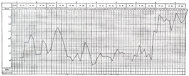
The chart W. H. (Fig. 37) is from a man aged fifty who fell in the street while returning from work at noon, and whose axillary temperature was taken at 5 P.M. and every two hours thereafter until death. The hemiplegia was not very marked, but the hemorrhage was extensive, involving the pons and left crus cerebri, the external capsule, left crus cerebelli, and medulla, bursting through into the fourth ventricle.
FIG. 38.
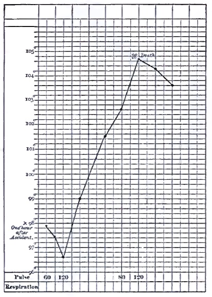
The chart M. M. (Fig. 38), as taken from Bourneville, represents the course of the temperature in a rapid case: each perpendicular line denotes an hour.
The difference in the temperature of the two sides has been variously stated, and probably depends on a number of factors besides the length of time that has elapsed since the first attack. There is probably, however, a tendency to excess of heat on the paralyzed side soon after the attack, owing to vaso-motor paralysis; and this difference will be more marked in the hands than in the axillæ. After a length of time which may be from days to months the temperature becomes equalized, or more frequently the relation is reversed, the paralyzed side being colder as atrophy takes place. Lepine19 gives a case where the axillary temperatures of the two sides continued the same within a small fraction of a degree for three days, and then separated very slowly, until at death the paralyzed side was six-tenths of a degree (Cent.) hotter than the other, in both being inferior to the rectal (107° Cent.).20
19 Mémoires de Société de Biol., 1867.
20 The chart in the original, and as reproduced by Bourneville, is wrongly lettered. The text says that the left side was the hotter.
FIG. 39.
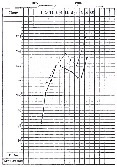
The chart C. M. (Fig. 39) shows the excess of temperature in a case of meningeal hemorrhage. The dotted line is from the paralyzed side. The first observation was made two and a half hours after the attack.
A very interesting case is reported by Johnson21 of crossed hemiplegia, where the temperature was about a degree higher on the paralyzed side of the body, and, corresponding to this, the sphygmograph showed a great diminution of tension; the lesion is supposed to have been a hemorrhage in the pons. Johnson, in commenting on the statement of Lorain that in all cases of hemiplegia the pulse is more full on the paralyzed side, says that it is incorrect for ordinary cases of hemorrhage into the corpus striatum, though true in his own case.
21 Brit. Med. Journ., Jan. 6, 1877.
The most marked differences of temperature have been observed where the lesion has been in the neighborhood of the pons, crus cerebri, or medulla oblongata. In a case reported by Allbutt there was a difference of 1.6°; the radial pulse was softer and fuller on the paralyzed side, and the cheek upon that side was flushed.22 The pulmonary hemorrhages which have been noticed by Brown-Séquard and others in animals after cerebral lesions, and the extravasation, congestion, subpleural ecchymoses noted by Ollivier23 in cerebral apoplexy, are probably to be referred to vaso-motor disturbances.
22 Med. Times and Gaz., Dec. 4, 1869.
23 Archives générales, 1873, 167.
Much more attention has been paid to the pulse than to the temperature, but it is less easy to lay down definite rules in regard to it. It may vary in either direction. When the case is approaching a fatal termination the pulse is apt to accompany the temperature in a general way in its rise, though not necessarily following exactly, as is seen in the chart in Fig. 38.
The throbbing or bounding of the arteries often described may indicate increased activity of heart, but means at the same time vaso-motor relaxation. The urine and feces are often passed involuntarily.
In some rare cases symptoms closely resembling those produced in animals by section of the sympathetic have been seen. These are false ptosis, narrowing of the palpebral opening and sinking of the globe of the eye into the orbit, diminution in the size of the pupil, higher temperature on the paralyzed side of the face and the corresponding ear, abnormal secretion of the eye, nose, and mouth on the same side.24 They are supposed to indicate a paralysis of the sympathetic.
24 Nothnagel, quoted by Grasset.
The condition of general relaxation may be so profound as to cover up everything else, but in many cases true paralytic symptoms may be discovered or provoked, which even at an early period give us information as to the locality and nature of the lesion.
A greater degree of muscular relaxation may be manifest on one side of the face than the other; the forehead may be a little smoother on one side, the corner of the mouth drooping, the downward line from the ala of the nose flattened, and the cheek flapping. There may be a little greater resistance to passive motion of the limbs on one side; one hand on being raised may drop helplessly back to the bed, while the other is laid slowly down; the right hand when pinched lies motionless and without power to escape the pain until the left comes to its assistance. Irregularity of the pupils, if present, is an important sign, but its absence signifies nothing.
One of the most significant signs is the conjugate deviation of the eyes, both eyes and the head being turned strongly to one side or the other. When the lesion is above the pons and is irritative, as in the early stage of hemorrhage, the deviation is toward the side of the body affected and away from the lesion; when paralysis is established, away from the paralysis and toward the lesion. Below the pons the rule is reversed. The spastic stage of conjugate deviation may coincide with stiffness (early rigidity) of the paralyzed limbs. This deviation must not be mistaken for an accidental position of the head. The patient should be addressed from the side away from which he is looking. Sometimes the eyes can be brought to the median line, and not beyond. An attempt to turn the head forcibly beyond the median line occasionally causes pain. The value of this symptom in diagnosis has been denied, but a part at least of the apparent contradictions have arisen from the neglect to notice whether it were of a paralytic or spastic character.
As the condition of unconsciousness gradually passes off, the face regaining, at least in part, its natural and more intelligent expression, the eyes trying to follow the movements of surrounding persons, an attempt being made, perhaps only by an unintelligible sound or by a nod, to answer questions, the tongue being protruded, or at least an attempt toward it made, and some motions being made with the limbs,—the exact extent and intensity of the paralysis become more apparent. Conjugate deviation, if it have existed, may disappear before the other symptoms, or, if it has been of the rigid form depending on an irritative lesion, it may become paralytic, and is then in the opposite direction. The patient is then usually found to be in a condition of hemiplegia, and at this point the history of hemorrhagic apoplexy becomes identical with that of paralysis from hemorrhage where no truly apoplectic condition has been present.
Lidell states that in more than one-third of all cases of cerebral hemorrhage hemiplegia is developed without loss of consciousness or coma. In some, the paralysis precedes unconsciousness, which then slowly supervenes.
Hemiplegia (ἥμι, half, πληγη blow) is a paralysis or paresis of a part of the voluntary muscles of one side of the body, and a few, in some cases, on the other, and is undoubtedly to be referred to a lesion interrupting the nervous communication between the cortical centres of motion and the nuclei of the motor nerves, cerebral and spinal; the conductors passing through the corpora striata, the internal capsule, the peduncles, and crossing in great part to the other side above or at the lower border of the medulla oblongata, and passing down the crossed pyramidal tracts of the cord, to be finally connected with the anterior gray columns of the cord. The portion which does not decussate passes down the inner border of the anterior columns under the name of columns of Türck. The amount of decussation which takes place varies somewhat, and the suggestion has been made, in order to explain certain cases of paralysis occurring on the same side with the lesion, that possibly in some rare cases there may be no decussation. It has never been shown, however, that this condition, highly exceptional if even it ever occurs, is present in such cases.
It may be said in a general way, although exceptions to the rule can be found, that it is those muscles trained to separate, specialized, or non-associated movements which are chiefly affected, while those which are habitually associated in function with those of the other side are less or not at all so. It would not, however, be in the least correct to say that specialized or educated movements of any set of muscles are alone paralyzed, since the fingers, which are trained to the most independent movements, are often just as incapable of making the slightest movement of simple flexion as of writing or sewing.
We have in ordinary hemiplegia more or less paralysis of the upper facial, the patient not being able to close his eye or to wink quite so well as on the paralyzed side. The forehead may be smoother on the paralyzed side. This condition is usually slight and of short duration, but varies in different cases. Paralysis of the lower facial angle of the mouth and cheek is usually better marked, but not absolute. The corner of the mouth droops, perhaps permits the saliva to escape; the naso-labial fold is less deep, and the glabella deviated away from the paralyzed side. The cheek flaps with respiration. The difference between this facial paralysis connected with hemiplegia and that dependent upon a lesion of the trunk or distribution of the nerve (Bell's), as in caries of the temporal bone or the so-called rheumatic paralysis, is very striking, the latter being so much more complete, and, by affecting the orbicularis palpebrarum so as to prevent closure of the eye, giving a very peculiar expression to the countenance. This distinction between the two portions of the facial seems to make an exception to the rule stated above, since in most persons the movements of the corner of the mouth and of the cheek are quite as closely bilaterally associated as those of the eyelids.
Paralyses of the third, fourth, and sixth pairs upon one side of the body are comparatively rare in hemiplegia, and when present are usually referable to localized lesions in the pons. They are to be looked upon as something superadded to the ordinary hemiplegia. These nerves, however, are affected in the peculiar way already spoken of as conjugate deviation, which phenomenon would seem to denote that muscles accomplishing combined movements in either lateral direction of both eyes, rather than all the muscles of each, are innervated from opposite sides—i.e. that the right rectus externus and the left rectus internus are innervated from the left motor centres, and vice versâ. Exactly the same remark will apply to the muscles of the neck which cause the rotation of the head seen together with the deviation of the eyes. The muscles controlling deviation to one side, although situated upon both sides of the median line, are apparently innervated from the side of the brain toward which the head is turned in paralysis.
The tongue is usually protruded with its point toward the paralyzed side; and this is simply for the reason that it is pushed out instead of pulled, and the stronger muscle thrusts the tongue away from it. The motor portion of the fifth is, according to Broadbent, affected to a certain extent, the bite upon the paralyzed side being less strong.
The hand and the foot are the parts most frequently and most completely affected, but one or the other may be partially or wholly spared, though the latter is rare. The muscles of the limbs nearer the trunk may be less affected, so that the patient may make shoulder or pelvis movements when asked to move hand or foot. In severe cases even the scapular movements may be paralyzed. The muscles of the trunk are but slightly affected, though Broadbent states that a difference in the abdominal muscles on the two sides may be perceived as the patient rises from a chair. The respiratory movements are alike on the two sides. A woman in the hospital service of the writer had a quite complete left hemiplegia at about the seventh month of pregnancy. There was some return of motion at the time of her confinement. None of the attendants could perceive any difference in the action of the abdominal muscles of the two sides, although, of course, the usual bracing of the hand and foot upon the left side was wanting. The pains were, however, generally inefficient, and she was delivered by turning. Muscular weakness often exists, and in some cases the non-paralyzed side shows a diminution of power.
The sphincters of the bladder and rectum frequently, and in severe cases almost invariably, lose their activity for a time. It is possible, however, that in some cases of alleged inability to retain urine and feces the defect is really mental, and akin to the dirty habits of the demented. The involuntary muscles probably take no part in hemiplegia, with the very important exception of the muscular coats of the arteries, which apparently share to a certain extent, and sometimes the iris.
Speech may be attempted, and the words be correct, so far as they can be understood, though the patient is apt to confine his remarks to the shortest possible answering of questions. It is, however, thick and indistinct, since the muscles of the tongue and lips are but imperfectly under the control of the will. This condition may be connected with paralysis of either side, and is to be sharply distinguished from aphasia or mental inability to select the proper word or to determine the necessary movements for its pronunciation. Aphasia is almost invariably connected with paralysis of the right side, and will be minutely spoken of hereafter. There is, of course, nothing to prevent the coexistence of the two conditions, but aphasia cannot well be shown to exist until we have reason to suppose, first, that the patient has ideas to express, and secondly, that the paralysis of the muscles of the lips and tongue has more or less completely disappeared. The patient may indistinctly mumble a word which, however, can be understood to be appropriate to the occasion (defective articulation, glosso-labial paralysis), or, on the other hand, pronounce with distinctness an entire wrong word or a number of sounds without meaning (aphasia).
Sensibility—that is, ordinary cutaneous sensation—and, so far as we can judge, the special senses, are not greatly affected after the deep coma has passed off, but exceptions to this rule will be noted later.
Having described this most typical but not most common form of cerebral hemorrhage—that is, the form in which both lesion and symptoms are most distinct and can be most clearly connected—we have a point of departure for conditions less clearly marked and less easily explained.
It is probable that cerebral hemorrhage is much less likely than cerebral embolism to take place without any disturbance of consciousness or abnormal sensations; but there can also be little doubt that a certain amount of paralysis is often accompanied by no other symptoms, and post-mortem appearances often show the remains of small hemorrhages which have passed unnoticed or are lightly estimated. It is highly probable that small hemorrhages may give rise to symptoms which pass for only a little accidental vertigo or a slight feeling of faintness, until a later and more serious attack gives a more definite explanation.
On the other hand, we have a set of cases in which all the symptoms of cerebral hemorrhage may be present without the lesion. Many of these are of course due to embolism, which will be considered later; but besides this condition, recognized as softening for many years, we find described under the head of simple, congestive, serous, and nervous apoplexy cases where sudden or rapid loss of consciousness occurs with general muscular relaxation, which, when fatal, show nothing beyond changes in the circulation—i.e. in the amount of blood in the cerebral vessels or of serum in the meshes of the pia or at the base of the brain.
Besides these, there are cases of temporary unconsciousness with complete recovery—the coup de sang of the French, or rush of blood to the head, which are attributed to congestion of the brain—a theory difficult to prove or disprove, but not in itself unreasonable. Trousseau, without denying the possibility, or even probability, of such a condition, says that which has been called apoplectiform cerebral congestion is in the greater number of cases an epileptic or eclamptic accident, sometimes a syncope. Simple epileptic vertigoes, vertigoes connected with a bad condition of the stomach or diseases of the ear, are wrongly considered as congestions of the brain. He speaks of various conditions, such as violent attacks of whooping cough, the expulsive efforts of women in labor, the congested faces of laborers under heavy burdens, to show that cerebral congestion does not give rise to an apoplectiform attack; and it is undoubtedly true that, as a rule, no long-continued attack is the result; but it must be within the personal experience of almost every one that decided cerebral disturbance is produced for a few moments by such efforts, as, for instance, blowing a fire with the head down. Besides this, a laborer under a heavy load is presumably healthy and accustomed to his work, so that his arteries may be supposed capable of maintaining a due balance between arterial and venous blood in the brain; and, again, although the ordinary efforts of women in labor do not cause unconsciousness, puerperal convulsions, involving a longer period of violent muscular action, may do so, and even give rise to hemiplegia.
Whatever name we may adopt for the temporary cases which recover, there are others, and fatal ones, which are not explained by any change in nomenclature. Epilepsy may, it is true, occur under such circumstances that no convulsion is observed, but, on the other hand, convulsions may accompany not only an attack of unconsciousness, but actual cerebral hemorrhage.
Cases of sudden death with no discoverable lesion furnish abundant opportunity and temptation for conjecture, and it is well known that too much dependence must not be placed upon the post-mortem appearances as to the amount of blood in the brain before death, and probably just as little upon the amount of serum, except as indicating a condition of atrophy.
Syncope, either from over-stimulation of the pneumogastric or from simple failure of the heart, may be put forward to explain some cases of sudden death, but seems to have no advantage as a universal theory over the older one, which meets with so little favor. Lidell gives no less than seventeen cases which he classifies as congestive or serous apoplexy. They are not all equally conclusive, and were almost all of alcoholics. In some of these there were absolutely no appearances which could account for death. The two most characteristic of congestive apoplexy were, first, a young negress who experienced a violent fit of passion, became unconscious, with stertorous breathing, and died, having had some tonic spasms. The brain contained a large amount of blood in the vessels, but no effusion. Second, a semi-intoxicated woman, aged thirty, became very angry, fell insensible, and expired almost immediately. The brain contained an excess of blood, with no effusion. In both these cases the patients were subject to fits under the influence of strong excitement, but in both the author took pains to inquire into and negative the probability of epilepsy of the ordinary kind; and a change of name does not go far toward clearing up the pathology.
Lidell's case (XXII.) was that of a man accustomed to alcohol, thin and pale, who had an apoplectic fit with coma and hemiplegia. He regained consciousness on the second day, and the hemiplegia disappeared in a fortnight. This rapid and complete recovery, exceptional to be sure, cannot be regarded as proof of the absence of hemorrhage or embolism. In fact, the latter is highly probable. It is possible that the clot may have been partially dislodged, so as to allow some blood to pass by it, or that an exceptionally favorable anastomosis allowed a better collateral circulation than usual to be established.
The following case occurred in the service of the writer: An elderly negress, who had extensive anasarca and signs of enfeebled action of the heart without any valvular lesion being detected, after washing her face was heard to groan, and found speechless and unable to swallow, with complete right hemiplegia. There was a slight improvement in a few hours, but she died two days later. The autopsy disclosed some hypertrophy and dilatation of the heart without valvular lesion. A careful search failed to discover any change in the brain or obstruction in its vessels, although there was chronic endarteritis.
The relations between epilepsy, apoplexy, and syncope are interesting and important, but are certainly far from clear. Little is gained by shifting obscure cases from one category to the other. If sudden deaths be synonymous with apoplexy, we shall certainly have to admit that apoplexy does not always demand for its cause cerebral changes sufficiently marked to be recognizable after death. If, on the other hand, we refer them to heart disease, we shall have to admit that a heart without valvular disease or extensive changes in its muscular substance may cease to beat under influences as yet not well understood.
Since the paralysis arising from hemorrhage resembles so closely in its progress that dependent upon occlusion of the cerebral vessels, a further description will be deferred until the latter lesion has been described; but this remark does not apply to the premonitory and initiative symptoms, which may be of great importance, and which are not always the same with the two or three sets of lesions. There are many of them, but, unfortunately, no one among them taken alone can be considered of high significance, unless we except what are sometimes called premonitory attacks, which are in all probability frequently genuine hemorrhages of so slight extent that they produce no unconsciousness, and but slight paralysis easily overlooked. A little indistinctness of speech or a forgetfulness of words, a droop of one angle of the mouth, or heaviness in the movement of a foot or hand, lasting but a few moments, may be real but slight attacks, which may be followed either by a much more severe one, by others of the same kind, or by nothing at all for a long time. They are sufficient to awaken apprehension, and to show in what direction danger lies, but they give little information as to the time of any future attack.