Volume 5 — DISEASES OF THE NERVOUS SYSTEM. (continued 2)
Reading edition
5 Ataxia is greatly aggravated by closing the eyes.
(c) Uncertainty in voluntary movements is sufficiently defined by the term employed and by the absence of ataxia. This is well illustrated in those rare cases in which the muscular sense is impaired or lost: in such a case volitional movements, such as placing the fore finger on an object or fastening a button, are fairly well done with the aid of sight, but without it the hand and fingers grope almost or quite hopelessly about the object. It is important to note that persistent trying to do the act does not aggravate the disorder, but that, on the other hand, success is often achieved after feeling about. In certain cases of blindness irregular movements of the eyeball are observed, due to semi-voluntary efforts to look or to direct the eyeballs in the direction of a person or object. In states of cutaneous anæsthesia when the eyes are closed the same disorder appears in muscular movements. Claude Bernard many years ago showed that section of the posterior roots of spinal nerves in an animal was followed, not by ataxia, but by vague inco-ordination and staggering. In human cases we find that where the sensibility of one hand is lost or greatly impaired, without paralysis, there is extreme awkwardness and uncertainty in delicate muscular movements, but no ataxia. The staggering exhibited by patients having plantar anæsthesia is largely of this type: they stand fairly well while their eyes are open, but oscillate or fall when they are closed. This variety of inco-ordination may result from toxic conditions (alcohol), peripheral, neural, or central nervous disease. A few cases are on record which would seem to show that there is a centre for muscular sense in the cortex of the brain, in a part intermediate between the caudal sensory area and the central motor one—viz. in the inferior parietal lobule, supramarginal gyrus (and angular gyrus?). It has also been shown (by Spitzka and others) that there is a conduction tract for muscular sense, dorsad of the pyramidal tract in the pons and oblongata (in the stratum intermedium and interolivary tract), lesions of which produce inco-ordination (ataxia?) without marked paralysis or anæsthesia.
(d) Titubation, or staggering, is the inability to stand erect or walk straight because of impaired equilibrium. There is neither tremor nor ataxia present, and paralysis and anæsthesia are not necessary factors. It is distinguished from vertigo and dizziness by absence of subjective sensations of movement. Staggering may show itself in a general way or in the shape of latero-, retro-, or propulsion (disease of the internal ear, paralysis agitans). A well-defined variety of staggering is the wrongly-termed cerebellar ataxia. In this the patient, having disease of the cerebellum involving its vermis superior, stands with feet widely separated to increase his base of support; the body is bent somewhat forward, and the arms and hands are used as balancing-rods to maintain a sort of equipoise. In walking, this attitude is exaggerated, and if the feet be bare it will be seen that the toes are unconsciously clutching the floor for support; there is no outward jerk of the leg or stamp of the heel as in the ataxic gait, and closing the eyes does not aggravate the attitude or walk. Besides, if the extremities be separately tested, it is found that with closed eyes the patient can perfectly well place his fore-finger on his nose or one heel on the opposite patella (lying down). The proper term for the disorder is cerebellar titubation. Yet it must be remembered that titubation also occurs from disease of the oblongata and pons, from lesions of the base of the brain in general, and from alcoholic, etc. intoxication. In many cases titubation occurs in connection with vertigo or dizziness.
(e) Inco-ordination more or less of the ataxic form often affects the muscles of articulation, phonation, and deglutition, giving to the symptoms dysarthria, dysphonia, and dysphagia. Dysarthria and dysphagia are probably often caused by lesions of the insula and subjacent white substance, as well as by those affecting the oblongata. There are two recent autopsies which would indicate that there may be a cerebral cortical centre for phonation laryngeal movements: in the ventral extremity of the right third frontal gyrus—a part homologous to the speech centre on the left side of the brain. In some cases, however, dysphonia and aphonia indicate a lesion of the laryngeal nerves or of the oblongata (nucleus of NN. x. and xi.).
(f) Doubtless the internal muscular organs and the blood-vessels are frequently the seat of inco-ordinate or quasi-ataxic movements, but our present knowledge of these conditions amounts to very little.
It may be permissible to consider vertigo under the general head of inco-ordination, because it usually finds a motor expression, either actual or subjective. Subjective vertigo consists in a sense of whirling or horizontal movement which is clearly referred to the brain by the patient. The sensation is variously expressed: in some the head seems to whirl around; in others external objects seem to whirl about the patient horizontally or vertically; in others still there is a sensation of falling in a given direction or of dropping into a hole. Dizziness is a minor degree of subjective motion conjoined with more or less confusion and other paræsthesiæ in the head. Static vertigo is that form in which there is actual loss of equilibrium, or falling. It is observed more especially in diseases of the internal auditory apparatus, and it may be produced artificially by galvanization of the brain. If the electrodes are placed exactly on the median line, and the current passed fronto-caudad through the brain, no loss of equilibrium or vertigo is produced. If, however, the current is passed transversely through the brain, there is produced at the moment of the closure and opening of the circuit a distinct tendency to fall or an actual fall to one side: in closing the current the fall is always away from the negative pole or cathode. The greatest amount of disturbance is produced when the electrodes are placed on the mastoid processes or near them. Whether the disturbance of equilibrium is caused by a change in electrotonus in the hemispheres, or by a change in the vascular supply of the hemispheres (in one anæmia, in the other relative hyperæmia), is uncertain. A third explanation is equally plausible—viz. that the vertigo is caused by unequal excitation of the internal acoustic organs and the two halves of the cerebellum. This view would be supported by the fact that in animals and man a lesion of the acoustic nerve and of the processus ad pontem on one side produces strong rotary movements about the long axis of the body.
ABNORMAL REFLEX MOVEMENTS.—Some reference has already been made to absence or exaggeration of reflexes in the preceding paragraphs, but the importance of these symptoms demands that they should be separately considered.
A reflex action, in its simplest conception, is a movement (muscular, vascular, or psycho-motor) or a secretion which is the result of the transformation of a centripetal impression into a centrifugal impulse. The apparatus required for the accomplishment of the reflex action consists essentially of a sensitive surface of skin or mucous membrane, of tendinous or other deep structure, to receive the impression; a sensory afferent nerve to convey it; a nervous centre of the simplest structure (one or two ganglion cells) to receive and transform it; a motor or efferent nerve to transmit the resulting impulse to a common muscle, to the vascular wall of a vessel or viscus, or to a gland. These various terminal organs execute their normal functions in response to the centrifugal impulse, and thus give external or tangible evidence that the reflex action or reflex is completed. This mechanism, the reflex arc, is illustrated by the diagram (Fig. 1), which will also serve for the explanation of many of the morbid reflex states.
FIG. 1.
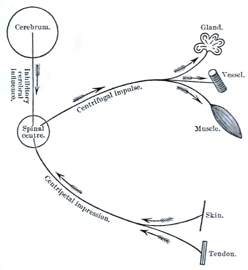
Diagram illustrating the Arc for Reflex Action. The centripetal and centrifugal paths, the receptive and terminal organs of the arc, are shown connected with the spinal centre, which is a portion of central gray matter containing ganglion cells. The inhibitory cerebral influence is constantly exerted in health to moderate reflexes.
In the healthy animal body many important functions are performed under the law of reflex action; for example, many organic or secretory functions, the movements of hollow viscera, vascular movements, muscular tonus, defensive and expressive muscular movements. In the process of education many acts which at first must be learned by oft-repeated efforts, such as walking, playing upon musical instruments, etc., later become habitual and automatic, and are then largely executed by reflex action unconsciously, co-ordinated by the spinal (?) apparatus. Much of our intellectual activity is governed by the same law operating between the psycho-sensory and the psycho-motor apparatus.
Reflex actions are only to a moderate extent under the control of volition—indeed, most of them are subconscious—but a strong restraining action is unconsciously exerted by the cerebrum (vide Fig. 1) upon the spinal reflex centres. This is the so-called inhibitory cerebral influence. The existence of such an influence is shown by experiments upon animals, and by human cases in which, the cerebral activity being reduced or abolished, we observe a great increase in the frequency and extent of reflex movements, particularly in the common muscular apparatus.
Clinically, we distinguish cutaneous or superficial, tendinous or deep reflexes, according to the seat of the original excitation or testing-point. Cerebral or psychic reflexes are, however, also to be borne in mind. These reflexes may be abolished or increased.
(a) Diminution and abolition of reflex action are frequent symptoms of disease of the nervous system. The fault or break may be anywhere in the reflex arc, so that each case must be analyzed by itself. Let us consider the phenomena as exhibited by two widely distant and different apparatuses.
First, in the eye. In case of atrophy of the optic nerve the pupillary reflex is lost, the reflex action failing because the receptive surface and efferent nerve are injured. In certain cases of spinal disease (posterior spinal sclerosis) the same pupillary immobility is observed (the Argyll-Robertson pupil); and in this case the lesion either affects indirect efferent spinal fibres destined for the iris, or it is situated in the centre for the reflex action—viz. the gray matter of the lobus opticus. There may be loss of pupillary reflex due to injury of the direct efferent fibres of the arc (paralysis of the motor oculi, N. iii.). Lastly, the iris itself may be so diseased as to be incapable of contracting, though it receive the reflex impulse properly; the lesion is then in the terminal organ of the arc.
Second, the patellar tendon reflex. In a healthy individual, sitting at ease with one leg thrown over the other (knee over knee), upon tapping the ligamentum patellæ of the overhanging or free leg a contraction of the quadriceps muscle occurs, causing a visible forward movement of the leg and foot. This is the well-known patellar reflex or knee-jerk. The arc in this case consists of the ligamentum patellæ with its included sensory fibres as receptive organs, sensory (afferent) fibres of the crural nerve, a segment of the lumbar gray matter of the cord as centre, motor (efferent) fibres of the crural nerve, supplying the quadriceps extensor femoris, which is the terminal organ. Theoretically (vide Fig. 1), we can conceive of numerous abnormal conditions of parts of this arc which would lead to abolition of the patellar reflex, but in practice the following are the principal lesions to be thought of: Disease (sclerosis) of the posterior root-zones of the lumbar enlargement of the cord, as exhibited in the pre-ataxic stage of tabes; disease of the posterior roots themselves through meningitis or meningo-myelitis, as in diphtheritic ataxia; lesion of the nervous centre, as is frequently observed in cases of infantile poliomyelitis; a lesion of the crural nerve, involving its efferent or afferent fibres, or both sets of fibres, would produce the same result, as would also, lastly, a severe myositis or cancerous infiltration of the quadriceps muscle.
It is therefore evident that there is, or can be, nothing pathognomonic in the loss of a given reflex: it simply indicates a break in the reflex arc, the seat and nature of which remain to be determined in each case. The topographical study of reflexes in disease is of very great importance, more especially as a help to the correct location and extent of neural and spinal diseases. To assist this study, and for use more especially in connection with the ensuing sections on localization, Gowers's excellent diagram and table are reproduced in Fig. 2.
FIG. 2.
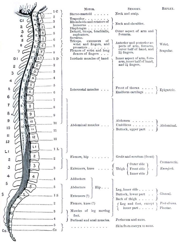
Diagram and Table showing the Approximate Relation to the Spinal Nerves of the Various Sensory and Reflex Functions of the Spinal Cord (after Gowers).
(b) Exaggeration of reflex actions. This is due, theoretically, to any condition which increases the excitability of the reflex arc, but practically and usually the cause of the increase is more or less complete removal of the inhibitory supraspinal or cerebral influence. The reflex arc itself remains structurally sound, but its function is abnormally active. Thus, we meet with the most exquisite exhibition of exaggerated reflexes in the paralyzed part after a transverse lesion of the dorsal part of the cord. The lesion cuts off all communication between the lumbar enlargement and the brain; no sensations pass to consciousness, and no volitional influence can be exerted upon the legs; yet the legs are abnormally active, the least touch on or pricking of the skin, tapping a tendon, handling a joint, the pressure of feces in the rectum, etc., produce more or less violent spasmodic movements of mixed clonic and tonic forms (spinal epilepsy). Increased knee-jerk, foot-clonus, and epileptoid trepidation are also present. Other proofs that removal of the inhibitory cerebral influences is the chief cause of increase of spinal reflexes are given by the history of various cerebral diseases. Thus, in hemiplegia with secondary contracture the cutaneous and tendinous reflexes on the paralyzed side are much exaggerated; in dementia paralytica, as a rule, the patellar reflex is increased on both sides; and in cerebral neurasthenia all reflexes are often extremely great.
A variety of increased tendon reflex is known as clonus—a series of rhythmic clonic muscular contractions set up by stretching the body and tendon of a muscle. This is best studied at the ankle. By holding the patient's leg nearly extended upon our left hand, and rather suddenly forcing back the foot (dorsal flexion) by pressing under the ball of the foot with our right hand, a series of oscillations or to-and-fro movements of the ankle-joint are produced for a few moments (ankle- or foot-clonus). Sometimes the same rhythmic clonic contractions may be produced in the quadriceps by quickly pulling down the patella with the leg semi-extended.
In some instances, together with great increase of tendon reflex, we observe a transfer of the centrifugal impulse to other nerves of the same extremity, or across the spinal cord to nerves and muscles of the homologous limb.
Another interesting manifestation of morbid reflex movements is the defensive tonic spasm of muscles about a diseased joint (of the hip-muscles in coxitis, of vertebral muscles in Potts' disease). This constitutes a valuable symptom for the differential diagnosis between articular and neural diseases in certain cases.
Clinically, increase of reflex action is not as valuable an indication as its absence, many healthy persons having high reflexes. Still, when studied comparatively as to topography (comparing the two sides of the body) and as to variations at different times in a given case, the symptom is of considerable assistance.
IV. Trophic Symptoms.
Our present knowledge of this great group of symptoms of nervous disease is in a somewhat chaotic state; and although we possess a large accumulation of facts of this category, their classification is incomplete and their explanation is for the most part theoretical and unsatisfactory. The following subdivisions may be made to include all the phenomena, and is sufficient for clinical purposes:
1. DEGENERATIVE ATROPHY, as distinguished from the simple atrophy of insufficient nutrition, of disuse of the parts, and of retarded growth, includes all those trophic symptoms in which, under the influence of nervous disease, the solid tissues exhibit histological and chemical alterations as well as reduction of volume. The principal factor of this group is—
(a) Muscular atrophy due to lesions of some part of the nervous apparatus (thus excluding directly traumatic or inflammatory atrophy), as seen in the course of progressive muscular atrophy, of various forms of poliomyelitis, of lead paralysis, of neuritis, and injuries of nerves. These muscular atrophies are characterized by reduction in the size of the affected muscles, by altered electrical reactions, and by histological changes. Reduction in size may affect one muscle or a muscular group shortly after paralysis (as in poliomyelitis, lead paralysis, affections of nerve-trunks), or without evident paralysis it may involve only small fasciculi of a muscle at a time (as in progressive muscular atrophy); so that we speak of diffused and fascicular atrophy, of secondary (post-paralytic) and primary atrophy. In some cases the wasting proceeds to such a degree that apparently no muscular tissue remains, and the skin seems to rest directly on the bones; in others the reduction in bulk is moderate, and is compensated by increase in the intra- and extra-muscular fat (thus in stout children the atrophy of severe poliomyelitis may be quite masked to the eye); in still another category the increase in connective tissue with fatty accumulation overbalances the muscular wasting, and we have the pseudo-hypertrophic condition. Alterations in electro-muscular contractility are most important signs of impaired muscular innervation and nutrition. They present numerous and somewhat complicated variations of value for diagnosis and prognosis, but which cannot here be noticed at length. The general principle underlying these variations is this: that a muscle deprived of its normal innervation (centrifugal or motor) soon loses its capacity to respond to the induced (faradic) current, and reacts in an abnormal way to the galvanic current. This constitutes the reaction of degeneration of Erb.6 The chief abnormality in the galvanic reaction of a paralyzed and diffusely atrophied muscle is its slow or wave-like contraction, which on a graphic, or even to the observer's eye, presents a striking contrast to the sharp, jerking, complete contraction of a healthy muscle. Besides, the reactions to the two poles become equal, or even reversed, so that we may obtain in testing by the polar method such formulas as CaCC = AnCC, or CaCC < AnCC. With reference to the failure of atrophied muscles to respond to the induced or faradic current, this qualification must be added: If the poles are applied on the moistened skin, as usual in testing or treating cases, no reaction occurs even with most intense currents; but if a needle be made one pole of the battery and inserted into the affected muscle, slight and limited contractions may be obtained with a moderate current for many months. Of this we have repeatedly satisfied ourselves in cases of poliomyelitis, lead paralysis, and traumatic peripheral paralysis. In the later stages of diffused paralytic atrophy, after an uncertain number of years, no reaction can be obtained with either electrical current. In fascicular non-paralytic muscular atrophy (as in progressive muscular atrophy) the loss of faradic contractility is only demonstrable in the affected fasciculi, good contractions being obtained in adjacent healthy fasciculi of the same muscle. The amount or extent of contraction varies pari passu with the progress of the atrophy. The galvanic reactions of this form of muscular atrophy are not yet well established. Muscles and muscular groups in a condition of impaired nutrition exhibit at an early period an interesting condition—viz. that they no longer contract by reflex excitation. Thus in a case of infantile poliomyelitis with paralysis of the muscles of the leg, these muscles no longer contract when the sole of the foot is tickled, and if the thigh-muscles are affected even so slightly as to appear of normal size and consistence, the patellar reflex is found wanting. In these and in traumatic cases the reflex act is prevented by lesion of the centrifugal motor nervous apparatus, and perhaps also by the associated muscular trophic alterations.
6 Conveniently expressed by the symbol De R.
Histological changes in atrophied muscles vary somewhat in character in different diseases, and vary much in degree at different periods of the atrophy. An early appearance is the presence of proteinaceous and of fatty granules or molecules in the sarcous substance. Later, the muscular fibres become reduced in size, lose their striation, and show inequalities; the interstitial connective tissue becomes active and increases in amount, at the same time that fat is deposited. The fatty change in some cases, in others the granular or proteinaceous transformation, ultimately completely destroys the muscular substance, so that in the place of a muscle we find abnormal connective and fatty tissues, blood-vessels, fatty and proteinaceous débris, the whole presenting a pale yellowish-white aspect. It is in this final stage of degenerative atrophy that all electrical reactions are lost. In the pseudo-hypertrophic state the interfibrillary and interfascicular connective-tissue growth is much more active, and the wasting muscular fibres are buried in masses of wavy and fatty connective tissue. In this condition also electrical reactions may be wholly absent.
(b) Atrophy due to defective innervation sometimes affects the skin and bones. In the former, after nerve-injuries more especially, we observe loss of thickness, glossiness, and perverted circulation and secretion; the hairs may fall out or grow abnormally; the nails are slow of growth, thick, rugose, incurvated, and brittle. In other cases, as in cerebral and spinal paralyses, the skin of the paralyzed part is abnormally dry, rough, and furfuraceous, and it loses its elasticity. In some varieties of so-called skin diseases the patches of altered nutrition (eczema, bullæ, herpes, psoriasis, leucoderma, scleroderma, etc.) are often, probably, dependent upon nervous lesions.
In the very rare disease known as progressive facial atrophy the skin, subcutaneous areolar tissue, and the bones undergo extreme atrophy. The initial lesion is usually a patch of scleroderma with or without neuralgic phenomena; the skin is thin, darker and smoother than normal; it soon adheres to the subjacent bone (maxilla or zygoma), which itself steadily diminishes in size. Almost the entire half of the face (including the palate and tongue) may ultimately show the atrophic changes. We are not yet prepared to state the causal nerve-lesion in this disease.
The bones are abnormally fragile (fragilitas ossium) in some nervous diseases, more especially in dementia paralytica and posterior spinal sclerosis; but whether this condition is due directly to the nervous disease or is the expression of more general malnutrition is now undecided.
The complex lesions of joints observed in the course of posterior spinal sclerosis, the spinal arthropathies of Charcot, probably belong to this category. The affected joint (knee, shoulder, or ankle) rather suddenly swells, and the swelling usually invades the rest of the member or extends to the next distal joint; it is a hard, semi-elastic swelling unlike common œdema. After its gradual subsidence it is found that changes have taken place in the articulation itself, and later distinct evidences of destructive disease, such as erosion of cartilages, relaxation of ligaments, swelling, are observed. In many cases extra-articular lesions appear in the shape of osteophitic formations from the adjacent bones. The hydrarthrosis may persist or disappear. So complete may be the destruction of the joint that—for example, in a case of arthropathy of the knee—the leg may be twisted about in all directions, and even over-extended so as to lie upon the anterior surface of the thigh. Examination of the joint post-mortem reveals non-suppurative destruction of all its component parts, cartilages, ligaments, and epiphyses; the eroded, deformed ends of the bones rub against one another in a false joint-cavity formed of the skin, connective tissue, tendons, and remains of ligaments. The absence of pain and tenderness in the course of arthropathies is a striking feature—so much so that when, in an adult patient, it is observed that manipulation or puncture of a diseased joint is painless, special inquiry should be made for symptoms of posterior spinal sclerosis.7
7 We have known one case in which the diagnosis of tabes dorsalis was made (and verified after death) in this way, after the surgeon in charge of the patient had mentioned the fact that puncturing a swollen diseased knee-joint was painless.
(c) Under the head of degenerative atrophies should be included the secondary changes which affect certain nerve-tracts within the nervous system itself. It is impossible in this introduction to treat fully of this interesting category of trophic changes; the following summary must suffice: (1) When the cerebral motor cortex or any part of the associated pyramidal (or motor) tract is destructively injured, there occurs, in from three to six weeks, a degeneration of the whole tract caudad of (below) the brain. The myeline becomes granular and disappears, the cylinder-axes are broken up and vanish, the connective tissue (neuroglia) increases in amount; the atrophied and degenerated area appears in a transection rather translucent in contrast to the pearly white of the normal medullary tissue, and when the preparation is stained with carmine the patch takes up an abnormal amount of pigment. This is the so-called descending degeneration, or centrifugal atrophy of the central nervous system. (2) After total transverse lesions of the spinal cord, besides the above-described centrifugal degeneration caudad of the lesion, we observe frontad of (above) it similar changes in the posterior median columns, and in the direct cerebellar tracts—centripetal degeneration. At present we have no knowledge of centripetal (ascending) degenerations in the cerebrum and in nerve-trunks; and if the results of von Gudden's experimental method be cited against this statement, it must be replied that its effects are best seen in newly-born animals, and that its pathology is yet unknown. (3) Lesions of the anterior gray matter (ventral cornua) of the spinal cord, and of mixed nerve-trunks produce only centrifugal or descending degeneration. All the nerve-fibres deriving their innervation from the injured area in the cord, or in case of nerves all fibres below the injury, perish—i.e. their myeline breaks up and undergoes granular and fatty degeneration, their cylinder-axes are segmented and disappear, while at the same time the connective tissue of the nerve becomes abnormally active and increases. Furthermore, in cases of this category there are, inevitably, degenerative and atrophic changes in the attached muscles, and peculiar electro-muscular reactions (vide supra). All these central and peripheral nervous degenerations, due to a local lesion, are conveniently grouped under the name of Wallerian degeneration.
2. ERUPTIONS AND ULCERATIONS.—(a) The cutaneous eruption about whose nervous origin there is the least doubt is that known as herpes or zona. This manifests itself, with or without paræsthesiæ (pain, itching, formication, etc.), as vesicles upon deeply-inflamed spots of skin distributed in the territory of one or more sensory nerves, and almost always unilaterally. The destructive process in the derma is so profound as to leave scars which are indelible as a rule. In general terms it might be stated that herpes may occur in the range of any sensory nerve distributed to the skin or mucous membranes. The neuralgia attending its development may be severe, and in some cases lasts for years after the healing of the eruption. The pathology of this affection appears to be inflammation of the ganglion of the posterior root of one or more spinal nerves (including the trigeminus and glosso-pharyngeal) or of their trunks. Herpes may appear in the course of spontaneous and traumatic neuritis; and in the last-named conditions a variety of eruptions have been observed in the area supplied by affected nerves, such as eczema, bullæ, etc.
(b) That ulceration may result directly from a nervous lesion is shown by the history of herpes, where a destructive process takes place in the derma under such conditions as to exclude the action of external agencies. But the same cannot be said of the ordinary ulcerations and gangrenous lesions observed in a number of nervous diseases, as the bed-sores of myelitis or of spinal injuries, the ulceration of the cornea in trigeminal anæsthesia, the digital ulcers and gangrene of lepra, asphyxia of the extremities, and nerve-injuries. As regards all these, the proper explanation is, it seems to us, that the anæsthesia existing as a predisposing cause (leading to imperfect protection of the part), the ulceration itself, is directly, actively caused by external agencies. Let me briefly cite a few instructive experimental and pathological facts bearing on this question.
It is well known that in animals and man lesions of the trigeminus nerve sufficient to produce anæsthesia (of its first branch more especially) are frequently, if not invariably, followed by ulceration and perforation of the cornea and phthisis bulbi. These were long held to be true dystropic changes, but about thirty and twenty years ago Snellen and von Gudden demonstrated independently and by different methods that these ocular lesions could be entirely prevented by absolute closure (perfect protection) of the eyelids prior to the experiment on the trigeminus. Not long afterward Brown-Séquard showed that the sloughing ulcers which occur about the foot of an animal whose sciatic nerve has been cut may be entirely prevented by care, cleanliness, and soft bedding. The conclusion to be drawn from these observations is that in almost all cases the ulcerations and sloughing observed in man during the course of a nervous disease sufficient to produce anæsthesia, such as traumatic neuritis, lepra anæsthetica, traumatic or simple myelitis transversa, disease of the trigeminal nerve, etc., are in reality produced by external agencies, injuries to the cuticle, action of filth, and, we think, the entrance of bacteria, which are well known to possess extraordinary powers of penetration into tissues whose protecting epithelial layer is removed. Clinical observation corroborates this view, for, with strict antiseptic treatment and under sealed dressings (collodion to ulcerated finger-tips), these ulcerations heal rapidly and completely, while the anæsthesia remains unchanged and the nervous lesions may even progress. At the same time, while we believe the above to be the pathology of so-called trophic ulcers, we would admit the possibility of spontaneous neuritic ulceration and gangrene, as shown more especially in herpes.
3. ALTERED SECRETIONS.—Under the influence of disordered innervation secretions may be altered in quantity and in quality. Symptoms of this class may be caused by neural or by central lesions, and the mechanism of their action may be direct or reflex.
(a) After section or lesion by disease of the cervical sympathetic nerve we observe increased activity of the entire skin in the affected area of the head and face: there is more perspiration, lachrymation, and more cerumen is found in the ear. In cases of lesions of a certain part of the oblongata we find one result to be polyuria. In many cases of nerve-injuries the sweat-glands and hair-follicles of the parts supplied by the affected nerve appear to be sometimes abnormally active, or in other cases inactive. In some rare cases of insanity profuse salivation and extraordinary growth of hair are striking symptoms. Lastly, in some functional nervous affections the various secretions and excretions may be altered to extraordinary extremes (e.g. the polyuria and anuria of hysteria).
(b) Qualitative modifications of secretions also occur, as exhibited in the many variations in the composition of the urine in various neuritic conditions, the watery urine of hysteria, the phosphatic urine of neurasthenia. Injuries of a part of the floor of the oblongata (and very probably other lesions of the nervous system) give rise to the appearance of sugar, and sometimes of albumen, in the urine. Under the influence of disease or of an emotion the breast-milk may become toxic to the nursing child, causing diarrhœa, convulsions, etc. In some forms of nervous dyspepsia large quantities of acid or of alkaline fluid may be rapidly poured out into the stomach as a result of disturbed glandular and vaso-motor action. In a few cases of neurosis malodorous or perfumed perspiration has been observed.
In a general way it may be stated that, inasmuch as the normal function of glands is largely, if not exclusively, under direct and reflex nervous control, their secretions may be quickly altered in quantity or quality by rapid nervous disease or by functional disorders. It is probable that much of our future progress in the semeiology of nervous diseases will be in this direction by the aid of improved medical chemistry.
4. ASSIMILATION, METAMORPHOSIS OF FOOD, TISSUE-LIFE, and BLOOD-MAKING are probably under nervous control to a certain extent, but our knowledge is not now in a state to speak positively and definitely of alterations in these processes as symptoms of nervous disease.
THE PRINCIPLES OF DIAGNOSIS.
Having in the preceding pages surveyed the field of neurological semeiology in an analytical way, it remains to briefly indicate how far and in what manner these data can be best utilized for logical diagnosis in the present state of science. If we may be allowed an illustration, we would say that the foregoing sections contain a nearly complete vocabulary of the language of nervous diseases, and that the following pages express an attempt at formulating its grammar.
In the first place, it is important to classify the symptoms observed according to their probable immediate cause in the nervous organs, whether they are due to irritation or to the destruction of parts. The extreme value of such a distinction in practice was first emphatically brought forward by Brown-Séquard in his famous Lectures on the Physiology and Pathology of the Central Nervous System (Philada., 1860); and to the observance of this law of genesis of symptoms we believe that a large part of the subsequent extraordinary progress of neurological science is due.
The symptoms produced by irritation of nervous organs are usually hyperkineses or paræsthesiæ in their various forms. The symptoms, spasm, pain, or numbness, are usually, though by no means always, intermittent. That paralysis and anæsthesia may occur from irritation, by an inhibitory process, is not to be denied, yet we must maintain the extreme rarity of such a mechanism. The phenomena of inhibition are so prominent in the normal nervous system that a physician who looks at nervous diseases from the standpoint of the physiologist may well be excused for seeing morbid inhibitory processes where others do not.
The symptoms due to destructive lesions—i.e. those indicating destruction of parts of the nervous system—are paralysis, hyperkinesis, and anæsthesia, of absolutely or relatively constant presence. Thus, for example, the paralysis of common hemiplegia due to destruction of the motor part of the internal capsule is constant and permanent, while the accompanying tonic spasm (late contracture), which is considered an even more positive sign of serious destructive injury to the cerebral motor tract, is permanent, but intermittent—i.e. it is absent in profound sleep, and reappears as soon as the patient awakes.
The association of symptoms of irritation and those of destruction is frequent but variable. In many cases, as in cerebral tremor and posterior spinal sclerosis, spasm and paræsthesiæ precede paralysis, anæsthesia, and ataxia. In other cases (in many at a certain period) they coexist. In a small group of cases the irritative symptoms follow those indicating destruction, sometimes occurring years afterward, as in post-hemiplegic epilepsy and neuralgia due to cicatrices. We would repeat that very often, more especially in organic nervous affections, much light is thrown on the diagnosis by careful noting of the topographic distribution and chronological order of appearance of the symptoms.
In the second place, it is necessary to group the symptoms of disease of the nervous system in two great classes—viz. those representing demonstrable lesions, macro- or microscopic, and those dependent upon perverted functions or molecular malnutrition of the nervous organs or elements. The first group is designated as organic diseases; the second as functional affections. Mental diseases, so called, can also be classified, according to their symptoms, in either of these groups.
The symptoms of the first group, that of organic nervous diseases, are characterized by definiteness of distribution, by permanency, by relative invariability, and by the predominance of objective signs. Another important characteristic of organic diseases is their progressive or fatal tendency, either with reference to life in general or to that of parts or organs. A third peculiarity of these diseases is that they do not occupy the patient's attention as strongly or as constantly as neuroses: in other words, the Ego is less involved.
The symptoms of the second group, that of functional nervous affections (neuroses and psychoses), are characterized by generality and indefiniteness of distribution, by relative variability, by easy removal or spontaneous disappearance, and by the preponderance of subjective symptoms. The affection may endure for many years or for a lifetime without fatal result and without special aggravation. The Ego is very strongly and deeply affected, fear, depression, and constant dwelling upon the symptoms being prominent features.
These are general statements intended to serve as guides for the preliminary study of a case. It must be remembered that they are all liable to exceptions, and that each patient must be separately considered. It should be borne in mind that what to-day appears as a functional affection, chiefly indicated by subjective symptoms, may in a few months present distinct signs of organic changes in the nervous system. Also, it must be added that in the present state of knowledge we sometimes are not sure as to the presence or absence of organic changes even after careful study of a case; as, for example, in some epilepsies and neuralgias.
Furthermore, allowance must be made for the following sources of error:
Anatomical variability; as, for example, in the distribution of peripheral nerves and in the amount of decussation of fibres of the pyramids;
The coincidence of diseases and multiplicity of lesions;
The toxic effects of drugs taken by the patient previous to our examination; as, for example, bromism.
I. The Diagnosis of Organic Diseases of the Nervous System.
This should invariably consist, in the observer's mind at least, of three separate diagnoses, each formed by the application of widely different sources of knowledge, and each requiring a different logical process. One diagnosis forms the indispensable preliminary to the others, and the last one, when correct, demonstrates that the neurologist is a physician as well as a specialist. A further utility of this procedure by the method of the threefold diagnosis is, in our opinion, that it constitutes the sure inductive reasoning to be employed in the search after the pathology of new diseases, and must prove of help in the future growth of neurological medicine.
These diagnoses are—
The diagnosis of the symptoms or symptom-group;
The diagnosis of the location of the lesion;
The diagnosis of the nature of the lesion or of the functional disorder.
1. The diagnosis of the symptom or symptom-group is to be made by—(a) careful inquiry into the manner of appearance, development, and chronological order of the symptoms as related by the patient or by his friends, and more especially by (b) an exact, and in some directions minute, determination of the symptoms, obvious or latent, present in the patient. This valuable method of exact observation is sometimes, no doubt, carried to a ridiculous extreme, entailing much loss of time without corresponding results; but in medical practice, as in all forms of professional work, there enters a sort of genius, partly innate, but largely developed by cultivation, which enables the observer to seize at once, apparently by intuition or in the course of a few minutes of study, the really valuable and suggestive phenomena; and it is in this line, the line of important and correlated facts, that observation cannot be too minute and exact. In following this method technical terms must be correctly used and definitions rigidly adhered to, as superficial and loose records nearly always mislead. Sometimes in the course of the examination a symptom is discovered or a hint is thrown out by the patient which suggests new lines of inquiry, and occasionally necessitates an entire rearrangement of the data obtained. Consequently, it is important that the observer should approach a case tabula rasa, and should, as far as possible, prevent his being biassed by anticipations of, and immature guesses at, the third or final diagnosis.
Besides exactness of observation, it is necessary that the physician should have a thorough knowledge of nervous nosology in order to complete the first diagnosis: he must be acquainted by book-study and by personal observation with the numerous types of symptom-groups which fill up our present classification of diseases, so that he can at once say, approximately at least, in what category the case before him belongs.
2. The second diagnosis, that of the location of the lesions, is to be arrived at by the application of the observer's knowledge of the anatomy and physiology of the nervous system; and therefore it is here that special training is of the greatest advantage. The anatomy and physiology of use in this connection are not the bare sciences as taught in ordinary text-books, but a higher sort of knowledge, corrected and extended by the teachings of pathology and pathological anatomy. The physician must be well versed in the recent revelations of experimentation and of autopsies bearing upon the architecture and functions of the central nervous system, and should be able to apply this knowledge deductively to the case in hand. In this manner the now abundant material grouped under the term Localization of Cerebral and Spinal Diseases (vide the next article) can be made of the greatest utility in every-day practice. The solution of the problem of localization of the lesion is much simplified if it be first accurately determined whether the lesion is peripheral, spinal, or cerebral.
3. The final diagnosis is with respect to the nature of the lesion. While the second diagnosis may be said to possess the greater scientific interest, this one must be admitted to possess paramount practical importance, as from it we derive the indications for rational treatment and the data of prognosis. The third diagnosis is to be made by the application of the observer's knowledge of general pathology and etiology; hence it is in this field that the best-trained physician succeeds—where the experienced practitioner may sometimes eclipse the brilliant specialist. The truth of this is maintained by those who hold, as we do, that it is unwise to embark in specialism without having had good hospital advantages and extensive general practice.
In order to arrive at the diagnosis of the nature of the lesion we must consider the family history, trace out predispositions, study the various causes of disease to which the patient has been exposed, and by a thorough examination of the various functions and the objective condition of the patient ascertain what pathological processes are active in him. Often the clue to the diagnosis is found in signs afforded by non-nervous organs, as nervous syphilis by nodes and cutaneous cicatrices, cerebral hemorrhage by renal disease and increased arterial tension, cerebral tuberculosis by pulmonary lesions, etc.
A purely empirical form of knowledge of some utility in proving the pathological diagnosis is that of the relative frequency of certain lesions in the two sexes, at different ages, in various professions, etc.—a statistical knowledge which is to be applied deductively to the case under study.
Considerable uncertainty sometimes remains even after the most careful analysis of a case, and often, after stating the first and second diagnoses quite positively, scientific caution and due regard for truth compel us to state the third diagnosis in alternative propositions or as a diagnosis of probability, to be finally settled by the appearance of new symptoms, or in some rare cases only by a post-mortem examination.
II. The Diagnosis of Functional Nervous Affections (Neuroses and Psychoses).
In some diseases of this class—as, for example, epilepsy—it is desirable to make the triple diagnosis as stated supra, but usually the two problems to be solved are—What is the symptom-group? and what is the pathology of the affection? The question of localization is less important and less easy of solution, as the symptoms are more usually generalized, often vague, and sometimes purely subjective.
1. The first diagnosis is to be made in the same manner as already stated, but besides, in many cases, a close psychological analysis is required to ascertain the emotional and mental state of the patient. Not only is this indispensable in cases of insanity, but it is often of great utility in other conditions, as hysteria, hypochondriasis, and simulation. In the course of this study we are frequently brought face to face with a most difficult problem—viz. the correct estimation of the degree of pain experienced by a patient. Is it a quasi-objective, correctly-portrayed sensation? is it magnified by abnormal sensitiveness or by true exaggeration? or is it simulated for a purpose? These questions demand the greatest freedom from prejudice and most delicate tact for their solution, and occasionally the most experienced physician is deceived. More especially are caution and scientific doubt to be exercised when this symptom (pain) stands alone or nearly so, as in some medico-legal cases and in certain hypochondriacal states where self-delusion seems to constitute the only real disease.
2. The diagnosis of the pathological nature of the functional disturbance (functional lesion) is to be made only by an exhaustive study of the patient's personal and family history and of his general condition. The following are the principal lines of inquiry to be followed:
(a) As to hereditary predisposition: direct or indirect inheritance of neurotic tendencies, of psychic peculiarities, and as to the presence of the various psychic and physical signs grouped under the term psychic degeneration.
(b) As to personal habits: overwork, masturbation, the abuse of tobacco, alcohol, or coitus, injudicious diet, abnormal postures, injurious avocations, etc.
(c) As to dyscrasic and hæmic conditions: uræmia, lithæmia, anæmia, malarial and syphilitic infection, etc.
(d) As to the condition of important organs: of the eye in connection with headaches and vertigo; of the ear in relation to vertigo and epileptiform attacks; of the heart (and arterial tension) in various head-symptoms; of the sexual organs in hysteria, hypochondriasis, epilepsy, etc.
A serious stumbling-block in this last line of inquiry is the ever-recurring question as to the causal relation between the symptoms observed. Is the asthenopia the cause of the headache, or does the neurasthenia, giving rise to the headache, cause the asthenopia? Does the extremely slow action of the heart in a given case produce the epileptoid attacks, or are both due to a lesion of the medulla? Does ovarian hyperæsthesia and neuralgia (with or without organic changes) cause the hystero-epilepsy, or is the ovaria one of the numerous peripherally projected sensory symptoms of the hysteric state? Does the lithæmia, oxaluria, azoturia, or phosphaturia found in a patient give rise to the nervous symptoms complained of, or are they (the morbid excretions) the result of defective innervation? The candid neurologist, looking at his cases from the standpoint of the general physician rather than from the loophole of narrow specialism, must admit that these questions vex him daily, and that they are often not to be resolved in the present state of knowledge except by a recurrence to the therapeutic test.
For this extremely difficult diagnosis of the pathology of functional nervous affections we would obtain much immediate assistance if observers had the courage to publish their cases in continuous series, instead of giving us successful cases, which often only serve to mislead. For example, how greatly would the question of the relation between ovarian symptoms and epilepsy (also hystero-epilepsy) be advanced if we had the final results of all cases of removal of the ovaries for these diseases at a period not less than one year after the operation! And so with the attempts made to cure headaches by the correction of errors of refraction and weakness of the interni. The profession has a right to demand a frank and full report of the experience of those who practice and teach in these directions.
THE LOCALIZATION OF LESIONS IN THE NERVOUS SYSTEM.
BY E. C. SEGUIN, M.D.
There are two ways in which this important subject may be treated. Of these, the more interesting and logical would be to systematically expose the results of anatomical researches and of physiological experiments which tend to demonstrate the organic independence and the functions of various parts of the nervous system, and to give a classified series of results of autopsies bearing on localization. This would be all the more satisfactory because the questions involved, although of much importance in practice, are in reality physiological. The localization of functions being known, the physician could from the symptoms (i.e. perverted or abolished functions) present make a deductive diagnosis of great exactness. A treatise on medicine, however, cannot allow the space necessary for such a treatment of the topic which is best suited for monographic writing. The other method of exposition, the one we will follow, is that of summary statement of the association of the symptoms with definite lesions, with occasional anatomical and physiological explanations. This will, after all, be a series of diagnostic propositions stated as concisely and classified as practically as possible. With this end in view we divide the subject into five parts:
The localization of lesions in the peripheral nervous system (including the cauda equina);
The localization of lesions in the spinal cord;
The localization of lesions in the medulla oblongata;
The localization of lesions in the encephalon.
Cranio-cerebral topography.
I. Localization of Lesions in the Peripheral Nervous System.
In general terms, it may be said that lesions of peripheral (cerebro-spinal) nerves give rise to various sensory symptoms in the area of cutaneous distribution of the affected nerves, and to a flaccid atrophic paralysis in muscles supplied by the same nerves. These muscles almost always exhibit the De R. in varying degrees, and other trophic and vaso-motor symptoms are common. Many of these symptoms also occur in cerebral and spinal diseases, so that, after all, the diagnosis of peripheral localization depends largely on a correct knowledge of the course and distribution of nerves; of the relative distribution of the sensory and motor filaments of nerve-trunks; and of the frequent anomalies which occur. The subject of collateral innervation at the periphery must also receive attention, as involving a source of error.
Of extreme importance is the law of the relative distribution of motor and sensory filaments derived from one nerve-trunk. This, Van der Kolk's law,1 has hardly received the attention it deserves from practical neurologists. Briefly stated, it is that of the two sorts of fibres in a mixed nerve the sensory filaments go to those parts which are moved by muscles innervated by the motor filaments of the same nerve. The reader can verify for himself the exactness of this law by making sketches of an extremity and tracing the motor and sensory distribution of its various nerves. There are partial and apparent exceptions to the formula, but this objection applies to almost all our medical laws. In the cranial system of nerves it is necessary to consider the trigeminus as the sensory companion of the six anterior motor nerves; the pneumogastric as the associate of the spinal accessory (in part).
1 Van der Kolk, On the Minute Structure and Functions of the Spinal Cord, etc., p. 7, New Sydenham Soc. transl., London, 1859; Hilton, On Rest and Pain, p. 101, Am. ed., N. Y., 1879.
(a) Irritative lesions of nerves, as tumors, punctured wounds, perineuritis, moderate pressure, etc., are indicated by pain, numbness, and other paræsthesiæ in their cutaneous distribution, and of spasm or cramp with paresis only, in the associated muscles. Pain is the most prominent symptom by far, and many cases of so-called neuralgia belong to this category. As a rule, there is no anæsthesia, and the electrical reactions of nerves and muscles remain normal, or at least they do not present De R. In some cases vaso-motor spasm (coldness, white or bluish appearance of parts) shows itself in the most peripheral distribution of the nerve. The cutaneous and tendinous reflexes are variable, but usually preserved.
(b) Destructive lesions of nerves, by section, severe pressure, true neuritis, etc., are characterized by anæsthesia with or without paræsthesiæ, by motor and vaso-motor paralysis, and by loss of superficial and deep reflexes. Later, there occurs degenerative atrophy of the paralyzed muscles with fully-developed De R., and dystrophic changes in the skin, etc. supplied by the sensory filaments of the injured nerve. In these anæsthetic parts ulceration is easily caused by traumatism and want of cleanliness.
The abnormal electrical reactions of the paralyzed muscles are of much importance for the diagnosis of the amount of injury done to the nerve and for the purpose of prognosis. They may be summarized as follows, assuming a case of complete section of a nerve-trunk:
α. The Faradic Reactions diminish rapidly in degree in both nerve and muscles from the third or fourth day, and in the nerve they are, as a rule, completely lost at the end of from ten to fifteen days. In the muscles complete loss of faradic reaction is noted only somewhat later, and is absolute. A return of musculo-faradic contractility is a most positive sign of recovery of the nerve.
β. The Galvanic Reactions.—In the nerve, distal of the lesion, the result is similar to that stated above—viz. after a few days (from five to fifteen) all reaction disappears. The anatomical cause of the complete loss of the faradic and galvanic reactions in the nerve is its disintegration by the Wallerian degeneration. In the attached muscles the phenomena are widely different, and present interesting and complicated variations. In the first place, during a variable number of days there is increased excitability—i.e. the paralyzed muscles, deprived of innervation, contract to a much weaker current then do the homologous normal muscles. This is best shown in cases of peripheral facial paralysis (Bell's palsy) in the second and third weeks, by placing the electrodes in the median line, one upon the cervical vertebræ, the other (a small testing interrupting electrode) on the chin; on closing a very weak current of from four to eight elements it will be seen that the muscles on the paralyzed side of the face (the lower muscles) contract distinctly, while those on the normal side remain quiet. In the course of time, many weeks usually, the excitability diminishes, and falls below the normal, and in some cases ultimately disappears. These are known as the quantitative changes in musculo-galvanic reactions. During the long period preceding recovery, or without it, various qualitative changes are also observed in the reaction. The normal general formula of CaCC > AnCC (with rapid, jerking and full contractions of the muscle) becomes CaCC = AnCC or CaCC < AnCC. Often, too, distinct opening contractions occur, usually AnOC. The muscular contractions also tend to the tonic type or tetanus, expressed as CaDT or AnDT, etc.
The form of the contractions obtained is much altered. Throughout a practically endless period in some cases, or until regeneration of the nerve takes place in others, it is observed that musculo-galvanic contractions are delayed, are slower, less jerking, or assume an undulating wave-like character, easily passing into tetanus. This change from the rapid, jerky, and full normal muscular contraction to one which is slow and wave-like we consider to be the most positive and reliable evidence of neuro-muscular degeneration and of the cutting off of the spinal-cord influence. Fig. 4 shows the characters of a human degenerative myogram contrasted with a normal one, Fig. 3.
FIG. 3.
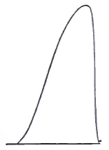
Contraction of Normal Abductor Indicis, CaCC, with strong current (Amidon2).
FIG. 4.
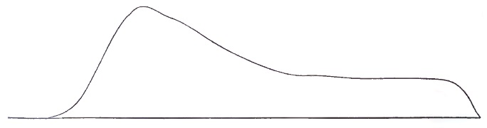
Contraction of Paralyzed Muscle on thirty-first day of Bell's palsy of the face. CaCC. 20 El. (Amidon). Shows retarded contraction and slow contraction, with tendency to tetanus.
2 “The Myography of Nerve-degeneration in Animals and Man,” Archives of Medicine, viii., No. 1, 1882.
In the period of recovery or regeneration of the nerve the musculo-galvanic reactions slowly reacquire their normal characters; the normal suddenness and completeness of the contractions gradually appear, and faradic excitation causes slight responses. Lastly, the nerve also begins to exhibit excitability under galvanism and faradism. These various abnormal electrical reactions, also frequently observed in diseases of the spinal cord, constitute the so-called reaction of degeneration, or De R. The subject is one of much practical importance, and for details the reader is referred to the treatises on electro-therapeutics of Erb and De Watteville.
Just as we depend upon the De R. to prove interruption of motor nerve-fibres (or, in other cases, destruction of ganglion cells in the anterior horns of the cord), so do we rely upon the demonstration of complete anæsthesia to prove interruption of sensory fibres. In seeking for the area of anæsthesia several points must be borne in mind: (1) The normal distribution of the principal nerve-trunks as taught by ordinary anatomical works; (2) the remarkable anomalies of distribution which sometimes occur; (3) that many nerves near their endings exchange filaments in very variable numbers; loops for collateral innervation, which will supply some sensibility to parts which, judging by ordinary anatomical rules, should be made anæsthetic by section of a given nerve-trunk; (4) another consideration is the degree of anæsthesia. Before pronouncing upon the complete and fatal division of a nerve-trunk (by injury or by disease), absolute anæsthesia should be demonstrated in its area of principal and isolated distribution. Ordinary tests are not, as a rule, sufficient for this purpose. The best means in our experience consists in the use of a very strong induction (faradic) current, as follows: The skin of the suspected region to be thoroughly dried and rubbed with chalk or powdered starch; one pole, consisting of an ordinary wet electrode, to be applied just above the part to be tested, and the other pole, consisting of a single wire, with which different parts of the anæsthetic area are to be touched. By this means partially insensible regions, not responding to pricking and burning, may be made to yield reaction, and the area of absolute anæsthesia be thereby much reduced. Malingering may also be readily exposed by this test, which presents another advantage—viz. of not causing local injury or scars as burning will.
It follows from the preceding statements that in cases of limited atrophic paralysis with De R. the diagnosis between a central lesion (destruction of ganglion cells of the anterior horns of the cord) and a strictly peripheral (or neural) lesion is to be chiefly based upon two considerations: (1) The distribution of the paralysis, which in the first case affects muscles which are physiologically grouped or associated, while in the second case the simple law of anatomical associations or grouping is observed; (2) by the state of sensibility, which is normal in disease of the anterior horns of the cord, and is frequently impaired or abolished in nerve lesions.
Lesions affecting the cauda equina cause all the above-mentioned symptoms of peripheral lesions in a limited (partial) paraplegic distribution. If the lesion or injury be in the sacro-coccygeal region, the symptoms will, as a rule, be found confined to parts below the knee, occasionally also involving the muscles on the posterior aspect of the thigh (flexors of the leg). Below the knees we find an atrophic paralysis with De R., anæsthesia of the foot and part of the leg, loss of plantar and achillis reflexes, and the sphincter ani will be paralyzed. The patellar reflex is preserved or exaggerated. In case the lesion be in the lumbar region, below the first lumbar vertebræ, the symptoms will be found to extend as high as the groin, involving also the buttocks and sphincter ani; state of the bladder variable. All reflexes will be lost in the paralyzed extremities, except in some cases the cremasteric reflex.
Strange as it may appear, physicians do not always remember that there is practically no lumbar spinal cord (vide Fig. 2.), and that injuries, etc. of the lumbar vertebræ and dura tend to affect nerves, and not a nervous centre.
II. Localization of Lesions in the Spinal Cord.
Diseases of the spinal cord are distinguished by the following general characters, positive and negative:
Positive Characters.—Tendency to primary bilateral or paraplegic distribution of all the symptoms. In the majority of cases preservation of electro-muscular excitability, and in one well-defined group of cases De R. more or less typically developed in the paralyzed parts. Frequent vesical and rectal paralysis, either of the retaining or of the ejecting apparatus. Pains and other paræsthesiæ in the extremities, the pains often possessing the electric or fulgurating character. Anæsthesia of paraplegic distribution. Sensations of constriction or cincture about a limb or about the body at various levels. Occurrence of ataxia without paralysis. Progressive muscular atrophy without actual paralysis. Easy production of eschars (bed-sores).
Negative Characters.—Absence of typical hemiplegic or monoplegic distribution of symptoms. Rarity of head symptoms, as headache, vertigo, mental disturbance; of lesions of the optic nerve. Absence of epileptiform convulsions. Absence of such grouping of motor and sensory symptoms as would exactly correspond to the area of distribution of one or more large nerve-trunks.
The above symptomatic indications are, of course, of the most general meaning only, and are liable to exceptions and subject to varying conditions.
The DIAGNOSIS of the exact localization of lesions in the spinal cord, considered from the clinical standpoint, is perhaps best arrived at by following an anatomical and physiological order of subdivision of the problem into three questions, as follows:
FIRST QUESTION.—BEING GIVEN SYMPTOMS INDICATING DISEASE LIMITED TO ONE OR MORE SYSTEMS OF THE SPINAL CORD, TO DECIDE WHICH ARE AFFECTED.
Physiology and the results of embryological and pathological researches justify us in making a general division of the spinal cord, for purposes of diagnosis, into two great systems, whose limits are fairly well known—viz. the æsthesodic or sensory system, and the kinesodic or motor system. The following outline diagram (Fig. 5) of section through the spinal cord exhibits the ascertained limits of the two systems.
FIG. 5.
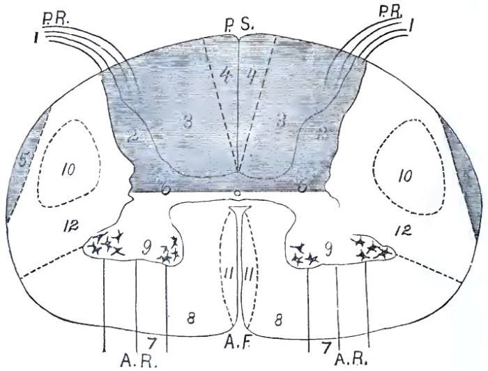
Diagram of a Transverse Section of the Spinal Cord through the Cervical Enlargement: The æsthesodic system is shaded, the kinesodic system unshaded. Parts of the æsthesodic system: (1) Posterior nerve-roots; (2) posterior gray horns; (3) fasciculi cuneati (columns of Burdach, inclusive of posterior root-zones); (4) fasciculi graciles (columns of Goll or posterior median columns); (5) ascending cerebellar fasciculi; (6) columns of Clarke. Parts of the kinesodic system: (7) Anterior roots; (8) anterior columns (inclusive of anterior root-zones); (9) anterior gray horns; (10) crossed pyramidal fasciculi; (11) direct pyramidal fasciculi (columns of Türck); [10 and 11 are the intraspinal prolongation of the cerebral motor tract]; (12) lateral columns, of ill-defined limits and unknown functions; P. R., posterior roots; P. S., posterior septum; A. R., anterior roots; A. F., anterior fissure.
A. Lesions of the Æsthesodic System.—Limits of the Æsthesodic System.—By this term we mean that combinations of ganglion-cells and nerve-fibres whose functions are locally sensory, and of those fibres which transmit impressions centripetally (frontad) to the encephalon. The following are the recognized parts of this system, as outlined on the diagram: (1) the posterior (dorsal) nerve-roots and attached ganglia; (2) the posterior gray horn and central gray substance to an unknown distance ventrad; (3) the fasciculi cuneati (columns of Burdach), whose lateral parts are more particularly designated as posterior root-zones; (4) the fasciculi graciles (columns of Goll) or posterior median columns; (5) the fasciculi ad cerebellum; (6) the vesicular columns of Clarke (most developed in the dorsal part of the cord). All of these parts have sensory functions, or at least transmit impressions centripetally, and they undergo secondary (Wallerian) degeneration toward the encephalon—i.e. frontad of a transverse lesion of the cord.3
3 There are some results of physiological experiments and a few isolated pathological facts which would seem to point to the existence of other sensory (centripetal) fasciculi in the lateral columns, but it would be wholly premature to make use of these facts in a practical consideration of the subject.
At the present time there is only one lesion of the æsthesodic system which can be diagnosticated during the patient's life from positive symptoms.
(a) Lesions of the fasciculi cuneati (posterior root-zones, 3). The symptoms of lesion (usually sclerosis) in this region are wholly sensory and ataxic. At an early stage acute pains, fulgurating pains, occur in the extremities; later paræsthesiæ, anæsthesia, and ataxia. The fulgurating pains caused by the slowly-progressive lesion of the posterior root-zones are very peculiar, and almost pathognomonic (vide preceding article for their description). In some cases paræsthesiæ precede the pains, which inversion of the usual order must be due to a difference in the starting-point of the sclerosis within the large fasciculi cuneati. Tendinous reflexes (especially the patellar) are lost at an early period in the disease, and by noting the disappearances of the different reflexes we can trace with some accuracy the longitudinal extension of the sclerosis (vide Fig. 2). In many cases the pupillary reflex is also abolished, constituting the Argyll-Robertson pupil.
As negative characters of lesions of the posterior root-zones (and of the rest of the æsthesodic system) we note absence of paralysis, contracture, atrophy, and De R.
In the present state of our knowledge of spinal physiology and pathology we think that a lesion in this location should be recognized by the physician early and positively—in some cases years before ataxia and other grosser symptoms make the diagnosis of locomotor ataxia obvious even to a layman's eye.
(b) Lesions of the fasciculi graciles (column of Goll, 4) cannot, we believe, be recognized directly by positive symptoms. There are a few cases on record of primary (?) sclerosis of these columns, in which during life vague sensory symptoms had been noted, but we cannot build upon such data. Indirectly, however, we can in many cases diagnosticate degeneration of these fasciculi, reasoning from the data of pathological anatomy. Thus, for example, in advanced cases of sclerosis of the fasciculi cuneati (posterior spinal sclerosis) we know that in the dorsal and cervical regions of the cord the columns of Goll are in a state of secondary degeneration. After complete transverse division of the cord by injury, extreme pressure, or focus of myelitis, etc. the same (centripetal) degeneration exists above the lesion.
The same remarks apply fairly to our clinical knowledge of the remaining parts of the æsthesodic system, columns of Clarke (6), and fasciculi ad cerebellum (5). We know absolutely nothing of lesions of the posterior horns (2) in their clinical and diagnostic relations.
B. Lesions of the Kinesodic System.—Limits of the Kinesodic System.—In general this includes the antero-lateral parts of the cord. In a trans-section of the cord (vide Figs. 5 and 6) the following columns and fasciculi are recognized, their location and limits being made known to us by embryology, descriptive and pathological anatomy: (7) The anterior (ventral) nerve-roots emerging from (8) the true anterior columns or anterior root-zones; (9) the ventral (anterior) gray horns with their groups of ganglion cells; (10) the crossed pyramidal fasciculus, which is the caudal continuation of the cerebral motor tract of the opposite hemisphere; (11) the direct pyramidal column (column of Türck), which is the caudal continuation of the cerebral motor part of the hemisphere on the same side; (12) the antero-lateral column. Fasciculi 10 and 11 bear an inverse relation to each other—i.e. 11 is larger in proportion as 10 is smaller.
FIG. 6.
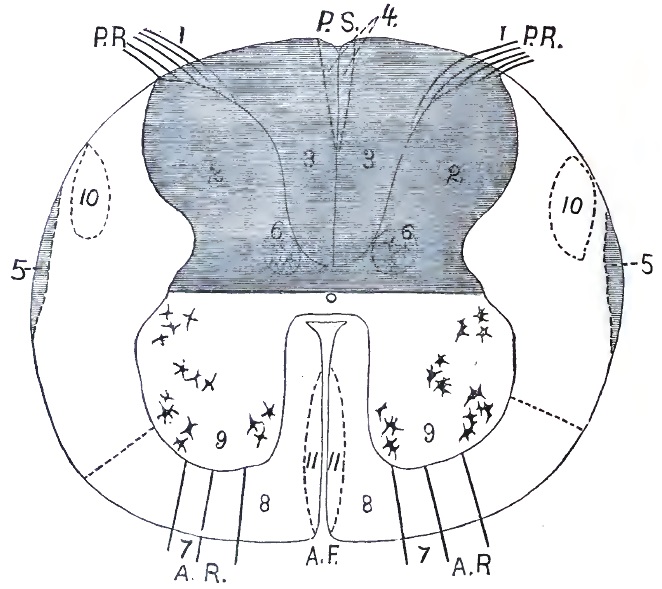
Diagram of a Transverse Section of the Spinal Cord through the Lumbar Enlargement: The æsthesodic system is shaded; the kinesodic system unshaded. Parts of the æsthesodic system: (1) Posterior nerve-roots; (2) posterior gray horns; (3) fasciculi cuneati (columns of Burdach, inclusive of posterior root-zones); (4) fasciculi graciles (columns of Goll, or posterior median columns); (5) ascending cerebellar fasciculi; (6) columns of Clarke. Parts of the kinesodic system: (7) Anterior roots; (8) anterior columns (inclusive of anterior root-zones); (9) anterior gray horns; (10) crossed pyramidal fasciculi; (11) direct pyramidal fasciculi (columns of Türck) [10 and 11 are the intraspinal prolongation of the cerebral motor tract]; (12) lateral columns of ill-defined limits and unknown functions; P. R., posterior roots; P. S., posterior septum; A. R., anterior roots; A. F., anterior fissure.
In a general way it may be stated that lesions of the kinesodic system are characterized positively by the isolated existence or predominance of motor symptoms, by impairment of muscular nutrition, and by De R.; also by contractures. The reflexes are almost never normal, being either exaggerated or lost. Negative symptoms are—absence of sensory symptoms, of ataxia, and of vesical or rectal symptoms.
(a) Lesions of the anterior gray horns (9) are revealed by most definite and characteristic symptoms. There occurs a flaccid paralysis involving more or less extensive groups of muscles in the extremities, rarely truncal muscles, and never those of organic life. In a few weeks the paralyzed muscles undergo atrophy, sometimes to an extreme degree, and various degrees of De R. are present. Cutaneous and tendinous reflexes are abolished. The bladder and rectum are normal. Sensory symptoms absent, and if present consist only of mild paræsthesiæ, which are probably due to postural pressure upon nerve-trunks or to disturbance of the peripheral circulation. There is no tendency to the formation of bed-sores, but circulation and calorification are reduced in the paralyzed members. It should be remembered that paralysis due to systematic lesion of the anterior gray horns is never typically paraplegic, with horizontal limit-line of sensory symptoms, a cincture feeling, and vesical paralysis.
The above symptom-grouping is characteristic of lesion of the anterior horns en masse; in other words, of poliomyelitis. In that form of systematic disease of the anterior horns which consists primarily and chiefly of a degeneration or molecular death of the ganglion cells there is no true paralysis; the atrophy of muscles is infinitely slower, and it proceeds in various muscles fasciculus by fasciculus, the wasting being usually preceded by fibrillary contractions, and being almost always symmetrically located on the two sides of the body (affecting analogous or homologous parts). The electrical reactions are abnormal, in that musculo-faradic reaction is lost in exact proportion to the wasting; so that in a large muscle one part may contract normally, while the adjacent fasciculi do not. It is as yet uncertain whether De R. occur in this disease (progressive muscular atrophy). Calorification and circulation are much less impaired than in poliomyelitis; the negative symptoms are much the same.
It is sometimes difficult to distinguish poliomyelitis from generalized neuritis. The diagnosis is to be made by the presence in the latter disease of marked sensory symptoms—neuro-muscular pains, numbness, slight anæsthesia—and by a grouping of symptoms coinciding with the distribution of nerve-trunks and branches. No assistance can be derived from electrical tests, as both diseases yield more or less typical De R.
(b) Lesions of the spinal pyramidal tract (of fasciculi 10 and 11) are followed by motor symptoms only—viz. paralysis and contracture—or, in other words, by a spastic paralysis. Sensibility is unaffected; the bladder, rectum, and truncal muscles are not distinctly paralyzed; the reflexes are much increased, and ankle-clonus is often present. The electrical reactions of the paralyzed muscles are normal, qualitatively at least. The diagnosis of localization may be pushed still farther by the following considerations:
(1) When the condition of spastic paralysis is unilateral, of hemiplegic distribution, and follows an attack of cerebral disease of some sort, we may feel sure that both the crossed and the direct fasciculi of the pyramidal (10 and 11) belonging to one cerebral motor tract are degenerated throughout the length of the spinal cord, the crossed fasciculus on the paralyzed side and the direct on the healthy side (same side as injured hemisphere). It would thus appear that lesion of the direct pyramidal fasciculus (11) produces no symptoms,4 except, of course, in those rare cases in which it is larger than its associated crossed fasciculus.
4 Unless it be the increase of reflexes which is so often observed on the non-paralyzed side in hemiplegics.
(2) The above symptoms may be bilateral, as observed in children as a congenital or early-acquired condition. In such cases the four fasciculi of the motor tract are degenerated or undeveloped, in correspondence with a symmetrical bilateral lesion of the motor area of the cerebrum—imperfect development or early destruction.
(3) When the legs alone are the seat of spastic paralysis, with increased reflexes, spastic or tetanoid gait, without sensory symptoms, the diagnosis of a primary sclerosis of both lateral columns (inclusive of 10) of the spinal cord is justified.
(c) Lesions of antero-lateral columns of the cord (8 and 12), primary and independent of lesions of the anterior horns or of the crossed pyramidal fasciculus, cannot now be diagnosticated during life. These large masses of fibres include fasciculi whose functions are probably motor; others (especially in 12) whose functions, according to recent experiments, may be sensory; and still others which are associating or commissural.
(d) Various combinations of the above lesions occur, and may be recognized positively during life: (1) Combined sclerosis of the posterior columns and of the crossed pyramidal fasciculi (3 and 10), indicated by ataxia with paralysis, absence of patellar reflex, tendency to contracture, pains, and anæsthesia less marked than in typical posterior spinal sclerosis. This symptom-group is usually found in children; it is pre-eminently a family disease, and is known as Friedreich's disease. Similar cases also occur in adults as a strictly personal disease. (2) Secondary degeneration of the pyramidal tract (10 and 11), with more or less localized atrophy of cells in the anterior horns (9) coexists in two forms: First, in a few cases of cerebral hemiplegia with contracture, and pathological atrophy of various muscles on the paralyzed side; second, as a distinctly spinal bilateral affection, characterized by a spastic or tetanoid state of the lower extremities, and a mixture of atrophic paralysis (vide (a)), with contracture in the arms and hands. This latter form is the amyotrophic lateral sclerosis of Charcot.
SECOND QUESTION.—BEING GIVEN SYMPTOMS INDICATING A TOTAL TRANSVERSE LESION OF THE SPINAL CORD, ONE INVOLVING ITS VARIOUS SYSTEMS AT A CERTAIN LEVEL, TO DETERMINE THE ELEVATION OR FRONTO-CAUDAL SITUATION OF THE LESION.
This question is usually easy of solution by the following method: Since a transverse lesion of the spinal cord gives rise to both motor and sensory symptoms of horizontal, paraplegic distribution caudad of the lesion, the first thing to do is to determine accurately the upper level of the symptom, either the line of anæsthesia or the limit of paralysis. The former is always much more definite than the latter, and usually serves as the guide to diagnosis, indicating accurately the uppermost part of the lesion. In the thorax the levels of sensory and motor symptoms nearly coincide, but in the extremities the operation of Van der Kolk's law of distribution of motor and sensory fibres of nerve-trunks must be borne in mind, although it does not apply as strictly in this case as in nerve lesions strictly speaking. Gowers's diagram will prove very serviceable in making a diagnosis of transverse lesions, and will also prove of use in the study of vertebral injury and disease, as it indicates with sufficient accuracy the relation between vertebræ (spinous processes) and segments of the spinal cord. (Vide Fig. 2.)
The following are the principal transverse localizations of disease which are usually recognized during life by the above procedure:
(a) Lesions of the cauda equina (by tumors, caries, or fracture of the bones, etc.) produce paralysis, anæsthesia, atrophy of muscles, with De R., in the range of distribution of the sciatic nerves mainly. The sphincter ani is paralyzed and relaxed, while the bladder remains normal as a rule. In all essential respects this paraplegiform, but not paraplegic, affection resembles that following injury to mixed nerve-trunks. It is in reality an intra-spinal peripheral paralysis. The more exact location of the lesion, in the absence of external physical signs (fracture, etc.), may be approximately determined by a study of the distribution of the symptoms and their relation to nerve-supply.
(b) Lesions of the lower end of the lumbar enlargement, or conus medullaris, behind the first lumbar vertebra, will give rise to the same symptoms as (a). The expression, lumbar part of the spinal cord, should be more carefully used than it is at present in the discussion of spinal injuries and spinal-cord diseases, disease of the lumbar enlargement being common enough, but disease of the lumbar part of the cord very rare. In the discussion of railway cases, more especially, it is often forgotten that the spinal cord practically ends behind the first lumbar vertebra.
(c) Lesions of the middle and upper parts (segments) of the lumbar enlargement are evidenced by true paraplegia, without paralysis of the abdominal muscles. In some cases the quadriceps group, supplied by the crural nerve, is not paralyzed. The constriction and the limit of anæsthesia are about the knees, at mid-thigh, or near the groin in different cases. The paralyzed muscles, as a rule, retain their irritability and show normal electrical reactions; the cutaneous and tendinous reflexes are preserved or increased. The sphincter is usually paralyzed, while the bladder is relatively unaffected.
(d) Transverse lesion of the dorsal spinal cord produces the classical type of paraplegia—i.e. paralysis and anæsthesia of all parts caudad of the lesion. The limit of anæsthesia and the constriction band are nearly horizontal, and their exact level varies with the height of the lesion, from the hypogastric region to above the nipples. Below the level of anæsthesia, which indicates by the number of the dorsal nerve the upper limit of the cord lesion, there are complete paralysis, retention of urine, constipation with relaxed sphincter ani, greatly exaggerated reflexes in the lower extremities, even to spinal epilepsy; the muscles preserve their volume fairly well, and their electrical reactions are normal—sensibility in all its modes is abolished; bed-sores are easily provoked. Retention of urine is an early symptom in lesion of the middle dorsal region of the cord—sometimes, in our experience, preceding symptoms in the legs.
(e) A transverse lesion of the cord at the level of the last cervical and first dorsal nerves—i.e. in the lower part of the cervical enlargement—gives rise to typical paraplegia with a sensory limit-line at or just below the clavicle, but also with some very peculiar symptoms superadded. These characteristic symptoms are in the upper extremities, and consist of paralysis and anæsthesia in the range of distribution of the ulnar nerves. In the arms the anæsthesia will be found along the lower ulnar aspect of the forearm, the ulnar part of the hands, the whole of the little fingers, and one half of the annuli. There will be paralysis (and in some cases atrophy with De R.) affecting the flexor carpi ulnaris, the hypothenar eminence, the interossei, and the ulnar half of the thenar group of muscles, producing in most cases a special deformity of the hand known as claw-hand or main-en-griffe. Another important symptom of a transverse lesion in this location is complete paralysis of all the intercostal and abdominal muscles, rendering respiration diaphragmatic and making coughing and expectoration impossible. The breathing is abdominal in type, and asphyxia is constantly impending.
(f) A transverse lesion of the upper part of the cervical enlargement, below the origin of the fourth cervical nerve, gives rise to symptoms designated as cervical or total paraplegia. The lower extremities and trunk are as in (d) and (e), but besides both arms are completely paralyzed and anæsthetic. The limit of anæsthesia usually extends along the clavicles to the acromion processes, or a little below, near the deltoid insertions. All reflexes caudad of this line are vastly increased, either with tonic or clonic contractions. In some cases of pressure upon the cervical cord by tumors, caries of vertebræ, etc. the tetanoid or spastic state of the extremities (the lower more especially) may precede paralysis for a long time; as the compression increases paralysis becomes more and more marked, while the reflexes remain high. This constitutes the clinical group we described in 1873 as tetanoid pseudo-paraplegia.
(g) Transverse lesions of the spinal cord from the decussation of the pyramids to the fourth cervical nerve are very rare, and usually of traumatic origin. They produce complete paralysis of the entire body, and also of the diaphragm (third and fourth cervical nerves), thus causing death by apnœa in a very short time.
(h) In ascending paralysis (Landry) the above symptom-groups, excepting (a), (b), are met with at successive stages of the disease, often by almost daily accession, until finally respiration ceases.
(i) The height of a transverse localized lesion (e.g. a stab-wound) of one lateral half of the spinal cord is to be determined by the various groupings of symptoms stated in the preceding paragraphs, the chief guide being the limit-line between the sensitive and anæsthetic portions of the body, measured vertically. The symptoms are, however, distributed in a very remarkable manner on either side of the median line. The paralysis will be found on the same side as the lesion, often accompanied by hyperæsthesia, vaso-motor paralysis, and loss of muscular sense, while the anæsthesia is on the other side of the body. When such a lesion occurs below the first dorsal nerve, the symptom-group is designated as hemiparaplegia; when the lesion is higher up, so as to paralyze the arms, the affection is termed spinal hemiplegia (Brown-Séquard).
Above the decussation of the pyramids total transverse lesions are practically unknown, so that the second question need not be followed farther.
By means of the data above given we are also enabled to determine the length—i.e. fronto-caudal extension—of the systematic lesions of the cord.
The symptoms of transverse lesions of the spinal cord are not exclusively caudad of the lesion, and some very striking ones are observable in the head. In lesions of the upper part of the dorsal cord and of the cervical enlargement (e, f, g) we observe vaso-motor and pupillary symptoms, due to injury of the spinal origin of the cervical sympathetic nerve; the pupils are contracted; the cheeks and ears congested and unnaturally warm; the cutaneous secretions are increased. In other words, the symptoms about the head, frontad of the lesion, are the same as those we produce experimentally in animals by section of the cervical sympathetic or of the lower cervical cord. In hemi-lesion of the cord, in man as in animals, these symptoms are unilateral, on the same side as the injury.
Another point to be remembered in the study of transverse lesions of the spinal cord is that the lesion may involve enough of the anterior gray horns to give rise to atrophic paralysis with De R. in some few muscles deriving their motor innervation from the focus of disease. This is not rarely seen in cervical paraplegia.
THIRD QUESTION.—BEING GIVEN VERY LIMITED MOTOR OR SENSORY SYMPTOMS OF SPINAL ORIGIN, TO DETERMINE THE EXACT LOCATION OF THE FOCAL LESIONS CAUSING THEM.
(a) In the range of sensory disturbances this question rarely presents itself for solution. Localized anæsthesia and pain of spinal origin (except the fulgurating pains of tabes) are rare, and we do not know the relation of cutaneous areas with the spinal segments as well as we know the motor innervations. It should be stated here, however, that the location of a fixed pain and of a zone of anæsthesia is often of great value in determining what spinal nerve is affected, just outside of the cord itself, by such directly-acting lesions as vertebral caries, spinal pachymeningitis, tumors upon the spinal cord, etc. Among the various symptoms of Pott's disease of the spine, fixed pains in one side of the trunk, in one thigh, or in the upper occipital region, etc. is a sign against which the physician should always be on his guard, as it is a very early and valuable indication of the existence of an affection which requires special treatment as soon as a diagnosis can be made.
(b) Localized motor symptoms of spinal origin calling for a diagnosis of the focal lesions causing them are frequent, and are mostly met with in two affections—viz. progressive muscular atrophy and poliomyelitis. The problem is now capable in many cases of an approximately exact solution by the deductive application of our knowledge of the intimate connection between certain muscles and muscular groups and limited portions or segments of the spinal cord (anterior gray horns more especially). This knowledge has been accumulated and organized from ordinary anatomy, physiological experiments, and more especially from carefully-made autopsies with microscopic examination of the cord. We cannot present this subject better than by reproducing a tabular statement of these results prepared by M. Allen Starr.5 Future autopsies may correct this table, and in making use of it the occurrence of anomalous nerve-distribution should be remembered:
LOCALIZATION OF FUNCTIONS IN THE VARIOUS SEGMENTS OF THE SPINAL CORD.
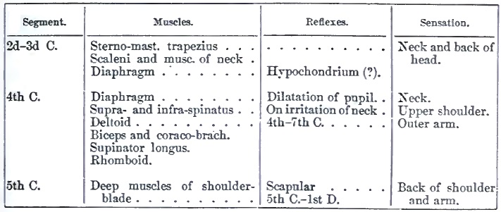
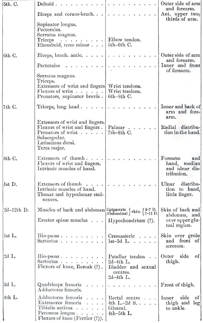
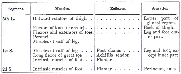
5 “The Localization of Functions of the Spinal Cord,” American Journal Neurology and Psychiatry, iii., 1884.
The above table should be studied in connection with Gowers's diagram of the vertebral column and spinal cord (Fig. 2, p. 53), for the thorough study of cases of neural and spinal localization. Additional details of much value with respect to the peripheral nerve distribution are accessible in the works of Ranney6 and Ross7.
6 The Applied Anatomy of the Nervous System, N. Y., 1881, p. 355 et seq.
7 Handbook of Diseases of the Nervous System, Am. ed., Philada., 1885, p. 356 et seq.
III. The Localization of Lesions in the Medulla Oblongata.
In general terms, lesions of the oblongata are characterized by the early appearance and prominence of motor symptoms in the mouth, throat, and larynx, and by bilaterality of the symptoms. Remote symptoms consist of disturbances in the cardiac action and in the functions of some intra-abdominal organs. There may also be more or less paralysis of all the extremities. These lesions may conveniently be classified, like those of the cord, into systematic and focal.
A. SYSTEMATIC LESIONS OF THE OBLONGATA.—1. Systematic lesions of the æsthesodic system of the oblongata are, for purposes of practical diagnosis, unknown at the present time.
2. Systematic lesions of the kinesodic system of the oblongata, on the other hand, are often positively recognizable during the patient's life.
(a) The most strictly systematic and most frequent of these lesions is that of secondary (Wallerian) degeneration of the pyramids, the prolongation of the cerebral motor tract. This morbid change gives rise to no distinct bulbar symptoms, and it can only be diagnosticated inferentially or inclusively by determining the existence of secondary degeneration of the entire pyramidal tract, from the occurrence of hemiplegia followed by contracture and increased reflexes. If the phenomena present be those of double spastic hemiplegia, there is surely degeneration of both pyramids.
(b) A systematic lesion affects the nuclei (origins) of the facial, hypoglossal, pneumogastric, spinal accessory, and the motor root of the trigeminus nerves, giving rise to a classical symptom-group. The symptoms thus produced are exclusively (?) motor and trophic, consisting of progressively increasing paresis, with atrophy of the muscles about the lips and cheeks, the intrinsic lingual muscles, the laryngeal and pharyngeal muscles. Later, the masseters, temporals, and pterygoids are sometimes involved; and, finally, extremely rapid action of the heart with pneumonic symptoms indicates the gravest extension of the morbid process. An early symptom in such cases is abnormal salivation. These affections, generally designated as varieties of bulbar paralysis, subacute or chronic, are usually readily recognized intra vitam, and recent discoveries in morbid anatomy have enabled us to correctly diagnosticate the seat of the lesion in its various extensions. The laryngeal paralysis represents disintegration (atrophy) of ganglion-cells in the bulbar origin of the spinal accessory nerve; lingual atrophic paralysis indicates the same lesion in the nuclei of the hypoglossal nerves; the labio-buccal symptoms are produced by lesion of the facial nerve nucleus (inferior facial nucleus of older writers); the paralysis of the muscles of mastication is due to extension of cell-degeneration to the motor nucleus of the trigeminus in the pons; and the final cardio-pulmonary symptoms indicate an extension of the lesion into the sensory (?) origin of the pneumogastric nerves.
It is evident that this systematic lesion of the oblongata is the equivalent or analogue of the various acute and chronic lesions of the anterior horns of the spinal cord, described supra, and in practice we sometimes find these bulbar and spinal lesions associated: progressive muscular atrophy of the extremities and amyotrophic lateral sclerosis being complicated with bulbar paralysis.
B. FOCAL LESIONS OF THE MEDULLA OBLONGATA, as at present known, occur mostly in the kinesodic system, but may also involve several fasciculi and nuclei at one time. The symptoms of such lesions are grouped in two principal types:
(a) Single symptoms, such as atrophy or atrophic paralysis of some one muscle or muscular group innervated by the hypoglossal, facial, or spinal accessory nerves. For example, unilateral atrophy of the tongue, when not due to neural injury, is quite surely the representative of destruction of one hypoglossal nucleus. It is possible that some cases of peripheral facial paralysis, so-called, or Bell's palsy, and cases of paralysis of the sterno-mastoid and trapezius, are not due to neural lesion, but to primary disease of the nuclei of the facial and spinal accessory nerves, either as poliomyelitis or as progressive cell-degeneration and atrophy.
A similar reserve must be used in speaking of the localization of small lesions in the oblongata, causing diabetes mellitus, polyuria, albuminuria, and salivation. From experiments upon animals and a few post-mortem examinations in human cases we know that such lesions may occur and cause the symptoms, but their recognition during the patient's life is at the present time next to impossible.
(b) The symptoms may be complex and belong to the general class of crossed paralysis, the mouth, face, tongue, and larynx being paralyzed (often with anæsthesia of the face) on one side, while the extremities are paralyzed on the other side. Such a symptom-group would indicate a large lesion (tumor) in one lateral half of the oblongata, more especially in its ventral aspect. These localizations will be studied more in detail farther on, under the general head of Crossed Paralysis due to Lesions of the Base of the Brain.
IV. Localization of Lesions in the Encephalic Mass.
Under this head, to prevent repetition and to facilitate condensation, we will consider the various localizations which can now be diagnosticated in the cerebrum, basis cerebri, and cerebellum.
It would be of the greatest advantage to preface these statements by a full summary of the anatomical and physiological data on which the localizations rest, but space is wanting for such an exposition, and the reader who is not already familiar with these branches of medical science will have to seek information in other accessible works.8
8 Ferrier, The Functions of the Brain, Am. ed., N. Y., 1876; Charcot, Lectures on Localization in Disease of the Brain, Am. ed., N. Y., 1878; Seguin, E. C., “Lectures on the Localization of Spinal and Cerebral Diseases,” N. Y. Medical Record, 1878; Ranney, Applied Anatomy of the Nervous System, N. Y., 1881; Meynert, Psychiatry: A Clinical Treatise on Diseases of the Fore-brain, Part I., Am. ed., 1885.
In a general way, encephalic lesions are distinguished by the following:
Positive Characters.—Tendency to strictly hemiplegic or bilaterally hemiplegic grouping of symptoms; frequency of contracture or of a spastic state of the paretic muscles; increase of reflexes in the affected extremities; spasmodic manifestations in remote muscular groups, but not in areas of nerve-distribution (forms of Jacksonian epilepsy); the presence of paralytic and anæsthetic symptoms in the range of distribution of cranial nerves; frequency of neuro-retinitis or atrophy of the optic nerves; occurrence of lateral hemianopsia; production of symptom-groups known as varieties of crossed paralysis; frequency of head symptoms, as headache, vertigo, apoplectic and epileptic seizures; mental symptoms of various sorts, dementia, coma, etc.
Negative Characters.—Absence of truly paraplegiform distribution of symptoms, even when they are bilateral; of cincture feelings; of pain or other paræsthesiæ and anæsthesia in the distribution of nerve-trunks; of muscular atrophy and De R. in paralyzed parts. Rarely do we observe visceral paralysis and bed-sores.
Pathological localizations in the encephalon may conveniently be grouped under two heads—viz. systematic and focal lesions.
A. SYSTEMATIC LESIONS OF THE ENCEPHALON.—The recent advances of embryology, anatomy, physiology, and pathological anatomy have conclusively established the existence, and fairly well defined the limits, of a sensory (æsthesodic) and of a motor (kinesodic) tract in the brain; and certain lesions of these tracts produce such precise symptom-groups that their diagnosis during the patient's life is often possible, and that, too, with great exactness.
1. SYSTEMATIC LESIONS OF THE ÆSTHESODIC SYSTEM OF THE ENCEPHALON.—The limits of this system are as follows: Within the oblongata and pons it occupies a somewhat uncertain (from a clinical standpoint) location, its fasciculi and ganglia lying in a general way dorso-laterad of the motor or kinesodic system. In the crus cerebri the fibres of the sensory tract are more closely packed together, and constitute a dense fasciculus of white substance in the lateral part of the crus in its subcerebral extremity, estimated by Flechsig at about one-fifth of the entire crus, and thence it enters into the composition of what is known as the internal capsule. This narrow but all-important mass of white fibres, as revealed by a horizontal section of the hemisphere (vide Fig. 7), lies between the nucleus caudatus and the nucleus lentiformis, and between the latter and the thalamus, thus assuming a V or elbow shape. Of this, the caudal third of that part of the capsule behind the bend or angle contains all the sensory paths for the opposite half of the body, reinforced by paths for the special senses; it is the carrefour sensitif of French writers. From this region sensory fasciculi radiate to various parts of the cerebral cortex—in the parietal, temporal, and occipital gyri—in which physiological experiments and human cases of disease have shown sensory areas or centres. Of the fasciculi from special sense-organs which reinforce the internal capsule, the only one which is well defined and easily traceable is the fasciculus opticus (fasciculus of Gratiolet), derived in part directly from the optic tract of the same side and from the primary optic centres (lobus opticus, corpus geniculatum laterale, and pulvinar), proceeds, along with fibres of the internal capsule, dorso-laterad of the posterior cornu of the lateral ventricle, to the mesal aspect of the occipital lobe. A fasciculus from the olfactory apparatus doubtless also joins the internal capsule, but its course is unknown.
The following localization diagnoses are now possible in the æsthesodic system as above defined:
(a) A lesion of the internal capsule, just above the crus cerebri, involving its bend or knee and caudal segment, with or without injury to the nucleus lentiformis and thalamus, will give rise to the following symptoms: Complete typical hemiplegia and total hemianæsthesia on the opposite side of the body; the anæsthesia involves the special senses as well as the body. Vision is, however, unaffected, unless the lesion extend far enough back to involve the pulvinar and fasciculus opticus, when lateral hemianopsia occurs (dark half-fields on the side opposite the lesion). When this lesion is in the left hemisphere, sensory aphasia also occurs.
(b) A lesion involving the caudal part of the thalamus and of the internal capsule. With such a lesion the motor symptoms consist in transient paralysis, with usually persistent post-paralytic chorea or ataxia. The sensory symptoms are more marked, and resemble those produced by lesion (a). It may be determined with some degree of accuracy whether the lesion be in the thalamus border, or in the internal capsule near to the nucleus lentiformis by the absence in the latter case of lateral hemianopsia.