Volume 5 — DISEASES OF THE NERVOUS SYSTEM. (continued 24)
Reading edition
14 Charles West, M.D., Lectures on the Diseases of Childhood and Infancy, 6th ed., London, 1874, p. 62. These large cysts are much more frequently (perhaps solely) found in cases of hemorrhagic pachymeningitis. (See Barthez and Sanné, op. cit., vol. i. p. 157.)
DIAGNOSIS.—The distinction between meningeal hemorrhage and cerebral apoplexy is always difficult, and in the majority of cases impossible. Sudden pain in the head, vomiting, and lowering of the bodily temperature (the thermometer should be placed in the rectum), followed by loss of consciousness, are strongly suggestive of hemorrhage within the cranium, either cerebral or meningeal. If these symptoms are followed by coma and resolution without obvious paralysis, the diagnosis would be almost impossible between intra- and extra-cerebral extravasation. If the loss of consciousness be not complete, so that the presence or absence of paralysis can be ascertained, we can sometimes distinguish the situation of the hemorrhage. Right-sided hemiplegia, with paralysis of the face or tongue, or with aphasia, is most probably owing to hemorrhage or embolism somewhere in the left motor tract, and hence within the brain. If the absence of paralysis can be certainly ascertained, the probabilities are in favor of meningeal apoplexy. Convulsions affecting the face or limbs of one side would point to irritation of the cortical centres of those parts, and so far to extravasation on the surface of the brain (on the opposite side), as in Webber's case. Where the amount of hemorrhage is small it furnishes no diagnostic indications. In the case of new-born children the presumption is in favor of meningeal effusion.
PROGNOSIS.—If the effusion be considerable in amount, as indicated by prolonged coma with resolution, the issue is almost inevitably fatal, though life is occasionally prolonged for a surprising length of time. Slight hemorrhages are doubtless recovered from, but there are no means for their certain diagnosis.
TREATMENT.—The treatment, which is essentially the same as that for cerebral congestion, has for its object the arrest of the hemorrhage, and, if that can be effected, the absorption of the effused blood. In view of the former, the patient's head should be elevated and kept cool by the application of ice. Unless the bowels have previously been freely moved, saline laxatives, followed by enemata if necessary, must be given. The state of the bladder must be carefully attended to. Liquid nourishment alone, in moderate quantities at regular intervals, is permissible, with stimulants if there be signs of exhaustion. For the absorption of the effusion mild counter-irritation to the scalp and the administration of the iodide of potassium may be employed.
Congestion of the Cerebral Pia Mater.
The pia mater consists of two layers, separated by a loose connective tissue. The outer layer, being that which was formerly called the visceral layer of the arachnoid, is stretched smoothly over the convolutions of the brain without dipping into the sulci; the inner layer is closely connected with the surface of the brain, whose inequalities it follows. The two layers are more firmly united together over the convolutions than between them; in the latter situation the connection is loose, and the space which separates the surfaces is called the meshes of the pia. The membrane extends into the ventricles, investing the ependyma and the choroid plexuses, and over the medulla oblongata and spinal cord. In the normal condition it is loosely attached to the brain, from which it can be stripped off without loss of substance. The meshes of the pia, together with the ventricles, constitute a series of cavities connected with each other, containing a variable amount of cerebro-spinal fluid, and they communicate with the lymph-spaces surrounding the blood-vessels. Pressure within the cranial cavity, from congestion of the vessels or from the products of inflammation, is thus relieved in a measure by displacement of the cerebro-spinal fluid, which is driven out through the perivascular lymph-spaces.
Congestion or hyperæmia of the pia mater probably never occurs independently of that of the external surface of the brain, on account of the intimate vascular connection of the two. In the adult it can only exist to a limited extent, on account of the unyielding nature of the cranial walls and of the limited compensatory action of the cerebro-spinal fluid. In young children the incomplete ossification of the skull and the delicate structure of the vessels are more favorable to congestion.
ETIOLOGY.—The causes of hyperæmia of the pia mater are in the main the same as those of congestion of the dura.
SYMPTOMS.—Since congestion of the pia always coexists with that of the corresponding part of the external portion of the brain, it is impossible to separate the symptoms belonging to each. They are therefore usually included under the head of Cerebral Congestion, to which article the reader is referred.
PATHOLOGICAL ANATOMY.—Arterial hyperæmia of the pia is seldom discovered after death, the elasticity of the vessels causing transudation of the fluid part of the blood through their walls. Venous congestion of the pia is more frequently noticed, usually in connection with that of the dura, the sinuses with their accompanying veins being distended with blood, and in cases of long standing often containing thrombi. Simple hyperæmia of the pia being rarely or never fatal of itself, these appearances are usually accompanied by those of inflammation of the membrane or of the cortical layer of the brain (effusion of lymph or pus), or by hemorrhage.
TREATMENT.—In a case of suspected congestion of the pia the treatment would be the same as that of cerebral congestion.
Inflammation of the Cerebral Pia Mater.
SYNONYMS.—Meningitis, Leptomeningitis, Acute non-tubercular hydrocephalus.
Meningitis (by which is generally understood inflammation of the pia) appears under an acute, a chronic, and an epidemic form. The latter, being a zymotic disease, is described in a separate article, to which the reader is referred.
ETIOLOGY.—Meningitis occurs both as a primitive disease and as secondary to other affections. The former is rare, the latter is more frequent. The causes of idiopathic meningitis are for the most part unknown. Exposure to the sun's rays and excessive indulgence in alcoholic liquors are thought to excite it in some instances. It has been known to follow blows and falls on the head which have produced no injury to the skull. It is rather more commonly observed in early manhood than at other periods of life. Secondary meningitis may follow injury or disease of the cranial bones or of the dura, and of the brain. A frequent cause is extension of disease of the ear to the membranes and substance of the brain. The reader is referred to the article on MEDICAL OTOLOGY for information concerning the symptoms of that formidable complication. Certain diseases are known to be occasionally complicated with meningitis—acute articular rheumatism; erysipelas of the scalp and of the face; Bright's disease, especially the chronic interstitial form; peritonitis; ulcerative endocarditis; pyæmia; the eruptive fevers; the puerperal state; and syphilis. Meningitis following or complicating acute rheumatism is generally supposed to be not uncommon, but the number of cases in which the existence of inflammation of the meninges has been proved by autopsy is small. Fuller,15 along with three cases in which dissection showed suppurative inflammation of the pia, cites several others in which no cerebral disease was found after death, although the symptoms gave every indication of it. True meningitis is rare in chronic Bright's disease, the symptoms resembling it being caused, in the majority of cases, by uræmia. Meningitis complicating pneumonia is also rare, although cerebral symptoms are common enough in that disease, especially in young children with inflammation of the upper lobes. C. Neuwerk16 reports seventeen cases of purulent meningitis complicating acute pneumonia. It was more frequent in men, especially in alcoholic subjects, than in women. The meningitis was generally total. The lungs were in a state of gray or yellow hepatization.
15 H. W. Fuller, M.D., On Rheumatism, Rheumatic Gout, and Sciatica, 3d ed., Philada., 1864, p. 271. See also E. Leudet, Clinique médicale de l'Hôtel Dieu de Rouen, Paris, 1874, p. 133.
16 Deutsches Archiv für klin. Med., xxix., 1881, p. 1; and Schmidt's Jahrbücher, Band cxcviii., 1883, Nov. 5.
SYMPTOMS.—The symptoms of acute leptomeningitis vary much in the course of the disease. This is readily explained by the complex character of the functions of the parts involved in the inflammation. It may be assumed that the cortical layer of the brain is implicated in every case unless of the most transient and limited kind; the substance of the brain, cerebellum, and medulla are subjected to pressure from the afflux of blood, from the effused lymph and pus, and from the accumulation of serum in the ventricles; the cranial nerves are exposed to pressure from the deposit of lymph, which may give rise to irritation or to suspension of function or both; parts at a distance from the seat of lesion may be functionally disordered by reflex action through communicating filaments. Finally, the general system suffers from the effects of the high fever upon the blood and the nutrition.
It is customary to speak of a stage of excitement followed by one of depression as characteristic of the course of the disease; but although active symptoms generally prevail in the early period, to be succeeded later by coma and paralysis, this disposition is by no means uniform. Sometimes sopor and paralysis constitute almost the only symptoms throughout the disease—this is especially noticed in infants—or active delirium and convulsions may persist until the fatal termination. More frequently the two conditions alternate several times with each other. A prodromic period of short duration, a few hours or a day or two, is sometimes observed in primitive meningitis, the patients complaining of headache, vertigo, vomiting, restlessness, or lassitude. Infants are irritable or drowsy, with heat of the head, quick pulse, and occasional vomiting. In secondary meningitis this period is usually masked by the symptoms of the primitive disease. In the majority of cases the beginning of acute meningitis is abrupt. Rigor is sometimes the first symptom observed, and in children is usually represented by a convulsion. More commonly, however, the disease is ushered in by severe headache, which is often referred to the forehead. The head is hot, the face is flushed, the eyes are brilliant, the pupils are contracted, the pulse is quick and hard, the temperature high (104° F. or upward). The patient is wakeful, restless, and irritable, sensitive to light and to sound. The skin is hyperæsthetic, especially that of the legs. There may be wandering or even active delirium. Vomiting is not unfrequent. There is thirst, but no desire for food. The urine is scanty and high-colored, the bowels constipated. These symptoms gradually increase in intensity, especially the pain in the head and the delirium, and in many cases they are followed by convulsions, at first in the form of twitchings of the facial muscles or of the limbs, grinding the teeth, etc., which give place to tonic contractions of the limbs or of the trunk, often confined at first to one or both members of the same side, but afterward becoming general; the flexors of the forearms and of the legs are most usually affected. The upper dorsal muscles may become contracted, so that the head is drawn backward, and more rarely trismus occurs.
A diminution in the rapidity of the pulse, which may fall to the normal rate, or even below that, notwithstanding the persistence of the high temperature, indicates the beginning of the stage of depression. This change is sometimes sudden, though more often gradual in its approach. The activity of the delirium subsides, giving place to a somnolence which may seem to the inexperienced observer a favorable indication, but which soon deepens into coma. The face becomes pale, the features are sunken. Only an occasional grimace or a movement of the hand to the head shows that the patient is to some extent conscious of suffering. This condition may alternate with the previous one from time to time, the comatose state being interrupted by noisy delirium and tonic or clonic convulsions, or even a partial return of consciousness, giving rise to fallacious hopes on the part of the friends, and sometimes deceiving the physician himself into a belief that a favorable issue is at hand. Before long, however, the symptoms of brain-compression become permanent. The rigidity of the limbs gives place to complete resolution. The patient lies absolutely unconscious, with dilated pupils. The pulse becomes again rapid in consequence of compression of the medulla, and thread-like and irregular; for the same reason the respirations increase to 40, 50, or 60 in the minute. The sphincters are relaxed, and the patient dies without any recurrence of the active symptoms. In rare cases recovery takes place, although almost never after the symptoms of compression have continued without interruption for any considerable length of time. Moreover, it is seldom that recovery takes place in the adult without leaving some traces of permanent damage, such as general debility, paralysis of one or more limbs, deafness, mental weakness, epilepsy, etc. Many cases of general paralysis of the insane and other forms of so-called mental disease are the result of meningitis.
DURATION.—In the adult usually the disease lasts about a week or ten days; exceptionally, it may last two or three weeks.
COURSE.—In young children the course of meningitis differs somewhat from that which is observed in adults, though the symptoms are essentially the same. It is more sudden in its onset and shorter in its duration. The prodromal stage may be brief or hardly noticeable; but in older children restlessness, sensitiveness to light and sound, wakefulness, slight twitchings of the features or of the limbs, a half-open condition of the eyelids during sleep, occasional vomiting, etc., are more commonly noticed. Convulsions are more common than in the adult, and sometimes constitute the chief symptom. They may be confined to a single extremity, but in general they shift from one limb to another. The muscles of the eyeball are usually implicated, causing strabismus. Retraction of the head is rarely absent, especially in young infants. These convulsions are almost always tonic, but occasionally they alternate with clonic ones. Distension and increased pulsation of the anterior fontanel is always observed in infants a few months old affected with this disease. When meningitis is secondary to some other disease, the first symptom noticed in children is apt to be vomiting, with delirium. According to Steffen, pneumonia is the disease most frequently complicated with meningitis in children. As in tubercular meningitis, the most prominent symptom may be mere sopor, sometimes with intervals of intelligence. Simple meningitis in children is generally a rapid disease, proving fatal in most cases within a week, and sometimes even in a few hours. Exceptionally, it may last much longer. A case occurring in a girl six years old, the duration of which was fifty-five days, is reported by J. Bokai, Jr.;17 the diagnosis was substantiated by autopsy. Another case, which recovered after seven weeks, is mentioned by Henoch.18
17 Jahrb. f. Kinderkrankheiten, N. F., xviii. 1, p. 105; and Schmidt's Jahrb., 1882, No. 6, p. 269.
18 Eduard Henoch, Vorlesungen über Kinderkrankheiten, Berlin, 1881, p. 277.
PATHOLOGICAL ANATOMY.—The lesions, which are rarely general, may occupy a greater or less extent of the membrane. They are usually disposed symmetrically with regard to the two hemispheres, or occupy corresponding regions of the base. The vessels are in the beginning of the disease distended, the finest ramifications being injected, giving a red color to the membrane, which varies in different places from crimson to light pink. The perivascular spaces of the larger vessels are filled with a grayish or yellowish fluid composed of extravasated liquor sanguinis and white blood-corpuscles. The meshes between the two layers of the pia are soon infiltrated with pus, and the thickened membrane can be stripped off from the surface of the brain, which is, however, adherent to it in places and is torn in the process. Sometimes a thin layer of pus, which can be scraped off with the knife, is found upon the surface of the pia. The extent of the lesion varies much in different cases. It may be confined to a limited region of the hemispheres, or it may spread to the fissure of Sylvius, where two surfaces become adherent. Sometimes the concrete pus and fibrin are deposited in thick masses upon the base of the brain, often completely surrounding the cranial nerves, and even the medulla. The inflammation may extend to the lateral ventricles, which become filled with a turbid fluid containing pus-cells, and sometimes wholly purulent. The choroid plexuses are often covered with flecks of pus. When the distension of the ventricles is very great, the gyri of the brain are more or less flattened by compression against the cranium, and the outer layer of the cerebral substance is bloodless and œdematous. The cerebral sinuses are distended with blood, and frequently contain thrombi due to an early stage of the inflammatory process, besides recent coagula.
DIAGNOSIS.—The diagnosis of acute meningitis is often difficult, and sometimes impossible, especially in the early stages, when the line between congestion and inflammation cannot be drawn, and in complicated cases. The typical symptoms are sudden and acute pain in the head, with sensitiveness to light and sound, contracted pupils, rapid pulse, and vomiting, followed by delirium, convulsions, and coma. If these symptoms were observed in an individual previously in good health, they would be strongly suggestive of the disease, and yet many of them are often present in the beginning of pneumonia, erysipelas, typhoid, typhus, and other eruptive fevers, uræmia, and poisoning from narcotic substances. Hence it is important to eliminate these sources of error before coming to a conclusion, and a neglect of this precaution is a not infrequent source of error in the diagnosis. A careful examination of the urine will generally enable us to exclude uræmia. The presence or absence of the eruptive fevers can usually be determined by the attendant circumstances, and yet cases of scarlatina, typhoid fever, variola, etc., beginning with active cerebral symptoms, are sometimes hastily pronounced to be meningitis by inexperienced observers. In poisoning by narcotics the history will often aid us in the diagnosis; moreover, except in the case of opium, the pupils are dilated instead of being contracted. In traumatic cases, with fracture of the cranial bones, it is always difficult, and often impossible, to distinguish between the symptoms of meningitis and those due to other lesions. In concussion without fracture we must be guided in our diagnosis by the same rules as in idiopathic cases. The distinction between extreme congestion of the pia mater and meningitis must be based chiefly upon the duration of the symptoms. The former is usually brief in its course; the latter lasts one or two weeks, and in cases which recover is often followed by after-effects which are more or less permanent in their duration, such as paralysis or rigidity of the limbs, mental defects, etc. Rapid recovery from the acute symptoms would be strongly suspicious of congestion, and doubtless in many such cases the treatment has been credited with a success to which it was not entitled. The diagnosis from tubercular meningitis will be reserved for the article on that disease.
PROGNOSIS.—Acute meningitis is fatal in the majority of cases, though recovery is possible. A gradual diminution of the severity of the symptoms, especially in respect to temperature, pulse, pain in the head, and other cerebral phenomena, would afford encouragement, but we must not trust too much to the brief appearances of amendment so often observed.
TREATMENT.—The indications for treatment are threefold: 1st, to prevent or arrest the inflammation; 2d, to modify its violence and shorten its duration when arrest is no longer possible; and 3d, to place the patient in the best condition to withstand the violence of the disease and to recover from its effects. It is only by prompt action that we can hope to attain the first object, that of preventing the passage of hyperæmia into inflammation. The patient should be placed in a cool and well-ventilated apartment of good size, from which a bright light is excluded. His head, moderately raised, should be kept cool by means of pounded ice enclosed in a rubber bag or a bladder. One or more leeches, according to his age, should be applied behind the ears, or blood may be drawn from the temples or back of the neck by means of cupping. The bleeding should be encouraged by poultices if necessary, but with young children the abstraction of blood should be done with caution. An active purge should be given, such as ten grains each of calomel and jalap, followed by castor oil or infusion of senna; for children, from three to six grains, according to age, followed by oil, would be sufficient. The medicine should be repeated in a few hours if there be no effect. Counter-irritation by means of blisters is recommended by most authorities as a valuable aid in the first stage of the disease. Unless the application be very extensive, it is not likely to be of any avail, and extensive blistering would hardly fail to greatly reduce the strength of the patient, and also is likely to irritate the kidneys. There are no medicines which can be relied upon to arrest the inflammatory process. Nevertheless, the tincture of aconite-root, in the dose of from one to three drops, according to the age of the patient, every two hours, might be given early, with the view of fulfilling the second indication by its sedative property. The bromide of potassium or of sodium, combined with small doses of chloral hydrate or of sulphate of morphia, will also calm the excitement and pain, and diminish convulsions. The success which sometimes follows the employment of ergot in the epidemic cerebro-spinal meningitis warrants its trial. Bartholow recommends the wet sheet two or three times a day if the temperature is high. Steffen advises four grains of sulphate of quinine with one grain of salicylate of soda, from two to four times daily, for young children, and in double these doses for older ones. The alimentation of the patient should be carefully attended to during this stage. Nourishing liquid food, such as milk, gruel, broth, eggs, with stimulants if indicated, should be given at proper intervals, care being taken not to overload the stomach, as is frequently done. When the patient can no longer swallow the food must be given by the rectum. During the stage of compression it is useless, in the present state of our knowledge, to expect any benefit from the further administration of drugs, and the treatment then consists mainly in giving small quantities of food at regular intervals, and in such external applications as the bodily temperature may require. The bladder must be relieved by the catheter when necessary. Simple enemata are generally sufficient to prevent constipation.
Chronic Cerebral Meningitis.
Chronic inflammation of the pia mater rarely follows the acute form, but is generally secondary to other conditions, such as inflammation and tumors of the dura, tumors and abscess of the brain, disease of the vessels of the brain, suppurative otitis, and to constitutional diseases, especially alcoholism, syphilis, and pulmonary tuberculosis. It is one of the most common lesions found after death from general paralysis of the insane. As a distinct affection, unconnected with constitutional disease, it is extremely rare, though less so, according to Flint,19 than the acute form. He cites a case in which the symptoms were intermittent. The patient, fifteen years old, died after a month's illness. The autopsy showed cerebral hyperæmia, lymph at the base of the brain, and distension of the ventricles with transparent fluid. There were no tubercles. In most cases in which the results of chronic meningitis are found after death the cortical substance of the brain is involved in the disease; hence the difficulty in defining its symptoms, which are usually extremely vague, and not always distinctive of cerebral disease. The principal are pain in the head, vertigo, vomiting, impairment of the memory, mental apathy, drowsiness, and muscular weakness. The anatomical changes are thickening and opacity of the membrane by the deposit of lymph upon its surface and into the connective tissue, adhesions to the dura and to the cortical substances of the brain, together with hyperæmia of the latter. These appearances are usually distributed in irregular patches of greater or less extent.
19 Austin Flint, M.D., Principles and Practice of Medicine, 5th ed., Philada., 1881, p. 701.
The DIAGNOSIS of chronic meningitis is often obscure or impossible. Long-continued pain in the head, accompanied by vertigo, impairment of memory, drowsiness, mental apathy, etc., without paralysis, would be suggestive of it, especially if there were occasional intermissions of the symptoms. The probability would be greatly increased if the patient had a syphilitic or alcoholic history. The diagnosis should exclude tumor of the brain, chronic pachymeningitis, and chronic hydrocephalus, but as these diseases are often complicated with chronic meningitis, the distinction might be very difficult. As already stated, chronic meningitis is almost a constant lesion in general paralysis, as well as in other forms of chronic insanity, but there are no special symptoms by which its presence can be ascertained during life.
TREATMENT.—Our aim should be to relieve pain, diminish congestion, and favor absorption. Counter-irritation to the head and nucha by means of small blisters or croton oil should be employed with moderation. Bromide of potassium, or, if necessary, small doses of morphia, may be given if the pain be severe. Should there be symptoms of cerebral congestion, such as acute delirium, flushing, and heat of head, an ice-bag should be applied to the head and leeches behind the ears, or blood may be drawn from the temples or nucha by cupping. As an absorbent the iodide of potassium is much recommended, but it is not likely to be effectual, except in syphilitic cases. The bowels should be kept free, but without active purging. The general health of the patient should be promoted by suitable diet and regimen, by relief from excitement and fatigue, or by change of scene and of climate. For the treatment of chronic meningitis complicating syphilis, alcoholism, and tuberculosis, the reader is referred to the articles treating of those diseases.
TUBERCULAR MENINGITIS.
BY FRANCIS MINOT, M.D.
DEFINITION.—Inflammation of the pia mater of the brain, with effusion of lymph and pus, caused by the deposit of miliary tubercles upon its surface or into its substance.
SYNONYMS.—Scrofulous meningitis, Granular meningitis, Basilar meningitis, Acute hydrocephalus, Dropsy of the brain.
HISTORY.1—It is only within a comparatively recent time that tubercular meningitis has been distinguished from other cerebral diseases. Up to the eighteenth century the term hydrocephalus was employed not only for the dropsical diseases of the head, including internal and external hydrocephalus, but also for meningeal inflammations, both simple and tubercular, and for congestion of the brain and of the membranes; the accumulation of water in the ventricles or between the membranes being looked upon as the disease, and not as one of its consequences. The term was even applied to external tumors, as cephalæmatoma and caput succedaneum. We owe the first accurate account of the symptomatology of acute hydrocephalus, or ventricular dropsy, to Robert Whytt of Edinburgh, whose remarkable monograph, entitled Observations on the Dropsy of the Brain, first published in 1768, after his death, was founded upon the study of 20 cases with 10 autopsies. No addition of importance has been made by later observers to his graphic description of the disease or to his rules for its diagnosis. Whytt, however, had no clear notion of its pathogeny, and it was not till 1815 that Gölis pointed out that acute ventricular dropsy was a secondary condition depending upon previous inflammation of the membranes or vessels of the brain.
1 See W. Hughes Willshire's valuable paper, entitled “Historic Data on Scrofulous Meningitis,” in Brit. and For. Med.-Chir. Review, Oct., 1854.
In 1827, Guersant remarked that the inflammation of the meninges constituting acute hydrocephalus presented such peculiarities as led him to denominate it granular meningitis. He did not, however, connect the granular deposit with tubercle. This was left for Papavoine to effect, who in 1830 published two cases of tuberculous arachnitis, in one of which effusion into the ventricles, or hydrocephalus, existed. The meningeal granulations or tubercles were described with care, and their coincidence with tuberculous deposit elsewhere was remarked upon, as also the apparent occurrence of the former previous to the inflammatory action in the meninges, and in one case the existence of the tuberculous granules without the sequence of inflammation. The important pathological element of acute hydrocephalus thus clearly pointed out by Papavoine now became apparent to observers, and obtained almost universal assent. The attention of the profession in this country was first called to it by W. W. Gerhard of Philadelphia in 1833, in an admirable paper published in the American Journal of the Medical Sciences,2 containing the reports of thirty-two cases with autopsies. In every case but two tubercles were found in other organs besides the meninges. In one of these two, gangrenous cavities were found in the lungs without tubercles, though perfectly characterized miliary tubercles existed in the membranes; in the other case the lungs were not examined with care, Gerhard not being present at the autopsy.
2 Vols. xiii. and xiv., 1833-34.
Finally, the distinction between tubercular and simple meningitis was pointed out by Guersant in 1839, and clearly established by Barthez and Rilliet in 1843 in their systematic work on the diseases of children; and it was further elucidated by Rilliet in 1847.
ETIOLOGY.—The causes of tubercular meningitis are predisposing and exciting. Among the former are hereditary tendency to tuberculosis and to the so-called scrofulous diathesis; the previous existence of tubercle in any part of the body, especially in the lungs; and the presence of caseous degeneration in the bronchial, the mesenteric, and other glands, or in the parenchyma of various organs, as the lung, the testicle, the liver, the spleen, etc. The dependence of miliary tuberculosis of the pia upon previously-existing caseous or other inflammatory deposits in some part of the body is acknowledged by most modern pathologists. Seitz3 states that out of 130 cases, with autopsies, of adults, upon which his work is based, such deposits were found in 93.5 per cent. General constitutional weakness, either congenital or resulting from grave disease or from overwork, from insufficient or unwholesome food, and from bad hygienic surroundings, also favors the deposit of tubercle in the meninges. Sometimes two or more predisposing causes exist at once. Thus, a child born of tuberculous parents may be fed with artificial diet instead of being nursed, or may live in a house whose sanitary condition is bad. Hence the disease is common among the poor, although by no means rare in the higher classes of society. In some cases it is difficult or impossible to assign any predisposing cause. A single child out of a numerous family may be stricken with the disease, while the rest of the children, as well as the parents and other ascendants, are healthy. For instance, while writing this article I had under observation a little boy six years old whose parents are living and healthy, with no pulmonary disease in the family of either. The only other child, an older brother, is healthy. While apparently in perfect health the child was attacked with tubercular meningitis, and died in seventeen days with all the characteristic symptoms of the disease. At the autopsy there was found much injection of the cerebral pia everywhere, a large effusion of lymph at the base of the brain and extending down the medulla, abundance of miliary tubercles in the pia and accompanying the vessels in the lateral regions of the hemispheres, lateral ventricles distended with nearly clear fluid, ependyma smooth, choroid plexuses covered with granulations, convolutions of brain much flattened. Careful investigation, however, will usually enable us to detect some lurking primary cause, either in the family predisposition or in the history of the patient himself.
3 Die Meningitis Tuberculosa der Erwachsenen, von Dr. Johannes Seitz, Berlin, 1874, p. 317.
Season appears to have but little influence on the production of the disease. The largest number of cases is observed during winter and spring, owing doubtless to the influence of the temperature and weather, and to the exclusion from fresh air, in favoring the development of tubercle and the scrofulous diathesis. Males, both children and adults, are somewhat more frequently attacked than females.
In regard to the exciting causes it may be said that where a disposition to the deposit of tubercle exists, anything which tends to lower the vitality of the individual is likely to hasten the event. In infants with hereditary tendency to tubercle, an improper diet is especially liable to develop meningeal tubercle. In older children, besides unwholesome or insufficient food and unfavorable hygienic surroundings, the acute diseases common to that period of life, such as the eruptive fevers, intestinal disorders, whooping cough, etc., often act as immediate causes. Sometimes the development of the disease may be traced to over-stimulation of the nervous system by excessive study, often aided by imperfect ventilation or overheating of the school-room. Caries of the temporal bone from disease of the middle ear may act as an immediate cause of tubercular meningitis, although simple meningitis is of course a more frequent result of that condition. The disease has been known to follow injuries of the head from blows or falls. In a larger number of cases the exciting cause is not discoverable, especially when the meningeal affection is simply an extension of the disease from some other part of the body, as the lungs, the bronchial or mesenteric glands, etc. This is often the case, both in adults and in children, when tubercular meningitis complicates pulmonary consumption.
SYMPTOMS.—The disease is most frequently observed in children between the ages of two and seven years. It is much less common in adults, who are generally attacked between the ages of twenty and thirty years. In the majority of cases the invasion of the malady is preceded by a prodromic stage, usually occupying from a few days to several weeks, though sometimes extending over a considerably longer period. This stage probably represents the process of deposit of miliary tubercles in the pia mater before their presence has given rise to much structural change in the tissue. The characteristic symptoms of the prodromic stage consist chiefly in an alteration of the character and disposition of the patient, varying in extent in different cases. In general, it may be said that he becomes sad, taciturn, apathetic, irritable, indisposed to play, often sitting apart from his companions, gazing in a strange way into vacancy. There is diminution or loss of appetite and some emaciation. He is restless at night, is disturbed by nightmare, or grinds his teeth. The digestion is deranged. Usually there is constipation, but occasionally diarrhœa, or these conditions may alternate with each other. Squinting and twitching of the facial muscles are sometimes noticed. Headache may occur early in this stage, but it is usually observed later, and it then forms a prominent symptom. Vomiting is also frequent, usually not preceded by nausea, sometimes provoked by sudden movement, as in sitting up in bed, and is apt to occur when the stomach contains little or no food. These symptoms vary much in degree, and they are often so slight that they pass unnoticed by the parents or friends. Occasionally the patient, if a child, will manifest a strange perversity or an unusual disobedience, for which he is perhaps punished under the belief that his misconduct is intentional. In older children and in adults delirium, especially at night, sometimes followed by delusions which may be more or less permanent, is frequent at this stage. The above symptoms often remit from time to time, and during the interval the patient may seem to have recovered his health. The prodromic symptoms are rarely altogether wanting in children, although they may have escaped notice from lack of opportunity of observation on the part of the physician. On the other hand, as Steffen4 justly observes, the most characteristic symptoms may be present, and lead even an experienced observer to a confident diagnosis of tubercular meningitis during the early stage of a case of typhoid fever or of cerebral congestion without tuberculosis.
4 “Meningitis Tuberculosa,” by A. Steffen, in Gerhardt's Handb. der Kinderkrankheiten, 5 B., 1ste Abth., 2te Hälfte, p. 465.
For convenience of description it is customary to divide the disease proper, after the prodromal period, into three stages—viz. of irritation, compression, and collapse. In some cases it is not difficult to observe these divisions, but it must be borne in mind that in others the symptoms do not follow any regular sequence, so that no division is possible. In infants profound slumber may be the only morbid manifestation throughout the entire disease. Steffen records such a case, and I have seen two similar ones.
First Stage: The interval between the prodromic period and the first stage is usually so gradual that no distinction between the two can be detected. In other cases the disease is ushered in suddenly by some striking symptom, such as an attack of general convulsions, with dilated pupils and loss of consciousness. This is not often repeated, though partial twitchings of the limbs or of the muscles of the face may follow at intervals. In young children a comatose condition, with unequal pupils, is apt to take the place of these symptoms. The principal phenomena of the first stage are headache, sensitiveness to light and sound, vomiting, and fever. The latter varies much in intensity from time to time, but is not usually high, the temperature seldom rising above 103° F., and usually, but not always, higher at night than in the morning; but there is no characteristic curve. The pulse varies in rate, but is usually slow and irregular or intermittent. The respiration is irregular, with frequent sighing. The tongue is dryish and covered with a thin white coat. The bowels are costive. Delirium is frequent at night, and the sleep is disturbed, the patient tossing about and muttering or crying out. The eyes are half open during sleep. These symptoms become more marked from day to day. The pain in the head is more frequent and severe; the patient presses the hands to the forehead or rests the head against some support if sitting up. During sleep he occasionally utters a loud, sharp cry, without waking. There is increasing apathy, and some intolerance of light, shown by an inclination to turn toward the wall of the room or to lie with the face buried in the pillow. The appetite is lost, the constipation becomes more obstinate, the slowness and irregularity of the pulse persist. With the rapid emaciation the belly sinks in, so that the spinal column can be easily felt. Soon the child falls into a state of almost continual somnolence, from which, however, he can be awakened in full consciousness, and will answer correctly, generally relapsing again immediately into slumber. His restlessness diminishes or ceases altogether, and he lies continuously on the back with the head boring into the pillow. He becomes more passive under the physician's examination, in strong contrast to his previous irritability. At the end of a week or more from the beginning of this stage symptoms of irritation of some of the cerebral nerves begin to show themselves, in consequence of pressure from the increasing exudation at the base of the brain and into the ventricles. Strabismus (usually convergent), twitching of the facial muscles and grimaces, grinding of the teeth, or chewing movements of the mouth are noticed. The somnolence deepens into sopor, from which it becomes more and more difficult to arouse the patient, who gradually becomes completely insensible.
Notwithstanding the alarming and often hopeless condition which this assemblage of symptoms indicates, intervals of temporary amendment not unfrequently take place. The child may awake from his lethargy, recognize those about him, converse rationally, take his food with relish, and exhibit such symptoms of general improvement that the parents and friends are led to indulge in fallacious hopes, and sometimes the physician himself ventures to doubt the accuracy of his diagnosis. Such hopes are of short duration; the unfavorable symptoms always return after a brief interval. The duration of the first stage may be reckoned at about one week.
Second Stage: This period is not separated from the preceding one by any distinct change in symptoms. The patient lies in a state of complete insensibility, from which he can no longer be aroused by any appeal. The face is pale or of an earthen tint, the eyes are half closed. If the anterior fontanel be still open, the integument covering it is distended by the pressure beneath. Often one knee is flexed, the opposite leg extended; one hand applied to the genitals, the other to the head. Sometimes one leg or arm is alternately flexed and extended. The head is apt to be retracted and bores into the pillow. The pupils are dilated, though often unequal and insensible to light: the sclerotica are injected; a gummy exudation from the Meibomian glands forms on the edges of the lids. The patient sighs deeply from time to time, and occasionally utters a loud, piercing cry. Paralysis, and sometimes rigidity of one or more of the extremities, are often observed, and occasionally there is an attack of general convulsions. The pulse continues to be slow and irregular, the emaciation progresses rapidly, and the abdomen is deeply excavated. The discharges from the bladder and rectum are involuntary. The average duration of the second stage is one week.
Third Stage: No special symptoms mark the passage of the second stage into the third, which is characterized by coma, with complete resolution of the limbs. The constipation frequently gives place to moderate diarrhœa. The distended fontanel subsides, and often sinks below the margin of the cranial bones. A striking feature of this stage is a great increase in the rate of the pulse, the heart being released from the inhibitory influence of the par vagum in consequence of the complete paralysis of the latter from pressure. The pulse varies in rapidity from 120 to 160 or more in the minute. For the same reason the respiration also increases in frequency, though not to the same degree. The eyelids are widely open; the pupils are dilated and generally motionless, even when exposed to a bright light. The eyes are rolled upward, so that only the lower half of the iris is visible; the sclerotica is injected from exposure to the air and dust. Convulsions may occur from time to time. Death terminates the painful scene, usually in from twenty-four to forty-eight hours, but sometimes the child lives on for days, unconscious, of course, of suffering, though the afflicted parents and friends can with difficulty be brought to believe it.
Certain points in the symptomatology of tubercular meningitis demand especial consideration.
I have already observed that the division of the disease into definite stages is purely arbitrary, and is employed here merely for convenience of description; in fact, few cases pursue the typical course. A period of active symptoms and another of depression can often be observed, but these frequently alternate. Stupor and paralysis may characterize the early stage, and symptoms of irritation, with restlessness, screaming, and convulsions, predominate toward the end. Certain characteristic symptoms may be wholly or in part wanting, such as vomiting, constipation, or stupor.
The temperature shows no changes which are characteristic of the disease. Throughout its whole course it varies from time to time, without uniformity, except that it usually rises somewhat toward night. It seldom exceeds 102° or 103° F., unless shortly before death, when it may rise to 104° F., or even higher, and may continue to rise for a short time after death.
During the premonitory stage the pulse offers no unusual characteristics. Its frequency is often increased, as is usual in any indisposition during the period of childhood, but it preserves its regularity. Toward the close of this period, and especially during the first stage of the disease proper, a remarkable change takes place. It becomes slow and irregular, the rate often diminishing below that in health. The irregularity varies in character; sometimes the pulse intermits, either at regular or irregular intervals. An inequality in the strength of different pulsations is also observed. These peculiarities of the circulation are due to the irritation of the medulla and the roots of the par vagum, by which the inhibitory function of that nerve upon the action of the heart is augmented. During the last period, on the other hand, the increasing pressure on the vagus paralyzes its function, and the heart, freed from its control, takes on an increased action, the pulse rising to 120 beats, and often many more, in the minute. Robert Whytt, in his interesting memoir,5 dates the beginning of the second stage from the time that the pulse, being quick but regular, becomes slow and irregular; the change again to the normal frequency, or beyond it, marking the commencement of the third stage.
5 “An Account of the Symptoms in the Dropsy of the Ventricles of the Brain,” in the Works of Robert Whytt, M.D., published by his son, Edinb., 1768, p. 729.
In the early stage the respiration presents nothing abnormal, but when the pulse becomes slow and irregular the breathing is similarly affected. Sighing is very common in the prodromal period and first stage. Toward the end of the second stage the increasing paralysis of the respiratory centre gives rise to the phenomena known as the Cheyne-Stokes respiration, consisting of a succession of respiratory acts diminishing in force until there is a complete suspension of the breathing, lasting from a quarter to three-quarters of a minute, when the series begins again with a full inspiration. In general, the variations in the rate of the respiration follow those of the pulse, though the correspondence is not always exact.
In the early stage of the disease the pupils are usually contracted and unequal. They are sluggish, but still respond to the stimulus of light. At a later period they become gradually dilated, and react even more slowly to light or not at all, the two eyes often differing in this respect. Ophthalmoscopic examination frequently shows the appearance of choked disc and commencing neuro-retinitis. In rare cases tubercles are seen scattered over the fundus of the eye. They are about the size of a small pin's head, of a yellowish color, and of sharply-defined contour. Neuro-retinitis and choked disc are not, of course, pathognomonic of tubercular meningitis, and choroidal tubercles are so rarely seen as to be of little avail in diagnosis. In fact, they are less frequent in this disease than in general tuberculosis without meningitis. In twenty-six cases of tubercular meningitis examined by Garlick at the London Hospital for Sick Children they were found only once.6 The effect upon the conjunctiva of the unclosed lids has been already described.
6 W. R. Gowers, M.D., Manual and Atlas of Medical Ophthalmoscopy, Philada., 1882, p. 148. See, also, Seitz, op. cit., p. 347; Steffen, op. cit., pp. 452 and 472; and “Tubercle of the Choroid,” Med. Times and Gazette, Oct. 21, 1882, p. 498.
The tongue is somewhat coated soon after the beginning of the disease, and the breath is offensive. The appetite is lost, and there is decided emaciation in many cases during the prodromic period. The thirst is usually moderate. Vomiting is one of the most constant symptoms during the first period, and its occurrence on an empty stomach is characteristic of tubercular meningitis. It is not usually preceded by nausea, and often takes place without effort, by mere regurgitation, the rejected fluid consisting chiefly of bile mixed with mucus. Although constipation is the most common condition in the early stage, and is often rebellious to treatment, yet in some cases diarrhœa is observed, which may mislead the physician in respect to the diagnosis. From the beginning of the second stage, and sometimes earlier, the discharges from the bowels and the bladder are involuntary.
DURATION.—The duration of tubercular meningitis, apart from the prodromic period, which often can hardly be determined, averages from two weeks to two weeks and a half. In exceptional cases death may take place in a few days or a week, and occasionally a patient may linger for several weeks,7 the difference being apparently due to the rapidity of the tubercular deposit and of the resulting inflammation and exudation. The patient usually takes to his bed at the beginning of the first stage, but he may be up during a part of the day until the beginning of the second. In rare instances the child will be about, and even out of doors, until a few days before death.
7 Such a case is reported by Michael Collins in the London Lancet, March 8, 1884.
PATHOLOGICAL ANATOMY.—The essential lesion of tubercular meningitis consists in a deposit of miliary tubercles in the pia mater of the brain, giving rise to inflammation of that membrane and exudation of serum and pus. In the early stage both surfaces of the pia are reddened and more or less thickened, and present an opaline appearance, while between them—that is, in the meshes of the pia—we find a colorless and transparent fluid which is effused in greater or smaller amount, resembling jelly when viewed through the arachnoid. These conditions are sometimes observable on the convexity of the hemispheres, but are much more abundant on the lateral surfaces, and especially at the base. More distinct evidence of inflammation is shown by the presence of a yellowish or greenish-yellow creamy deposit on the surface of the pia, consisting chiefly of pus, which is also much more abundant at the base than elsewhere, especially about the optic commissure, infundibulum, pons Varolii, and the anterior surface of the medulla. The cranial nerves may be deeply imbedded in the deposit, which often extends into the fissure of Sylvius, gluing together the adjacent surfaces of the lobes, and accompanies the vessels, forming narrow streaks along the sides of the brain up to the convexity.
The miliary tubercles or granulations consist of semi-transparent bodies, grayish or whitish in color, varying in size from that of the head of the smallest pin, indeed almost invisible to the naked eye, to that of a millet-seed (whence their name). Larger masses are frequently seen, formed by the aggregation of smaller granulations. The tubercles are usually found on the inner surface of the pia, always in the immediate neighborhood of the blood-vessels, which they accompany in their ramifications, and are also scattered, in greater or less numbers, throughout the purulent exudation from the surface of the pia. They are most abundant at the base of the brain, ascending the sides along the course of the vessels. Sometimes, though rarely, they are more abundant on the convexity. The total number varies; it is usually very large, but sometimes only a limited number exists, even in well-marked cases, and along with intense inflammation of the pia. The granulations are found in different degrees of development—sometimes all of them similar in color, size, and consistency, at others in various stages of fatty degeneration. The distribution may be symmetrical in the two hemispheres or irregular. Under the microscope (after suitable preparation of the part) the bacillus tuberculosus in considerable numbers may be found in the pia, in places adjacent to the arterioles.8
8 See a case reported by Y. Dawson in the London Lancet, April 12, 1884, in which tubercles were visible only by the microscope with numerous bacilli.
The ventricles of the brain are usually distended with a clear or opalescent, rarely bloody, fluid, the amount of which generally corresponds to the intensity and extent of the meningeal inflammation, although sometimes it is not above the normal quantity. The two lateral ventricles are affected in an equal degree; the third and fourth ventricles are more rarely implicated. According to Huguenin,9 it is doubtful whether acute inflammation of the ependyma takes place in tubercular meningitis. Steffen also10 says that the ependyma is not inflamed, and that it is not the seat of the deposit of tubercles. This latter statement is denied by other authorities, and Huguenin is inclined to believe that they may exist in that membrane. In the following case, under my care, abundant granulations were found on the surface of the ependyma:
9 G. Huguenin, op. cit., p. 499.
10 Op. cit., p. 449.
Olaf M—— (male), æt. 8 years, born in Denmark, entered Massachusetts General Hospital Sept. 13, 1881. Maternal grandmother died of consumption; paternal grandfather lived to the age of ninety-five years. One brother had some disease of hip. Patient was the child of poor parents and lived in an unhealthy suburb of Boston. During the two preceding winters he had a bad cough. He was apparently well till four weeks before his entrance, when he complained of bellyache, and became listless, but he was out of doors ten days before he came to the hospital. It was noticed that he was sensitive to sound. No vomiting, no diarrhœa, no epistaxis, no cry; some cough. He had been somnolent, and was observed to swing his arm over his head while asleep. June 14, when first seen by me, he was lying on his back, unconscious, eyes half closed, pupils dilated, jaw firmly closed, much emaciated, belly retracted, left leg occasionally flexed and extended. No priapism. The optic discs were reddened. June 15, there is some intelligence, he answers questions; keeps one hand on the genitals. June 16, pupils contracted, does not swallow. June 18, left eye divergent, conjunctiva injected, whole surface livid, cries out occasionally. Died at midnight.
FIG. 30.
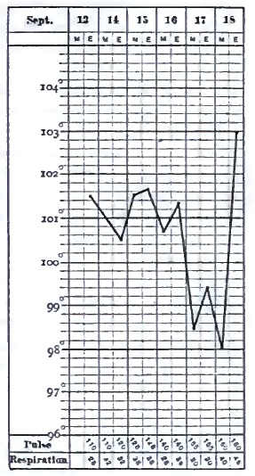
Autopsy.—General lividity of surface, much emaciation. Much fine arborescent injection on outer surface of dura mater. Numerous Pacchionian bodies. Yellow matter beneath arachnoid along course of vessels on each side of anterior lobes. Abundant fine granulations along course of vessels on each anterior lobe, on upper margins of median fissure, along fissure of Sylvius, and on choroid plexuses. Very little lymph at base of brain. Six or eight ounces of serum from lateral ventricles, and abundant fine transparent granules over ependyma of both. Numerous opaque granulations in pia mater of medulla oblongata. Surface of right pleura universally adherent. Mucous membrane of bronchia much injected; a considerable amount of pus flowed from each primary bronchus. No tubercles in lungs nor in peritoneum. No ulcerations in intestines. No other lesions.
The choroid plexuses are generally involved in the inflammatory process, and are sometimes covered with yellow purulent exudations. As in the above case, large numbers of tubercles may be found in them, notwithstanding the opinion of Huguenin that their number is always small.
The substance of the brain in the vicinity of the tubercular deposit is generally found in a more or less œdematous condition, owing to the obstruction of the circulation resulting from compression of the vessels by the tubercles and effused lymph. Softening, sometimes even to diffluence, not unfrequently occurs in the neighborhood of the deposit, probably from ischæmia (necrobiosis). If there be any considerable amount of exudation in the ventricles, the convolutions are flattened by compression against the cranial bones.
The above-described lesions are not confined to the brain, but may extend to the cerebellum, the pons, the medulla, and the spinal cord. If examinations of the latter were more frequent in autopsies of this disease, we should doubtless find, as has been done in some instances, that the membranes often show the characteristic alterations of tubercular meningitis, and even the presence of granulations in the cord itself. The lesions may extend throughout the cord, and are especially noticed in the dorsal region and in the vicinity of the cauda equina. Their presence explains some of the symptoms evidently due to spinal origin, such as retraction of the head with rigidity of the neck and of the trunk, contractions of the limbs, tetanic spasms, priapism, paralysis of the bladder and rectum, etc., which are common in simple spinal meningitis.
The deposit of miliary tubercles in the pia mater, with little or no accompanying meningitis, is met with in rare instances. The tubercles are few in number, but vary in dimensions, being sometimes united together in masses of considerable size, which are frequently encysted. Beyond thickening and opacity of the membrane, their presence seems to excite but little inflammatory reaction, but they are generally accompanied by ventricular effusion which by its pressure gives rise to characteristic symptoms.
The principal lesions found in other organs of the body consist of tubercle in various stages of development, caseous matter, diseases of the bones, etc. Miliary granulations are chiefly seen in the lungs, peritoneum, intestinal mucous membrane, pleura, spleen, liver, and kidneys. The bronchial and mesenteric glands often contain caseous masses, some of which are broken down and suppurating. The testicles sometimes present the same appearances. In adults, the most frequent lesion which is found external to the brain is pulmonary tuberculosis in a more or less advanced stage. Tubercles are also sometimes present in the eye. Angel Money11 states that out of 44 examinations made at the Hospital for Sick Children, London, the meninges were the seat of gray granulations in 42. The choroid (one or both) showed tubercles 14 times (right 3, left 5, both 6), and 11 times there were undoubted evidences of optic neuritis. Twice the choroid was affected with tubercle when the meninges were free; in one of these instances there was a mass of crude tubercle in the cerebellum; in the other, although there were tubercles in the belly and chest, there were none in the head. So that 12 times in 42 cases of tubercles in the meninges there were tubercles in the choroid—i.e. about 31 per cent.
11 “On the Frequent Association of Choroidal and Meningeal Tubercle,” Lancet, Nov. 10, 1883.
DIAGNOSIS.—In many cases tubercular meningitis offers but little difficulty in the diagnosis. Although the symptoms, taken singly, are not pathognomonic, yet their combination and succession, together with their relation to the age, previous health, and antecedents of the patient, are usually sufficient to lead us to a correct opinion. The prodromic period of altered disposition (irritability of temper or apathetic indifference), headache, constipation, vomiting, and emaciation, followed by irregularity and slowness of the pulse, sighing respiration, sluggishness and irregularity of the pupils; the progress from somnolence to unconsciousness and coma; the sudden lamentable cry; the convulsions and paralysis; the return of rapid pulse and respiration in the last stage,—are characteristic of no other disease. Our chief embarrassment arises during the insidious approach of the malady, before its distinctive features are visible or when some important symptom is absent. Its real nature is then apt to be overlooked, and, in fact, in some cases it is impossible to decide whether the symptoms are indicative of commencing cerebral disease, or, on the other hand, are owing to typhoid fever, to a simple gastro-intestinal irritation from error in diet, to worms in the alimentary canal, to overwork in school, or to some other cause. Under these circumstances the physician should decline giving a positive opinion until more definite signs make their appearance. It must be remembered that very important symptoms may be absent in cases which are otherwise well marked. In all doubtful cases the family history should, if possible, be obtained, especially whether one or both parents or other near relatives have been consumptive or have shown symptoms of scrofula or tuberculosis in any form, and whether the patient himself has signs of pulmonary tuberculosis, of enlarged or suppurating glands, or obstinate skin eruptions. The presence or history of those conditions would add greatly to the probability of tubercular meningitis.
The diseases for which tubercular meningitis is most liable to be mistaken are acute simple meningitis, typhoid fever, acute gastro-intestinal affections, eclampsia of infants and children, worms in the intestines or stomach, the hydrencephaloid disease of Marshall Hall, and cerebro-spinal meningitis.
Acute meningitis is distinguished from the tubercular disease by its sudden invasion without prodromatous stage, by the acuteness and intensity of the symptoms, the severity of the headache, the activity of the delirium, the greater elevation of the temperature, and by its brief duration, which rarely exceeds one week. In those exceptional cases of tubercular meningitis in which the prodromal period is absent or not observed and the course is unusually rapid, it would be perhaps impossible to distinguish between the two diseases. A family history of tubercle, or the discovery of the granulations in the choroid by ophthalmoscopic examination, might save us from error under such circumstances. The great rarity of idiopathic simple meningitis should be remembered. Meningitis from disease of the ear sometimes resembles the tubercular affection, but the history of the attack, usually beginning with local pain and otorrhœa, will in most cases prevent any confusion between the two forms of disease.
The early period of typhoid often bears considerable resemblance to that of tubercular meningitis. Headache, languor, restlessness, and mild delirium are common to both. Typhoid can be distinguished by the coated tongue, the diarrhœa, the enlargement of the spleen, the tympanites, abdominal tenderness and gurgling, the eruption, and, above all, by the characteristic temperature-curve, which, if accurately observed, is conclusive. The course of typhoid fever is comparatively uniform, while that of tubercular meningitis is often extremely irregular. It should not be forgotten that the two diseases may coexist.
The presence of worms in the alimentary canal may cause symptoms somewhat like those of tubercular meningitis, and the symptoms of the latter disease are occasionally erroneously attributed to those parasites. The administration of an anthelmintic, which should never be omitted in doubtful cases, will clear up all uncertainty.
Cerebro-spinal meningitis is usually an epidemic, and therefore not likely to be confounded with the tubercular disease. In sporadic cases it can be recognized by its sudden onset and acute character, by the eruption, and by the prominence of the spinal symptoms.
The so-called hydrencephaloid disease of Marshall Hall is a condition of exhaustion and marasmus belonging to infancy, caused by insufficient or unsuitable nourishment, by diarrhœa, and by the injudicious depletive treatment so much in vogue in former times, when the affection was much more common than at present. Some of its symptoms, such as sighing respiration, stupor, pallor, and dilated pupils, bear a certain resemblance to those of tubercular meningitis, though it would be more easily confounded with chronic hydrocephalus. The absence of constipation, headache, convulsions, and vomiting, and the favorable results of suitable nourishment and stimulants, serve to distinguish it from cerebral disease.
Eclampsia, or sudden convulsion, is common in infants and young children, and, since the occurrence of a fit may be the first or the most striking symptom in tubercular meningitis, it is important to ascertain its origin. In the majority of cases convulsions in children arise from some peripheral irritation, such as difficult dentition, worms in the alimentary canal, constipation, fright, etc., acting through the reflex function of the spinal cord, which is unusually sensitive in the early period of life. The absence of previous symptoms, and the discovery of the source of the irritation, with the favorable effect of its removal by appropriate treatment, will in most cases suffice to eliminate structural disease of the brain. In others we must withhold a positive opinion for a reasonable time in order to ascertain whether more definite symptoms follow. Convulsions also occasionally form the initial symptom of the eruptive fevers, especially scarlatina. Here the absence of prodromal symptoms, and the speedy appearance of those belonging to the exanthematous affection, will remove all sources of doubt. Convulsions, with or without coma, occurring in the early stage of acute renal inflammations, may simulate the symptoms of tubercular meningitis. An examination of the urine will show the true nature of the disease.
In addition to the above diseases there are some cerebral affections of uncertain pathology which resemble tubercular meningitis, but which are not generally fatal. As Gee justly remarks,12 “Every practitioner from time to time will come across an acute febrile disease accompanied by symptoms which seem to point unmistakably to some affection of the brain, there being every reason to exclude the notion of suppressed exanthemata or analogous disorders. After one or several weeks of coma, delirium, severe headache, or whatever may have been the prominent symptom, the patient recovers, and we are left quite unable to say what has been the matter with him. To go more into detail, I could not do otherwise than narrate a series of cases which would differ from each other in most important points, and have nothing in common excepting pyrexia and brain symptoms. There is, generally, something wanting which makes us suspect that we have not to do with tubercular meningitis. Brain fever is as good a name as any whereby to designate these different anomalies; cerebral congestion, which is more commonly used, involves an explanation which is probably often wrong, and certainly never proved to be right.” No doubt such cases are occasionally cited as examples of recovery from tubercular meningitis.
12 “Tubercular Meningitis,” by Samuel Jones Gee, M.D., in Reynolds's System of Medicine, Philada., 1879, vol. i. p. 832.
PROGNOSIS.—Although there are on record undoubted instances of recovery from tubercular meningitis, yet their number is so small that practically the prognosis is fatal. It is safe to say that in almost all the reported cases of recovery the diagnosis was erroneous.13 Even should the patient survive the attack, he is usually left with paralyzed limbs and impaired mental faculties, and dies not long afterward from a recurrence of the disease or from tuberculosis of the lungs or other organs.
13 Hahn, “Recherches sur la Méningite tuberculeuse et sur le Traitement de cette Maladie” (Arch. gén. de méd., 4e Série, vols. xx. and xxi.), claims to have cured 7 cases, but of 5 of them there is no evidence that they were examples of tubercular meningitis at all. The subject of the curability of tubercular meningitis is ably treated by Cadet de Gassicourt (Traité clinique des Maladies de l'Enfance, vol. iii., Paris, 1884, p. 553 et seq.). His conclusion is that most of the alleged cures are cases of meningitis of limited extent, arising from the presence of tubercular tumors, syphilitic gummata, cerebral scleroses, and neoplasms of various kinds.
TREATMENT.—In view of the fatality of the disease, and of its frequent occurrence in childhood, the prophylactic treatment is of great importance. Every effort should be made to protect children whose parents or other near relatives are tuberculous or scrofulous, and who are themselves delicate, puny, or affected with any constitutional disorder, from tubercular meningitis, by placing them in the best possible hygienic conditions. Pure air, suitable clothing, wholesome and sufficient food, and plenty of out-of-door exercise are indispensable. Sedentary amusements and occupations should be sparingly allowed. Especial pains should be taken to prevent fatigue by much study, and school-hours should be of short duration. The hygiene of the school-room is of paramount importance, and if its ventilation, temperature, and light are not satisfactory, the child should not be permitted to enter it. The bed-chamber should be well ventilated night and day. A sponge-bath, cold or tepid according to the season or to the effect on the patient, should be given daily, followed by friction with a towel. The bowels must be kept regular by appropriate diet if possible, or by simple laxatives, such as magnesia or rhubarb. For delicate, pale children some preparation of iron will be useful. The choice must be left to the practitioner, but one of the best in such cases is the tartrate of iron and potassium, of which from two to six grains, according to the age, may be given three times daily after meals. Cod-liver oil is invaluable for scrofulous patients or where there is a lack of nutrition. A teaspoonful, given after meals, is a sufficient dose, and it is usually taken without difficulty by children, or if there be much repugnance to it some one of the various emulsions may be tried in proportionate dose. Along with this, iodide of iron will in many cases be found useful or as a substitute for the oil when the latter cannot be borne. It is best given in the form of the officinal syrup, in the dose of from five to twenty drops. Change of air is useful in stimulating the nutritive functions, and a visit to the seashore or mountains during warm weather will often be followed by general improvement.
Since it is not possible to arrest the disease when once begun, the efforts of the physician must be directed toward relieving the sufferings of the patient as far as possible. In the early period the restlessness at night and inability to sleep will call for sedatives, such as the bromide of sodium or of potassium, in the dose of ten or fifteen grains at bedtime or oftener. This should be well diluted with water, sweetened if necessary. The addition of five to twenty drops of the tincture of hyoscyamus increases the effect. Sometimes chloral hydrate, either alone or combined with the bromide when the latter fails, will procure quiet sleep. From five to ten grains may be given at a dose, according to the age. Compresses wet with spirit and water or an ice-cap may be applied to the head if there be much pain in that region, or it may be necessary to give opium in some of its forms by the mouth, such as the tincture or fluid extract, in doses of from one to five drops. Constipation is best overcome by means of calomel in three- to five-grain doses, to which may be added, when necessary, an equal amount of jalap powder, or an enema of soapsuds may be administered. Active purging should be avoided. Liquid nourishment, such as milk, gruel of oatmeal, farina, or barley, beef-tea, broths, etc., must be given in moderate quantities at intervals of a few hours so long as the patient is able to swallow. Occasional sponging of the whole surface with warm or cool water, and scrupulous attention to cleanliness after defecation, especially when control of the sphincters is lost, will add to his comfort. He should occupy a large and well-ventilated chamber, from which all persons whose presence is not necessary for his care and comfort should be excluded. He should be protected from noise and from bright light, and should lie on a bed of moderate width for convenience of tending.
There is no specific treatment at present known which is likely to be of any benefit in this disease, any more than in tuberculosis of other organs than the brain. Common experience has shown that mercury, which formerly had so high a reputation in the treatment of cerebral diseases of early life, not only fails completely, but adds to the sufferings of the patient when pushed to salivation. The iodide of potassium is recommended by almost all writers, but, so far as I know, there is no proof that it possesses any virtue in meningeal tuberculosis. It has the high authority of Charles West,14 however, who thinks the remedy is more encouraging than any other, and who mentions one instance in which recovery took place under its employment. He recommends that two grains be given every four hours to a child three years old, the bowels being kept free. Most authorities recommend much higher doses, such as ten or fifteen grains, three or four times daily.
14 Op. cit., p. 102.
Counter-irritation to the head or back of the neck was formerly much employed, but is now generally abandoned, as giving rise to much discomfort without obvious beneficial effect. In the cases reported by Hahn, already alluded to under the head of Prognosis, recovery is attributed to the energetic application of tartar-emetic ointment to the scalp, producing extensive ulceration, which in one of them lasted more than ten months before cicatrization took place. A careful examination of the reports of these cases satisfies me that but two out of the seven were really examples of tubercular meningitis. How far the recovery in the successful cases is to be attributed to the treatment is very doubtful. Small blisters applied to the vertex or back of the neck are alluded to favorably by West, but he quotes no observations in which they were followed by benefit.
Tubercular Meningitis in the Adult.
Tubercular meningitis may occur at any age, but after the period of childhood it is most frequent between the ages of sixteen and thirty years. About 75 per cent. of the patients are males, and 25 per cent. females.15 The disease does not differ essentially in its course and symptoms from that in children. A family history of tuberculosis is common, or the patient may be already suffering from phthisis, scrofulous glands, cheesy deposits in various organs, caries of the bone, syphilis, or other constitutional affections. According to Seitz, in 93.5 per cent. out of 130 cases with autopsies chronic inflammatory conditions or caseous deposits were found in various organs of the body. Many cases are examples of acute tuberculosis in which the brunt of the disease has fallen upon the brain rather than the other organs.
15 Seitz, op. cit., p. 9.
When cerebral symptoms supervene upon acute disease, such as typhoid fever, pericarditis, acute rheumatism, pneumonia, the exanthemata, etc., the diagnosis between tubercular and simple meningitis is important, because the latter is not necessarily fatal, while the former is almost never recovered from. If the patient were previously healthy and presumably free from tubercular disease, the chances would be in favor of the simple form. A rapid course of the disease would also speak for acute meningitis, and recovery would almost certainly preclude tubercular meningitis. In some cases the diagnosis is difficult or impossible in the beginning.
In cases beginning without acute antecedents—in adults as in children—there is no pathognomonic symptom, but the combination and succession of the phenomena are usually sufficient for the diagnosis. Headache, depression or irritability of temper, delirium, half-closed eyes, ptosis of one lid, squinting, inequality and sluggishness of the pupils, moderate fever, sunken belly, vomiting, constipation, slow and irregular pulse, sopor gradually deepening to coma, with occasional convulsions or paralysis of the limbs, followed in the course of two or three weeks by death, especially if occurring in a patient who has already presented signs of tuberculosis in other organs, point almost unmistakably to tubercular meningitis. Some of the symptoms may not be strongly pronounced, one or two may be absent, but the general picture will suggest no other disease. According to Seitz, tubercle of the choroid is rare in tubercular meningitis of the adult. It is usually associated with tuberculosis of other organs, the pia being free. The subjoined cases are illustrations of the disease in adolescence and adult life:
Case I.—A lad sixteen years old, always somewhat delicate, with a cough and morning expectoration of some months' standing, exerted himself immoderately in gymnastic exercise on the afternoon of Sept. 24, 1875. That night he was awakened by cough and hæmoptysis. Signs of consolidation were found at the apices of both lungs. In three months there were swelling, induration, and suppuration of one testicle. Some months later, pain in the right arm, stiffness of the shoulder-joint, and an abscess communicating with the joint, from which small spicula of bone were discharged. He was about, and even attended school, for more than a year from the time of the attack of hæmoptysis. About Jan. 1, 1877, he began to complain of severe pain in the forehead, with nausea (but no vomiting) and constipation. Jan. 27th he took to his bed, complaining chiefly of pain in the forehead and eyes. Feb. 1st he was drowsy, irritable, and delirious. Feb. 3d incontinence of urine and constant delirium. Up to this time the pulse had not been above 76 in the minute, and on this day it was 64. The next day, Feb. 4th, he was wholly unconscious; pulse 96, pupils dilated. Feb. 5th, left hand in constant motion; pulse 112. From this time the pulse steadily increased in frequency. Feb. 6th he swallowed food, notwithstanding his stupor. Feb. 7th he answered questions; there was oscillation of the eyeballs, and epistaxis from constant picking of the nose. He died Feb. 9th, the pulse being 144 and the respirations 60 in the minute for several hours previous. The temperature never rose above 101.7° F. There was never any vomiting. The duration of the case was thirteen days, in addition to the prodromic period of twenty-seven days.—Autopsy by R. H. Fitz: The pia of the base of the brain from the medulla to the optic thalamus contained a large number of gray miliary tubercles, old and recent, and the same condition was found in the surfaces of the fissure of Sylvius. Ventricles distended with fluid, ependyma thick and translucent. A moderate-sized cavity in the apex of the left lung, with cheesy contents. Both lungs contained an abundance of hard, gray, miliary tubercles. Left kidney contained a wedge-shaped, cheesy mass of the size of a walnut, with numerous tubercles. Left testicle contained a cheesy mass of the size of a walnut; both epididymes were cheesy. The mucous membrane of the bladder contained tubercles near the neck. The vesiculæ seminales contained softened cheesy masses with openings into the urethra. There was a fistulous opening into the right shoulder-joint.
Case II.—Emeline K. L——, 32 years old, single, nurse, had become much exhausted by taking care of a difficult case, and entered Massachusetts General Hospital March 6, 1883, complaining since four days of a little cough, slight expectoration, and chilliness, but no rigor. She seemed hysterical. There was complaint of severe pain in the head, chest, and abdomen. A slight systolic murmur was found at the apex of the heart, and a few moist râles at the base of the chest on both sides behind. The urine was normal. Temperature, 103° F. Three weeks after entrance she began to be delirious, especially at night. April 13th, five weeks and three days after entrance, she was semi-conscious, but would put out her tongue and open her eyes when requested to do so; the abdomen was distended, the pupils were dilated and unequal; there was twitching of the muscles of the right side of the face. Careful examination only disclosed occasional fine râle at the base of right chest. She died April 16th, having been completely unconscious for twenty-four hours. There was no vomiting throughout the case. The temperature was very irregular, ranging between 100° and 103° F., and once as high as 104° F.; it was usually one or two degrees higher at night than in the morning. Pulse, generally from 100 to 110; it rose steadily during the last few days to 160 at the time of death.—Autopsy: Pia mater of brain œdematous, slightly opaque; its lower surface, especially at the base of the brain, showed numerous minute gray tubercles; enlarged cheesy glands at the base of the neck; small, opaque, gray tubercles scattered rather sparsely throughout both lungs. There was also miliary tuberculosis of the liver, spleen, and kidneys.
CHRONIC HYDROCEPHALUS.
BY FRANCIS MINOT, M.D.
SYNONYMS.—Dropsy of the brain, Dropsy of the head.
DEFINITION.—A gradual accumulation of serous fluid in the brain, occupying either the ventricles or the cavity of the arachnoid, or both, occurring chiefly in infants or very young children.
The term hydrocephalus, which was applied by the older writers to accumulations of serous fluid both within and also without the cranium, termed distinctively internal and external hydrocephalus, is now restricted to dropsical effusions either between the meninges or within the ventricular cavities. These may be acute or chronic, and they arise from the same conditions which are followed by the effusion of serum in other parts of the body; that is to say, from an alteration in the serous membrane lining a cavity, from an obstruction in the capillary circulation with increased tension in the larger vessels, from an altered condition of the blood, etc.
Chronic hydrocephalus is almost entirely confined to young children, and is probably due to an arrested development of the brain, as shown by its being usually congenital, by the dwarfed intellectual condition of the patient, and by its frequent association with spina bifida. The pathogeny of the disease is, however, still obscure. Whether the abnormal accumulation of serous fluid is to be ascribed to a chronic alteration of the ventricular walls or of the choroid plexuses, allied to inflammation, such as occurs in the pleura, for example, causing an increased secretion, or to a closure of the communication between the ventricles and the spinal cavity, as suggested by the late John Hilton, resulting in dropsy by retention, or to some other cause, is not yet determined. Hilton says:1 “In almost every case of internal hydrocephalus which I have examined after death I found that this cerebro-spinal opening [between the fourth ventricle and the spinal canal] was so completely closed that no cerebro-spinal fluid could escape from the interior of the brain; and, as the fluid was being constantly secreted, it necessarily accumulated there, and the occlusion formed, to my mind, the essential pathological element of internal hydrocephalus.” Sieveking, commenting upon Hilton's theory, says:2 “While giving these facts due weight, it must be pointed out that we are yet far from understanding either how the fluid is poured into the cerebral cavities or how it is removed, and that we do not positively know that the spinal canal has any better means of getting rid of an excess of fluid than the cerebral cavities have.” An arrest of the growth of the brain is supposed by some pathologists to account for ventricular as well as arachnoidal dropsy by the creation of a vacuum in the cavity of the cranium, which is filled by exudation of the more fluid portion of the blood from the vessels or of lymph from the lymphatics.
1 John Hilton, Lectures on Rest and Pain, etc., 2d ed., New York, 1879, p. 22.
2 Jones and Sieveking's Pathological Anatomy, 2d ed., London, 1875, p. 248.
ETIOLOGY.—That chronic hydrocephalus in young children is in a large proportion of cases an hereditary disease is shown by the fact that it is frequently congenital, that more than one child in a family is occasionally affected by it, and that while one child is hydrocephalic others may be idiotic. A scrofulous taint in the family history is noticeable in many cases, the evil effect of which is frequently enhanced by unfavorable sanitary conditions of life, especially by residence in dark, damp, ill-ventilated, and badly-drained apartments and by insufficient or unwholesome food. Hence the disease is more frequently met with among the poor than in the well-to-do classes of society. There is a difference of opinion as to whether it ever arises in consequence of external violence, such as a blow on the head, or of some strong impression, like fright or grief, acting on the mother of the child during pregnancy. The probability is that such causes would not be efficient except in cases where a predisposition to hydrocephalus had already existed. West3 mentions the case of a healthy child five months old who fell out of the arms of her nurse, and was taken with a fit the same day. She apparently recovered, but when a year old had frequent returns of convulsions. At the age of fifteen months the head began to enlarge, and it continued to increase in size until she was three years old, when she was attacked by measles, and died in a few days with convulsions and coma. The symptoms, both bodily and mental, were typical of hydrocephalus, and the diagnosis was fully confirmed by the autopsy.
3 Op. cit., p. 127.
The causes of chronic hydrocephalus in older subjects and in adults are, in addition to the above mentioned, chiefly mechanical. Any lesion which hinders the exit of venous blood from the cranium may be followed by dropsy of the arachnoid cavity or ventricles. The principal ones are tumors of the brain or its membranes, the effusion of lymph in the neighborhood of veins, thrombosis of the cerebral sinuses, compression of numerous small vessels by tubercular granulations, obstructions outside the cranium, including tumors of the neck, aneurisms, obstructive disease of the heart, emphysema and cirrhosis of the lungs, besides diseases giving rise to general dropsy, as the different forms of nephritis, marasmus, the various cachexiæ, etc.
SYMPTOMS.—When the disease is already somewhat advanced before birth, the head is often abnormally large, the cranial bones are separated, the fontanels are distended and fluctuating, and it occasionally happens that delivery of the distended head can only be effected after the fluid has been evacuated by puncture. Even when there is no abnormal increase in the size of the head, indications of cerebral disturbance are sometimes apparent from birth, such as strabismus or convulsions recurring with more or less frequency, along with signs of failure of the general health. In the course of some weeks, or, it may be, months, the attention of the parents is attracted to the prominence of the child's forehead, strongly contrasting with the comparatively diminutive size of the face. Soon an enlargement of the fontanels is perceived, the sutures between the cranial bones become broader, and the head assumes a globular shape from the pressure of the contained fluid. The separation of the bones at the vertex of the skull causes the os frontis to protrude forward, the parietals backward and outward, and the occipital backward and downward. The orbital plates of the frontal bone yield to the pressure of the fluid behind them, and from a horizontal position tend to assume a vertical one, protruding the eyeballs, which have a peculiar downward direction. The sclerotica is visible above the iris, and the latter is partially covered by the lower eyelid. The enlargement of the head is progressive, though not uniformly so, there being pauses of weeks or months during which it is arrested. If it be considerable, the cranial bones are usually thinned, so that the skull becomes translucent when opposite a bright light. When there is a wide separation of the bones there is an unusual development of ossa triquetra in the sutures.
In cases in which the ossification of the cranium is considerably advanced before the beginning of the disease, the enlargement of the head is apt to be correspondingly less. If all the sutures are consolidated, there may be no increase in size, and this is especially true of the mechanical dropsy of adult life. The dropsical effusion, which is then moderate in amount, finds room through compression of the brain-substance, and part of it escapes into the spinal canal.
The size of the hydrocephalic head varies considerably. Where there has been an early arrest of the disease it may be but slightly above the normal. On the other hand, the dimensions are sometimes enormous. In the Warren Museum of Harvard University is a cranium which measures 27½ inches in its greatest circumference, and 20½ inches from one auditory foramen to the other over the top of the head. Its internal capacity is 257 cubic inches. The patient, who died at the age of three years, was never able to sit up and support the head, or even to turn it from the left side, on which she continually lay; she never spoke, and seemed to have no intelligence. The skull of the celebrated James Cardinal measured 32¼ inches in its largest circumference.4 Even more remarkable instances are on record. The dimensions of the fontanels, particularly the anterior, usually correspond with those of the skull in general. During any strong muscular effort on the part of the patient the membrane covering them is seen to be bulged out by pressure from the fluid beneath. The enlargement is not always uniform; it may be in part or wholly confined to one side, owing to consolidation of the sutures of the opposite side, or because only one of the lateral ventricles is affected. The scalp is traversed by numerous distended veins. The hair is very scanty. The head has a soft, fluctuating feel, and the walls sometimes seem to crackle beneath the fingers, like parchment.
4 Reports of Medical Cases, etc., by Richard Bright, M.D., London, 1831, vol. i. p. 431.
In cases of moderate severity there may be few or no symptoms of active cerebral disturbance, such as convulsions or paralysis, but the child does not learn to talk, to walk, or to control the sphincters even at the age of three or four years, and the signs of imperfect mental development are evident. He is apt to be irritable or mischievous, and even when not actually idiotic is very backward in evincing signs of intelligence. There are, however, not a few exceptions to this, and some children with large hydrocephalic heads are intelligent and amiable. The cranium of a girl who died at the age of sixteen years is preserved in the Warren Museum. It measures 24¼ inches in circumference and 17¼ inches from one auditory meatus to the other over the vertex, and the bone is in no place more than one-eighth of an inch in thickness. The child was an inmate of a house of industry, where she was instructed in the usual branches of knowledge taught in our common schools, until at length, such was her capacity, she was entrusted with the teaching of the other pauper children, and she had an excellent character for intelligence and moral worth. She died of phthisis. On post-mortem examination “the brain was found floating, as it were, in a large collection of water.” As in other chronic diseases, there are often pauses, from time to time, of variable duration, in which there is some improvement in the condition of the patient, as well as a temporary arrest in the enlargement of the head. The growth of the child in stature is often retarded, and when life is prolonged the individual is more or less dwarfed. In some cases there is a considerable increase in the amount of adipose tissue, and the appetite is often voracious. L. Fürst reports5 the case of a hydrocephalic girl, sixteen years old, whose height (or, rather, length, for she was unable to stand) was 81 centimeters (nearly 32 inches), corresponding to the stature of a child between three and four years old. The periphery of the head measured 511/5 centimeters (20¼ inches). The anterior fontanel was still open. The age was verified by reference to the registry of birth at the police-office.
5 “Exquisite Wachsthumshemmung bei Hydrocephalus chronicus,” von Dr. Livius Fürst, Virchow's Archiv, June, 1884.
The symptoms of cerebral disturbance in chronic hydrocephalus are much less striking than one would expect, doubtless because the increase of pressure upon the brain is so gradual. Actual paralysis, especially of the limbs, is rare, but convulsions are not infrequent, as is the spasmodic constriction of the glottis known as laryngismus stridulus or spasmodic croup. A general state of uneasiness and restlessness is common. Vision is often impaired and sometimes wholly lost. Strabismus is frequently present, or there may be an involuntary rolling movement of the eyeballs. The pupils are often dilated and insensible to light. In consequence of the increased weight of the head, the child is unable to support it, and in most cases is compelled to keep the bed. Vomiting is common. The digestive functions are disturbed. Constipation is an almost constant symptom, and the sphincters are relaxed in the advanced stages of the disease. Although in some cases, as already stated, the child may grow fat, the reverse of this is the rule; there is usually progressive emaciation, especially of the lower extremities.
The duration of the disease varies much in different cases. The earlier the characteristic symptoms manifest themselves the more rapid is its course. Most children born hydrocephalic survive but a few days or weeks. In cases which are more slow in their development the patient may live some years, and in rare instances attain adult life. But his feeble vitality usually makes him an easy prey to the ordinary complaints of childhood, and a large proportion of cases succumb to inflammation of the lungs or of other organs, to diarrhœa, whooping cough, or the eruptive diseases. Most of those who escape these intercurrent maladies perish from defective nutrition, the result of malassimilation of food, or else are cut off by acute cerebral inflammation, with convulsions, etc.
PATHOLOGICAL ANATOMY.—It is rarely that a large amount of serum is found in the cavity of the arachnoid unless a free communication has been established between it and the ventricles by the destruction of cerebral tissue. In the pia the quantity may be more abundant than normal, filling the subarachnoid spaces, separating the convolutions from each other, and occasionally forming little sac-like elevations on the surface of the membrane; but it should be borne in mind that a certain amount of serous infiltration of the tissue of the pia is by no means rare in cases of death from various diseases, owing to the obstruction to the circulation during the last hours of life, and should not be considered pathological unless it exceed the usual limits. The amount of effusion into the ventricles varies between very wide limits, from some ounces to two or three pints. It may be transparent, or turbid from particles of cerebral tissue or epithelial or pus-cells, and is occasionally slightly tinged with blood. Its specific gravity is nearly identical with that of water, and it contains a trace of albumen. The effusion usually occupies the two lateral and the middle ventricles. Less frequently the dropsy is unilateral, in consequence, apparently, of an obliteration of the foramen of Monro. The tissues composing the walls of the ventricles are compressed and hardened. The ependyma of the walls and of the plexus is thickened and roughened by the formation of minute elevations, which the microscope shows to be composed of proliferated and sclerosed connective tissue. The brain in cases of large effusion is reduced to a membranous sac, flattened, with hardly any trace of its original structure at first sight apparent. In the case of James Cardinal, before alluded to, Bright says, “The brain lay at its base [of the skull] with its hemispheres opened outward like the leaves of a book.” Closer inspection shows that all the parts are present, although atrophied by pressure. The convolutions are flattened and the brain-substance is pale and softened. The cranial nerves are often softened and flattened.
In the dropsy of the head in adults which is the result of mechanical pressure or of cachexia the appearances are widely different. The effusion may occupy the cavity of the arachnoid, and even the space between the dura and the skull, as well as the ventricles. The amount of fluid is much less than in the chronic hydrocephalus of children. The ventricular walls present no signs of inflammatory changes.
DIAGNOSIS.—Chronic hydrocephalus is usually recognized without difficulty. The chief points of diagnostic importance are the progressive enlargement of the head, the separation of the cranial bones, with their peculiar change of position, as already described, and the evident signs of arrested intellectual development. If the head be but little enlarged, the case might be mistaken for that of chronic hypertrophy of the brain, but this is a very rare disease, and is not accompanied with defective mental development.
PROGNOSIS.—The elements of prognosis include the size of the head at birth, its rate of enlargement, the general condition of the child, both physically and intellectually, his hereditary antecedents, and the hygienic influences to which he may be subjected. A large proportion of children born hydrocephalic live but a short time; a few survive one or more years. The number of those who reach adult life is extremely small. The favorable indications are a tardy appearance of the dropsy and its slow progress, without marked evidence of defective mental and bodily development.