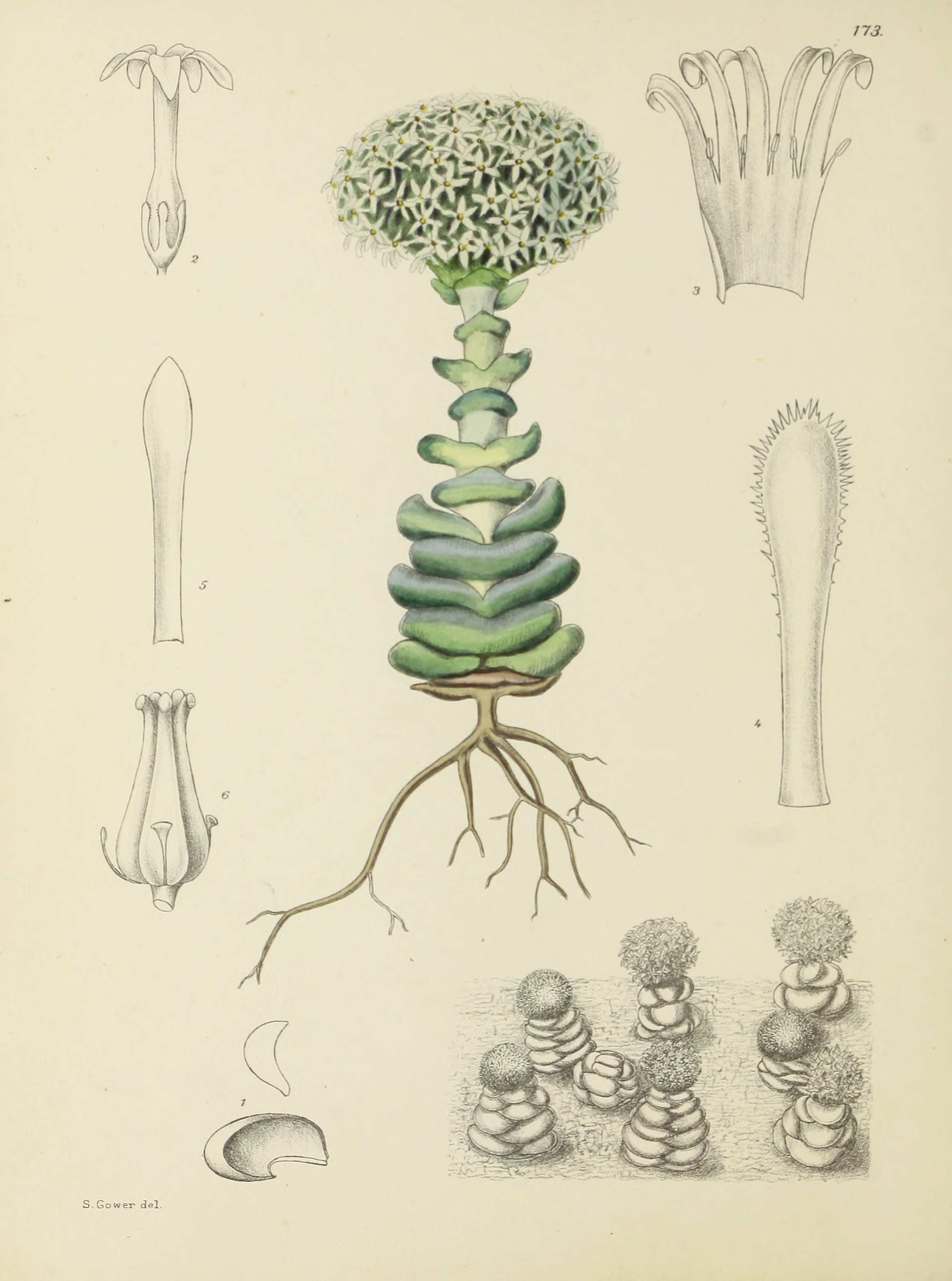
Plate 172.
Reading edition
HYPOXIS rooperi. Basutoland, Cape Province, Natal, Transvaal. Amaryllidaceae. Tribe Hypoxideae. Hypoxis, Linn.; Benth. et Hook. f. Gen. Plant. vol. iii. p. 717.
Hypoxis Rooperi, Moore in Gard. Comp. 1, 65, cum icone; Fl. Cap. vol. vi. p. 188.
The accompanying plate is our first illustration of a characteristic South African genus, namely, Hypoxis. The genus contains over sixty species, of which number more than forty are found in the Union, the remainder being natives of tropical Africa, tropical Asia, Australia and America.
The species is acaulescent, with a large underground corm crowned with a ring of bristles, and bears a number of distichous leaves. The flower-stalks arise from the axils of the leaves.
It is quite a common plant in the south-eastern portion of the Cape Province, and extends through East Griqualand into Natal and northwards into the Drakensbergen round Barberton. It has, however, also been recorded from the Potchefstroom District in the Transvaal.
In winter the leaves die down, but appear again as soon as the frosts cease, and sometimes long before the summer rains commence the plants are in full bloom in the veld.
Our plate was prepared from specimens flowering at the Division of Botany, Pretoria.
Description:—Leaves up to 30 cm. long, 3·2 cm. broad, strap-shaped, narrowing upwards, falcate, folded from the midrib, closely and distinctly ribbed, almost glabrous on the upper surface, softly pilose on the back and margins. Peduncles much shorter than the leaves, villous. Bracts 1·3 cm. long, linear, acute, villous on the back. Pedicels up to 1·6 cm. long, villous. Outer perianth-segments 2·2 cm. long, 8 mm. broad, oblong, obtuse; inner segments 2·1 cm. long, 1·5 cm. broad, elliptic, obtuse. Filaments shorter than the anthers. Ovary subglobose, villous, stigmas 3-lobed, papillose on the margins.
Plate 172.—Fig. 1, plant, much reduced; Fig. 2, portion of peduncle showing bracts and part of pedicels; Fig. 3, flower, surface view; Fig. 4, flower, back view; Fig. 5, anthers; Fig. 6, stigma, side and top views; Fig. 7, transverse section of ovary.
F.P.S.A., 1925.