LAMENESS: ITS CAUSES AND TREATMENT.
Reading edition
By A. Liautard, M. D., V. M.,
Formerly principal of the American Veterinary College, New York.
[Revised by John R. Mohler, A. M., V. M. D.]
It is as living, organized, locomotive machines that the horse, camel, ox, and their burden-bearing companions are of practical value to man. Hence the consideration of their usefulness and consequent value to their human masters ultimately and naturally resolves itself into an inquiry concerning the condition of that special portion of their organism which controls their function of locomotion. This is especially true in regard to the members of the equine family, the most numerous and valuable of all the beasts of burden, and it naturally follows that with the horse for a subject of discussion the special topic and leading theme of inquiry, by an easy lapse, will become an inquest into the condition and efficiency of his power for usefulness as a carrier or traveler. There is a great deal of abstract interest in the study of that endowment of the animal economy which enables its possessor to change his place at will and convey himself whithersoever his needs or his moods may incline him; how much greater, however, the interest that attaches to the subject when it becomes a practical and economic question and includes within its purview the various related topics which belong to the domains of physiology, pathology, therapeutics, and the entire round of scientific investigation into which it is finally merged as a subject for medical and surgical consideration—in a word, of actual disease and its treatment. It is not surprising that the intricate and complicated apparatus of locomotion, with its symmetry and harmony of movement and the perfection and beauty of its details and adjuncts, by students of creative design and attentive observers or nature and her marvelous contrivances and adaptations, should be admiringly denominated a living machine.
Of all the animal tribe the horse, in a state of domesticity, is the largest sharer with his master in his liability to the accidents and dangers which are among the incidents of civilized life. From his exposure to the missiles of war on the battlefield to his chance of picking up a nail from the city pavement there is no hour when he is not in danger of incurring injuries which for their repair may demand the best skill of the veterinary practitioner. This is true not alone of casualties which belong to the class of external and traumatic cases, but includes as well those of a kind perhaps more numerous, which may result in lesions of internal parts, frequently the most serious and obscure of all in their nature and effects.
The horse is too important a factor in the practical details of human life and fills too large a place in the business and pleasure of the world to justify any indifference to his needs and physical comfort or neglect in respect to the preservation of his peculiar powers for usefulness. In entering somewhat largely, therefore, upon a review of the subject, and treating in detail of the causes, the symptoms, the progress, the treatment, the results, and the consequences of lameness in the horse, we are performing a duty which needs no word of apology or justification. The subject explains and justifies itself, and is its own vindication and illustration, if any are needed.
The function of locomotion is performed by the action of two principal systems of organs, known in anatomical and physiological terminology as passive and active, the muscles performing the active and the bones the passive portion of the movement. The necessary connection between the cooperating parts of the organism is effected by means of a vital contact by which the muscle is attached to the bone at certain determinate points on the surface of the latter. These points of attachment appear sometimes as an eminence, sometimes as a depression, sometimes a border or an angle, or again as a mere roughness, but each perfectly fulfilling its purpose, while the necessary motion is provided for by the formation of the ends of the long bones into the requisite articulations, joints, or hinges. Every motion is the product of the contraction of one or more of the muscles, which, as it acts upon the bony levers, gives rise to a movement of extension or flexion, abduction or adduction, rotation or circumduction. The movement of abduction is that which passes from and that of adduction that which passes toward the median line, or the center of the body. The movements of flexion and extension are too well understood to need defining. It is the combination and rapid alterations of these movements which produce the different postures and various gaits of the animal, and it is their interruption and derangement, from whatever causes, which constitute the pathological condition known as lameness.
A concise examination of the general anatomy of these organs, however, must precede the consideration of the pathological questions pertaining to the subject. A statement, such as we have just given, containing only the briefest hint of matters which, though not necessarily in their ultimate scientific minutiæ, must be clearly comprehended in order to acquire a symmetrical and satisfactory view of the theme as a practical collation of facts to be remembered, analyzed, applied, and utilized.
It was the great Bacon who wrote: "The human body may be compared, from its complex and delicate organization, to a musical instrument of the most perfect construction, but exceedingly liable to derangement." In its degree the remark is equally applicable to the equine body, and if we would keep it in tune and profit by its harmonious action we must at least acquaint ourselves with the relations of its parts and the mode of their cooperation.
ANATOMY.
The bones, then, are the hard organs which in their connection and totality constitute the skeleton of an animal (see Plate XXIII). They are of various forms, three of which—the long, the flat, and the small—are recognized in the extremities. These are more or less regular in their form, but present upon their surfaces a variety of aspects, exhibiting in turn, according to the requirement of each case, a roughened or smooth surface, variously marked with grooves, crests, eminences, and depressions, for the necessary muscular attachments, and, as before mentioned, are connected by articulations and joints, of which some are immovable and others movable.
The substance of the bone is composed of a mass of combined earthy and animal matter surrounded by a fine, fibrous enveloping membrane (the periosteum) which is intimately adherent to the external surface of the bone, and is, in fact, the secreting membrane of the bony structure. The bony tissue proper is of two consistencies, the external portion being hard and "compact," and called by the latter term, while the internal, known as the "spongy" or "areolar tissue," corresponds to the descriptive terms. Those of the bones that possess this latter consistency contain also, in their spongy portion, the medullary substance known as marrow, which is deposited in large quantities in the interior of the long bones, and especially where a central cavity exists, called, for that reason, the medullary cavity. The nourishment of the bones is effected by means of what is known as the nutrient foramen, an opening established for the passage of the blood vessels which convey the nourishment necessary to the interior of the organ. Concerning the nourishment of the skeleton, there are other minutiæ, such as the venous arrangement and the classification of their arterial vessels into several orders, which, though of interest as an abstract study, are not of sufficient practical value to refer to here.
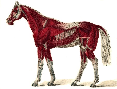
The active organs of locomotion, the muscles (see Plate XXIII), speaking generally, form the fleshy covering of the external part of the skeleton and surround the bones of the extremities. They vary greatly in shape and size, being flat, triangular, long, short, or broad, and are variously and capriciously named, some from their shape, some from their situation, others from their use; and thus we have abductors and adductors—the pyramidal, orbicular, the digastricus, the vastus, and so on. Those which are under the control of the will, known as the voluntary muscles, appear in the form of fleshy structures, red in color, and with fibers of various degrees of fineness, and are composed of fasciculi, or bundles of fibers, united by connective or cellular tissue, each fasciculus being composed of smaller ones but united in a similar manner to compose the larger formations, each of which is enveloped by a structure of similar nature known as the sarcolemma. Many of the muscles are united to the bones by the direct contact of their fleshy fibers, but in other instances the body of the muscle is more or less gradually transformed into a cordy or membranous structure known as the tendon or sinew, and the attachment is made by the very short fibrous threads through the medium of a long tendinous band, which, passing from a single one to several others of the bones, effects its object at a point far distant from its original attachment. In thus carrying its action from one bone to another, or from one region of a limb to another, these tendons must necessarily have smooth surfaces over which to glide, either upon the bones themselves or formed at their articulations, and this need is supplied by the secretion of the synovial fluid, a yellowish, unctuous substance, furnished by a peculiar tendinous synovial sac designed for the purpose.
Illustrations in point of the agency of the synovial fluid in assisting the sliding movements of the tendons may be found under their various forms at the shoulder joint, at the upper part of the bone of the arm, at the posterior part of the knee joint, and also at the fetlocks, on their posterior part.
As the tendons, whether singly or in company with others, pass over these natural pulleys they are retained in place by strong, fibrous bands or sheaths, which are by no means exempt from danger of injury, as will be readily inferred from a consideration of their important special use as supports and reenforcements of the tendons themselves, with which they must necessarily share the stress of whatever force or strain is brought to bear upon both or either.
We have referred to that special formation of the external surface of a bone by which it is adapted to form a joint or articulation, either movable or fixed, and a concise examination of the formation and structure of the movable articulations will here be in place. These are formed generally by the extremities of the long bones, or may exist on the surfaces of the short ones. The points or regions where the contact occurs are denominated the articular surface, which assumes from this circumstance a considerable variety of aspect and form, being in one case comparatively flat and another elevated; or as forming a protruding head or knob, with a distinct convexity; and again presenting a corresponding depression or cavity, accurately adapted to complete, by their coaptation, the ball-and-socket joint. The articulation of the arm and shoulder is an example of the first kind, while that of the hip with the thigh bone is a perfect exhibition of the latter.
The structure whose office it is to retain the articulating surfaces in place is the ligament. This is usually a white, fibrous, inelastic tissue; sometimes, however, it is elastic in character and yellowish. In some instances it is funicular shaped or corded, serving to bind more firmly together the bones to which its extremities are attached; in others it consists of a broad membrane, wholly or partially surrounding the broad articulations, and calculated rather for the protection of the cavity from intrusion by the air than for other security. This latter form, known as capsular, is usually found in connection with joints which possess a free and extended movement. The capsular and funicular ligaments are sometimes associated, the capsular appearing as a membranous sac wholly or partially inclosing the joint, the funicular, here known as an interarticular ligament, occupying the interior, and thus securing the union of the several bones more firmly and effectively than would be possible for the capsular ligament unassisted.
The universal need which pertains to all mechanical contrivances of motion has not been forgotten while providing for the perfect working of the interesting piece of living machinery which performs the function of locomotion, as we are contemplating it, and nature has consequently provided for obviating the evils of attrition and friction and insuring the easy play and smooth movement of its parts by the establishment of the secretion of the synovia, the vital lubricant of which we have before spoken, as a yellow, oily, or rather glairy secretion, which performs the indispensable office of facilitating the play of the tendons over the joints and certain given points of the bones. This fluid is deposited in a containing sac, the lining (serous) membrane of which forms the secreting organ. This membrane is of an excessively sensitive nature, and while it lines the inner face of the ligaments, both capsular and fascicular, it is attached only upon the edges of the bones, without extending upon their length, or between the layers of cartilage which lie between the bones and their articular surfaces.
Our object in thus partially and concisely reviewing the structure and condition of the essential organs of locomotion has been rather to outline a sketch which may serve as a reference chart of the general features of the subject than to offer a minute description of the parts referred to. Other points of interest will receive proper attention as we proceed with the illustration of our subject and examine the matters which it most concerns us to bring under consideration. The foundation of facts which we have thus far prepared will be found sufficiently broad, we trust, to include whatever may be necessary to insure a ready comprehension of the essential matters which are to follow as our review is carried forward to completion. What we have said touching these elementary truths will probably be sufficient to facilitate a clear understanding of the requirements essential to the perfection and regularity which characterize the normal performance of the various movements that result in the accomplishment of the action of locomotion. So long as the bones, the muscles and their tendons, the joints with their cartilages, their ligaments, and their synovial structure, the nerves and the controlling influences which they exercise over all, with the blood vessels which distribute to every part, however minute, the vitalizing fluid which sustains the whole fabric in being and activity—so long as these various constituents and adjuncts of animal life preserve their normal exemption from disease, traumatism, and pathological change, the function of locomotion will continue to be performed with perfection and efficiency.
On the other hand, let any element of disease become implanted in one or several of the parts destined for combined action, any change or irregularity of form, dimensions, location, or action occur in any portion of the apparatus—any obstruction or misdirection of vital power take place, any interference with the order of the phenomena of normal nature, any loss of harmony and lack of balance be betrayed—and we have in the result the condition of lameness.
DEFINITION OF LAMENESS.
Physiology.—Comprehensively and universally considered, then, the term lameness signifies any irregularity or derangement of the function of locomotion, irrespective of the cause which produced it or the degree of its manifestation. However slightly or severely it may be exhibited, it is all the same. The nicest observation may be demanded for its detection, and it may need the most thoroughly trained powers of discernment to identify and locate it, as in cases in which the animal is said to be fainting, tender, or to go sore. On the contrary, the patient may be so far affected as to refuse utterly to use an injured leg, and under compulsory motion keep it raised from the ground, and prefer to travel on three legs rather than to bear any portion of his weight upon the afflicted member. In these two extremes, and in all the intermediate degrees, the patient is simply lame—pathognomonic minutiæ being considered and settled in a place of their own.
This last condition of disabled function—lameness on three legs—and many of the lower degrees of simple lameness are very easy of detection, but the first, or mere tenderness or soreness, may be very difficult to identify, and at times very serious results have followed from the obscurity which has enveloped the early stages of the malady. For it may easily occur that in the absence of the treatment which an early correct diagnosis would have indicated, an insidious ailment may so take advantage of the lapse of time as to root itself too deeply into the economy to be subverted, and become transformed into a disabling chronic case, or possibly one that is incurable and fatal. Hence the impolicy of depreciating early symptoms because they are not accompanied with distinct and pronounced characteristics, and from a lack of threatening appearances inferring the absence of danger. The possibilities of an ambush can never be safely ignored. An extra caution costs nothing, even if wasted. The fulfillment of the first duty of a practitioner, when introduced to a case, is not always an easy task, though it is too frequently expected that the diagnosis, or "what is the matter" verdict, will be reached by the quickest and surest kind of an "instantaneous process" and a sure prognosis, or "how will it end," guessed at instanter.
Usually the discovery that the animal is becoming lame is comparatively an easy matter to a careful observer. Such a person will readily note the changes of movements which will have taken place in the animal he has been accustomed to drive or ride, unless they are indeed slight and limited to the last degree. But what is not always easy is the detection, after discovering the fact of an existing irregularity, of the locality of its point of origin, and whether its seat be in the near or off leg, or in the fore or the hind part of the body. These are questions too often wrongly answered, notwithstanding the fact that with a little careful scrutiny the point may be easily settled. The error, which is too often committed, of pronouncing the leg upon which the animal travels soundly as the seat of the lameness, is the result of a misinterpretation of the physiology of locomotion in the crippled animal. Much depends upon the gait with which the animal moves while under examination. The act of walking is unfavorable for accurate observation, though, if the animal walks on three legs, the decision is easy to reach. The action of galloping will often, by the rapidity of the muscular movements and their quick succession, interfere with a nice study of their rhythm, and it is only under some peculiar circumstances that the examination can be safely conducted while the animal is moving with that gait. It is while the animal is trotting that the investigation is made with the best chances of an intelligent decision, and it is while moving with that gait, therefore, that the points should be looked for which must form the elements of the diagnosis.
PLATE XXIII. SKELETON OF HORSE.
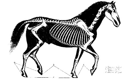
PLATE XXIV. SUPERFICIAL LAYER OF MUSCLES.
Our first consideration should be the physiology of normal or healthy locomotion, that thence we may the more easily reach our conclusions touching lameness, or that which is abnormal, and by this process we ought to succeed in obtaining a clew to the solution of the first problem, to wit, in which leg is the seat of the lameness?
A word of definition is here necessary, in order to render that which follows more easily intelligible. In veterinary nomenclature each two of the legs, as referred to in pairs, is denominated a biped. Of the four points occupied by the feet of the animal while standing at rest, forming a square, the two fore legs are known as the anterior biped; the two hinder, the posterior; the two on one side, the lateral: and one of either the front or hind biped with the opposite leg of the hind or front biped will form the diagonal biped.
Considering, as it is proper to do, that in a condition of health each separate biped and each individual leg is required to perform an equal and uniform function and to carry an even or equal portion of the weight of the body, it will be readily appreciated that the result of this distribution will be a regular, evenly balanced, and smooth displacement of the body thus supported by the four legs, and that therefore, according to the rapidity of the motion in different gaits, each single leg will be required at certain successive moments to bear the weight which had rested upon its congener while it was itself in the air, in the act of moving; or, again, two different legs of a biped may be called upon to bear the weight of the two legs of the opposite biped while also in the air in the act of moving.
To simplify the matter by an illustration, the weight of an animal may be placed at 1,000 pounds, of which each leg, in a normal and healthy condition, supports while at rest 250 pounds. When one of the fore legs is in action, or in the air, and carrying no weight, its 250 pounds share of the weight will be thrown upon its congener, or partner, to sustain. If the two legs of a biped are both in action and raised from the ground, their congeners, still resting in inaction, will carry the total weight of the other two, or 500 pounds. And as the succession of movements continues, and the change from one leg to another or from one biped to another, as may be required by the gait, proceeds, there will result a smooth, even, and equal balancing of active movements, shifting the weight from one leg or one biped to another, with symmetrical precision, and we shall be presented with an interesting example of the play of vital machanics in a healthy organization.
Much may be learned from the accurate study of the action of a single leg. Normally, its movements will be without variation or failure. When at rest it will easily sustain the weight assigned to it without showing hesitancy or betraying pain, and when it is raised from the ground in order to transfer the weight to its mate it will perform the act in such manner that when it is again placed upon the ground to rest it will be with a firm tread, indicative of its ability to receive again the burden to be thrown back upon it. In planting it upon the ground or raising it again for the forward movement while in action, and again replanting it upon the earth, each movement will be the same for each leg and for each biped, whether the act is that of walking or trotting, or even of galloping. In short, the regular play of every part of the apparatus will testify to the existence of that condition of orderly soundness and efficient activity eloquently suggestive of the condition of vital integrity which is simply but comprehensively expressed by the terms health and soundness.
But let some change, though slight and obscure, occur among the elements of the case; some invisible agency of evil intrude among the harmonizing processes going forward; any disorder occur in the relations of cooperating parts; anything appear to neutralize the efficiency of vitalizing forces; any disability of a limb to accept and to throw back upon its mate the portion of the weight which belongs to it to sustain—present itself, whether as the effect of accident or otherwise; in short, let anything develop which tends to defeat the purpose of nature in organizing the locomotive apparatus and we are confronted at once by that which may be looked upon as a cause of lameness.
Not the least of the facts which it is important to remember is that it is not sufficient to look for the manifestation of an existing discordance in the action of the affected limb alone, but that it is shared by the sound one and must be searched for in that as well as the halting member, if the hazard of an error is to be avoided. The mode of action of the leg which is the seat of the lameness will vary greatly from that which it exhibited when in a healthy condition, and the sound leg will also offer important modifications in the same three particulars before alluded to, to wit, that of resting on the ground, that of its elevation and forward motion, and that of striking the ground again when the full action of stepping is accomplished. Inability in the lame leg to sustain weight will imply excessive exertion by the sound one, and lack of facility or disposition to rest the lame member on the ground will necessitate a longer continuance of that action on the sound side. Changes in the act of elevating the leg, or of carrying it forward, or in both, will present entirely opposite conditions between the two. The lame member will be elevated rapidly, moved carefully forward, and returned to the ground with caution and hesitancy, and the contact with the earth will be effected as lightly as possible, while the sound limb will rest longer on the ground, move boldly and rapidly forward, and strike the ground promptly and forcibly. All this is due to the fact that the sound member carries more than its normal, healthy share of the weight of the body, a share which may be in excess from 1 to 250 pounds, and thus bring its burden to a figure varying from 251 to 500 pounds, all depending upon the degree of the existing lameness, whether it is simply a slight tenderness or soreness, or whether the trouble has reached a stage which compels the patient to the awkwardness of traveling on three legs.
That all this is not mere theory, but rests on a foundation of fact may be established by observing the manifestations attending a single alteration in the balancing of the body. In health the support and equilibrium of that mass of the body which is borne by the fore legs is equalized and passes by regular alternations from the right to the left side and vice versa. But if the left leg, becoming disabled, relieves itself by leaning, as it were, on the right, the latter becomes, consequently, practically heavier and the mass of the body will incline or settle upon that side. Lameness of the left side, therefore, means dropping or settling on the right and vice versa. We emphasize this statement and insist upon it, the more from the frequency of the instances of error which have come under our notice, in which persons have insisted upon their view that the leg which is the seat of the lameness is that upon which he drops and which the animal is usually supposed to favor.
HOW TO DETECT THE SEAT OF LAMENESS.
Properly appreciating the remarks which have preceded, and fully comprehending the modus operandi and the true pathology of lameness, but little remains to be done in order to reach an answer to the question as to which side of the animal is the seat of the lameness, except to examine the patient while in action. We have already stated our reasons for preferring the movement of trotting for this purpose. In conducting such an examination the animal should be unblanketed, and held by a plain halter in the hands of a man who knows how to manage his paces, and the trial should always be made over a firm, hard road whenever it is available. He is to be examined from various positions—from before, from behind, and from each side. Watching him as he approaches, as he passes by, and as he recedes, the observer should carefully study that important action which we have spoken of as the dropping of the body upon one extremity or the other, and this can readily be detected by attending closely to the motions of the head and of the hip. The head drops on the same side on which the mass of the body will fall, dropping toward the right when the lameness is in the left fore leg, and the hip dropping in posterior lameness, also on the sound leg, the reversal of the conditions, of course, producing reversed effects. In other words, when the animal in trotting exhibits signs of irregularity of action, or lameness, and this irregularity is accompanied with dropping or nodding the head, or depressing the hip on the right side of the body, at the time the feet of the right side strike the ground, the horse is lame on the left side. If the dropping and nodding are on the near side the lameness is on the off side.
In a majority of cases, however, the answer to the first question relating to the lameness of a horse is, after all, not a very difficult task. There are two other problems in the case more difficult of solution and which often require the exercise of a closer scrutiny, and draw upon all the resources of the experienced practitioner to settle satisfactorily. That a horse is lame in a given leg may be easily determined, but when it becomes necessary to pronounce upon the query as to what part, what region, what structure is affected, the easy part of the task is over, and the more difficult and important, because more obscure, portion of the investigation has commenced—except, of course, in cases of which the features are too distinctly evident to the senses to admit of error. It is true that by carefully noting the manner in which a lame leg is performing its functions, and closely scrutinizing the motions of the whole extremity, and especially of the various joints which enter into its structure; by minutely examining every part of the limb; by observing the outlines; by testing the change, if any, in temperature and the state of the sensibility—all these investigations may guide the surgeon to a correct localization of the seat of trouble, but he must carefully refrain from the adoption of a hasty conclusion, and, above all, assure himself that he has not failed to make the foot, of all the organs of the horse the most liable to injury and lesion, the subject of the most thorough and minute examination of all the parts which compose the suffering extremity.
The greater liability of the foot than of any other part of the extremities to injury from casualties, natural to its situation and use, should always suggest the beginning of an inquiry, especially in an obscure case of lameness at that point. Indeed the lameness may have an apparent location elsewhere when that is the true seat of the trouble, and the surgeon who, while examining his lame patient, discovers a ringbone, and convincing himself that he has encountered the cause of the disordered action suspends his investigation without subjecting the foot to a close scrutiny, at a later day when regrets will avail nothing, may deeply regret his neglect and inadvertence. As in human pathological experience, however, there are instances when inscrutable diseases will deliver their fatal messages, while leaving no mark and making no sign by which they might be identified and classified, so it will happen that in the humbler animals the onset and progress of mysterious and unrecognizable ailments will at times baffle the most skilled veterinarian, and leave our burden-bearing servants to succumb to the inevitable, and suffer and perish in unrelieved distress.
DISEASES OF BONES.
PERIOSTITIS, OSTITIS, AND EXOSTOSIS.
From the closeness and intimacy of the connection existing between the two principal elements of the bony structure while in health, it frequently becomes exceedingly difficult, when a state of disease has supervened, to discriminate accurately as to the part primarily affected and to determine positively whether the periosteum or the body of the bone is originally implicated. Yet a knowledge of the fact is often of the first importance, in order to obtain a favorable result from the treatment to be instituted. It is, however, quite evident that in a majority of instances the bony growths which so frequently appear on the surface of their structure, to which the general term of exostosis is applied, have had their origin in an inflammation of the periosteum, or enveloping membrane, and known as periostitis. However this may be, we have as a frequent result, sometimes on the body of the bone, sometimes at the extremities, and sometimes involving the articulation itself, certain bony growths, or exostoses, known otherwise by the term of splint, ringbone, and spavin, all of which, in an important sense, may be finally referred to the periosteum as their nutrient source and support, at least after their formation, if not for their incipient existence.
Cause.—It is certain that inflammation of the periosteum is frequently referable to wounds and bruises caused by external agencies, and it is also true that it may possibly result from the spreading inflammation of surrounding diseased tissues, but in any case the result is uniformly seen in the deposit of a bony growth, more or less diffuse, sometimes of irregular outline, and at others projecting distinctly from the surface from which it springs, as so commonly presented in the ringbone and the spavin.
Symptoms.—This condition of periostitis is often difficult to determine. The signs of inflammation are so obscure, the swelling of the parts so insignificant, any increase of heat so imperceptible, and the soreness so slight, that even the most acute observer may fail to find the point of its existence, and it is often long after the discovery of the disease itself that its location is positively revealed by the visible presence of the exostosis. Yet the first question had been resolved, in discovering the fact of the lameness, while the second and third remained unanswered, and the identification of the affected limb and the point of origin of the trouble remained unknown until their palpable revelation to the senses.
Treatment.—When, by careful scrutiny, the ailment has been located, a resort to treatment must be had at once, in order to prevent, if possible, any further deposit of the calcareous structure and increase of the exostotic growth. With this view the application of water, either warm or cold, rendered astringent by the addition of alum or sugar of lead, will be beneficial. The tendency to the formation of the bony growth, and the increase of its development after its actual formation, may often be checked by the application of a severe blister of Spanish fly. The failure of these means and the establishment of the diseased process in the form of chronic periostitis cause various changes in the bone covered by the disordered membrane, and the result may be softening, degeneration, or necrosis, but more usually it is followed by the formation of the bony growths referred to, on the cannon bone, the coronet, the hock, etc.
SPLINTS.
We first turn our attention to the splint, as certain bony enlargements that are developed on the cannon bone, between the knee or the hock and the fetlock joint, are called. (See Plate XXV.) They are found on the inside of the leg, from the knee, near which they are frequently found, downward to about the lower third of the principal cannon bone. They are of various dimensions, and are readily perceptible both to the eye and to the touch. They vary considerably in size, ranging from that of a large nut downward to very small proportions. In searching for them they may be readily detected by the hand if they have attained sufficient development in their usual situation, but must be distinguished from a small, bony enlargement that may be felt at the lower third of the cannon bone, which is neither a splint nor a pathological formation of any kind, but merely the buttonlike enlargement at the lower extremity of the small metacarpal or splint bone.
We have said that splints are to be found on the inside of the leg. This is true as a general statement, but it is not invariably so, for they occasionally appear on the outside. It is also true that they appear most commonly on the fore legs, but this is not exclusively the case, because they may at times be found on both the inside and outside of the hind leg. Usually a splint forms only a true exostosis, or a single bony growth, with a somewhat diffuse base, but neither is this invariably the case. In some instances they assume more important dimensions, and pass from the inside to the outside of the bone, on its posterior face, between that and the suspensory ligament. This form is termed the pegged splint, and constitutes a serious and permanent deformity, in consequence of its interference with the play of the fibrous cord which passes behind it, becoming thus a source of continual irritation and consequently of permanent lameness.
Symptoms.—A splint may thus frequently become a cause of lameness though not necessarily in every instance, but it is a lameness possessing features peculiar to itself. It is not always continuous, but at times assumes an intermittent character, and is more marked when the animal is warm than when cool. If the lameness is near the knee joint, it is very liable to become aggravated when the animal is put to work, and the gait acquires then a peculiar character, arising from the manner in which the limb is carried outward from the knees downward, which is done by a kind of abduction of the lower part of the leg. Other symptoms, however, than the lameness and the presence of the splint, which is its cause, may be looked for in the same connection as those which have been mentioned as pertaining to certain evidences of periostitis, in the increase of the temperature of the part, with swelling and probably pain on pressure. This last symptom is of no little importance, since its presence or absence has in many cases formed the determining point in deciding a question of difficult diagnosis.
Cause.—A splint being one of the results of periostitis, and the latter one of the effects of external hurts, it naturally follows that the parts which are most exposed to blows and collisions will be those on which the splint will most commonly be found, and it may not be improper, therefore, to refer to hurts from without as among the common causes of the lesion. But other causes may also be productive of the evil, and among these may be mentioned the over-straining of an immature organism by the imposition of excessive labor upon a young animal at a too early period of his life. The bones which enter into the formation of the cannon are three in number, one large and two smaller, which, during the youth of the animal, are more or less articulated, with a limited amount of mobility, but which become in maturity firmly joined by a rigid union and ossification of their interarticular surface. If the immature animal is compelled, then, to perform exacting tasks beyond his strength, the inevitable result will follow in the muscular straining, and perhaps tearing asunder of the fibers which unite the bones at their points of juncture, and it is difficult to understand how inflammation or periostitis can fail to develop as the natural consequence of such local irritation. If the result were deliberately and intelligently designed, it could hardly be more effectually accomplished.
The splint is an object of the commonest occurrence—so common, indeed, that in large cities a horse which can not exhibit one or more specimens upon some portion of his extremities is one of the rarest of spectacles. Though it is in some instances a cause of lameness, and its discovery and cure are sometimes beyond the ability of the shrewdest and most experienced veterinarians, yet as a source of vital danger to the general equine organization, or even of functional disturbance, or of practical inconvenience, aside from the rare exceptional cases which exist as mere samples of possibility, it can not be considered to belong to the category of serious lesions. The worst stigma that attaches to it is that in general estimation it is ranked among eyesores and continues indefinitely to be that and nothing different. The inflammation in which they originated, acute at first, either subsides or assumes the chronic form, and the bony growth becomes a permanence—more or less established, it is true, but doing no positive harm and not hindering the animal from continuing his daily routine of labor. All this, however, requires a proviso against the occurrence of a subsequent acute attack, when, as with other exostoses, a fresh access of acute symptoms may be followed by a new pathological activity, which shall again develop, as a natural result, a reappearance of the lameness.
Treatment.—It is, of course, the consideration of the comparative harmlessness of splints that suggests and justifies the policy of noninterference, except as they become a positive cause of lameness. And a more positive argument for such noninterference consists in the fact that any active and irritating treatment may so excite the parts as to bring about a renewed pathological activity, which may result in a reduplication of the phenomena, with a second edition, if not a second and enlarged volume, of the whole story. For our part, our faith is firm in the impolicy of interference, and this faith is founded on an experience of many years, during which our practice has been that of abstention.
Of course, there will be exceptional conditions which will at times indicate a different course. These will become evident when the occasions present themselves, and extraordinary forms and effects of inflammation and growth in the tumors offer special indications. But our conviction remains unshaken that surgical treatment of the operative kind is usually useless, if not dangerous. We have little faith in the method of extirpation except under very special conditions, among which that of diminutive size has been named; this seems in itself to constitute a sufficient negative argument. Even in such a case a resort to the knife or the gouge could scarcely find a justification, since no operative procedure is ever without a degree of hazard, to say nothing of the considerations which are always forcibly negative in any question of the infliction of pain and the unnecessary use of the knife.
PLATE XXV. Splint.
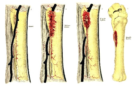
PLATE XXVI. SOUND FOOT. RINGBONE.
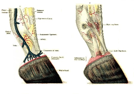
PLATE XXVII. VARIOUS TYPES OF SPAVIN.
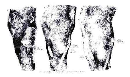
PLATE XXVIII. Bone Spavin. Hocks, with Skin Removed.
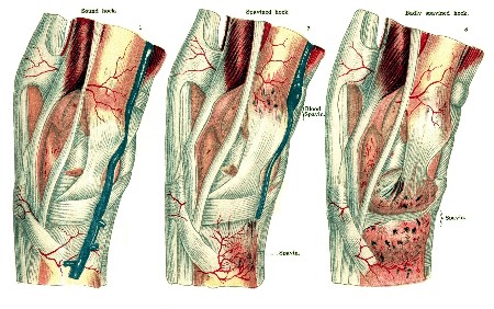
PLATE XXIX. BONE SPAVIN.
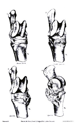
If an acute periostitis of the cannon bone has been readily discovered, the treatment we have already suggested for that ailment is at once indicated, and the astringent lotions may be relied upon to bring about beneficial results. Sometimes, however, preference may be given to a lotion possessing a somewhat different quality, the alterative consisting of tincture of iodin applied to the inflamed spot several times daily. If the lameness persists under this mild course of treatment, it must, of course, be attacked by other methods, and we must resort to the cantharides ointment or Spanish-fly blister, as we have before recommended. Besides this, and producing an analogous effect, the compounds of biniodid of mercury are favored by some. It is prepared in the form of an ointment, consisting of 1 dram of the biniodid to 1 ounce of either lard or vaseline. It forms an excellent blistering and alterative application, and is of special advantage in newly formed or recently discovered exostosis.
It remains a pertinent query, however, and one which seems to be easily answered, whether a tumor so diminutive in size that it can be detected only by diligent search, and which is neither a disfigurement nor an obstruction to the motion of the limb, need receive any recognition whatever. Other modes of treatment for splints are recommended and practiced which belong strictly to the domain of operative veterinary surgery; among these are to be reckoned actual cauterization, or the application of the fire iron and the operation of periosteotomy. These are frequently indicated in the treatment of splints which have resisted milder means.
The mode of the development of their growth; their intimacy, greater or less, with both the large and the small cannon bones; the possibility of their extending to the back of these bones under the suspensory ligament; the dangerous complications which may follow the rough handling of the parts, with also a possibility, and, indeed, a probability, of their return after removal—these are the considerations which have influenced our judgment in discarding from our practice and our approval the method of removal by the saw or the chisel, as recommended by certain European veterinarians.
RINGBONES.
Ringbone is the designation of the exostosis which is found on the coronet and in the digital and phalangeal regions. (See Plate XXVI.) The name is appropriate, because the growth extends quite around the coronet, which it encircles in the manner of a ring, or perhaps because it often forms upon the back of that bone a regular osseous arch, through which the back tendons obtain a passage. The places where these growths are usually developed have caused their subdivision and classification into three varieties, with the designations of high, middle, and low, though much can be said as to the importance of the distinction. It is true that the ringbone or phalangeal exostosis may be found at various points on the foot, in one case forming a large bunch on the upper part and quite close to the fetlock joint; in another around the upper border of the hoof, or perhaps on the extreme front or on the very back of the coronet. The shape in which they commonly appear is favorable to their easy discovery, their form when near the fetlock usually varying too much from the natural outlines of the part when compared with those of the opposite side to admit of error in the matter. (See also page 439.)
A ringbone, when on the front of the foot, even when not very largely developed, assumes the form of a diffused convex swelling. If situated on the lower part, it will form a thick ring, encircling that portion of the foot immediately above the hoof; when found on the posterior part, a small, sharp osseous growth somewhat projecting, sometimes on the inside and sometimes on the outside of the coronet, may comprise the entire manifestation.
Cause.—As with splints, ringbones may result from severe labor in early life, before the process of ossification has been fully perfected; or they may be referred to bruises, blows, sprains, or other violence; injuries of tendons, ligaments, or joints also may be among the accountable causes.
It is certain that they may commonly be traced to diseases and traumatic lesions of the foot, and their appearance may be reasonably expected among the sequelæ of an abscess of the coronet; or the cause may be a severe contusion resulting from calking, or a deep-punctured wound from picking up a nail or stepping upon any hard object of sufficiently irregular form to penetrate the sole.
Moreover, a ringbone may originate in heredity. This is a fact of no little importance in its relation to questions connected with the extensive interests of the stock breeder and purchaser.
That the hereditary transmission of constitutional idiosyncrasies is an active cause with regard to diseases in general, it would be absurd to assert, but we do say that a predisposition to contract ringbone through faulty conformation, such as long, thin pasterns with narrow joints and steep fetlocks, may be inherited in many cases, and in a smaller proportion of cases this predisposition may act as a secondary cause in the formation of ringbone.
The importance of this point when considered in reference to the policy which should be observed in the selection of breeding stock is obvious, and, as the whole matter is within the control of the owners and breeders, it will be their own fault if the unchecked transmission of ringbones from one equine generation to another is allowed to continue. It is our belief that among the diseases which are known for their tendency to perpetuate and repeat themselves by individual succession, those of the bony structures stand first, and the inference from such fact which would exclude every animal of doubtful soundness in its osseous apparatus from the stud list and the brood farm is too plain for argument.
Symptoms.—Periostitis of the phalanges is an ailment requiring careful exploration and minute inspection for its discovery, and is very liable to result in a ringbone of which lameness is the result. The mode of its manifestation varies according to the state of development of the diseased growth as affected by the circumstances of its location and dimensions. It is commonly of the kind which, in consequence of its intermittent character, is termed lameness when cool, having the peculiarity of exhibiting itself when the animal starts from the stable and of diminishing, if not entirely disappearing after some distance of travel, to return to its original degree, if not indeed a severer one, when he has again cooled off in his stable. The size of the ringbone does not indicate the degree to which it cripples the patient, but the position may, especially when it interferes with the free movement of the tendons which pass behind and in front of the foot. While a large ringbone will often interfere but little with the motion of the limb, a smaller growth, if situated under the tendon, may become the cause of considerable and continued pain.
A ringbone is doubtless a worse evil than a splint. Its growth, its location, its tendency to increased development, its exposure to the influence of causes of renewed danger, all tend to impart an unfavorable cast to the prognosis of a case and to emphasize the importance and the value of an early discovery of its presence and possible growth. Even when the discovery has been made, it is often the case that the truth has come to light too late for effectual treatment. Months may have elapsed after the first manifestation of the lameness before a discovery has been made of the lesion from which it has originated, and there is no recall for the lapsed time. And by the uncompromising seriousness of the discouraging prognosis must the energy and severity of the treatment and the promptness of its administration be measured. The periostitis has been overlooked; any chance that might have existed for preventing its advance to the chronic stage has been lost; the osseous formation is established; the ringbone is a fixed fact, and the indications are urgent and pressing.
Treatment.—The preventive treatment consists in keeping colts well nourished and in trimming the hoof and shoeing to balance the foot properly and thus prevent an abnormal strain on the ligaments. Even after the ringbone has developed, a cure may sometimes be occasioned by proper shoeing directed toward straightening the axis of the foot as viewed from the side by making the wall of the hoof from the coronet to the toe continuous with the line formed by the front of the pastern. So long as inflammation of the periosteum and ligaments remains, a sharp blister of biniodid of mercury and cantharides may do good if the animal is allowed to rest for four or five weeks. If this fails, some success may be accomplished by point firing in two or three lines over the ringbone. It is necessary to touch the hot iron well into the bone, as superficial firing does little good. When all these measures have failed to remove the lameness, or when the animal is not worth a long and uncertain treatment, a competent veterinarian should be engaged to perform double neurectomy, high or low, of the plantar nerves, or neurectomy of the median nerve as indicated by the seat of the lesion.
SIDEBONES.
On each side of the bone of the hoof—the coffinbone—there are normally two supplementary organs which are called the cartilages of the foot. They are soft, and though in a degree elastic, yet somewhat resisting, and are implanted on the lateral wings of the coffinbone. Evidently their office is to assist in the elastic expansion and contraction of the posterior part of the hoof, and their healthy and normal action doubtless contributes in an important degree to the perfect performance of the functions of that part of the leg. These organs are, however, liable to undergo a process of disease which results in an entire change in their properties, if not in their shape, by which they acquire a character of hardness resulting from the deposit of earthy substance in the intimate structure of the cartilage, and it is this change, when its consummation has been effected, that brings to our cognizance the diseased growth which has received the designation of sidebones. They are situated on one or both sides of the leg, bulging above the superior border of the hoof in the form of two hard bodies composed of ossified cartilage, irregularly square in shape and unyielding under the pressure of the fingers.
Cause.—Sidebones may be the result of a low inflammatory condition or of an acute attack as well, or may be caused by sprains, bruises, or blows; or they may have their rise in certain diseases affecting the foot proper, such as corns, quarter cracks, or quittor. The deposit of calcareous matter in the cartilage is not always uniform, the base of that organ near its line of union with the coffinbone being in some cases its limit, while at other times it is diffused throughout its substance, the size and prominence of the growth varying much in consequence.
Symptoms.—It would naturally be inferred that the degree of interference with the proper functions of the hoof which must result from such a pathological change would be proportioned to the size of the tumor, and that as the dimensions increase the resulting lameness would be the greater in degree. This, however, is not the fact. A small tumor while in a condition of acute inflammation during the formative stage may cripple a patient more severely than a much larger one in a later stage of the disease. In any case the lameness is never wanting, and with its intermittent character may usually be detected when the animal is cooled off after labor or exercise. The class of animals in which this feature of the disease is most frequently seen is that of the heavy draft horse and others similarly employed. There is a wide margin of difference in respect to the degrees of severity which may characterize different cases of sidebone. While one may be so slight as to cause no inconvenience, another may develop elements of danger which may involve the necessity of severe surgical interference.
Treatment.—The curative treatment should be similar to the prophylactic, and such means should be used as would tend to prevent the deposit of bony matters by checking the acute inflammation which causes it. The means recommended are the free use of the cold bath; frequent soaking of the feet, and at a later period treatment with iodin, either by painting the surface with the tincture several times daily or by applying an ointment made by mixing 1 dram of the crystals with 2 ounces of vaseline, rubbed in once a day for several days. If this proves to be ineffective, a Spanish-fly blister to which a few grains of biniodid of mercury have been added will effect in a majority of cases the desired result and remove the lameness. If finally this treatment is ineffectual the case must be relegated to the surgeon for the operation of neurectomy, or the free and deep application of the firing iron.
SPAVIN.
(Pls. XXVII-XXIX.)
This affection, popularly termed bone spavin, is an exostosis of the hock joint. The general impression is that in a spavined hock the bony growth should be seated on the anterior and internal part of the joint, and this is partially correct, as such a growth will constitute a spavin in the most nearly correct sense of the term. But an enlargement may appear on the upper part of the hock also, or possibly a little below the inner side of the lower extremity of the shank bone, forming what is known as a high spavin; or, again, the growth may form just on the outside of the hock and become an outside or external spavin. And, finally, the entire under surface may become the seat of the osseous deposit, and involve the articular face of all the bones of the hock, which again is a bone spavin. There would seem, then, to be but little difficulty in comprehending the nature of a bone spavin, and there would be none but for the fact that there are similar affections which may confuse one if the diagnosis is not very carefully made.
But the hock may be "spavined," while to all outward observation it still retains its perfect form. With no enlargement perceptible to sight or touch the animal may yet be disabled by an occult spavin, an anchylosis in fact, which has resulted from a union of several of the bones of the joint, and it is only those who are able to realize the importance of its action to the perfect fulfillment of the function of locomotion by the hind leg who can comprehend the gravity of the only prognosis which can be justified by the facts of the case—a prognosis which is essentially a sentence of serious import in respect to the future usefulness and value of the animal. For no disease, if we except those acute inflammatory attacks upon vital organs to which the patient succumbs at once, is more destructive to the usefulness and value of a horse than a confirmed spavin. Serious in its inception, serious in its progress, it is an ailment which, when once established, becomes a fixed condition which there is no known means of dislodging.
Cause.—The periostitis, of which it is nearly always a termination, is usually the effect of a traumatic cause operating upon the complicated structure of the hock, such as a sprain which has torn a ligamentous insertion and lacerated some of its fibers, or a violent effort in jumping, galloping, or trotting, to which the victim has been compelled by the torture of whip and spur while in use as a gambling implement by a sporting owner, under the pretext of "improving his breed"; the extra exertion of starting an inordinately heavy load, or an effort to recover his balance from a misstep, slipping upon an icy surface, or sliding with worn shoes upon a bad pavement, and other kindred causes. We can repeat here what we have before said concerning bones, in respect to heredity as a cause. From our own experience we know of equine families in which this condition has been transmitted from generation to generation, and animals otherwise of excellent conformation have been rendered valueless by the misfortune of a congenital spavin.
Symptoms.—The evil is one of the most serious character for other reasons, among which may be specified the slowness of its development and the insidiousness of its growth. Certain indefinite phenomena and alarming changes and incidents furnish usually the only portents of approaching trouble. Among these signs may be mentioned a peculiar posture assumed by the patient while at rest, and becoming at length so habitual that it can not fail to suggest the action of some hidden disorder. The posture is due to the action of the adductor muscles, the lower part of the leg being carried inward, and the heel of the shoe resting on the toe of the opposite foot. Then an unwillingness may be noticed in the animal to move from one side of the stall to the other. When driven he will travel, but stiffly, with a sort of sidelong gait between the shafts, and after finishing his task and resting again in his stall will pose with the toe pointing forward, the heel raised, and the hock flexed. Considerable heat and inflammation soon appear. The slight lameness which appears when backing out of the stall ceases to be noticeable after a short distance of travel.
A minute examination of the hock may then reveal the existence of a bony enlargement which may be detected just at the junction of the hock and the cannon bone, on the inside and a little in front, and tangible both to sight and touch. This enlargement, or bone spavin, grows rapidly and persistently and soon acquires dimensions which renders it impossible to doubt any longer its existence or its nature. Once established, its development continues under conditions of progress similar to those to which we have before alluded in speaking of other like affections. The argument advanced by some that because these bony deposits are frequently found on both hocks they are not spavins is fallacious. If they are discovered on both hocks, it proves merely that they are not confined to a single joint.
The characteristic lameness of bone spavin, as it affects the motion of the hock joint, presents two aspects. In one class of cases it is most pronounced when the horse is cool, in the other when he is at work. The first is characterized by the fact that when the animal travels the toe first touches the ground, and the heel descends more slowly, the motion of flexion at the hock taking place stiffly, and accompanied with a dropping of the hip on the opposite side. In the other case the peculiarity is that the lameness increases as the horse travels; that when he stops he seeks to favor the lame leg, and when he resumes his work soon after he steps much on his toe, as in the first variety.
As with sidebones, though for a somewhat different reason, the dimensions of the spavin and the degree of the lameness do not seem to bear any determinate relation, the most pronounced symptoms at times accompanying a very diminutive growth. The distinction between the two varieties of cool and warm, however, may easily be determined by remembering the fact that in most cases the first, or cool, is due to a simple exostosis, while the second is generally connected with disease of the articulation, such as ulceration of the articular surface—a condition which, as we proceed further, will receive our attention when we reach the subject of stringhalt.
An excellent test for spavin lameness, which may be readily applied, consists in lifting the affected leg from the ground for one or two minutes and holding the foot high so as to flex all the joints. An assistant, with the halter strap in his hand, quickly starts the animal off in a trot, when, if the hock joint is affected, the lameness will be so greatly intensified as to lead readily to a diagnosis.
Prognosis.—Having thus fully considered the history of bone spavin, we are prepared to give due weight to the reasons that exist for the adverse prognosis which we must usually feel compelled to pronounce when encountering it in practice, as well as to realize the importance of early discovery. It is but seldom, however, that the necessary advantage of this early knowledge can be obtained, and when the true nature of the trouble has become apparent it is usually too late to resort to the remedial measures which, if duly forewarned, a skillful practitioner might have employed. We are fully persuaded that but for the loss of the time wasted in the treatment of purely imaginary ailments very many cases of bone spavin might be arrested in their incipiency and their victims preserved for years of comfort for themselves and valuable labor to their owners.
Treatment.—To consider a hypothetical case: An early discovery of lameness has been made; that is, the existence of an acute inflammation—of periostitis—has been detected. The increased temperature of the parts has been observed, with the stiffened gait and the characteristic pose of the limb, and the question is proposed for solution, What is to be done? Even with only these comparatively doubtful symptoms—doubtful with the nonexpert—we should direct our treatment to the hock in preference to any other joint, since of all the joints of the hind leg it is this which is most liable to be attacked, a natural result from its peculiarities of structure and function. And in answer to the query, What is the first treatment indicated? We should answer rest—emphatically, and as an essential condition, rest. Whether only threatened, suspected, or positively diseased, the animal must be wholly released from labor, and it must be no partial or temporary quiet of a few days. In all stages and conditions of the disease, whether the spavin is nothing more than a simple exostosis, or whether accompanied with the complication of arthritis, there must be a total suspension of effort until the danger is over. Less than a month's quiet ought not to be thought of—the longer the better.
Good results may also be expected from local applications. The various lotions which cool the parts, the astringents which lower the tension of the blood vessels, the tepid fomentations which accelerate the circulation in the engorged capillaries, the liniments of various composition, the stimulants, the opiate anodynes, the sedative preparations of aconite, the alterative frictions of iodin—all these are recommended and prescribed by one or another. We prefer counterirritants, for the reason, among many others, that by the promptness of their action they tend to prevent the formation of the bony deposits. The lameness will often yield to the blistering action of cantharides, in the form of ointment or liniment, and to the alterative preparations of iodin or mercury. If the owner of a "spavined" horse really succeeds in removing the lameness, he has accomplished all that he is justified in hoping for; beyond this let him be well persuaded that a "cure" is impossible.
For this reason, moreover, he will do well to be on his guard against the patented "cures" which the traveling horse doctor may urge upon him, and withhold his faith from the circular of the agent who will deluge him with references and certificates. It is possible that nostrums may in some exceptional instances prove serviceable, but the greater number of them are capable of producing only injurious effects. The removal of the bony tumor can not be accomplished by any such means, and if a trial of these unknown compounds should be followed by complications no worse than the establishment of one or more ugly, hairless cicatrices, it will be well for both the horse and his owner.
Rest and counterirritation, with the proper medicaments, constitute, then, the prominent points in the treatment designed for the relief of bone spavin. Yet there are cases in which all the agencies and methods referred to seem to lack effectiveness and fail to produce satisfactory results. Either the rest has been prematurely interrupted or the blisters have failed to modify the serous infiltration, or the case in hand has some undiscernible characteristics which seem to have rendered the disease neutral to the agencies used against it. An indication of more energetic means is then presented, and free cauterization with the firing iron becomes necessary.
At this point a word of explanation in reference to this operation of firing may be appropriate for the satisfaction of any among our readers who may entertain an exaggerated idea of its severity and possible cruelty.
The operation is one of simplicity, but is nevertheless one which, in order to secure its benefits, must be reserved for times and occasions of which only the best knowledge and highest discretion should be allowed to judge. It is not the mere application of a hot iron to a given part of the body which constitutes the operation of firing. It is the methodical and scientific introduction of heat into the structure with a view to a given effect upon a diseased organ or tissue by an expert surgeon. The first is one of the degrees of mere burning. The other is scientific cauterization, and is a surgical manipulation which should be committed exclusively to the practiced hand of the veterinary surgeon.
Either firing alone or stimulation with blisters is of great efficacy for the relief of lameness from bone spavin. Failure to produce relief after a few applications and after allowing a sufficient interval of rest should be followed by a second or, if needed, a third firing.
In case of further failure there is a reserve of certain special operations which have been tried and recommended, among which those of cunean tenotomy, periosteotomy, the division of nervous branches, etc., may be mentioned. These, however, belong to the peculiar domain of the veterinary practitioner, and need not now engage our attention.
FRACTURES.
In technical language a fracture is a "solution of continuity in the structure or substance of a bone." It ranks among the most serious of the lesions to which the horse—or any animal—can be subject. It is a subject of special interest to veterinarians and horse owners in view of the fact that it occurs in such a variety of forms and subjects the patient to much loss of time, resulting in the suspension of his earning capacity. Though of less serious consequence in the horse than in man, it is always a matter of grave import. It is always slow and tedious in healing and is frequently of doubtful and unsatisfactory result.
This solution of continuity may take place in two principal ways. In the most numerous instances it includes the total thickness of the bone and is a complete fracture. In other cases it involves only a portion of the thickness of the bone, and for that reason is described as incomplete. If the bone is divided into two separate portions and the soft parts have received no injury, the fracture is a simple one, or it becomes compound if the soft parts have suffered laceration, and comminuted if the bones have been crushed or ground into fragments, many or few. The direction of the break also determines its further classification. Broken at a right angle, it is transverse; at a different angle it becomes oblique, and it may be longitudinal or lengthwise. In a complete fracture, especially of the oblique kind, there is a condition of great importance in respect to its effect upon the ultimate result of the treatment in the fact that from various causes, such as muscular contractions or excessive motion, the bony fragments do not maintain their mutual coaptation, but become separated at the ends, which makes it necessary to add another descriptive term—with displacement. These words again suggest the negative and introduce the term without displacement, when the facts justify that description. Furthermore, a fracture may be intra-articular or extra-articular, as it extends into a joint or otherwise, and, once more, intra-periosteal when the periosteum remains intact. Finally, there is no absolute limit to the use of descriptive terminology in the case.
The condition of displacement is largely influential in determining the question of treatment and as affecting the final result of a case of fracture. This, however, is dependent upon its location or whether its seat is in one or more of the axes of the bone, in its length, its breadth, its thickness, or its circumference. An incomplete fracture may also be either simple or comminuted. In the latter case the fragments are held together by the periosteum when it is intact; in that case the fracture belongs to the intra-periosteal class. At times, also, there is only a simple fissure or split in the bone, making a condition of much difficulty of diagnosis.
Causes.—Two varieties of originating cause may be recognized in cases of fracture. They are the predisposing and the occasional. As to the first, different species of animals differ in the degree of their liability. That of the dog is greater than that of the horse, and in horses the various questions of age, the mode of labor, the season of the year, the portion of the body most exposed, and the existence of ailments, local and general, are all to be taken into account.
Among horses, those employed in heavy draft work or that are driven over bad roads are more exposed than light-draft or saddle horses, and animals of different ages are not equally liable. Dogs and young horses, with those which have become sufficiently aged for their bones to have acquired an enhanced degree of frangibility, are more liable than those which have not exceeded the time of their prime. The season of the year is undoubtedly, though in an incidental way, an important factor in the problem of the etiology of these accidents, for though they may be observed at all times, it is during the months when the slippery condition of the icy roads renders it difficult for both men and beasts to keep their feet that they occur most frequently. The long bones, those especially which belong to the extremities, are most frequently the seat of fractures, from the circumstance of their superficial position, their exposure to contact and collision, and the violent muscular efforts involved both in their constant, rapid movement and their labor in the shafts or at the pole of heavy and heavily laden carriages.
The relation between sundry idiosyncrasies and diathesis and a liability to fractures is too constant and well-established a pathological fact to need more than a passing reference. The history of rachitis, of melanosis, and of osteoporosis, as related to an abnormal frangibility of the bones, is a part of our common medical knowledge. There are few persons who have not known of cases among their friends of frequent and almost spontaneous fractures, or at least of such as seem to be produced by the slightest and most inadequate violence, and there is no tangible reason for doubting an analogous condition in dividuals of the equine race. Among local predisposing causes mention must not be omitted of such bony diseases as caries, tuberculosis, and others of the same class.
Exciting, occasional, or "efficient" causes of fracture are in most instances external traumatisms, as violent contacts, collisions, falls, etc., or sudden muscular contractions. These external accidents are various in their character, and are usually associated with quick muscular exertion. A violent, ineffectual effort to move too heavy a load; a semispasmodic bracing of the frame to avoid a fall or resist a pressure; a quick jump to escape a blow; stopping too suddenly after speeding; struggling to liberate a foot from a rail, perhaps to be thrown in the effort—all these are familiar and easy examples of accidents happening hourly by which our equine servants become sufferers. We may add to these the fracture of the bones of the vertebræ, occurring when casting a patient for the purpose of undergoing a surgical operation, quite as much as the result of muscular contraction as of a preexisting diseased condition of the bones. A fracture occurring under these circumstances may be called with propriety indirect, while one which has resulted from a blow or a fall differently caused is of the direct kind.
Symptoms.—We now return to the first items in our classification of the varieties of fractures for the purpose of bringing them in turn under an orderly review, and our first examination will include those which belong to the first category, or the complete kind. Irregularity in the performance of the functions of the apparatus to which the fractured bone belongs is a necessary consequence of the existing lesion, and this is lameness. If the broken bone belongs to one of the extremities, the impossibility of the performance of its natural function in sustaining the weight of the body and contributing to the act of locomotion is usually complete, though the degree of disability will vary according to the kind of fracture and the bone which is injured. For example, a fracture of the cannon bone without displacement, or of one of the phalanges, which are surrounded and sustained by a complex fibrous structure, is, in a certain degree, not incompatible with some amount of resting on the foot. On the contrary, if the shank bone, or that of the forearm is the implicated member, it would be very difficult for the leg to exercise any agency whatever in the support of the body, and in a fracture of the lower jaw it would be obviously unreasonable to expect it to contribute materially to the mastication of feed.
It seldom happens that a fracture is not accompanied with a degree of deformity, greater or less, of the region or the leg affected. This is due to the exudation of the blood into the meshes of the surrounding tissues and to the displacement which occurs between the fragments of the bones, with subsequently the swelling which follows the inflammation of the surrounding tissues. The character of the deformity will mainly depend upon the manner in which the displacement occurs.
In a normal state of things the legs perform their movements with the joints as their only centers or bases of action, with no participation of intermediate points, while with a fracture the flexibility and motion which will be observed at unnatural points are among the most strongly characteristic signs of the lesion. No one need be told that, when the shaft of a limb is seen to bend midway between the joints, with the lower portion swinging freely, the leg is broken. There are still some conditions, however, in which the excessive mobility is not easy to detect. Such are the cases in which the fracture exists in a short bone, near a movable joint, or in a bone of a region where several short and small bones are united in a group, or even in a long bone the situation of which is such that the muscular covering prevents the visible manifestation of the symptom.
If the situation of a fracture precludes its discovery by means of this abnormal flexibility, other modes of detection remain. There is one method which is absolute and positive and which can be applied in by far the most, though not in all cases. This is crepitation, or the peculiar effect which is produced by the friction of the fractured surfaces one against another. Though discerned by the organs of hearing it can scarcely be called a sound, for the grating of the parts as the rubbing takes place is more felt than heard; however, there is no mistaking its import in cases favorable for the application of the test. The conditions in which it is not available are those of incomplete fracture, in which the mobility of the part is lacking, and those in which the whole array of phenomena are usually obscure. To obtain the benefit of this pathognomonic sign requires deliberate, careful, and gentle manipulation. Sometimes the slightest of movements will be sufficient for its development, after much rougher handling has failed to discover it. Perhaps the failure in the latter case is due to a sort of defensive spasmodic rigidity caused by the pain resulting from the rude interference.
More or less reactive fever is a usual accompaniment of a fracture. Ecchymosis in the parts is but a natural occurrence, and is more easily discovered in animals possessing a light-colored and delicate skin than in those of any other character.
There are difficulties in the way of the diagnosis of an incomplete fracture, even sometimes when there is a degree of impairment in the function of locomotion, with evidences of pain and swelling at the seat of lesion. There should then be a careful examination for evidences of a blow or other violence sufficient to account for the fracture, though very often a suspicion of its existence can be converted into a certainty only by a minute history of the patient if it can be obtained up to the moment of the occurrence of the injury. A diagnosis ought not to be hastily pronounced, and where good ground for suspicion exists it ought not to be rejected upon any evidence less than the best. We too often read of serious and fatal complications following careless conclusions in similar cases, among which we may refer to one instance of a complete fracture manifesting itself in an animal during the act of rising in his stall after a decision had been pronounced that he had no fracture at all.
Fractures are of course liable to complications, especially those which are of a traumatic character, such as extensive lacerations, tearing of tissues, punctures, contusions, etc. Unless these are in communication with the fracture itself the indication is to treat them simply as independent lesions upon other parts of the body. A traumatic emphysema at times causes trouble, and abscesses, more or less deep and diffused, may follow. In some cases small, bony fragments from a comminuted fracture, becoming loose and acting as foreign bodies, give rise to troublesome fistulous tracts. A frequent complication is hemorrhage, which often becomes of serious consequence. A fracture in close proximity to a joint may be accompanied with dangerous inflammations of important organs, and induce an attack of pneumonia, pleurisy, arthritis, etc., especially if near the chest; it may also cause luxations, or dislocations. Gangrene, as a consequence of contusions or of hemorrhage or of an impediment to the circulation, caused by unskillfully applied apparatus, must not be overlooked among the occasional incidents; nor must lockjaw, which is not an uncommon occurrence. Even founder, or laminitis, has been met with as the result of forced and long-continued immobility of the feet in the standing posture, as one of the involvements of unavoidably protracted treatment.
When a simple fracture has been properly treated and the broken ends of the bone have been securely held in coaptation, one of two things will occur. Either—and this is the more common event—there will be a union of the two ends by a solid cicatrix, the callus, or the ends will continue separated or become only partially united by an intermediate fibrous structure. In the first instance the fracture is consolidated or united; in the second there is a false articulation, or pseudarthrosis.
The time required for a firm union or true consolidation of a fracture varies with the character of the bone affected, the age and constitution of the patient, and the general conditions of the case. The union will be perfected earlier in a young than in an adult animal, and sooner in the latter than in the aged, and a general healthy condition is, of course, in every respect, an advantage.
The mode of cicatrization, or method of repair in lesions of the bones, has been a subject of much study among investigators in pathology, and has elicited various expressions of opinion from those high in authority. The weight of evidence and preponderance of opinion are about settled in favor of the theory that the law of reparation is the same for both the hard and the soft tissues. In one case a simple exudation of material, with the proper organization of newly formed tissue, will bring about a union by the first intention, and in another the work will be accompanied with suppuration, or union by the second intention, a process so familiar in the repair of the soft structures by granulation.
Considering the process in its simplest form, in a case in which it advances without interruption or complication to a favorable result, it may probably be correctly described in this wise:
On the occurrence of the injury an effusion of blood takes place between the ends of the bone. The coagulation of the fluid soon follows, and this, after a few days, undergoes absorption. There is then an excess of inflammation in the surrounding structure, which soon spreads to the bony tissue, when a true ostitis is established, and the compact tissue of the bone becomes the seat of a new vascular organization, and of a certain exudation of plastic lymph, appearing between the periosteum and the external surface of the bone, as well as on the inner side of the medullary cavity. After a few days the ends of the bone thus surrounded by this exudate become involved in it, and the lymph, becoming vascular, is soon transformed into cartilaginous, and in due time into bony, tissue.
Thus the time required for the consolidation of the fractured segments is divisible into two distinct periods. In the first they are surrounded by an external bony ring, and the medullary cavity is closed by a bony plug or stopper, constituting the period of the provisional callus. This is followed by the period of permanent callus, during which the process of converting the cartilaginous into the osseous form is going forward.
The restorative process is sooner completed in the carnivorous than in the herbivorous tribes. In the former the temporary callus may attain sufficient fineness of consistency for the careful use of the limb within four weeks, but with the latter a period of from six weeks to two months is not too long to allow before removing the supporting apparatus from the limb.
This, in general terms, represents the fact when the resources of nature have not been thwarted by untoward accidents, such as a want of vigor in the constitution of the patient or a lack of skill on the part of the practitioner, and especially when, from any cause, the bony fragments have not been kept in a state of perfect immobility and the constant friction has prevented the osseous union of the two portions. Failures and misfortunes are always more than possible, and instead of a solid and practicable bony union the sequel of the accident is sometimes a false joint, composed of mere flexible cartilage, a poor pseudarthrosis. The explanation of this appears to be that, first, the sharp edges of the ends of the bone disappear by becoming rounded at their extremities by friction and polishing against each other. Then follows an exudation of a plastic nature which becomes transformed into a cartilaginous layer of a rough, articular aspect. In this bony nuclei soon appear, and the lymph secreted between the segments thus transformed, instead of becoming truly ossified, is changed into a sort of fibrocartilaginous pouch, or capsular sac, in which a somewhat albuminous secretion, or pseudo-synovia, permits the movement to take place. Most commonly, however, in our animals, the union of the bony fragments is obtained wholly through the medium of a layer of fibrous tissue, and it is because the union has been accomplished by a ligamentous formation only that motion becomes practicable.
Prognosis.—The prognosis in a case of fracture in an animal is one of the gravest vital import to the patient, and therefore of serious pecuniary concern to his owner. The period has not long elapsed when to have received such a hurt was quite equivalent to undergoing a sentence of death for the suffering animal, and perhaps to-day a similar verdict is pronounced in many cases in which the exercise of a little mechanical ingenuity, with a due amount of careful nursing, might secure a contrary result and insure the return of the patient to his former condition of soundness and usefulness.
Treatment.—Considered per se, a fracture in an animal is in fact no less amenable to treatment than the same description of injury in any other living being. But the question of the propriety and expediency of treatment is dependent upon certain specific points of collateral consideration.
(1) The nature of the lesion is a point of paramount importance. A simple fracture occurring in a bone where the ends can be firmly secured in coaptation presents the most favorable condition for successful treatment. If it is that of a long bone, it will be the less serious if situated at or near the middle of its length than if it were in close proximity to a joint, from the fact that perfect immobility can rarely, in the latter case, be secured without incurring the risk of subsequent rigidity of the joint.
A simple is always less serious than a compound fracture. A comminuted is always more dangerous than a simple, and a transverse break is easier to treat than one which is oblique. The most serious are those which are situated on parts of the body in which it is difficult to obtain perfect immobility, and especially those which are accompanied with severe contusions and lacerations in the soft parts; the protrusion of fragments through the skin; the division of blood vessels by the broken ends of the bone; the existence of an articulation near the point to which inflammation is liable to extend; the luxation of a fragment of the bone; laceration of the periosteum; the presence of a large number of bony particles, the result of the crushing of the bone—all these are circumstances which discourage a favorable prognosis, and weigh against the hope of saving the patient for future usefulness.
Fractures which may be accounted curable are those which are not conspicuously visible, as those of the ribs, where displacements are either very limited or do not occur, the parts being kept in situ by the nature of their position, the shape of the bones, the articulations they form with the vertebra, the sternum, or their cartilages of prolongation; those of transverse processes of the lumbar vertebra; those of the bones of the face; those of the ilium; and that of the coffinbones. To continue the category, the following are evidently curable when their position and the character of the patient contribute to aid the treatment: Those of the cranium, in the absence of cerebral lesions; those of the jaws; of the ribs, with displacement; of the hip; and those of the bones of the leg in movable regions, but where their vertical position admits of perfect coaptation.
On the contrary, a compound, complicated, or comminuted, fracture, in whatever region it may be situated, may be counted incurable.
In treating fractures time is an important element and "delays are dangerous." Those of recent occurrence unite more easily and more regularly than older ones.
(2) As a general rule, fractures are less serious in animals of the smaller species than in those of more bulky dimensions. This influence of species will be readily appreciated when we realize that the difficulties involved in the treatment of the latter class have hardly any existence in connection with the former. The difference in weight and size, and consequent facility in handling and making the necessary applications of dressings and other appliances for the purpose of securing the indispensable immobility of the parts, and usually a less degree of uneasiness in the deportment of the patients are considerations in this connection of great weight.
(3) In respect to the utilization of the animal, the most obvious point in estimating the gravity of the case in a fracture accident is the certainty of the total loss of the services of the patient during treatment—certainly for a considerable period of time; perhaps permanently. For example, the fracture of the jaw of a steer just fattening for the shambles will involve a heavier loss than a similar accident to a horse. Usually the fracture of the bones of the extremities in a horse is a very serious casualty, the more so proportionately as the higher region of the limb is affected. In working animals it is exceedingly difficult to treat a fracture in such manner as to restore a limb to its original perfection of movement. A fracture of a single bone of an extremity in a breeding stallion or mare will not necessarily impair the value of the animal as a breeder. Other specifications under this head, though pertinent and more or less interesting, may be omitted.
(4) Age and temper are important factors of cure. A young, growing, robust patient whose vis vitæ is active is amenable to treatment which one with a waning constitution and past mature energies would be unable to endure, and a docile, quiet disposition will act cooperatively with remedial measures which would be neutralized by the fractious opposition of a peevish and intractable sufferer.
The fulfillment of three indications is indispensable in all fractures. The first is the reduction, or the replacement, of the parts as nearly as possible in their normal position. The second is their retention in that position for a period sufficient for the formation of the provisional callus, and the third, which, in fact, is but an incident of the second, the careful avoidance of any accidents or causes of miscarriage which might disturb the curative process.
In reference to the first consideration, it must be remembered that the accident may befall the patient at a distance from his home, and his removal becomes the first duty to be attended to. Of course, this must be done as carefully as possible. If he can be treated on the spot, so much the better, though this is seldom practicable, and the method of removal becomes the question calling for settlement. But two ways present themselves—he must either walk or be carried. If the first, it is needless to say that every caution must be observed in order to obviate additional pain and to avoid any aggravation of the injury. Led slowly, and with partial support, if practicable, the journey will not always involve untoward results. If he is carried, it must be by means of a wagon, a truck, or an ambulance; the last being designed and adapted to the purpose, would, of course, be the most suitable vehicle. As a precaution which should never be overlooked, a temporary dressing should first be applied. This may be so done as for the time to answer all the purposes of the permanent adjustment and bandaging. Without thus securing the patient, a fracture of an inferior degree may be transformed to one of the severest kind, and, indeed, a curable changed to an incurable injury. We recall a case in which a fast-trotting horse, after running away in a fright caused by the whistle of a locomotive, was found on the road limping with excessive lameness in the off fore leg, and walked with comparative ease some 2 miles to a stable before being seen by a surgeon. His immediate removal in an ambulance was advised, but before that vehicle could be procured the horse lay down, and upon being made to get upon his feet was found with a well-marked comminuted fracture of the os suffraginis, with considerable displacement. The patient, however, after long treatment, made a comparatively good recovery and though with a large, bony deposit, a ringbone, was able to trot in the forties.
The two obvious indications in cases of fracture are reduction, or replacement, and retention.
In an incomplete fracture, where there is no displacement, the necessity of reduction does not exist. With the bone kept in place by an intact periosteum, and the fragments secured by the uninjured fibrous and ligamentous structure which surrounds them, there is no dislocation to correct. Reduction is also at times rendered impossible by the seat of the fracture itself, by its dimensions, alone, or by the resistance arising from muscular contraction. That is illustrated even in small animals, as in dogs, by the exceeding difficulty encountered in bringing together the ends of a broken femur or humerus, the muscular contractions being even in these animals sufficiently forcible to renew the displacement.