DISEASES OF THE DIGESTIVE ORGANS. (continued 2)
Reading edition
HERNIA, OR RUPTURE.
There are several kinds or hernias that require notice, not all of which, however, produce serious symptoms or results. Abdominal hernias, or ruptures, are divided into reducible, irreducible, and strangulated, according to condition; and into inguinal, scrotal, ventral, umbilical, and diaphragmatic, according to their situation. A hernia is reducible when the displaced organ can be returned to its natural location. It consists of a soft swelling, without heat, pain, or any uneasiness, generally larger on full feed, and decreases in size as the bowels become empty. An irreducible hernia is one that can not be returned into the abdomen, and yet does not cause any pain or uneasiness. Strangulated hernia is one in which the contents of the sac are greatly distended, or when from pressure upon the blood vessels of the imprisoned portion the venous circulation is checked or stopped, thereby causing congestion, swelling, inflammation, and, if not relieved, gangrene of the part and death of the animal. According to the time or mode of origin, hernias may be congenital or acquired.
Congenital scrotal hernia.—Not a few foals are noticed from birth to have an enlarged scrotum, which gradually increases in size until about the sixth month, sometimes longer. Sometimes the scrotum of a six-months-old colt is as large as that of an adult stallion, and operative treatment is considered. This is unnecessary in the great majority of cases, as the enlargement often disappears by the time the colt has reached his second year. Any interference, medicinal or surgical, is worse than useless. If the intestine contained within the scrotum should at any time become strangulated, it must then be treated the same as in an adult horse.
Scrotal hernia is caused by dilatation of the sheath of the testicle, combined with relaxation of the fibrous tissues surrounding the inguinal ring, thus allowing the intestine to descend to the scrotum. At first this is intermittent, appearing during work and returning when the horse is at rest. For a long time this form of hernia may not cause the least uneasiness or distress. In course of time, however, the imprisoned gut becomes filled with feces, its return into the abdominal cavity is prevented, and it becomes strangulated. While the gut is thus filling the horse often appears dull, is disinclined to move, appetite is impaired, and there is rumbling and obstruction of the bowels. Colicky symptoms now supervene. Strangulation and its consequent train of symptoms do not always follow in scrotal hernia, for often horses have this condition for years without suffering inconvenience.
Inguinal hernia is but an incomplete scrotal hernia, and, like the latter, may exist and cause no signs of distress, or, again, it may become strangulated and cause death. Inguinal hernia is seen mostly in stallions, next in geldings, and very rarely in the mare. Bearing in mind that scrotal hernia is seen only in entire horses, we may proceed to detail the symptoms of strangulated, inguinal, and scrotal hernia at the same time. When, during the existence of colicky symptoms, we find a horse kicking with his hind feet while standing or lying upon his back, we should look to the inguinal region and scrotum. If scrotal hernia exists, the scrotum will be enlarged and lobulated; by pressure we may force a portion of the contents of the gut back into the abdomen, eliciting a gurgling sound. If we take a gentle but firm hold upon the enlarged scrotum and then have an assistant cause the horse to cough, the swelling will be felt to expand and as quickly contract again.
The history of these cases will materially aid us, as the owner can often assure us of preceding attacks of "colic," more or less severe, that have been instantaneously relieved in some (to him) unaccountable manner. The colicky symptoms of these hernias are not diagnostic, but, probably, more closely resemble those of enteritis than any other bowel diseases. In many cases the diagnosis can be made only by a veterinarian, when he has recourse to a rectal examination; the bowels can here be felt entering the internal abdominal ring.
Treatment of inguinal hernia.—If the reader is sure of the existence of hernia, he should secure the horse upon its back, and, with a hand in the rectum, endeavor to catch hold of the wandering bowel and pull it gently back into the cavity of the abdomen. Pressure should be made upon the scrotum during this time. If this fails, a veterinarian must be called to reduce the hernia by means of incising the inguinal ring, replacing the intestines, and to castrate, using clamps and performing the "covered operation."
Ventral hernia.—In this form of hernia the protrusion is through some accidental opening or rupture of the abdominal wall. It may occur at any part of the belly except at the umbilicus, and is caused by kicks, blows, hooks, severe jumping or pulling, etc. Ventral hernia is most common in pregnant mares, and is here due to the weight of the fetus or to some degenerative changes taking place in the abdominal coats. It is recognized by the appearance of a swelling, at the base of which can be felt the opening or rent in the abdominal tunics, and from the fact that the swelling containing the intestines can be made to disappear when the animal is placed in a favorable position.
Treatment of ventral hernia.—In many instances there is no occasion for treatment, and again, where the hernial sac is extensive, treatment is of no avail. If the hernia is small, a cure may be attempted by the methods to be described in treating of umbilical hernia. If one is fortunate enough to be present when the hernia occurs, and particularly if it is not too large, he may, by the proper application of a pad and broad bandage, effect a perfect cure.
Umbilical hernia is the passing of any portion of the bowel or omentum ("caul") through the navel, forming a "tumor" at this point. This is often congenital in our animals, and is due to the imperfect closure of the umbilicus and to the position of the body. Many cases of umbilical hernia, like inguinal and scrotal of the congenital kind, disappear entirely by the time the animal reaches its second or third year. Advancing age favors cure in these cases from the fact that the omentum (swinging support of the bowels) is proportionally shorter in adults than in foals, thus lifting the intestines out of the hernial sac and allowing the opening in the walls to close. Probably one of the most frequent causes of umbilical hernia in foals is the practice of keeping them too long from their dams, causing them to fret and worry, and to neigh, or cry, by the hour. The contraction of the abdominal muscles and pressure of the intestines during neighing seem to open the umbilicus and induce hernia. Accidents may cause umbilical hernia in adults in the same manner as ventral hernia is produced, though this is very rare.
Treatment of umbilical hernia.—In the treatment of umbilical hernia it should be remembered that congenital hernias are often removed with age, but probably congenital umbilical hernias less frequently than others. Among the many plans of treatment are to be mentioned the application of a pad over the tumor, the pad being held in place by a broad, tight bandage placed around the animal's body. The chief objection to this is the difficulty in keeping the pad in its place. Blisters are often applied over the swelling, and, as the skin hardens and contracts by the formation of scabs, an artificial bandage or pressure is produced that at times is successful. Another treatment that has gained considerable repute of late years consists in first clipping off the hair over the swelling. Nitric acid is then applied with a small brush, using only enough to moisten the skin. This sets up a deep-seated, adhesive inflammation, which, in very many cases, closes the opening in the navel. Still another plan is to inject a solution of common salt by means of the hypodermic syringe at three or four points about the base of the swelling. This acts in the same manner as the preceding, but may cause serious injury if the syringe or solution is not sterile.
Others, again, after keeping the animal fasting for a few hours, cast and secure it upon its back; the bowel is then carefully returned into the abdomen. The skin over the opening is pinched up and one or two skewers are run through the skin from side to side as close as possible to the umbilical opening. These skewers are kept in place by passing a cord around the skin between them and the abdomen and securely tying it. Great care must be taken not to draw these cords too tight, as this would cause a speedy slough of the skin, the intestines would extrude, and death result. If properly applied, an adhesion is established between the skin and the umbilicus, which effectually closes the orifice. Special clamps are provided for taking up the fold of the skin covering the hernial sac and holding it until the adhesion is formed.
Diaphragmatic hernia.—This consists of the passage of any of the abdominal viscera through a rent in the diaphragm (midriff) into the cavity of the thorax. It is a rather rare accident, and one often impossible to diagnose during life. Colicky symptoms, accompanied with great difficulty in breathing, and the peculiar position so often assumed (that of sitting upon the haunches), are somewhat characteristic of this trouble, though these symptoms, as we have already seen, may be present during diseases of the stomach or anterior portion of the bowels. Even could we diagnose with certainty this form of hernia, there is little or nothing that can be done. Leading the horse up a very steep gangway or causing him to rear up may possibly cause the hernial portion to return to its natural position. This is not enough, however; it must be kept there.
PERITONITIS.
Peritonitis is an inflammation of the serous membrane lining the cavity of and covering the viscera contained within the abdomen. It is very rare to see a case of primary peritonitis. It is, however, somewhat common as a secondary disease from extension of the inflammatory action involving organs covered by the peritoneum. Peritonitis is often caused by injuries, as punctured wounds of the abdomen, severe blows or kicks, or, as is still more common, following the operation of castration. It follows strangulated hernia, invagination, or rupture of the stomach, intestines, liver, or womb.
Symptoms.—Peritonitis is mostly preceded by a chill; the horse is not disposed to move, and, if compelled to do so, moves with a stiff or sore gait; he paws with the front feet and may strike at his belly with the hind ones; lies down very carefully; as the pain is increased while down, he maintains the standing position during most of the time; he walks uneasily about the stall. Constipation is usually present. Pressure on the belly causes acute pain, and the horse will bite, strike, or kick if so disturbed; the abdomen is tucked up; the extremities are fine and cold. The temperature is higher than normal, reaching from 102° to 104° F. The pulse in peritonitis is rather characteristic; it is quickened, beating from 70 to 90 beats a minute, and is hard and wiry. This peculiarity of the pulse occurs in inflammation of the serous membrane, and if accompanied with colicky symptoms, and, in particular, if following any injuries, accidental or surgical, of the peritoneum, there is reason to think that peritonitis is present. Peritonitis in the horse is mostly fatal when it is at all extensive. If death does not occur in a short time, the inflammation assumes a chronic form, in which there is an extensive effusion of water in the cavity of the belly, constituting what is known as ascites, and which, as a rule, results in death.
Treatment.—The treatment of peritonitis is somewhat like that of enteritis. Opium in powder, 1 to 2 drams, with calomel, one-half dram, is to be given every two, three, or four hours, and constitutes the main dependence in this disease. Extensive counterirritants over the belly, consisting of mustard plasters, applications of mercurial ointment, turpentine stupes, or even mild blisters, are recommended. Purgatives must never be given during this complaint. Should we desire to move the bowels, it can be done by gentle enemas, though it is seldom necessary to resort even to this.
ASCITES, OR DROPSY OF THE ABDOMEN.
This is seen as a result of subacute or chronic peritonitis, but may be due to diseases of the liver, kidneys, heart, or lungs. There will be found, on opening the cavity of the belly, a large collection of yellowish or reddish liquid; from a few quarts to several gallons may be present. It may be clear in color, though generally it is yellowish or of a red tint, and contains numerous loose flakes of coagulable lymph.
Symptoms.—There is slight tenderness on pressure; awkward gait of the hind legs; the horse is dull, and may have occasional very slight colicky pains, shown by looking back and striking at the belly with the hind feet. Oftener, however, these colicky symptoms are absent. Diarrhea often precedes death, but during the progress of the disease the bowels are alternately constipated and loose. On percussing the abdominal walls we find that dullness exists to the same height on both sides of the belly; by suddenly pushing or striking the abdomen we can hear the rushing or flooding of water. If the case is an advanced one, the horse is potbellied in the extreme, and dropsical swellings are seen under the belly and upon the legs.
Treatment is, as a rule, unsatisfactory. Saline cathartics, as Epsom or Glauber's salt, and diuretics, ounce doses of saltpeter, may be given. If a veterinarian is at hand he will withdraw the accumulation of water by tapping and then endeavor to prevent its recurrence (though this is almost sure to follow) by giving three times a day saltpeter 1 ounce and iodid of potash 1 dram, and by the application of mustard or blisters over the abdominal walls. Tonics, mineral and vegetable, are also indicated. Probably the best tonic is one consisting of powdered sulphate of iron, gentian, and ginger in equal parts; a heaping tablespoonful of the mixture is given as a drench or mixed with the feed, twice a day. Good nutritious feeds and gentle exercise complete the treatment.
DISEASES OF THE LIVER.
In the United States the liver of the horse is but rarely the seat of disease, and when we consider how frequently the liver of man is affected this can not but appear strange. The absence of the gall bladder may account to a certain extent for his freedom from liver diseases, as overdistention of this and the presence in it of calculi (stones) in man is a frequent source of trouble. In domestic animals, as in man, hot climates tend to produce diseases of the liver, just as in cold climates lung diseases prevail. Not only are diseases of the liver rare in horses in temperate climates, but they are also very obscure, and in many cases pass totally unobserved until after death. There are some symptoms, however, which, when present, should make us examine the liver as carefully as possible. These are jaundice (yellowness of the mucous membranes of the mouth, nose, and eyes) and the condition of the dung, it being light in color and pasty in appearance.
HEPATITIS, OR INFLAMMATION OF THE LIVER.
This disease may be general or local, and may assume an acute or chronic form.
Symptoms.—The symptoms of acute hepatitis are: Dullness; the horse is suffering from some internal pain, but not of a severe type; constipated and clay-colored dung balls; scanty and high-colored urine; and general febrile symptoms. If lying down, he is mostly found on the left side; looks occasionally toward the right side, which, upon close inspection, may be found to be slightly enlarged over the posterior ribs, where pain upon pressure is also evinced. Obscure lameness in front, of the right leg mostly, may be a symptom of hepatitis. The horse, toward the last, reels or staggers in his gait and falls backward in a fainting fit, during one of which he finally succumbs. Death is sometimes due to rupture of the enveloping coat of the liver or of some of its blood vessels.
Causes.—Among the causes that lead to this disease we must mention first the stimulating effect of overfeeding, particularly during hot weather. Horses that are well fed and receive but little exercise are the best subjects for diseases of this organ. We must add to these causes the more mechanical ones, as injuries on the right side over the liver, worms in the liver, gallstones in the biliary ducts, foreign bodies—as needles or nails that have been swallowed and in their wanderings have entered the liver—and, lastly, in some instances, the extension of inflammation from neighboring parts, thus involving this organ. Acute hepatitis may terminate in chronic inflammation, abscesses, rupture of the liver, or may disappear, leaving behind no trace of disease whatever.
Treatment.—This should consist, at first, of the administration of 1 ounce of Barbados aloes or other physic. General blood-letting, if had recourse to early, must prove of much benefit in acute inflammation of the liver. The vein in the neck (jugular) must be opened, and from 4 to 6 quarts of blood may be drawn. Saline medicines, as Glauber's salt or the artificial Carlsbad salt, are indicated. These may be given with the feed in tablespoonful doses. The horse is to be fed sparingly on soft feed, bran mashes chiefly. If treatment proves successful and recovery takes place, see to it that the horse afterwards gets regular exercise and that his feed is not of a too highly nutritious character and not excessive.
JAUNDICE, ICTERUS, OR THE YELLOWS.
This is a condition caused by the retention and absorption of bile into the blood. It was formerly considered to be a disease of itself, but is now regarded as a symptom of disorder of the liver. "The yellows" is observed by looking at the eyes, nose, and mouth, when it will be seen that these parts are yellowish instead of the pale-pink color of health. In white or light-colored horses the skin even may show this yellow tint. The urine is saffron colored, the dung is of a dirty-gray color, and constipation is usually present. Jaundice may be present as a symptom of almost any inflammatory disease. We know that when an animal has fever the secretions are checked, the bile may be retained and absorbed throughout the system, and yellowness of the mucous membranes follows. Jaundice may also exist during the presence of simple constipation, hepatitis, biliary calculi, abscesses, hardening of the liver, etc.
Treatment.—When jaundice exists we must endeavor to rid the system of the excess of bile, and this is best accomplished by giving purgatives that act upon the liver. Calomel, 2 drams, with aloes, 7 drams, should be given. Glauber's salt in handful doses once or twice a day for a week is also effective. May apple, rhubarb, castor oil, and other cathartics that act upon the first or small bowels may be selected. We must be careful to see that the bowels are kept open by avoiding hard, dry, bulky feeds.
RUPTURE OF THE LIVER.
This is known to occur at times in the horse, most frequently in old, fat horses and those that get but little exercise. Horses that have suffered from chronic liver disease for years eventually present symptoms of colic and die quite suddenly. Upon post-mortem examination we discover that the liver has ruptured. The cicatrices, or scars, that are often found upon the liver indicate that this organ may suffer small rupture and yet the horse may recover from it. This can not be the result, however, if the rent or tear is extensive, since in such cases death must quickly follow from hemorrhage, or, later, from peritonitis. Enlarged liver is particularly liable to rupture.
The immediate causes of rupture appear to be excessive muscular exertion, as leaping a fence, a fall, a blow from a collision, a kick from a horse, or sudden distention of the abdomen with gas.
The symptoms of rupture of the liver will depend upon the extent of the laceration. If slight, there will be simply the symptoms of abdominal pain, looking back to the sides, lying down, etc.; if extensive, the horse is dull and dejected, has no appetite, breathing becomes short and catching, he sighs or sobs, visible mucous membranes are pale, extremities cold, pulse fast, small, and weak or running down. Countenance now shows much distress, he sweats profusely, totters in his gait, props his legs wide apart, reels, staggers, and falls. He may get up again, but soon falls dead. The rapid running-down pulse, paleness of the eyes, nose, and mouth, sighing, stertorous breathing, tottering gait, etc., are symptoms by which we know that the animal is dying from internal hemorrhage.
Treatment.—But little can be done in the way of treatment. Opium in powder, in doses of 2 drams every two or three hours, may be given, with the idea of preventing as much as possible all movements of internal organs. If there is reason to suspect internal bleeding, we should give large and frequent doses of white-oak bark tea, dram doses of tannic or gallic acid, or the same quantity of sugar of lead, every half hour or hour. Fluid extract of ergot or tincture of the chlorid of iron, in ounce doses, may be selected. Cold water dashed upon the right side or injected into the rectum is highly spoken of as a means of checking the hemorrhage.
BILIARY CALCULI, OR GALLSTONES.
These are rarely found in the horse, but may occupy the hepatic ducts, giving rise to jaundice and to colicky pains. There are no absolutely diagnostic symptoms, but should one find a horse that suffers from repeated attacks of colic, accompanied with symptoms of violent pain, and that during or following these attacks the animal is jaundiced, it is possible that gallstones are present. There is little or nothing to be done except to give medicines to overcome pain, trusting that these concretions may pass on to the bowels, where, from their small size, they will not occasion any inconvenience.
DISEASES OF THE PANCREAS AND SPLEEN.
Diseases of the pancreas and spleen are so rare, or their symptoms so little understood, that it is impossible to write anything concerning either of these organs and their simple diseases that will convey to the reader information of practical value.
GASTROINTESTINAL PARASITES.
[By Maurice C. Hall, Ph. D., D. V. M.]
Horses are subject to infestation by a number of species of worms, these worms being especially numerous at certain points in the alimentary canal.
The tapeworms of the horse are relatively unimportant and not very common. There are three species, the smallest about two inches long and the largest about eight inches long. These two occur in the small intestine; a form intermediate in size may also be found in the cecum and colon. These are flat, segmented worms with the head at the smaller end.
Flukes occur in horses elsewhere, but have apparently never been reported in the United States.
Roundworms, or nematodes, constitute the most important group of parasitic worms in the horse. The more important of these are as follows:
Roundworm (Ascaris equorum).—This is the common large, yellowish roundworm (Pl. V, fig. 5), about the size of a lead pencil or larger, which may be found in horses almost anywhere in the United States. It occurs in the intestine and probably occasions little damage as a rule, except when present in large numbers, in which case it will probably be found in the droppings. The symptoms occasioned by it are rather obscure and are such as might arise from a number of other causes, namely, colicky pains, depraved appetite, diarrhea or constipation, and general unthriftiness. In a general way, the presence of parasites may be suspected when an animal shows no fever but is unthrifty, debilitated, and shows disordered bowel movements in cases where there is no evident explanation in the way of feed, care, and surroundings.
Treatment for the removal of this worm consists in the use of anthelmintics such as tartar emetic, turpentine, and carbon bisulphid, but as these remedies are essentially poisons intended to kill the worm, and as their use by persons unused to determining conditions unfavorable for their use is dangerous and likely to result in the death of the animal or in permanent injury to the kidneys or other organs, it is advisable to call in a veterinarian in such cases.
Pinworm (Oxyuris equi).—This is a rather large worm (Pl. V, fig. 1), somewhat smaller than the foregoing and readily distinguishable from it by the presence of a long, slender tail. It also occurs generally throughout the United States, and except when present in large numbers probably does very little damage. It inhabits the large intestine and hence is difficult to reach with medicines administered by the mouth. The use of a half ounce of gentian on the feed night and morning for a week has been recommended, but the use of rectal enemas will give more prompt and perhaps more certain results. These enemas may be made up with one or two tablespoonfuls of salt to the pint, or infusions of quassia chips, a half pound to the gallon of water, and injected into the rectum once or twice a day.
Stomach worms of the horse (Habronema spp.).—These worms (Pl. V, fig. 4) occur in nodules in the mucous lining of the horse's stomach and are credited with doing more or less damage. Their presence is not likely to be diagnosed in the present state of our knowledge, but in case their presence is determined or suspected in connection with the summer sores noted later, tartar emetic is recommended. At least one of these worms has an intermediate stage in the ordinary housefly, the fly becoming infested while it is a larva developing in horse manure. Obviously, therefore, any measures looking toward the eradication of the fly or the proper disposal of manure will aid in the control and eradication of this worm. The United States Bureau of Entomology has shown that fly maggots travel downward through a manure pile as it comes time for the maggot to enter the ground and pupate, and an excellent maggot trap, consisting of an exposed manure platform raised on posts which are set in a concrete basin extending under the platform and filled with three or four inches of water, has been devised. As maggots work down they come to the platform and escape through the spaces between the boards, left open for the purpose, to the water in the concrete basin, where they are drowned. In this way the exposed manure pile serves to attract flies with a deceptive proffer of a breeding place.
Apparently it is the young forms of these stomach worms which develop at times on the skin, causing a cutaneous habronemiasis known as summer sores. This is discussed under diseases of the skin.
Strongyles (Strongylus spp. and Cylicostomum spp.).—These worms (Pl. V, figs. 2 and 3) live in the large intestines of the horse as adult worms and are often present in enormous numbers. Many of them are very small, and the largest are less than two inches long. The adult worms do considerable damage, but the immature or larval worms do even more.
The larva of Strongylus vulgaris enters the blood vessels of the intestinal wall and finally attaches in the great mesenteric artery, where it causes aneurisms; here it transforms to an adult without sexual organs, which passes to the walls of the cecum and encysts, giving rise to small cysts or abscesses; these cysts finally discharge to the interior of the cecum, setting the worms, now mature, at liberty in the lumen of the intestines.
The larvæ of Strongylus equinus are found principally in the liver, lungs, and pancreas.
The larvæ of Strongylus edentatus may be met with almost anywhere, especially under the serous membranes, the pleura and peritoneum.
The embryos and larvæ of species of Cylicostomum are found in the mucosa of the large intestine.
Aneurisms impede the circulation of the blood, and may give rise to intermittent lameness. The aneurism may rupture, since it constitutes a weak place in the wall of the blood vessel, and the horse die of the resulting hemorrhage. Particles of blood clots in the aneurisms may break off and plug a blood vessel at the point where they lodge, thereby causing the death of the part from which the blood is shut off and occasioning a type of colic which often terminates fatally. The larvæ of Cylicostomum form cysts in the walls of the large intestine, and when these open they give rise to small sores; when they are numerous they cause a thickening and hardening which impair the proper functioning of the intestine. Abscesses sometimes perforate, causing death. The adult worm attacks the intestinal wall, causing bleeding which results in anemia. The numerous small sores thus caused allow bacteria to get into the circulation, sometimes resulting in localized abscesses or in septic arthritis or joint disease.
PLATE V. INTESTINAL WORMS.
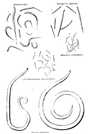
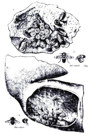
PLATE VI. BOTS 1. Bots in the stomach. 2. Bots in the duodenum.
The disease due to these worms is quite common. The worms enter the body as immature forms in the spring, when the animal is turned out on pasture. The first symptoms show in November or December, the disease being in a latent stage during the development of the worms. The first symptoms are diarrhea, loss of appetite, and emaciation. The animal becomes anemic. Secondary symptoms are edema and such complications as joint infection, colic due to embolism, and accidents from falls, hemorrhage from ruptured aneurisms, or perforation at the site of abscess. The animal may die, recover, or become a chronic sufferer, the internal injuries failing to make a satisfactory recovery even with the removal of the worms in chronic cases.
Treatment calls for the expulsion of the adult worms from the intestine, the development of the body resistance to repair the damage wrought by the developing worms, and the combating of complications. For the expulsion of the worms the use of carbon bisulphid in gelatin capsules, 2 to 5 grams, according to the size of the patient, for five days, followed by magnesium sulphate the sixth day, has been recommended. Owing to the difficulty and danger in the administration of carbon bisulphid in capsule, it is advisable to call in a veterinarian. Tonic treatment consists in the subcutaneous administration of artificial serum and caffein. The various complications of bacterial infection, colic, heart depression, etc., call for the attention of a veterinarian. Preventive measures consist in avoiding reinfection with worms so far as possible by using dry upland pasture in preference to low, wet land, and by rotating pastures or rotation of the stock on a given pasture. Horses may be alternated with cattle, sheep, or hogs to advantage, so far as parasites are concerned. Another feature, always of importance, is the provision of a pure, potable drinking water.
Bots (Gastrophilus spp.).—Bots (Pl. VI) are quite common in the stomach and upper part of the small intestine of the horse anywhere in the United States, one kind being occasionally found in the rectum. They attach to that portion of the mucous lining of the stomach nearest the esophagus or sometimes around the pyloric opening to the intestine or even in the upper intestine, and undoubtedly interfere with the proper functioning of the stomach and the health of the animal to a certain extent. The symptoms are rather vague as a rule, but the general result is a condition of unthriftiness.
A treatment which has been found effective consists in feeding lightly on the day preceding treatment, withholding food in the evening and giving an ounce of Barbados aloes or a pint of linseed oil. The next day give 3 drams of carbon bisulphid in a gelatin capsule at 6 o'clock, repeat the dose at 7 o'clock, and again at 8 o'clock, making a total of 9 drams altogether for an adult horse; half that amount will be sufficient for a yearling colt. As previously noted, there is some little difficulty and danger of accident in the administration of treatments of this character and it is advisable to call in a veterinarian.
Unless destroyed by treatment, the bots in the stomach of the horse pass out in the manure in the spring and burrow down into the soil an inch or two. Here they undergo a certain amount of development and finally emerge as adult flies. These bot flies mate and during the summer the eggs are deposited by the female on the forelegs and shoulders or around the chin, mouth and nostrils of the horse, the location and appearance of the eggs varying somewhat with different species of bot flies. These eggs or the young maggots escaping from them are ingested by the horse in licking the portions irritated by the movement of the escaping maggots, and when swallowed develop to form bots in the stomach. Careful currying, especially around the forequarters, is an aid in keeping down bot infestation, but this is not commonly feasible with horses on pasture, the ones most liable to become infested.