LAMENESS: ITS CAUSES AND TREATMENT. (continued 2)
Reading edition
It is generally, therefore, only fractures of the long bones, and then at points not in close proximity to the trunk, that may be considered to be amenable to reduction. It is true that some of the more superficial bones, as those of the head, of the pelvis, and of the thoracic walls, may in some cases require special manipulations and appliances for their retention in their normal positions; hence the treatment of these and of a fractured leg can not be the same.
The methods of accomplishing reduction vary with the features of each case, the manipulations being necessarily modified to meet different circumstances. If the displacement is in the thickness of the bone, as in transverse fracture, the manipulation of reduction consists in applying constant pressure upon one of the fragments, while the other is kept steady in its place, the object of the pressure being the reestablishment of the exact coincidence of the two bony surfaces. If the displacement has taken place at an angle it will be sufficient in order to effect the reduction to press upon the summit, or apex, of the angle until its disappearance indicates that the parts have been brought into coaptation. This method is often practiced in the treatment of a fractured rib. In a longitudinal fracture, or when the fragments are pressed together by the contraction of the muscles to which they give insertion until they so overlap as to correspond by certain points of their circumference, the reduction is to be accomplished by effecting the movements of extension, counter extension, and coaptation. Extension is accomplished by making traction upon the lower portion of the limb. Counter extension consists in firmly holding or confining the upper or body portion in such manner, that it shall not be affected by the traction applied to the lower part. In other words, the operator, grasping the limb below the fracture, draws it down or away from the trunk, while he seeks not to draw away, but simply to hold the upper portion still until the broken ends of bone are brought to their natural relative positions, when the coaptation, which is thus effected, has only to be made permanent by the proper dressings to perfect the reduction.
In treating fractures in small animals the strength of the hand is usually sufficient for the required manipulations. In the fracture of the forearm of a dog, for example, while the upper segment is firmly held by one hand the lower may be grasped by the other and the bone itself made to serve the purpose of a lever to bring about the desired coaptation. In such case that is sufficient to overcome the muscular contraction and correct the overlapping or other malposition of the bones. If, however, the resistance can not be overcome in this way, the upper segment may be committed to an assistant for the management of the counter extension, leaving to the operator the free use of both hands for the further manipulation of the case.
If the reduction of fractures in small animals is an easy task, however, it is far from being so when the patient is a large animal whose muscular force is largely greater than that of several men combined. In such case resort must be had not only to superior numbers for the necessary force, but in many cases to mechanical aids. A reference to the manner of proceeding in a case of fracture with displacement of the forearm of a horse will illustrate the matter. The patient is first to be carefully cast, on the uninjured side, with ropes or a broad, leather strap about 18 feet long passed under and around his body and under the axilla of the fractured limb and secured at a point opposite to the animal and toward his back. This will form the mechanical means of counter extension. Another rope will then be placed around the inferior part of the leg below the point of fracture, with which to produce extension, and this will sometimes be furnished with a block and pulleys, in order to augment the power when necessary; there is, in fact, always an advantage in their use, on the side of steadiness and uniformity, as well as of increased power. It is secured around the fetlock or the coronet or, what is better, above the knee and nearer the point of fracture, and is committed to assistants. The traction on this should be firm, uniform, and slow, without relaxing or jerking, while the operator carefully watches the process. If the bone is superficially situated he is able, by the eye, to judge of any changes that may occur in the form or length of the parts under traction, and discovering, at the moment of its happening, the restoration of symmetry in the disturbed region he gently but firmly manipulates the place until all appearance of severed continuity has vanished. Sometimes the fact and the instant of restoration are indicated by a peculiar sound or "click" as the ends of the bones slip into contact, to await the next step of the restorative procedure.
The process is the same when the bones are covered with thick muscular masses except that it is attended with greater difficulties from the fact that the finger must be substituted for the eye and taxis must take the place of sight.
It frequently happens that perfect coaptation is prevented by the interposition, between the bony surfaces, of such substances as a small fragment of detached bone or a clot of blood; sometimes the extreme obliquity of the fracture, by permitting the bones to slip out of place, is the opposing cause. These are difficulties which can not always be overcome, even in small-sized animals, and still it is only when they are mastered that a correct consolidation can be looked for. Without it the continuity between the fragments will be by a deformed callus, the union will leave a shortened, crooked, or angular limb, and the animal will be disabled.
If timely assistance can be obtained, and the reduction accomplished immediately after the occurrence of the accident, that is the best time for it, but if it can not be attended to until inflammation has become established and the parts have become swollen and painful, time must be allowed for the subsidence of these symptoms before attempting the operation. A spasmodic, muscular contraction which sometimes interposes a difficulty may be easily overcome by subjecting the patient to general anesthesia, and need not, therefore, cause any loss of time. A tendency to this may also be overcome by the use of sedatives and antiphlogistic remedies.
The reduction of the fracture having been accomplished, the problem which follows is that of retention. The parts which have been restored to their natural position must be kept there, without disturbance or agitation, until the perfect formation of a callus, and it is here that ample latitude exists for the exercise of ingenuity and skill by the surgeon in the contrivance of the necessary apparatus. One of the most important of the conditions which are available by the surgeon in treating human patients is denied to the veterinarian in the management of those which belong to the animal tribes. This is position. The intelligence of the human patient cooperates with the instructions of the surgeon, in the case of the animal sufferer there is a continual antagonism between the parties, and the forced extension and fatiguing position which must for a considerable period be maintained as a condition of restoration require special and effective appliances to insure successful results. To obtain complete immobility is scarcely possible, and the surgeon must be content to reach a point as near as possible to that which is unattainable. For this reason, as will subsequently be seen, the use of slings and the restraint of patients in very narrow stalls is much to be preferred to the practice sometimes recommended of allowing entire freedom of motion by turning them loose in box stalls. Temporary and movable apparatus are not usually of difficult use in veterinary practice, but the restlessness of the patients and their unwillingness to submit quietly to the changing of the dressings render it obligatory to have recourse to permanent and immovable bandages, which should be retained without disturbance until the process of consolidation is complete.
The materials composing the retaining apparatus consist of oakum, bandages, and splints, with an agglutinating compound which forms a species of cement by which the different constituents are blended into a consistent mass to be spread upon the surface covering the locality of the fracture. Its components are black pitch, rosin, and Venice turpentine, blended by heat. The dressing may be applied directly to the skin, or a covering of thin linen may be interposed. A putty made with powdered chalk and the white of egg is recommended for small animals, though a mixture of sugar of lead and burnt alum with the albumen is preferred by others. Another formula is spirits of camphor, Goulard's extract, and albumen. Another recommendation is to saturate the oakum and bandages with an adhesive solution formed with gum arabic, dextrin, flour paste, or starch. This is advised particularly for small animals, as is also the silicate of soda. Dextrin mixed while warm with burnt alum and alcohol cools and solidifies into a stony consistency, and is preferable to plaster of Paris, which is less friable and has less solidity, besides being heavier and requiring constant additions as it becomes older. Starch and plaster of Paris form another good compound.
In applying the dressing the leg is usually padded with a cushion of oakum thick and soft enough to equalize the irregularities of the surface and to form a bedding for the protection of the skin from chafing. Over this the splints are placed. The material for these is, variously, pasteboard, thin wood, bark, laths, gutta-percha, strips of thin metal, as tin or perhaps sheet iron. They should be of sufficient length not only to cover the region of the fracture but to extend sufficiently above and below to render the immobility more nearly complete than in the surrounding joints. The splints, again, are covered with cloth bandages—linen preferably—soaked in a glutinous mixture. These bandages are to be carefully applied, with a perfect condition of lightness. They are usually made to embrace the entire length of the leg in order to avoid the possibility of interference with the circulation of the extremity as well as for the prevention of chafing. They should be rolled from the lower part of the leg upward and carefully secured against loosening. In some instances suspensory bandages are recommended, but except for small animals our experience does not justify a concurrence in the recommendation.
These permanent dressings always need careful watching with reference to their immediate effect upon the region they cover, especially during the first days succeeding that of their application. Any manifestation of pain, or any appearance of swelling above or below, or any odor suggestive of suppuration should excite suspicion, and a thorough investigation should follow without delay. The removal of the dressing should be performed with great care, and especially so if time enough has elapsed since its application to allow of a probability of a commencement of the healing process or the existence of any points of consolidation. With the original dressing properly applied in its entirety in the first instance, the entire extremity will have lost all chance of mobility, and the repairing process may be permitted to proceed without interference. There will be no necessity and there need be no haste for removal or change except under such special conditions as have just been mentioned, or when there is reason to judge that solidification has become perfect, or for the comfort of the animal, or for its readaptation in consequence of the atrophy of the limb from want of use. Owners of animals are often tempted to remove a splint or bandage prematurely at the risk of producing a second fracture in consequence of the failure of the callus properly to consolidate.
The method of applying the splints which we have described refers to the simple variety only. In a compound case the same rules must be observed, with the modification of leaving openings through the thickness of the dressing, opposite the wound, in order to permit the escape of pus and to secure access to the points requiring the application of treatment.
FRACTURE OF CRANIAL BONES.
Fractures of the cranial bones in large animals are comparatively rare, though the records are not destitute of cases. When they occur, it is as the result of external violence, the sufferers being usually run-aways which have come in collision with a wall or a tree or other obstruction, or it may occur in those which in pulling upon the halter have broken it with a jerk and been thrown backward, as may occur in rearing too violently. Under these conditions we have witnessed fractures of the parietal, of the frontal, and of the sphenoid bones. These fractures may be of both the complete and the incomplete kinds, which indeed is usually the case with those of the flat bones, and they are liable to be complicated with lacerations of the skin, in consequence of which they are easily brought under observation. When the fact is otherwise and the skin is intact, however, the diagnosis becomes difficult.
Symptoms.—The incomplete variety may be unaccompanied with any special symptoms, but in the complete kind one of the bony plates may be so far detached as to press upon the cerebral substance with sufficient force to produce serious nervous complications. When the injury occurs at the base of the cranium hemorrhage may be looked for, with paralytic symptoms, and when these are present the usual termination is death. It may happen, however, that the symptoms of an apparently very severe concussion may disappear, resulting in an early and complete recovery, and the surgeon will therefore do well to avoid undue haste in venturing upon a prognosis. In fractures of the orbital or the zygomatic bones the danger is less pressing than with injuries otherwise located about the head.
Treatment.—The treatment of cranial fractures is simple, though involving the best skill of an experienced surgeon. When incomplete hardly any interference is needed; even plain bandaging may usually be dispensed with. In the complete variety the danger to be combated is compression of the brain, and attention to this indication must not be delayed. The means to be employed are the trephining of the skull over the seat of the fracture and the elevation of the depressed bone or the removal of the portion which is causing the trouble. Fragments of bone in comminuted cases, bony exfoliations, collections of fluid, or even protruding portions of the brain substance must be carefully cleansed away and a simple bandage so applied as to facilitate the application of subsequent dressings.
FRACTURES OF THE BONES OF THE FACE.
In respect to their origin—usually traumatic—these injuries rank with the preceding, and are commonly of the incomplete variety. They may easily be overlooked, and may even sometimes escape recognition until the reparative process has been well established and the wound is discovered owing to the prominence caused by the presence of the provisional callus which marks its cure. When the fracture is complete it will be marked by local deformity, mobility of the fragments, and crepitation. Nasal hemorrhage, roaring, frequent sneezing, loosening or loss of teeth, difficulty of mastication, and inflammation of the cavities of the sinuses are varying complications of these accidents. The object of the treatment should be the restoration of the depressed bones as nearly as possible to their normal position and their retention in place by protecting splints, which should cover the entire facial region. Special precautions should be observed to prevent the patient from disturbing the dressing by rubbing his head against surrounding objects, such as the stall, manger, rack, etc. Clots of blood in the nasal passages must be washed out, collections of pus removed from the sinuses, and, if the teeth are loosened and liable to fall out, they should be removed. If roaring is threatened, tracheotomy is indicated.
FRACTURES OF THE PREMAXILLARY BONE.
These are mentioned by continental authors and are usually encountered in connection with fractures of the nasal bone, and may take place either in the width or the length of the bone.
The deformity of the upper lip, which is drawn sidewise in this lesion, renders it easy of diagnosis. The abnormal mobility and the crepitation, with the pain manifested by the patient when undergoing examination, are concurrent symptoms. Looseness of the teeth, abundant salivation, and entire inability to grasp the feed complete the symptomatology of these accidents. In the treatment splints of gutta-percha or leather are sometimes used, but they are of difficult application. Our own judgment and practice are in favor of the union of the bones by means of metallic sutures.
FRACTURES OF THE LOWER JAW.
A fracture here is not an injury of infrequent occurrence. It involves the body of the bone, at its symphysis, or back of it, and includes one or both of its branches, either more or less forward, or at the posterior part near the temporomaxillary articulation, at the coronoid process.
Falls, blows, or other external violence, or powerful muscular contractions during the use of the speculum, may be mentioned among the causes of this lesion. The fracture of the neck, or that portion formed by the juncture of the two opposite sides, and of the branches in front of the cheeks, causes the lower jaw, the true dental arch, to drop, without the ability to raise it again to the upper, and the result is a peculiar and characteristic physiognomy. The prehension and mastication of feed become impossible; there is an abundant escape of fetid and sometimes bloody saliva, especially if the gums have been wounded; there is excessive mobility of the lower end of the jawbone; and there is crepitation, and frequently paralysis of the under lip. Although an animal suffering with a complete and often compound and comminuted fracture of the submaxilla presents at times a serious aspect, the prognosis of the case is comparatively favorable, and recovery is usually only a question of time. The severity of the lesion corresponds in degree to that of the violence to which it is due, also to the resulting complications and the situation of the wound. It is simple when at the symphysis, but becomes more serious when it affects one of the branches, and most aggravated when both are involved. Fracture of the coronoid process becomes important principally as an evidence of the existence of a morbid diathesis, such as osteoporosis, or the like.
The particular seat of the injury, with its special features, will, of course, determine the treatment. For a simple fracture, without displacement, provided there is no laceration of the periosteum, an ordinary supporting bandage will usually be sufficient, but when there is displacement the reduction of the fracture must first be accomplished, and for this special splints are necessary. In a fracture of the symphysis or of the branches the adjustment of the fragments by securing them with metallic sutures is the first step necessary, to be followed by the application of supports, consisting of splints of leather or sheets of metal, the entire front of the head being then covered with bandages prepared with adhesive mixtures. During the entire course of treatment a special method of feeding becomes necessary. The inability of the patient to appreciate the situation, of course, necessitates a resort to an artificial mode of introducing the necessary feed into his stomach; this is accomplished by forcing between the commissures of the lips, in a liquid form, by means of a syringe, the milk or nutritive gruels selected for his sustenance until the consolidation is sufficiently advanced to permit the ingestion of feed of a more solid consistency. The callus will usually be sufficiently hardened in two or three weeks to allow of a change of diet to mashes of cut hay and scalded grain, until the removal of the dressing restores the animal to its old habit of mastication.
FRACTURES OF VERTEBRÆ.
These are not very common, but when they do occur the bones most frequently injured are those of the back and loins.
Causes.—The ordinary causes of fracture are responsible here as elsewhere, such as heavy blows on the spinal column, severe falls while conveying heavy loads, and especially violent efforts in resisting the process of casting. Although occurring more or less frequently under the latter circumstances, the accident is not always attributable to carelessness or error in the management. It may, of course, sometimes result from such a cause as a badly prepared bed, or the accidental presence of a hard body concealed in the straw, or to a heavy fall when the movements of the patient have not been sufficiently controlled by an effective apparatus and its skillful adaptation, but it is quite as liable to be caused by the violent resistance and the consequent powerful muscular contraction by the frightened patient. The simple fact of the overarching of the vertebral column, with excessive pressure against it from the intestinal mass, owing to the spasmodic action of the abdominal muscles, may account for it, and so also may the struggles of the animal to escape from the restraint of the hobbles while frantic under the pain of an operation without anesthesia. In these cases the fracture usually occurs in the body or the annular part, or both, of the posterior dorsal or the anterior lumbar vertebra. When the transverse processes of the last-named bones are injured, it is probably in consequence of the heavy concussion incident to striking the ground when cast. The diagnosis of a fracture of the body of a vertebra is not always easy, especially when quite recent, and more especially when there is no accompanying displacement.
Symptoms.—There are certain peculiar signs accompanying the occurrence of the accident while an operation is in progress which should at once excite the suspicion of the surgeon. In the midst of a violent struggle the patient becomes suddenly quiet; the movement of a sharp instrument, which at first excited his resistance, fails to give rise to any further evidence of sensation; perhaps a general trembling, lasting for a few minutes, will follow, succeeded by a cold, profuse perspiration, particularly between the hind legs, and frequently there will be micturition and defecation. Careful examination of the vertebral column may then detect a slight depression or irregularity in the direction of the spine, and there may be a diminution or loss of sensation in the posterior part of the trunk, while the anterior portion continues to be as sensitive as before. In making an attempt to get upon his feet, however, upon the removal of the hobbles, only the fore part of the body will respond to the effort, a degree of paraplegia being present, and while the head, neck, and fore part of the body will be raised, the hind quarters and hind legs will remain inert. The animal may perhaps succeed in rising and probably may be removed to his stall, but the displacement of the bone will follow, converting the fracture into one of the complete kind, either through the exertion of walking or by a renewed attempt to rise after another fall before reaching his stall. By this time the paralysis is complete, and the extension of the meningitis, which has become established, is a consummation soon reached.
To say that the prognosis of fracture of the body of the vertebra is always serious is to speak very mildly. It would be better, perhaps, to say that occasionally a case may recover. Fractures of the transverse processes are less serious.
Treatment.—Instead of stating the indication in this class of cases as if assuming them to be amenable to treatment, the question naturally would be: Can any treatment be recommended in a fracture of the body of a vertebra? The only indication in such a case, in our opinion, is to reach the true diagnosis in the shortest possible time and to act accordingly. If there is displacement, and the existence of serious lesions may be inferred from the nervous symptoms, the destruction of the suffering animal appears to suggest itself as the one conclusion in which considerations of policy, humanity, and science at once unite.
If, however, it is fairly evident that no displacement exists; that pressure upon the spinal cord is not yet present; that the animal with a little assistance is able to rise upon his feet and to walk a short distance—it may be well to experiment upon the case to the extent of placing the patient in the most favorable circumstances for recovery and allow nature to operate without further interference. This may be accomplished by obtaining immobility of the whole body as much as possible, and especially of the suspected region, by placing the patient in slings, in a stall sufficiently narrow to preclude lateral motion, and covering the loins with a thick coat of agglutinative mixture. Developments should be watched and awaited.
FRACTURE OF THE RIBS.
The different regions of the chest are not equally exposed to the violence that causes fractures of the ribs, and they are therefore either more common or more easily discovered during life at some points than at others. The more exposed regions are the middle and the posterior, while the front is largely covered and defended by the shoulder. A single rib may be the seat of fracture, or a number may be involved, and there may be injuries on both sides of the chest at the same time. It may take place lengthwise, in any part of the bone, though the middle, being the most exposed, is the most frequently hurt. Incomplete fractures are usually lengthwise, involving a portion only of the thickness of one or other of the surfaces. The complete kind may be either transverse or oblique, and are most commonly denticulated. The fracture may be comminuted, and a single bone may show one of the complete and one of the incomplete kind at different points. The extent of surface presented by the thoracic region, with its complete exposure at all points, explains the liability of the ribs to suffer from all the forms of external violence.
Symptoms.—In many instances fractures, especially the incomplete variety, of these bones continue undiscovered, without displacement, though the evidences of local pain, a certain amount of swelling, and a degree of disturbance of the respiration, if noticed during the examination of a patient, may suggest a suspicion of their existence. Abnormal mobility and crepitation are difficult of detection, even when present, and they are not always present. When there is displacement the deformity which it occasions will betray the fact, and when such an injury exists the surgeon, in view of possible and probable complications of thoracic trouble, of course will become vigilant and prepare himself for an encounter with a case of traumatic pleuritis or pneumonia. Fatal injuries of the heart are recorded. Subcutaneous emphysema is a common accompaniment of broken ribs, and I recall the death, from this cause, of a patient of my own which had suffered a fracture of two ribs in the region of the withers, under the cartilages of the shoulder, and of which the diagnosis was made only after the fatal ending of the case.
These hurts are not often of a very serious character, though the union is never so solid and complete as in other fractures, the callus being usually imperfect and of a fibrous character, with an amphiarthrosis formation. Still, complications occur which may impart gravity to the prognosis.
Treatment.—Fractures with but a slight or no displacement need no reduction. All that is necessary is a simple application of a blistering nature as a preventive of inflammation or for its subjugation when present, and in order to excite an exudation which will tend to aid in the support and immobilization of the parts. At times, however, a better effect is obtained by the application of a bandage placed firmly around the chest, although, while this limits the motion of the ribs, it is liable to render the respiration more labored.
If there is displacement, with much accompanying pain and evident irritation of the lungs, the fracture must be reduced without delay. The means of effecting this vary according to whether the displacement is outward or inward. In the first case the bone may be straightened by pressure from without, while in the second the end of it must be raised by a lever, for the introduction of which a small incision through the skin and intercostal spaces will be necessary. When coaptation has been effected it must be retained by the external application of an adhesive mixture, with splints and bandages around the chest.
FRACTURES OF THE BONES OF THE PELVIS.
These fractures will be considered under their separate denominations, as those of the sacrum and the os innominatum, or hip, which includes the subdivisions of the ilium, the pubes, and the ischium.
The sacrum.—Fractures of this bone are rarely met with among solipeds. Among cattle, however, it is of common occurrence, being attributed not only to the usual varieties of violence, as blows and other external hurts, but to the act of coition and violent efforts in parturition. It is generally of the transverse kind and may be recognized by the deformity which it occasions. This is due to the dropping of the bone, with a change in its direction and a lower attachment of the tail, which also becomes more or less paralyzed. The natural and spontaneous relief which usually interposes in these cases has doubtless been observed by the extensive cattle breeders of the West, and their practice and example fully establish the inutility of interference. Still, cases may occur in which reduction may be indicated, and it then becomes a matter of no difficulty. It is effected by the introduction of a round, smooth piece of wood into the rectum as far as the fragment of bone and using it as a lever, resting upon another as a fulcrum placed under it outside. The bone, having been thus returned, may be kept in place by the ordinary external means in use.
The os innominatum.—Fracture of the ilium may be observed either at the angle of the hip or at the neck of the bone; those of the pubes may take place at the symphysis, or in the body of the bone; those of the ischium on the floor of the bone, or at its posterior external angle. Or, again, the fracture may involve all three of these constituent parts of the hip bone by having its situation in the articular cavity—the acetabulum by which it joins the femur or thigh bone.
Symptoms.—Some of these fractures are easily recognized, while others are difficult to identify. The ordinary deformity which characterizes a fracture of the external angle of the ilium, its dropping and the diminution of that side of the hip in width, unite in indicating the existence of the condition expressed by the term "hipped." An incomplete fracture, however, or one that is complete without displacement, or even one with displacement, often demands the closest scrutiny for its discovery. The lameness may be well marked, and an animal may show it but little while walking, though upon being urged into a trot will manifest it more and more, until presently it will cease to use the crippled limb altogether, and travel entirely on three legs. The acute character of the lameness will vary in degree as the seat of the lesion approximates the acetabulum. In walking, the motion at the hip is very limited, and the leg is dragged; while at rest it is relieved from bearing its share in sustaining the body. An intelligent opinion and correct conclusion will depend largely upon a knowledge of the history of the case, and while in some instances that will be but a report of the common etiology of fractures, such as blows, hurts, and other external violence, the simple fact of a fall may furnish in a single word a satisfactory solution of the whole matter.
With the exception of the deformity of the ilium in a fracture of its external angle, and unless there has been a serious laceration of tissues and infiltration of blood, or excessive displacement, there are no very definite external symptoms in a case of a fracture of the hip bone. There is one, however, which, in a majority of cases, will not fail—it is crepitation. This evidence is attainable by both external and internal examination—by manipulation of the gluteal surface and by rectal taxis. Very often a lateral motion, or balancing of the hinder parts by pressing the body from one side to the other, will be sufficient to render the crepitation more distinct—a slight sensation of grating, which may be perceived even through the thick coating of muscle which covers the bone—and the sensation may not only be felt, but to the expert may even become audible. This external manifestation is, however, not always sufficient in itself, and should invariably be associated with the rectal taxis for corroboration. It is true that this may fail to add to the evidence of fracture, but till then the simple testimony afforded by the detection of crepitation from the surface, though a strong confirmatory point, is scarcely sufficiently absolute to establish more than a reasonable probability or strong suspicion in the case.
In addition to the fact that the rectal examination brings the exploring hand of the surgeon into near proximity to the desired point of search, and to an accurate knowledge of the situation of parts, both pro and con as respects his own views, there is another advantage attendant upon it which is well entitled to appreciation. This is the facility with which he can avail himself of the help of an assistant, who can aid him by manipulating the implicated limb and placing it in various positions, so far as the patient will permit, while the surgeon himself is making explorations and studying the effect from within. By this method he can hardly fail to ascertain the character of the fracture and the condition of the bony ends. By the rectal taxis, as if with eyes in the finger ends, he will "see" what is the extent of the fracture of the ilium or of the neck of that bone; to what part of the central portion of the bone (the acetabulum) it reaches; whether this is free from disease or not, and in what location on the floor of the pelvis the lesion is situated. By this method we have frequently been able to detect a fracture at the symphysis, which, from its history and symptoms and an external examination, could only have been guessed at. Yet, with all its advantages, the rectal examination is not always necessary, as, for example, when the fracture is at the posterior and external angle of the ischium, when by friction of the bony ends the surgeon may discern the crepitation without it.
Every variety of complication, including muscular lacerations with the formation of deep abscesses and injuries to the organs of the pelvic cavity, the bladder, the rectum, and the uterus, may be associated with fractures of the hip bone.
Prognosis.—The prognosis of these lesions will necessarily vary considerably. A fracture of the most superficial part of the bone of the ilium or of the ischium, especially if there is little displacement, will unite rapidly, leaving a comparatively sound animal often quite free from subsequent lameness. If there is much displacement, however, only a ligamentous union will take place, with much deformity and more or less irregularity in the gait. Other fractures may be followed by complete disability of the patient, as, for example, when the cotyloid cavity is involved, or when the reparatory process has left bony deposits in the pelvic cavity at the seat of the union, which may, in the case of the female, interfere with the steps of parturition, or induce some local paralysis by pressure upon the nerves which govern the muscles of the hind legs. This is a condition not infrequently observed when the callus has been formed on the floor of the pelvis near the obturator foramen, pressing upon the course or involving the obturator nerve.
Treatment.—In our estimation, the treatment of all fractures of the hip bone should be of the simplest kind. Rendered comparatively immovable by the thickness of the muscles by which the region is enveloped, one essential indication suggests itself, and that is to place the animal in a position which, so far as possible, will be fixed and permanent. For the accomplishment of this purpose the best measure, as we consider it, is to place the horse in a stall of just sufficient width to admit him, and to apply a set of slings, snugly, but comfortably. (See Plate XXXI.) This will fulfill the essential conditions of recovery—rest and immobility. Blistering applications would be injurious, though the adhesive mixture might prove in some degree beneficial.
The minimum period allowable for solid union in a fractured hip is, in our judgment, two months, and we have known cases in which that was too short a time.
As we have said before, there may be cases in which the treatment for fracture at the floor of the pelvis has been followed by symptoms of partial paralysis, the animal, when lying down, being unable to regain his feet, but moving freely when placed in an upright position. This condition is owing to the interference of the callus with the functions of the obturator nerve, which it presses upon or surrounds. By my experience in similar cases I feel warranted in cautioning owners of horses in this condition to exercise due patience, and to avoid a premature sentence of condemnation against their invalid servants; they are not all irrecoverably paralytic. With alternations of moderate exercise, rest in the slings, and the effect of time while the natural process of absorption is taking effect upon the callus, with other elements of change that may be so operating, the horse in due time may become able once more to earn his subsistence and serve his master.
FRACTURE OF THE SCAPULA.
This bone is seldom fractured, its comparative exemption being due to its free mobility and the protection it receives from the superimposed soft tissues. Only direct and powerful causes are sufficient to effect the injury, and when it occurs the large rather than the smaller animals are the subjects.
Cause.—The causes are heavy blows or kicks and violent collisions with unyielding objects. Those which are occasioned by falls are generally at the neck of the bone, and of the transverse and comminuted varieties.
Symptoms.—The diagnosis is not always easy. The symptoms are inability to rest the leg on the ground and to carry weights, and they are present in various degrees from slight to severe. The leg rests upon the toe, seems shortened, and locomotion is performed by jumps. Moving the leg while examining it and raising the foot for inspection seem to produce much pain and cause the animal to rear. Crepitation is readily felt with the hand upon the shoulder when the leg is moved. If the fracture occurs in the upper part of the bone, overlapping of the fragments and displacement will be considerable.
The fracture of this bone is usually classed among the more serious accidents, though cases may occur which are followed by recovery without very serious ultimate results, especially when the seat of the injury is at some of the upper angles of the bone or about the acromion crest. But if the neck and the joint are the parts involved, complications which are likely to disable the animal for life are liable to be present.
Treatment.—If there is no displacement, a simple adhesive dressing to strengthen and immobilize the parts will be sufficient. A coat of black pitch dissolved with wax and Venice turpentine, and kept in place over the region with oakum or linen bands, will be all the treatment required, especially if the animal is kept quiet in the slings.
Displacement can not be remedied, and reduction is next to impossible. Sometimes an iron plate is applied over the parts and retained by bandages, as in the dressing of Bourgelat (Plate XXX); this may be advantageously replaced by a pad of thick leather. In smaller animals the parts are retained by figure-8 bandages, embracing both the normal and the diseased shoulders, crossing each other in the axilla and covered with a coating of adhesive mixture.
FRACTURES OF THE HUMERUS.
These are more common in small than in large animals, and are always the result of external traumatism, such as falls, kicks, and collisions. They are generally very oblique, are often comminuted, and though more usually involving the shaft of the bone will in some cases extend to the upper end and into the articular head.
Symptoms.—There is ordinarily considerable displacement in consequence of the overlapping of the broken ends of the bone, and this of course causes more or less shortening of the limb. There will also be swelling, with difficulty of locomotion, and crepitation will be easy of detection. This fracture is always a serious damage to the patient, leaving him with a permanently shortened limb and an incurable, lifelong lameness.
Treatment.—If treatment is determined on, it will consist in the reduction of the fracture by means of extension and counter extension, to accomplish which the animal must be thrown. If successful in the reduction, then follows the application and adjustment of the apparatus of retention, which must be of the most perfect and efficient kind. Finally, this, however skillfully contrived and carefully adapted, will often fail to effect any good purpose whatever.
FRACTURES OF THE FOREARM.
A fracture in this region may also involve the radius or the ulna, the latter being broken at times in its upper portion above the radio-ulnar arch at the olecranon. If the fracture occurs at any part of the forearm from the radio-ulnar arch down to the knee, it may involve either the radius alone or the radius and the cubitus, which are there intimately united.
Cause.—Besides having the same etiology with most of the fractures, those of the forearm are, nevertheless, more commonly due to kicks from other animals, especially when crowded together in large numbers in insufficient space. It is a matter of observation that under these circumstances fractures of the incomplete kind are those which occur on the inside of the leg, the bone being in that region almost entirely subcutaneous, while those of the complete class are either oblique or transverse. The least common are the longitudinal, in the long axis of the bone.
Symptoms.—This variety of fracture is easily recognized by the appearance of the leg and the different changes it undergoes. There is inability to use the limb; impossibility of locomotion; mobility below the injury; the ready detection of crepitation—in a word, the assemblage of all the signs and symptoms which have been already considered as associated with the history of broken bones.
The fracture of the ulna alone, principally above the radio-ulnar arch, may be ascertained by the aggravated lameness, the excessive soreness on pressure, and perhaps a certain increase of motion, with a very slight crepitation if tested in the usual way. Displacement is not likely to take place except when it is well up toward the olecranon or its tuberosity, the upper segment of the bone being in that case likely to be drawn upward. For a simple fracture of this region there is a fair chance of recovery, but in a case of the compound and comminuted class there is less ground for a favorable prognosis, especially if the elbow joint has suffered injury. A fracture of the ulna alone is not of serious importance, except when the same conditions prevail. A fracture of the olecranon is less amenable to treatment, and promises little better than a ligamentous union.
Treatment.—Considering all the various conditions involving the nature and extent of these lesions, the position and direction of the bones of the forearm are such as to render the chances for recovery from fracture as among the best. The reduction, by extension and counterextension; the maintenance of the coaptation of the segments; the adaptation of the dressing by splints, oakum, and agglutinative mixtures; in fact, all the details of treatment may be here fulfilled with a degree of facility and precision not attainable in any other part of the organism. An important, if not an essential, point, however, must be emphasized in regard to the splints. Whether they are of metal, wood, or other material, they should reach from the elbow joint to the ground, and should be placed on the posterior face and on both sides of the leg. This is then to be so confined in a properly constructed box as to preclude all possibility of motion, while yet it must sustain a certain portion of the weight of the body. The iron splint (represented in Plate XXX) recommended by Bourgelat is designed for fractures of the forearm, of the knee, and of the cannon bone, and will prove to be an appliance of great value. For small animals the preference is for an external covering of gutta-percha, embracing the entire leg. A sheet of this substance of suitable thickness, according to the size of the animal, softened in lukewarm water, is, when sufficiently pliable, molded on the outside of the leg, and when suddenly hardened by the application of cold water forms a complete casing sufficiently rigid to resist all motion. Patients treated in this manner have been able to use the limb freely, without pain, immediately after the application of the dressing. The removal of the splint is easily effected by cutting it away, either wholly or in sections, after softening it by immersing the leg in a warm bath.
FRACTURE OF THE KNEE.
This accident, happily, is of rare occurrence, but when it takes place is of a severe character, and always accompanied with synovitis, with disease of the joint.
Cause.—It may be caused by falling upon a hard surface, and is usually compound and comminuted. Healing seldom occurs, and when it does there is usually a stiffness of the joint from arthritis.
Symptoms.—As a result of this fracture there is inability to bear weight on the foot. The leg is flexed as in complete radial paralysis, or fracture of the ulna. There is abnormal mobility of the bones of the knee, but crepitation is usually absent.
Prognosis.—Healing is hard to effect, as one part of the knee is drawn upward by the two flexor muscles which separate it from the lower part. The callus which forms is largely fibrous, and if the animal is put to work too quickly this callus is liable to rupture. In favorable cases healing takes place in two or three months. Many horses during the treatment develop founder, with consequent drop sole in the sound leg, as a result of pressure due to continuous standing.
Treatment.—Place the animal in the slings, bring the pieces of bone together if possible, and try to keep them in place by a tight plaster-of-Paris dressing about the leg, extending down to the fetlock. Place the animal in a roomy box stall well provided with bedding so that he can lie down, to prevent founder.
FRACTURE OF THE FEMUR.
The protection which this bone receives from the large mass of muscles in which it is enveloped does not suffice to invest it with immunity in regard to fractures.
Cause.—It contributes its share to the list of accidents of this description, sometimes in consequence of external violence and sometimes as the result of muscular contraction; sometimes it takes place at the upper extremity of the bone; sometimes at the lower; sometimes at the head, when the condyles become implicated; but it is principally found in the body or diaphysis. The fracture may be of any of the ordinary forms, simple or compound, complete or incomplete, transverse or oblique, etc. A case of the comminuted variety is recorded in which 85 fragments of bone were counted and removed.
The thickness of the muscular covering sometimes renders the diagnosis difficult by interfering with the manipulation, but the crepitation test is readily available, even when the swelling is considerable, and which is liable to be the case as the result of the interstitial hemorrhage which naturally follows the laceration of the blood vessels of the region involved.
Symptoms.—If the fracture is at the neck of the bone the muscles of that region (the gluteal) are firmly contracted, and the leg seems to be shortened in consequence. Locomotion is impossible. There is intense pain and violent sweating at first. Crepitation may in some cases be discerned by rectal examination, with one hand resting over the coxo-femoral (hip) articulation. Fractures of the tuberosities of the upper end of the bone, the great trochanter, may be identified by the deformity, the swelling, the impossibility of rotation, and the dragging of the leg in walking. Fracture of the body is always accompanied with displacement, and as a consequence a shortening of the leg, which is carried forward. The lameness is excessive, the foot being moved, both when raising it from the ground and when setting it down, very timidly and cautiously. The manipulations for the discovery of crepitation always cause much pain. Lesions of the lower end of the bone are more difficult to diagnosticate with certainty, though the manifestation of pain while making heavy pressure upon the condyles will be so marked that only crepitation will be needed to turn a suspicion into a certainty.
Treatment.—The question as to treatment in fractures of this description resolves itself into the query whether any treatment can be suggested that will avail anything practically as a curative measure; whether, upon the hypothesis of reduction as an accomplished fact, any permanent or efficient device as a means of retention is within the scope of human ingenuity. If the reduction were successfully performed, would it be possible to keep the parts in place by any known means at our disposal? At the best the most favorable result that could be anticipated would be a reunion of the fragments with a considerable shortening of the bone and a helpless, limping, crippled animal to remind us that for human achievement there is a "thus far and no farther."
In small animals, such as dogs and cats, however, attempts at treatment are justifiable, and we are convinced that in many cases of difficulty in the application of splints and bandages a patient may be placed in a condition of undisturbed quiet and left to the processes of nature for "treatment" as safely and with as good an assurance of a favorable result as if he had been subjected to the most heroic secundum artem doctoring known to science. As a case in point, mention may be made of the case of a pregnant bitch which suffered a fracture of the upper end of the femur by being run over by a light wagon. Her "treatment" consisted in being tied up in a large box and let alone. In due time she was delivered of a family of puppies, and in three weeks she was running in the streets, limping very slightly, and nothing the worse for her accident.
FRACTURE OF THE PATELLA.
This, fortunately, is a rare accident, and can result only from direct violence, as a kick or other blow. The lameness which follows it is accompanied with enormous tumefaction of the joint, pain, inability to bear weight upon the foot, and finally disease of the articulation. Crepitation is absent, because the hip muscles draw away the upper part of the bone. The prognosis is unavoidably adverse, destruction being the only termination of this incurable and very painful injury. Most of the reported cases of cures are based upon a wrong diagnosis.
FRACTURES OF THE TIBIA.
Of all fractures these are probably more frequently encountered than any others among the class of accidents we are considering. As with injuries of the forearm of a like character, they may be complete or incomplete; the former when the bone is broken in the middle or at the extremities, and transverse, oblique, or longitudinal. The incomplete kind are more common in this bone than in any other.
Symptoms.—Complete fractures are easy to recognize, either with or without displacement. The animal is very lame, and the leg is either dragged or held clear from the ground by flexion at the stifle, while the lower part hangs down. Carrying weight or moving backward is impossible. There is excessive mobility below the fracture, and well-marked crepitation. If there is much displacement, as in an oblique fracture, there will be considerable shortening of the leg.
While incomplete fractures can not be recognized in the tibia with any greater degree of certainty than in any other bone, there are some facts associated with them by which a diagnosis may be justified. The hypothetical history of a case may serve as an illustration:
An animal has received an injury by a blow or a kick on the inside of the bone, perhaps without showing any mark. Becoming very lame immediately afterwards, he is allowed a few days' rest. If taken out again, he seems to have recovered his soundness, but within a day or two he betrays a little soreness, and this increasing he becomes very lame again, to be furloughed once more, with the result of a temporary improvement, and again a return to labor and again a relapse of the lameness; and this alternation seems to be the rule. The leg being now carefully examined, a local periostitis is readily discovered at the point of the injury, the part being warm, swollen, and painful. What further proof is necessary? Is it not evident that a fracture has occurred, first superficial—a mere split in the bony structure, which, fortunately, has been discovered before some extra exertion or a casual misstep had developed it into one of the complete kind, possibly with complications? What other inference can such a series of symptoms thus repeated establish?
The prognosis of fracture of the tibia, as a rule, must be unfavorable.
Treatment.—The difficulty of obtaining a union without shortening, and consequently without lameness, is proof of the futility of ordinary attempts at treatment, but though this may be true in respect to fractures of the complete kind, it is not necessarily so with the incomplete variety, and with this class the simple treatment of the slings is all that is necessary to obtain consolidation. A few weeks of this confinement will be sufficient.
With dogs and other small animals there are cases which may be successfully treated. If the necessary dressings can be successfully applied and retained, a cure will follow.
FRACTURES OF THE HOCK.
Injuries of the astragalus which had a fatal termination have been recorded. Fractures of the os calcis have also been observed, but never with a favorable prognosis, and attempts to induce recovery, as might have been expected, have proved futile.
FRACTURES OF THE CANNON BONES.
Whether these occur in the fore or hind legs, they appear either in the body or near their extremities. If in the body as a rule the three metacarpal or metatarsal bones are affected, and the fracture is generally transverse and oblique. On account of the absence of soft tissue and tightness of the skin, the broken bones pierce the skin and render the fracture a complicated one. The diagnosis is easy when all the bones are completely broken, but the incomplete fracture can be only suspected.
Symptoms.—There is no displacement, but excessive mobility, crepitation, inability to sustain weight, and the leg is kept off the ground by the flexion of the upper joint.
No region of the body affords better facilities for the application of treatment, and the prognosis on this account is usually favorable. We recall a case, however, which proved fatal, though under exceptional circumstances. The patient was a valuable stallion of highly nervous organization, with a compound fracture of one of the cannon bones, and his unconquerable resistance to treatment, excited by the intense pain of the wound, precluded all chance of recovery, and ultimately caused his death.
Treatment.—The general form of treatment for these lesions will not differ from that which has been already indicated for other fractures. Reduction, sometimes necessitating the casting of the patient; coaptation, comparatively easy by reason of the subcutaneous situation of the bone; retention, by means of splints and bandages—applied on both sides of the region, and reaching to the ground as in fractures of the forearm—these are always indicated. We have obtained excellent results by the use of a mold of thick gutta-percha, composed of two sections and made to surround the entire lower part of the leg as in an inflexible case.
FRACTURE OF THE FIRST PHALANX.
The hind extremity is more liable than the fore to this injury. It is usually the result of a violent effort, or of a sudden misstep or twisting of the leg, and may be transverse, or, as has usually been the case in our experience, longitudinal, extending from the upper articular surface down to the center of the bone, and generally oblique and often comminuted. The symptoms are the swelling and tenderness of the region, possibly crepitation; a certain abnormal mobility; an excessive degree of lameness, and in some instances a dropping back of the fetlock, with perhaps a straightened or upright condition of the pastern.
The difficulty of reduction and coaptation in this accident, and the probability of bony deposits, as of ringbones, resulting in lameness, are circumstances which tend to discourage a favorable prognosis.
The treatment is that which has been recommended for all fractures, so far as it can be applied. The iron splint which has been mentioned gives excellent results in many instances, but if the fracture is incomplete and without displacement, a form of treatment less energetic and severe should be attempted. One case is within our knowledge in which the owner lost his horse by his refusal to subject the animal to treatment, the post-mortem revealing only a simple fracture with very slight displacement.
FRACTURES OF THE SECOND PHALANX (CORONET).
Though these are generally of the comminuted kind, there are often conditions associated with them which justify the surgeon in attempting their treatment. Though crepitation is not always easy to detect, the excessive lameness, the soreness on pressure, the inability to carry weight, the difficulty experienced in raising the foot, all these suggest, as the solution of the question of diagnosis, the fracture of the coronet, with the accompanying realization of the fact that there is yet, by reason of the situation of the member, immobilized as it is by its structure and its surroundings, room left for a not unfavorable prognosis. Only a slight manipulation will be needed in the treatment of this lesion. To render the immobility of the region more fixed, to support the bones in their position by bandaging, and to establish forced immobility of the entire body with the slings is usually all that is required. Ringbone, being a common sequela of the reparative process, must receive due attention subsequently. One of the severest complications liable to be encountered is an immobile joint (anchylosis). Neurectomy of the median nerve may relieve lameness after a fracture of the phalanges.
FRACTURES OF THE THIRD PHALANX (OS PEDIS).
These lesions may result from a penetrating street nail, or follow plantar or median neurectomy. In the latter instance it is caused by the animal setting the foot down carelessly and too violently, and partly due to degeneration of bone tissue which follows nerving.
Though these fractures are not of very rare occurrence, their recognition is not easy, and there is more of speculation than of certainty pertaining to their diagnosis. The animal is very lame and spares the injured foot as much as possible, sometimes resting it upon the toe alone and sometimes holding it from the ground. The foot is very tender, and the exploring pinchers of the examining surgeon cause much pain. During the first 24 hours there is no increased pulsation in the digital and plantar arteries, but on the second day it is apparent.
There is nothing to encourage a favorable prognosis, and a not unusual termination is an anchylosis with either the navicular bone or the coronet.
No method of treatment needs to be suggested here, the hoof performing the office of retention unaided. Local treatment by baths and fomentations will do the rest. It may be months before there is any mitigation of the lameness.
An ultimate recovery depends to a great extent upon whether the other foot can support the weight during the healing process without causing a drop sole in the supporting foot.
FRACTURE OF THE SESAMOID BONES.
This lesion has been considered by veterinarians, erroneously, we think, as one of rare occurrence. We believe it to be more frequent than has been supposed. Many observations and careful dissections have convinced us that fractures of these little bones have been often mistaken for specific lesions of the numerous ligaments that are implanted upon their superior and inferior parts, and which have been described as a "giving way" or "breaking down" of these ligaments. In my post-mortem examinations I have always noted the fact that when the attachments of the ligaments were torn from their bony connections minute fragments of bony structure were also separated, though we have failed to detect any diseased process of the fibrous tissue composing the ligamentous substance.
Cause.—From whatever cause this lesion may arise, it can hardly be considered as of a traumatic nature, no external violence having any apparent agency in producing it, and it is our belief that it is due to a peculiar degeneration or softening of the bones themselves, a theory which acquires plausibility from the consideration of the spongy consistency of the sesamoids. The disease is a peculiar one, and the suddenness with which different feet are successively attacked, at short intervals and without any obvious cause, seems to prove the existence of some latent, morbid cause which has been unsuspectedly incubating. It is not peculiar to any particular class of horses, nor to any special season of the year, having fallen under our observation in each of the four seasons.
Symptoms.—The general fact is reported in the history of most cases that it makes its appearance without premonition in animals which, after enjoying a considerable period of rest, are first exercised or put to work, though in point of fact it may manifest itself while the horse is still idle in his stable. A hypothetical case, in illustration, will explain our theory: An animal which has been at rest in his stable is taken out to work, and it will be presently noticed that there is something unusual in his movement. His gait is changed, and he travels with short, mincing steps, without any of his accustomed ease and freedom. This may continue until his return to the stable, and then, after being placed in his stall, he will be noticed shifting his weight from side to side and from one leg to another, continuing the movement until rupture of the bony structure takes place. But it may happen that the lameness in one or more of the extremities, anterior or posterior, suddenly increases, and it becomes evident that the rupture has taken place in consequence of a misstep or a stumble while the horse is at work. Then, upon coming to a standstill, he will be found with one or more of his toes turned up; he is unable to place the affected foot flat on the ground. The fetlock has dropped and the leg rests upon this part, the skin of which may have remained intact or may have been more or less extensively lacerated. It seldom happens that more than one toe at a time will turn up, yet still the lesion in one will be followed by its occurrence in another. Commonly two feet, either the anterior or posterior, are affected, and we recall one case in which the two fore and one of the hind legs were included at the same time. The accident, however, is quite as liable to happen while the horse is at rest in his stall, and he may be found in the morning standing on his fetlocks. One of the earliest of the cases occurring in my own experience had been under care for several weeks for suspected disease of the fetlocks, the nature of which had not been made out, when, apparently improved by the treatment which he had undergone, the patient was taken out of the stable to be walked a short distance into the country, but had little more than started when he was called to a halt by the fracture of the sesamoids of both fore legs.
While there are no positive premonitory symptoms of these fractures known, we believe that there are signs and symptoms which come but little short of being so, and the appearance of which will always justify a strong suspicion of the truth of the case. These have been indicated when referring to the soreness in standing, the short, mincing gait, and the tenderness betrayed when pressure is made over the sesamoids on the sides of the fetlock, with others less tangible and definable.
Prognosis.—These injuries can never be accounted less than serious, and in our judgment will never be other than fatal. If our theory of their pathology is the correct one, and the cause of the lesions is truly the softening of the sesamoidal bony structure and independent of any changes in the ligamentous fibers, the possibility of a solid osseous union can hardly be considered admissible.
Treatment.—In respect to the treatment to be recommended and instituted it can be employed only with any rational hope of benefit during the incubation, and with the anticipatory purpose of prevention. It must be suggested by a suspicion of the verities of the case, and applied before any rupture has taken place. To prevent this and to antagonize the causes which might precipitate the final catastrophe—the elevation of the toes—resort must be had to the slings and to the application of firm bandages or splints, perhaps of plaster of Paris, with a high shoe, as about the only indications which science and nature are able to offer. When the fracture is an occurred event, and the toes, one or more, are turned up, any further resort to treatment will be futile.
DISEASES OF JOINTS.
Three classes of injury will be considered under this head. These are, affections of the synovial sacs, those of the joint structures, or of the bones and their articular surfaces, and those forms of solution of continuity known as dislocations or luxations.
DISEASES OF THE SYNOVIAL SACS.
Two forms of affection here present themselves, one being the result of an abnormal secretion which induces a dropsical condition of the sac without any acute, inflammatory action, while the other is characterized by excessive inflammatory symptoms, with their modifications, constituting synovitis.
SYNOVIAL DROPSIES.
We have already considered in a general way the presence of these peculiar oil bags in the joints, and in some regions of the legs where the passage of the tendons takes place, and have noticed the similarity of structure and function of both the articular and the tendinous bursæ, as well as the etiology of their injuries and their pathological history, and we will now treat of the affections of both.
WINDGALLS.
This name is given to the dilated bursæ found at the posterior part of the fetlock joint. They have their origin in a dropsical condition of the bursæ of the joint itself, also of the tendon which slides behind it, and are therefore further known by the designations of articular and tendinous windgalls, or puffs. (See also p. 401.)
They appear in the form of soft and somewhat symmetrical tumors, of varying dimensions, and generally well defined in their circumference. They are more or less tense, according to the quantity of secretion they contain, apparently becoming softer as the foot is raised and the fetlock flexed. Usually they are painless and only cause lameness under certain conditions, as when they begin to develop themselves under the stimulus of inflammatory action, or when large enough to interfere with the functions of the tendons, or again when they have undergone certain pathological changes, such as calcification, which is among their tendencies.
Cause.—Windgalls may be attributed to external causes, such as severe labor or strains resulting from heavy pulling, fast driving, or jumping, or they may be among the sequelæ of internal disorders, such as strangles or the resultants of a pleuritic or pneumonic attack.
Unnecessary anxiety is sometimes experienced respecting these growths, with much questioning touching the expediency of their removal, all of which might be spared, for, while they constitute a blemish, their unsightliness will not hinder the usefulness of the animal, and in any case they rarely fail to show themselves easily amenable to treatment.
Treatment.—When in their acute stage, and when the dropsical condition is not excessive, the inflammation may be checked during the day by continuous, cold-water irrigation by means of a hose or soaking tub and at night by applying a moderately tight-roller bandage. Later absorption may be promoted by a Priessnitz bandage,[2] pressure by roller bandages, sweating, the use of liniments, or if necessary by a sharp blister of biniodid of mercury. This treatment should subdue the inflammation, abate the soreness, absorb the excess of secretion, strengthen the walls of the sac, and finally cause the windgalls to disappear, provided the animal is not too quickly returned to labor and exposed to the same factors that occasioned them at first.
If the inflammation has become chronic, however, and the enlargement has been of considerable duration, the negative course will be the wiser one. If any benefit results from treatment it will be of only a transient kind, the dilatation returning when the patient is again subjected to labor, and it will be a fortunate circumstance if inflammation has not supervened.
Notwithstanding the generally benignant nature of the swelling there are exceptional cases, usually when it is probably undergoing certain pathological changes, which may result in lameness and disable the animal, in which case surgical treatment will be indicated, especially if repeated blisters have failed to improve the symptoms. Line firing is then a preeminent suggestion, and many a useful life has received a new lease as the result of this operation timely performed. Another method of firing, which consists in emptying the sac by means of punctures through and through, made with a red-hot needle or wire, and the subsequent injection of certain irritating and alterative compounds into the cavity, designed to effect its closure by exciting adhesive inflammation, such as tincture of iodin, may be commended. But they are all too active and energetic in their effects and require too much special attention and intelligent management to be trusted to any hands other than those of an expert veterinarian.
BLOOD SPAVIN, BOG SPAVIN, AND THOROUGHPIN.
The blood spavin is situated in front and to the inside of the hock and is merely a varicose or dilated condition of the saphena vein. It occurs directly over the point where the bog spavin is found, and has thus been frequently confused with the latter.
The complicated arrangement of the hock joint, and the powerful tendons which pass on the posterior part, are lubricated with the product of secretion from one tendinous synovial and several articular synovial sacs. A large articular sac contributes to the lubrication of the shank bone (the tibia) and one of the bones of the hock (the astragalus). The tendinous sac lies back of the articulation itself and extends upward and downward in the groove of that joint through which the flexor tendons slide. The dilatation of this articular synovial sac is what is denominated bog spavin, the term thoroughpin being applied to the dilatation of the tendinous capsule.
The bog spavin is a round, smooth, well-defined, fluctuating tumor situated in front and a little inward of the hock. On pressure it disappears at this point to reappear on the outside and just behind the hock. If pressed to the front from the outside it will then appear on the inside of the hock. On its outer surface it presents a vein which is quite prominent, running from below upward, and it is to the preternatural dilatation of this blood vessel that the term blood spavin is applied.
The thoroughpin is found at the back and on the top of the hock in that part known as the "hollows," immediately behind the shank bone. It is round and smooth, but not so regularly formed as the bog spavin, and is most apparent when viewed from behind. The swelling is usually on both sides and a little in front of the so-called hamstring, but may be more noticeable on the inside or on the outside.
In their general characteristics bog spavins and thoroughpins are similar to windgalls, and one description of the origin, symptoms, pathological changes, and treatment will serve for all equally, except that it is possible for a bog spavin to cause lameness, and thus to involve a verdict of unsoundness in the patient, a circumstance which will, of course, justify its classification by itself as a severer form of a single type of disease.
We have already referred to the subject of treatment and the means employed—rest, of course—with liniments, blisters, etc., and what we esteem as the most active and beneficial of any, early, deep, and well-performed cauterization. There are, besides, commendatory reports of a form of treatment by the application of pressure pads and peculiar bandages upon the hocks, and it is asserted that the removal of the tumors has been effected by their use. Our experience with this apparatus, however, has not been accompanied with such favorable results as would justify our indorsement of the flattering representations which have sometimes appeared in its behalf.
OPEN JOINTS, BROKEN KNEES, SYNOVITIS, AND ARTHRITIS.
The close relationship which exists among these several affections, their apparently possible connection as successive developments of a similar, if not an essentially identical, origin, together with the advantage gained by avoiding frequent repetitions in the details of symptoms, treatment, etc., are our reasons for treating under a single head the ailments we have grouped together in the present section.
Cause.—The great, comprehensive, common cause of, sometimes permanent, sometimes only transient, disability of the horse is external traumatism.
Blows, bruises, hurts by nearly every known form of violence, falls, kicks, lacerations, punctures—we may add compulsory speed in racing and cruel overloading of draft animals—cover the entire ground of causation of the diseases and injuries of the joints now receiving our consideration.
In one case, a working horse making a misstep stumbles, and falling on his knees receives a hurt, variously severe, from a mere abrasion of the skin to a laceration, a division of the tegument, a slough, mortification, and the escape of the synovial fluid, with or without exposure of the bones and their articular cartilages.
In another case, an animal, from one cause or another, perhaps an impatient temper, has formed the habit of striking or pawing his manger with his fore feet until inflammation of the knee joint is induced, first as a little swelling, diffused, painless; then as a periostitis of the bones of the knee; later as bony deposits, then lameness, and finally the implication of the joint, with all the various sequelæ of chronic inflammation of the knee joint.
In another case, a horse has received a blow with a fork from a careless hostler on or near a joint, or has been kicked by a stable companion, with the result of a punctured wound, at first mild-looking, painless, apparently without inflammation, and not yet causing lameness, but which, in a few hours, or it may be only after a few days, becomes excessively painful, grows worse, the entire joint swells, presently discharges, and at last a case of suppurative synovitis is presented, with perhaps disease of the joint proper, and arthritis as a climax. The symptoms of articular injuries vary not only in the degrees of the hurt but in the nature of the lesion.
Or the condition of broken knees, resulting as we have said, may have for its starting point a mere abrasion of the skin—a scratch, apparently, which disappears without a scar. The injury may, however, have been more severe, the blow heavier, the fall aggravated by occurring upon an irregular surface, or sharp or rough object, with tearing or cutting of the skin, and this laceration may remain. A more serious case than the first is now brought to our notice.
Another time, immediately following the accident, or possibly as a sequel of the traumatism, the tendinous sacs may be opened, with the escape of the synovia, or, worse, the tendons which pass in front of the knee are torn, the inflammation spreads, the joint and leg are swollen, the animal is becoming very lame; synovitis has set in. With this the danger becomes very great, for soon suppuration will be established, then the external coat of the articulation proper becomes ulcerated, if it is not already in that state, and we find ourselves in the presence of an open joint with suppurative synovitis—that is, with the worst among the conditions of diseased processes, because of the liability of the suppuration to become infiltrated into every part of the joint, macerating the ligaments and irritating the cartilages, soon to be succeeded by their ulceration, with the destruction of the articular surface—or the lesion of ulcerative arthritis, one of the gravest among all the disorders known to the animal economy.
Ulcerative arthritis and suppurative synovitis may be developed otherwise than in connection with open joints; the simplest and apparently most harmless punctures may prove to be sufficient cause. For example, a horse may be kicked, perhaps, on the inside of the hock; there is a mark and a few drops of blood to indicate the spot; he is put to work apparently free from pain or lameness and performs his task with his usual ease and facility. On the following morning, however, the hock is found to be a little swollen and there is some stiffness. A little later on he betrays a degree of uneasiness in the leg, and shrinks from resting his weight upon it, moving it up and down for relief. The swelling has increased and is increasing; the pain is severe; and finally, at the spot where the kick inpinged, there is an oozing of an oily liquid mixed with whitish drops of suppuration. The mischief is done; a simple, harmless, punctured wound has expanded into a case of ulcerative arthritis and suppurative synovitis.
Prognosis.—From ever so brief and succinct description of this traumatism of the articulations, the serious and important character of these lesions, irrespective of which particular joint is affected, will be readily understood. Yet there will be modifications in the prognosis in different cases, in accordance with the peculiarities of structure in the joint specially involved, as, for example, it is obvious that a better result may be expected from treatment when but a single joint, with only its plain articular surfaces, is the place of injury, than in one which is composed of several bones, united in a complex formation, as in the knee or hock. As severe a lesion as suppurative synovitis always is, and as frequently fatal as it proves to be, still cases arise in which, the inflammation assuming a modified character and at length subsiding, the lesion terminates favorably and leaves the animal with a comparatively sound and useful joint. There are cases, however, which terminate in no more favorable a result than the union of the bones and occlusion of the joint, to form an anchylosis, which is scarcely a condition to justify a high degree of satisfaction, as it insures a permanent lameness with very little capacity for usefulness.
Appreciating now the dangers associated with all wounds of articulations, however simple and apparently slight, and how serious and troublesome are the complications which are liable to arise during their progress and treatment, we are prepared to understand and realize the necessity and the value of early and prompt attention upon their discovery and diagnosis.
Treatment.—For simple bruises, like those which appear in the form of broken knees or of carpitis, simple remedies, such as warm fomentations or cold-water applications and compresses of astringent mixtures, suggest themselves at once. Injuries of a more complicated character, as lacerations of the skin or tearing of soft structures, will also be benefited by simple dressings with antiseptic mixtures, as those of the carbolic-acid order. The escape of synovia should suggest the prompt use of collodion dressings to check the flow and prevent the further escape of the fluid. But if the discharge is abundant and heavily suppurative, little can be done more than to put in practice the "expectant" method with warm fomentations, repeatedly applied, and soothing, mucilaginous poultices. Improvement, if any is possible, will be but slow to manifest itself. The most difficult of all things to do, in view of varying interests and opinions—that is, in a practical sense—is to abstain from "doing" entirely, and yet in the cases we are considering we are firmly convinced that noninterference is the best and wisest policy.
In cases which are carried to a successful result the discharge will diminish by degrees, the extreme pain will gradually subside, the convalescent will begin timidly to rest his foot upon the ground, and presently to bear weight upon it, and perhaps, after a long and tedious process of recuperation, he may be returned to his former and normal condition of usefulness. When the discharge has wholly ceased and the wounds are entirely healed, a blister covering the whole of the joint for the purpose of stimulating the absorption of the exudation will be of great service. If, on the contrary, there is no amelioration of symptoms and the progress of the disease resists every attempt to check it; if the discharge continues to flow not only without abatement but in an increased volume, and not alone by a single opening but by a number of fistulous tracts which have successively formed; if it seems evident that this drainage is rapidly and painfully sapping the suffering animal's vitality, and a deficient vis vitæ fails to cooperate with the means of cure—all rational hope of recovery may be finally abandoned. Any further waiting for chances, or time lost in experimenting, will be mere cruelty and there need be no hesitation concerning the next step. The poor beast is under sentence of death, and every consideration of interest and of humanity demands an anticipation of nature's evident intent in the quick and easy execution of the sentence.
PLATE XXX. DISLOCATION OF SHOULDER AND ELBOW Bourgelat's apparatus
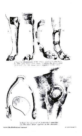
PLATE XXXI. THE SLING IN USE
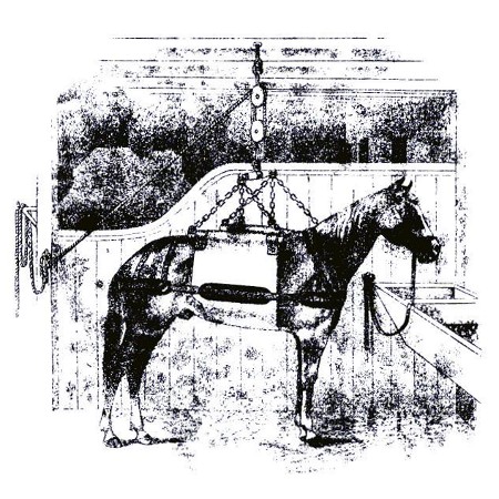
One of the essentials of treatment, and probably an indispensable condition when recovery is in any wise attainable, is the suspension of the patient in slings. He should be continued in them so long as he can be made to submit quietly to their restraint.
DISLOCATIONS.
Dislocations and luxations are interchangeable terms, meaning the separation and displacement of the articulating surfaces of the bones entering into the formation of a joint. This injury is rarely encountered in our large animals on account of the combination of strength and solidity in the formation of their joints. It is met with but seldom in cattle and less so in horses, while dogs and smaller animals are more often the sufferers.
Cause.—The accident of a luxation is less often encountered in the animal races than in man. This is not because the former are less subject to occasional violence involving powerful muscular contractions, or are less often exposed to casualties similar to those which result in luxations in the human skeleton, but because it requires the cooperation of conditions—anatomical, physiological, and perhaps mechanical—present in the human race and lacking in the others, which, however, can not in every case be clearly defined. Perhaps the greater relative length of the bony levers in the human formation may constitute a cause of the difference.
Among the predisposing causes in animals may be enumerated caries of articular surfaces, articular abscesses, excessive dropsical conditions, degenerative softening of the ligaments, and any excessive laxity of the soft structures.
Symptoms and diagnosis.—Three signs of dislocation must usually be taken into consideration. They are: (1) An alteration in the shape of the joint and in the normal relationship of the articulating surfaces; (2) an alteration in the length of the limb, either shortening or lengthening; (3) an alteration in the movableness of the joint, usually an unnatural immobility. Only the first, however, can be relied upon as essential. Luxations are not always complete; they may be partial; that is, the articulating surfaces may be displaced but not separated. In such cases several symptoms may not be present. And not only may the third sign be absent, but the mobility of the first be greatly increased when the character of the injury has been such as to produce extensive lacerations of the articular ligaments.
In addition to the above signs, a luxation is usually characterized by pain, swelling, hemorrhage beneath the skin from damaged or ruptured blood vessels, and even paralysis, when important nerves are pressed on by the displaced bones.
Sometimes a bone is fractured in the immediate vicinity of a joint. The knowledge of this fact requires us to be able to diagnose between a dislocation and such a fracture. In this we generally have three points to assist us: (1) The immobility of a dislocated joint as against the apparently remarkable freedom of movement in fracture; (2) in a dislocation there is no true crepitus—that peculiar grating sensation heard as well as felt on rubbing together the rough ends of fractured bones; however, it must be remembered that in a dislocation two or three days old the inflammatory changes around the joint may give rise to a crackling sensation similar to that in fracture; (3) as a rule, in luxations, if the ligamentous and muscular tissues about the joint are not badly torn, the displacement, when reduced, does not recur.
Prognosis.—The prognosis of a luxation is comparatively less serious than that of a fracture, though at time the indications of treatment may prove to be so difficult to apply that complications of a very severe character may arise.
Treatment.—The treatment of luxations must, of course, be similar to that of fractures. Reduction, naturally, will be the first indication in both cases, and the retention of the replaced parts must follow. The reduction involves the same steps of extension and counter extension, performed in the same manner, with the patient subdued by anesthetics.
The difference between the reduction of a dislocation and that of a fracture consists in the fact that in the former the object is simply to restore the bones to their true, normal position, with each articular surface in exact contact with its companion surface, the apparatus necessary afterwards to keep them in situ being similar to that which is employed in fracture cases, and which will usually require to be retained for a period of from 40 to 50 days, if not longer, before the ruptured retaining ligaments are sufficiently firm to be trusted to perform their office unassisted. A variety of manipulations are to be used by the surgeon, consisting in pushing, pulling, pressing, rotating, and, indeed, whatever movement may be necessary, until the bones are forced into such relative positions that the muscular contraction, operating in just the right directions, pulls the opposite matched ends together in true coaptation—a head into a cavity, an articular eminence into a trochlea, as the case may be. The "setting" is accompanied with a peculiar, snapping sound, audible and significant, as well as a visible return of the surface to its normal symmetry.
Special dislocations.—While all the articulations of the body are liable to this form of injury, there are three in the large animals which may claim a special consideration, viz:
THE SHOULDER JOINT.
We mention this displacement without intending to imply the practicability of any ordinary attempt at treatment, which is usually unsuccessful, the animal whose mishap it has been to become a victim to it being disabled for life. The superior head of the arm bone as it is received into the lower cavity of the shoulder blade is so situated as to be liable to be forced out of place in four directions. It may escape from its socket, according to the manner in which the violence affects it—outward, inward, backward, or forward—and the deformity which results and the effects which follow will correspondingly differ. We have said that treatment is generally unsuccessful. It may be added that the difficulties which interpose in the way of reduction are nearly insurmountable, and that the application of means for the retention of the parts after reduction would be next to impossible. The prognosis, from any point of view, is sufficiently grave for the luckless animal with a dislocated shoulder.
THE HIP JOINT.
This joint partakes very much of the characteristics of the humero-scapular articulation, but is more strongly built. The head of the thigh bone is more separated, or prominent and rounder in form, and the cup-like cavity, or socket, into which it fits is much deeper, forming together a deep, true ball-and-socket joint, which is, moreover, reenforced by two strong cords of funicular ligaments, which unite them. It will be easily comprehended, from this hint of the anatomy of the region, that a luxation of the hip joint must be an accident of comparatively rare occurrence; yet cases are recorded in which the head of the bone has been affirmed to slip out of its cavity and assume various positions—inward, outward, forward, or backward.
The indications of treatment are those of all cases of dislocation. When the reduction is accomplished the surgeon will be apprised of the fact by the peculiar, snapping sound usually heard on such occasions.
PSEUDO-LUXATIONS OF THE PATELLA.
This is not a true dislocation. The stifle bone is so peculiarly articulated with the thigh bone that the means of union are of sufficient strength to resist the causes which usually give rise to luxations, yet there is sometimes discovered a peculiar, pathological state in the hind legs of animals, the effect of which is closely to simulate the manifestation of many of the general symptoms of dislocations. This condition originates in muscular cramps, the action of which is seen in a certain change in the coaptation of the articular surfaces of the stifle and thigh bone, resulting in the exhibition of a sudden and alarming series of symptoms which have suggested the phrase of "stifle out" as a descriptive term.
Symptoms.—The animal so affected stands quietly and firmly in his stall, or perhaps with one of his hind legs extended backward, and resists every attempt to move him backward. If urged to move forward he will either refuse or comply with a jump, with the toe of the disabled leg dragging on the ground and brought forward by a second effort. There is no flexion at the hock and no motion at the stifle, while the circular motion of the hip is quite free. The leg appears to be much longer than the other, owing to the straightened position of the thigh bone, which forms almost a straight line with the tibia from the hip joint down. The stifle joint is motionless, and the motions of all the joints below it are more or less interfered with. External examination of the muscles of the hip and thigh reveals a certain degree of rigidity, with perhaps some soreness, and the stifle bone may be seen projecting more or less on the outside and upper part of the joint.
This state of things may continue for some time and until treatment is applied, or it may spontaneously and suddenly terminate, leaving everything in its normal condition, but perhaps to return again.
Cause.—Pseudo-dislocation of the patella is liable to occur under many of the conditions which cause actual dislocation, and yet it may often occur in animals which have not been exposed to the ordinary causes, but which have remained at rest in their stables. Sometimes these cases are assignable to falls in a slippery stall, or perhaps slipping when endeavoring to rise; sometimes to weakness in convalescing patients; sometimes to lack of tonicity of structure and general debility; sometimes to relaxation of tissues from want of exercise or use. A straight leg, sloping croup, and the young are predisposed to this dislocation.
Treatment.—The reduction of these displacements of the patella is not usually attended with difficulty. A sudden jerk or spasmodic action will often be all that is required to spring the patella into place, when the flexion of the leg at the hock ends the trouble for the time. But this is not always sufficient, and a true reduction may still be indicated. To effect this the leg must be drawn well forward by a rope attached to the lower end, and the patella, grasped with the hand, forcibly pushed forward and inward and made to slip over the outside border of the trochlea of the femur. The bone suddenly slips into position, the excessive rigor of the leg ceases with a spasmodic jerk, and the animal may walk or trot away without suspicion of lameness. Though this may end the trouble for the time, and the restoration seem to be perfect and permanent, a repetition of the entire transaction may subsequently take place, and perhaps from the loss of some proportion of tensile power which would naturally follow the original attack in the muscles involved the lesion might become a habitual weakness.
Warm fomentations and douches with cold water will often promote permanent recovery, and liberty in a box stall or in the field will in many cases insure constant relief. The use of a high-heeled shoe is recommended by European veterinarians. The use of stimulating liniments, with frictions, charges, or even severe blisters, may be resorted to in order to prevent the repetition of the difficulty by strengthening and toning up the parts.
DISEASES OF MUSCLES AND TENDONS.
SPRAINS.
This term expresses a more or less complete laceration or yielding of the fibers of the muscles, tendons, or the sheaths surrounding and supporting them. The usual cause of a sprain is external violence, such as a fall or a powerful exertion of strength, with following symptoms of soreness, heat, swelling, and a suspension of function. Their termination varies from simple resolution to suppuration, and commonly fibrinous exudation difficult to remove. None of the muscles or tendons of the body are exempt from liability to this lesion, though naturally from their uses and the exposure of their situation the extremities are more liable than other regions to become their seat. The nature of the prognosis will be determined by a consideration of the seat of the injury and the complications likely to arise.