DISEASES OF THE RESPIRATORY ORGANS.
Reading edition
By W. S. Harbaugh, V. S.
[Revised by Leonard Pearson, B. S., V. M. D.]
The organs pertaining to the respiratory function may be enumerated in natural order as follows: The nasal openings, or nostrils; the nasal chambers, through which the air passes in the head; the sinuses in the head, communicating with the nasal chambers; the pharynx, common to the functions of breathing and swallowing; the larynx, at the top of the windpipe; the trachea, or windpipe; the bronchi (into which the windpipe divides), two tubes leading from the windpipe to the right and left lungs, respectively; the bronchial tubes, which penetrate and convey air to all parts of the lungs; the lungs.
The pleura is a thin membrane that envelops the lung and lines the walls of the thoracic cavity. The diaphragm is a muscular structure, completely separating the contents of the thoracic cavity from those of the abdominal cavity. It is essentially a muscle of inspiration, and the principal one. Other muscles aid in the mechanism of respiration, but the diseases or injuries of them have nothing to do with the diseases under consideration.
Just within the nasal openings the skin becomes gradually but perceptibly finer, until it is succeeded by the mucous membrane. Near the junction of the skin and membrane is a small hole, presenting the appearance of having been made with a punch; this is the opening of the lachrymal duct, a canal that conveys the tears from the eyes. Within and above the nasal openings are the cavities, or fissures, called the false nostrils. The nasal chambers are completely separated, the right from the left, by a cartilaginous partition, the nasal septum. Each nasal chamber is divided into three continuous compartments by two thin, scroll-like turbinated bones.
The mucous membrane lining the nasal chambers, and, in fact, the entire respiratory tract, is much more delicate and more frequently diseased that the mucous membrane of any other part of the body. The sinuses of the head are compartments which communicate with the nasal chambers and are lined with a continuation of the same membrane that lines the nasal chambers; their presence increases the volume and modifies the form of the head without increasing its weight.
The horse, in a normal condition, breathes exclusively through the nostrils. The organs of respiration are quite liable to become diseased, and, as many of the causes which lead to these attacks can be avoided, it is both important and profitable to know and study the causes.
CAUSES OF DISEASES OF RESPIRATORY ORGANS.
The causes of many of the diseases of these organs may be given under a common head, because even a simple cold, if neglected or badly treated, may run into the most complicated lung disease and terminate fatally. In the spring and fall, when the animals are changing their coats, there is a marked predisposition to contract disease, and consequently at those periods care should be taken to prevent other exciting causes.
Badly ventilated stables are a frequent source of disease. It is a mistake to think that country stables necessarily have purer air than city stables. Stables on some farms are so faultily constructed that it is almost impossible for the foul air to gain an exit. All stables should have a sufficient supply of pure air, and be so arranged that strong drafts can not blow directly on the animals. In ventilating a stable, it is best to arrange to remove air from near the floor and admit it through numerous small openings near the ceiling. The reason for this is that the coldest and most impure air in the stable is near the floor, while that which is warmest and purest, and therefore can least be spared, is near the top of the room. In summer, top exits and cross currents should be provided to remove excessive heat. Hot stables are almost always poorly ventilated, and the hot stable is a cause of disease on account of the extreme change of temperature that a horse is liable to when taken out, and extreme changes of temperature are to be avoided as certain causes of disease.
A cold, close stable is invariably damp, and is to be avoided as much as the hot, close, and foul one. Horses changed from a cold to a warm stable are more liable to contract cold than when changed from a warm to a cold one. Pure air is more essential than warmth, and this fact should be especially remembered when the stable is made close and foul to gain the warmth. It is more economical to keep the horse warm with blankets than to prevent the ingress of pure air in order to make the stable warm.
Stables should be well drained and kept clean. Some farmers allow large quantities of manure to accumulate in the stable. This is a pernicious practice, as the decomposing organic matter evolves gases that are predisposing or exciting causes of disease. When a horse is overheated, it is not safe to allow him to dry by evaporation; rubbing him dry and gradually cooling him out is the wisest treatment. When a horse is hot—covered with sweat—it is dangerous to allow him to stand in a draft; it is the best plan to walk him until his temperature moderates. In such cases a light blanket thrown over the animal may prevent a cold. Overwork or overexertion often causes the greater number of fatal cases of congestion of the lungs. Avoid prolonged or fast work when the horse is out of condition or unaccustomed to it. Animals that have been working in cold rains should be dried and cooled out and not left to dry by evaporation. When the temperature of the weather is at the extreme, either of heat or cold, diseases of the organs of respiration are most frequent.
It is not to be supposed that farmers can give their horses the particular attention given to valuable racing and pleasure horses, but they can most assuredly give them common-sense care, and this may often save the life of a valuable animal. If the owner properly considers his interests, he will study the welfare of his horses so that he may be able to instruct the servant in details of stable management.
WOUNDS ABOUT THE NOSTRILS.
Wounds in this neighborhood are common, and are generally caused by snagging on a nail or splinter or by the bite of another horse; or by getting "run into," or by running against something. Occasionally the nostril is so badly torn and lacerated that it is impossible to effect a cure without leaving the animal blemished for life, but in the majority of instances the blemish, or scar, is the result of want of conservative treatment. As soon as possible after the accident the parts should be brought together and held there by stitches. If too much time is allowed to elapse, the swelling of the parts will considerably interfere. Never cut away any skin that may be loose and hanging, or else a scar will certainly remain. Bring the parts in direct apposition and place the stitches from a quarter to a half-inch apart, as circumstances may demand. It is not necessary to have special surgeons' silk and needles for this operation; good linen thread or ordinary silk thread will answer. The wound afterwards only requires to be kept clean. For this purpose it should be cleansed and discharges washed away daily with a solution made of carbolic acid 1 part in 40 parts of water. If on account of the irritability the horse is inclined to rub the wound against some object, his head should be tied by means of two halter ropes attached to the opposite sides of the stall to prevent him from opening the wound. Except when at work or eating, the head should be so tied about 10 days.
TUMORS WITHIN THE NOSTRILS.
A small, globular tumor is sometimes found within the false nostril, under that part of the skin that is seen to puff or rise and fall when a horse is exerted and breathing hard. These tumors contain matter of a cheesy consistency.
Treatment.—If the tumor is well opened and the matter squeezed out, nature will perform a cure. If the opening is made from the outside through the skin, it should be at the most dependent part, but much the best way to open the tumor is from the inside. Quiet the animal, gently insert your finger up in the direction of the tumor, and you will soon discover that it is much larger inside than it appears to be on the outside. If necessary put a twitch on the ear of the horse to quiet him; run the index finger of your left hand against the tumor; now, with the right hand, carefully insert the knife by running the back of the blade along the index finger of the left hand until the tumor is reached; with the left index finger guide the point of the blade quickly and surely into the tumor; make the opening large. A little blood may flow for a while, but it is of no consequence. Squeeze out the matter and keep the part clean.
COLD IN THE HEAD, OR NASAL CATARRH.
Catarrh is an inflammation of a mucous membrane. It is accompanied with excessive secretion. In nasal catarrh the inflammation may extend from the membrane lining the nose to the throat, the inside of the sinuses, and to the eyes. The causes are the general causes of respiratory disease enumerated above. It is especially common in young horses and in horses not acclimated.
Symptoms.—The membrane at the beginning of the attack is dry, congested, and irritable; it is of a deeper hue than natural, pinkish red or red. Soon a watery discharge from the nostrils makes its appearance; the eyes may also be more or less affected and tears flow over the cheeks. The animal has some fever, which may be easily detected by means of a clinical thermometer inserted in the rectum or, roughly, by placing the finger in the mouth, as the feeling of heat conveyed to the finger will be greater than natural.
To become somewhat expert in ascertaining the changes of temperature in the horse it is only necessary to place the finger often in the mouths of horses known to be healthy. After you have become accustomed to the warmth of the mouth of the healthy animal you will have no difficulty in detecting a marked increase of the temperature. The animal may be dull; he sneezes or snorts, but does not cough unless the throat is affected; he expels the air forcibly through his nostrils, very often in a manner that may be aptly called "blowing his nose." A few days after the attack begins the discharge from the nostrils changes from a watery to that of a thick, mucilaginous state, of a yellowish-white color, and may be more or less profuse. Often the appetite is lost and the animal becomes debilitated.
Treatment.—This disease is not serious, but inasmuch as neglect or bad treatment may cause it to lead to something worse or become chronic it should receive proper attention. The animal should not be worked for a time. A few days of rest, with pure air and good feed, will be of greater benefit than most medication. The value of pure air can not be overestimated, but drafts must be avoided. The benefit derived from the inhalation of steam is considerable. This is effected by holding the horse's head over a bucketful of boiling water, so that the animal will be compelled to inhale steam with every inhalation of air. Stirring the hot water with a wisp of hay causes the steam to arise in greater abundance. One may cause the horse to put his nose in a bag containing cut hay upon which hot water has been poured, the bottom of the bag being stood in a bucket, but the bag must be of loose texture, as gunny sack, or, if of canvas, holes must be cut in the side to admit fresh air.
The horse may be made to inhale steam four or five times a day, about 15 or 20 minutes each time.
Particular attention should be paid to the diet. Give bran mashes, scalded oats, linseed gruel, and grass, if in season. If the horse evinces no desire for this soft diet, it is better to allow any kind of feed he will eat, such as hay, oats, corn, etc., than to keep him on short rations.
If the animal is constipated, relieve this symptom by injections (enemas) of warm water into the rectum three or four times a day, but do not administer purgative medicines, except of a mild character.
For simple cases the foregoing is all that is required, but if the appetite is lost and the animal appears debilitated and dull, give 3 ounces of the solution of acetate of ammonia and 2 drams of powdered chlorate of potassium diluted with a pint of water three times a day as a drench. Be careful when giving the drench; do not pound the horse on the gullet to make him swallow; be patient, and take time, and do it right.
If the weather is cold, blanket the animal and keep him in a comfortable stall. If the throat is sore, treat as advised for that ailment, to be described hereafter.
If, after 10 days or 2 weeks, the discharge from the nostrils continues, give one-half dram of reduced iron three times a day. This may be mixed with damp feed. Common cold should be thoroughly understood and intelligently treated in order to prevent more dangerous diseases.
CHRONIC CATARRH (OR NASAL GLEET, OR COLLECTION IN THE SINUSES).
This is a subacute or chronic inflammation of some part of the membrane affected in common cold, the disease just described. It is manifested by a persistent discharge of a thick white or yellowish-white matter from one or both nostrils. The commonest cause is a neglected or badly treated cold, and it usually follows those cases where the horse has suffered exposure, been overworked, or has not received proper feed, and, as a consequence, has become debilitated. It may occur as a sequel to influenza.
Other but less frequent causes for this affection are: Fractures of the bones that involve the membrane of the sinuses, and even blows on the head over the sinuses. Diseased teeth often involve a sinus and cause a fetid discharge from the nostril. Violent coughing is said to have forced particles of feed into the sinus, which acted as a cause of the disease. Tumors growing in the sinuses are known to have caused it. It is also attributed to disease of the turbinated bones. Absorption of the bones forming the walls of the sinuses has been caused by the pressure of pus collecting in them and by tumors filling up the cavity.
Symptoms.—Great caution must be exercised when examining these cases, for the horse may have glanders, while, on the other hand, horses have been condemned as glandered when really there was nothing ailing them but nasal gleet. This is not contagious, but may stubbornly resist treatment and last for a long time. In most cases the discharge is from one nostril only, which may signify that the sinuses on that side of the head are affected. The discharge may be intermittent; that is, quantities may be discharged at times and again little or none for a day or so. Such an intermittent discharge usually signifies disease of the sinuses. The glands under and between the bones of the lower jaw may be enlarged. The peculiar ragged-edged ulcer of glanders is not to be found on the membrane within the nostrils, but occasionally sores are to be seen there. If there is any doubt about it, the symptoms of glanders should be well studied in order that one may be competent to form a safe opinion.
The eye on the side of the discharging nostril may have a peculiar appearance and look smaller than its fellow. There may be an enlargement, having the appearance of a bulging out of the bone over the part affected, between or below the eyes. The breath may be offensive, which indicates decomposition of the matter or bones or disease of the teeth. A diseased tooth is further indicated by the horse holding his head to one side when eating, or by dropping the feed from the mouth after partly chewing it. When the bones between the eyes, below the eyes, and above the back teeth of the upper jaw are tapped on, a hollow, drumlike sound is emitted, but if the sinus is filled with pus or contains a large tumor the sound emitted will be the same as if a solid substance were struck; by this means the sinus affected may be located in some instances. The hair may be rough over the affected part, or even the bone may be soft to the touch and the part give somewhat to pressure or leave an impression where it is pressed upon with the finger.
Treatment.—The cause of the trouble must be ascertained before treatment is commenced. In the many cases in which the animal is in poor condition (in fact, in all cases) he should have the most nutritive feed and regular exercise. The feed, or box containing it, should be placed on the ground, as the dependent position of the head favors the discharge.
The cases that do not require a surgical operation must, as a rule, have persistent medical treatment. Mineral tonics and local medication are of the most value. For eight days give the following mixture: Reduced iron, 3 ounces; powdered nux vomica, 1 ounce. Mix and make into 16 powders; one powder should be mixed with the feed twice a day. Arsenious acid (white arsenic) in doses of from 3 to 6 grains three times daily is a good tonic for such cases. Sulphur burnt in the stable while the animal is there to inhale its fumes is also a valuable adjunct. Care should be taken that the fumes of the burning sulphur are sufficiently diluted with air so as not to suffocate the horse. Chlorid of lime sprinkled around the stall is good. Also keep a quantity of it under the hay in the manger so that the gases will be inhaled as the horse holds his head over the hay while eating. Keep the nostrils washed and the discharge cleaned away from the manger and stall. The horse may be caused to inhale the vapor of compound tincture of benzoin by pouring 2 ounces of this drug into hot water and fumigating in the usual way.
If the nasal gleet is the result of a diseased tooth, the latter must be removed. Trephining is the best possible way to remove it in such cases, as the operation immediately opens the cavity, which can be attended to direct. In all those cases of nasal gleet in which sinuses contain either tumors or collections of pus the only relief is by the trephine; and, no matter how thoroughly described, this is an operation that will be seldom attempted by the nonprofessional. It would therefore be a waste of time to give the modus operandi.
An abscess involving the turbinated bones is similar to the collection of pus in the sinuses and must be relieved by trephining.
THICKENING OF THE NASAL MEMBRANE.
This is sometimes denoted by a chronic discharge, a snuffling in the breathing, and a contraction of the nostril. It is a result of common cold and requires the same treatment as prescribed for nasal gleet, namely, the sulphate of iron, sulphate of copper, iodid of potassium, etc. The membranes of both sides may be affected, but one side only is the rule; the affected side may be easily detected by holding the hand tightly over one nostril at a time. When the healthy side is closed in this manner the breathing through the affected side will demonstrate a decreased caliber or an obstruction.
NASAL POLYPUS.
Tumors with narrow bases (somewhat pear-shaped) are occasionally found attached to the membrane of the nasal chambers, and are obstructions to breathing through the side in which they are located. They vary much in size; some are so small that their presence is not manifested, while others almost completely fill the chamber, thereby causing a serious obstruction to the passage of air. The stem, or base, of the tumor is generally attached high in the chamber, and usually the tumor can not be seen, but occasionally it increases in size until it can be observed within the nostril. Sometimes, instead of hanging down toward the nasal opening, it falls back into the pharynx. It causes a discharge from the nostril, a more or less noisy snuffling sound in breathing, according to its size, a discharge of blood (if it is injured), and sneezing. The side that it occupies can be detected in the same way as described for the detection of the affected side when the breathing is obstructed by a thickened membrane.
The only relief is removal of the polypus, which, like all other operations, should be done by an expert when it is possible to obtain one. The operation is performed by grasping the base of the tumor with suitable forceps and twisting it round and round until it is torn from its attachment, or by cutting it off with a noose of wire. The resulting hemorrhage is checked by the use of an astringent lotion, such as a solution of the tincture of iron, or by packing the nostrils with surgeon's gauze.
PHARYNGEAL POLYPUS.
This is exactly the same kind of tumor described as nasal polypus, the only difference being in the situation. Indeed, the stem of the tumor may be attached to the membrane of the nasal chamber, as before explained, or it may be attached in the fauces (opening of the back part of the mouth), and the body of the tumor then falls into the pharynx. In this situation it may seriously interfere with breathing. Sometimes it drops into the larynx, causing the most alarming symptoms. The animal coughs, or tries to cough, saliva flows from the mouth, the breathing is performed with the greatest difficulty and accompanied with a loud noise; the animal appears as if strangled and often falls exhausted. When the tumor is coughed out of the larynx the animal regains quickly and soon appears as if nothing were ailing. These sudden attacks and quick recoveries point to the nature of the trouble. The examination must be made by holding the animal's mouth open with a balling iron or speculum and running the hand back into the mouth. If the tumor is within reach, it must be removed in the same manner as though it were in the nose.
BLEEDING FROM THE NOSE.
This often occurs during the course of certain diseases, namely, influenza, bronchitis, purpura hemorrhagica, glanders, etc. But it also occurs independently of other affections and, as before mentioned, is a symptom of polypus, or tumor, in the nose.
Injuries to the head, exertion, violent sneezing—causing a rupture of a small blood vessel—also induce it. The bleeding is almost invariably from one nostril only, and is never very serious. The blood escapes in drops (seldom in a stream) and is not frothy, as when the hemorrhage is from the lungs. (See Bleeding from the lungs, p. 127.) In most cases bathing the head and washing out the nostril with cold water are all that is necessary. If the cause is known, you will be guided according to circumstances. If the bleeding continues, pour ice-cold water over the face, between the eyes and down over the nasal chambers. A bag containing ice in small pieces applied to the head is often efficient. If in spite of these measures the hemorrhage continues, plugging the nostrils with cotton, tow, or oakum, should be tried. A string should be tied around the plug before it is pushed up into the nostril, so that it can be safely withdrawn after 4 or 5 hours. If both nostrils are bleeding, only one nostril at a time should be plugged. If the hemorrhage is profuse and persistent, a drench composed of 1 dram of acetate of lead dissolved in 1 pint of water, or ergot, 1 ounce, should be given.
INFLAMMATION OF THE PHARYNX.
As already stated, the pharynx is common to the functions of both respiration and alimentation. From this organ the air passes into the larynx and thence onward to the lungs. In the posterior part of the pharynx is the superior extremity of the gullet, the canal through which the feed and water pass to the stomach. Inflammation of the pharynx is a complication of other diseases—namely, influenza, strangles, etc.—and is probably always more or less complicated with inflammation of the larynx. That it may exist as an independent affection there is no reason to doubt, and it is discussed as such with the diseases of the digestive tract.
SORE THROAT, OR LARYNGITIS.
The larynx is situated in the space between the lower jawbones just back of the root of the tongue. It may be considered as a box (somewhat depressed on each side), composed principally of cartilages and small muscles, and lined on the inside with a continuation of the respiratory mucous membrane. Posteriorly it opens into and is continuous with the windpipe. It is the organ of the voice, the vocal cords being situated within it; but in the horse this function is of little consequence. It dilates and contracts to a certain extent, thus regulating the volume of air passing through it. The mucous membrane lining it internally is so highly sensitive that if the smallest particle of feed happens to drop into it from the pharynx violent coughing ensues instantly and is continued until the source of irritation is ejected. This is a provision of nature to prevent foreign substances gaining access to the lungs. That projection called Adam's apple in the neck of man is the prominent part of one of the cartilages forming the larynx.
Inflammation of the larynx is a serious and sometimes fatal disease, and, as before stated, is usually complicated with inflammation of the pharynx, constituting what is popularly known as "sore throat." The chief causes are chilling and exposure.
Symptoms.—About the first symptom noticed is cough, followed by difficulty in swallowing, which may be due to soreness of the membrane of the pharynx, over which the feed or water must pass, or from the pain caused by the contraction of the muscles necessary to impel the feed or water onward to the gullet; or this same contraction of the muscles may cause a pressure on the larynx and produce pain. In many instances the difficulty in swallowing is so great that water, and in some cases feed, is returned through the nose. This, however, does not occur in laryngitis alone, but only when the pharynx is involved in the inflammation. The glands between the lower jawbones and below the ears may be swollen. Pressure on the larynx induces coughing. The head is more or less "poked out," and has the appearance of being stiffly carried. The membrane in the nose becomes red. A discharge from the nostrils soon appears. As the disease advances, the breathing may assume a more or less noisy character; sometimes a harsh, rasping snore is emitted with every respiration, the breathing becomes hurried, and occasionally the animal seems threatened with suffocation.
Treatment.—In all cases steam the nostrils, as has been advised for cold in the head. In bad cases cause the steam to be inhaled continuously for hours—until relief is afforded. Have a bucketful of fresh boiling water every fifteen or twenty minutes. In each bucketful of water put a tablespoonful of oil of turpentine, or compound tincture of benzoin, the vapor of which will be carried along with the steam to the affected parts and have a beneficial effect. In mild cases steaming the nostrils five, six, or seven times a day will suffice.
The animal should be placed in a comfortable, dry stall (a box stall preferred), and should have pure air to breathe. The body should be blanketed, and bandages applied to the legs. The diet should consist of soft feed—bran mashes, scalded oats, linseed gruel, and, best of all, fresh grass, if in season. The manger, or trough, should neither be too high nor too low, but a temporary one should be constructed at about the height he carries his head. Having to reach too high or too low may cause so much pain that the animal would rather forego satisfying what little appetite he may have than inflict pain by craning his head for feed or water. A supply of fresh water should be before him all the time; he will not drink too much, nor will the cold water hurt him. Constipation (if present) must be relieved by enemas of warm water, administered three or four times during the twenty-four hours.
A liniment composed of 2 ounces of olive oil and 1 each of solution of ammonia and tincture of cantharides, well shaken together, may be thoroughly rubbed in about the throat from ear to ear, and about 6 inches down over the windpipe, and in the space between the lower jaws. This liniment should be applied once a day for two or three days.
If the animal is breathing with great difficulty, persevere in steaming the nostrils, and dissolve 2 drams of chlorate of potassium in every gallon of water he will drink; even if he can not swallow much of it, and even if it is returned through the nostrils, it will be of some benefit to the pharynx as a gargle.
An electuary of acetate of potash, 2 drams, honey, and licorice powder may be spread on the teeth with a paddle every few hours. If the pain of coughing is great, 2 or 3 grains of morphin may be added to the electuary.
When the breathing begins to be loud, relief is afforded in some cases by giving a drench composed of 2 drams of fluid extract of jaborandi in half a pint of water. If benefit is derived, this drench may be repeated four or five hours after the first dose is given. It will cause a free flow of saliva from the mouth.
In urgent cases, when suffocation seems inevitable, the operation of tracheotomy must be performed. To describe this operation in words that would make it comprehensible to the general reader is a more difficult task than performing the operation, which, in the hands of the expert, is simple and attended with little danger.
The operator should be provided with a tracheotomy tube (to be purchased from any veterinary instrument maker) and a sharp knife, a sponge, and a bucket of clean cold water. The place to be selected for opening the windpipe is that part which is found, upon examination, to be least covered with muscles, about 5 or 6 inches below the throat. Right here, then, is the place to cut through. Have an assistant hold the animal's head still. Grasp your knife firmly in the right hand, select the spot and make the cut from above to below directly on the median line on the anterior surface of the windpipe. Make the cut about 2 inches long in the windpipe; this necessitates cutting three or four rings. One bold stroke is usually sufficient, but if it is necessary to make several other cuts to finish the operation, do not hesitate. Your purpose is to make a hole in the windpipe sufficiently large to admit the tracheotomy tube. It is quickly manifested when the windpipe is severed; the hot air rushes out, and when air is taken in it is sucked in with a noise. A slight hemorrhage may result (it never amounts to much), which is easily controlled by washing the wound with a sponge and cold water, but use care not to get any water in the windpipe. Do not neglect to instruct your assistant to hold the head down immediately after the operation, so that the neck will be in a horizontal line. This will prevent the blood from getting into the windpipe and will allow it to drop directly on the ground. If you have the self-adjustable tube, it retains its place in the wound without further trouble after it is inserted. The other kind requires to be secured in position by means of two tapes or strings tied around the neck. After the hemorrhage is somewhat abated, sponge the blood away and see that the tube is thoroughly clean, then insert it, directing the tube downward toward the lungs.
The immediate relief this operation affords is gratifying to behold. The animal, a few minutes before on the verge of death from suffocation, emitting a loud wheezing sound with every breath, with haggard countenance, body swaying, pawing, gasping, fighting for breath, now breathes tranquilly, and may be in search of something to eat.
The tube should be removed once a day and cleaned with carbolic-acid solution (1 to 20), and the discharge washed away from the wound with a solution of carbolic acid, 1 part to 40 parts water. Several times a day the hand should be held over the opening in the tube to test the animal's ability to breathe through the nostrils, and as soon as it is demonstrated that breathing can be performed in the natural way the tube should be removed, the wound thoroughly cleansed with carbolic-acid solution (1 to 40), and closed by inserting four or five stitches through the skin and muscle. Do not include the cartilages of the windpipe in the stitches. Apply the solution to the wound three or four times a day until healed. When the tube is removed to clean it the lips of the wound may be pressed together to ascertain whether or not the horse can breathe through the larynx. The use of the tube should be discontinued as soon as possible.
It is true that tracheotomy tubes are seldom to be found on farms, and especially when most urgently required. In such instances there is nothing left to be done but, with a strong needle, pass a waxed end or other strong string through each side of the wound, including the cartilage of the windpipe, and keep the wound open by tying the strings over the neck.
During the time the tube is used the other treatment advised must not be neglected. After a few days the discharge from the nostrils becomes thicker and more profuse. This is a good symptom and signifies that the acute stage has passed. At any time during the attack, if the horse becomes weak, give whisky or aromatic spirits of ammonia, 2 ounces in water. Do not be in a hurry to put the animal back to work, but give plenty of time for a complete recovery. Gentle and gradually increasing exercise may be given as soon as the horse is able to stand it. The feed should be carefully selected and of good quality. Tonics, as iron or arsenic, may be employed.
If abscesses form in connection with the disease they must be opened to allow the escape of pus, but do not rashly plunge a knife into swollen glands; wait until you are certain the swelling contains pus. The formation of pus may be encouraged by the constant application of poultices for hours at a time. The best poultice for the purpose is made of linseed meal, with sufficient hot water to make a thick paste. If the glands remain swollen for some time after the attack, rub well over them an application of the following: Biniodid of mercury, 1 dram; lard, 1 ounce; mix well. This may be applied once every day until the part is blistered.
Sore throat is also a symptom of other diseases, such as influenza, strangles, purpura hemorrhagica, etc., which diseases may be consulted under their proper headings.
After a severe attack of inflammation of the larynx the mucous membrane may be left in a thickened condition, or an ulceration of the part may ensue, either of which is liable to produce a chronic cough. For the ulceration it is useless to prescribe, because it can neither be diagnosed nor topically treated by the nonprofessional.
If a chronic cough remains after all the other symptoms have disappeared, it is advisable to give 1 dram of iodid of potassium dissolved in a bucketful of drinking water, one hour before feeding, three times a day for a month if necessary. Also rub in well the preparation of iodid of mercury (as advised for the swollen glands) about the throat, from ear to ear, and in the space between the lower jawbones. The application may be repeated every third day until the part is blistered.
SPASM OF THE LARYNX.
The symptoms are as follows: Sudden seizure by a violent fit of coughing; the horse may reel and fall, and after a few minutes recover and be as well as ever. The treatment recommended is this: Three drams of bromid of potassium three times a day, dissolved in the drinking water, or give as a drench in about a half pint of water for a week. Then give 1 dram of powdered nux vomica (either on the food or shaken with water as a drench) once a day for a few weeks.
CROUP AND DIPHTHERIA.
Neither of these diseases affects the horse, but these names are sometimes wrongly applied to severe laryngitis or pharyngitis, or to forage poisoning, in which the throat is paralyzed and becomes excessively inflamed and gangrenous.
THICK WIND AND ROARING.
Horses that are affected with chronic disease that causes a loud, unnatural noise in breathing are said to have thick wind, or to be roarers. This class does not include those affected with severe sore throat, as in these cases the breathing is noisy only during the attack of the acute disease.
Thick wind is caused by an obstruction to the free passage of the air in some part of the respiratory tract. Nasal polypi, thickening of the membrane, pharyngeal polypi, deformed bones, paralysis of the wing of the nostril, etc., are occasional causes. The noisy breathing of horses after having been idle and put to sudden exertion is not due to any disease and is only temporary. Very often a nervous, excitable horse will make a noise for a short time when started off, generally caused by the cramped position in which the head and neck are forced in order to hold him back.
Many other causes may occasion temporary, intermittent, or permanent noisy respiration, but chronic roaring is caused by paralysis of the muscles of the larynx; and almost invariably it is the muscles of the left side of the larynx that are affected.
In chronic roaring the noise is made when the air is drawn into the lungs; only when the disease is far advanced is a sound produced when the air is expelled, and even then it is not nearly so loud as during inspiration.
In a normal condition the muscles dilate the aperture of the larynx by moving the cartilage and vocal cord outward, allowing a sufficient volume of air to rush through. But when the muscles are paralyzed the cartilage and vocal cord that are normally controlled by the affected muscles lean into the tube of the larynx, so that when the air rushes in it meets this obstruction and the noise is produced. When the air is expelled from the lungs its very force pushes the cartilage and vocal cords out, and consequently noise is not produced in the expiratory act.
The paralysis of the muscles is due to derangement of the nerve that supplies them with energy. The muscles of both sides are not supplied by the same nerve; there is a right and a left nerve, each supplying its respective side. The reason why the muscles on the left side are the ones usually paralyzed is owing to the difference in the anatomical arrangement of the nerves. The left nerve is much longer and more exposed to interference than the right nerve.
In chronic roaring there is no evidence of any disease of the larynx other than the wasted condition of the muscles in question. The disease of the nerve is generally far from the larynx. Disease of parts contiguous to the nerve along any part of its course may interfere with its proper function. Enlargement of lymphatic glands within the chest through which the nerve passes on its way back to the larynx is the most frequent interruption of nervous supply, and consequently roaring. When roaring becomes confirmed, medical treatment is entirely useless, as it is impossible to restore the wasted muscle and at the same time remove the cause of the interruption of the nervous supply. Before roaring becomes permanent the condition may be benefited by a course of iodid of potassium, if caused by disease of the lymphatic glands. Electricity has been used with indifferent success. Blistering or firing over the larynx is, of course, not worthy of trial if the disease is due to interference of the nerve supply. The administration of strychnia (nux vomica) on the ground that it is a nerve tonic with the view of stimulating the affected muscles is treating only the result of the disease without considering the cause, and is therefore useless. The operation of extirpating the collapsed cartilage and vocal cord is believed to be the only relief, and, as this operation is critical and can be performed only by the skillful veterinarian, it will not be described here.
From the foregoing description of the disease it will be seen that the name "roaring," by which the disease is generally known, is only a symptom and not the disease. Chronic roaring is also in many cases accompanied with a cough. The best way to test whether a horse is a "roarer" is either to make him pull a load rapidly up a hill or over a sandy road or soft ground; or, if he is a saddle horse, gallop him up a hill or over soft ground. The object is to make him exert himself. Some horses require a great deal more exertion than others before the characteristic sound is emitted. The greater the distance he is forced, the more he will appear exhausted if he is a roarer; in bad cases the animal becomes utterly exhausted, the breathing is rapid and difficult, the nostrils dilate to the fullest extent, and the animal appears as if suffocation was imminent.
An animal that is a roarer should not be used for breeding purposes. The taint is transmissible in many instances.
Grunting.—A common test used by veterinarians when examining "the wind" of a horse is to see if he is a "grunter." This is a sound emitted during expiration when the animal is suddenly moved, or startled, or struck at. If he grunts he is further tested for roaring. Grunters are not always roarers, but, as it is a common thing for a roarer to grunt, such an animal must be looked upon with suspicion until he is thoroughly tried by pulling a load or galloped up a hill. The test should be a severe one. Horses suffering with pleurisy, pleurodynia, or rheumatism, and other affections accompanied with much pain, will grunt when moved, or when the pain is aggravated, but grunting under these circumstances does not justify the term of "grunter" being applied to the horse, as the grunting ceases when the animal recovers from the disease that causes the pain.
High blowing.—This term is applied to a noisy breathing made by some horses. It is distinctly a nasal sound, and must not be confounded with "roaring." The sound is produced by the action of the nostrils. It is a habit and not an unsoundness. Contrary to roaring, when the animal is put to severe exertion the sound ceases. An animal that emits this sound is called a "high blower." Some horses have naturally very narrow nasal openings, and they may emit sounds louder than usual in their breathing when exercised.
Whistling is only one of the variations of the sound emitted by a horse called a "roarer," and therefore needs no further notice, except to remind the reader that a whistling sound may be produced during an attack of severe sore throat or inflammation of the larynx, which passes away with the disease that causes it.
CHRONIC BRONCHITIS.
This may be due to the same causes as acute bronchitis or it may follow the latter disease. An attack of the chronic form is liable to be converted into acute bronchitis by a very slight cause. This chronic affection in most instances is associated with thickening of the walls of the tubes. Its course is slower, it is less severe, and is not accompanied with so much fever as the acute form. If the animal is exerted, the breathing becomes quickened and he soon shows signs of exhaustion. In many instances the animal keeps up strength and appearances moderately well, but in other cases the appetite is lost, flesh gradually disappears, and he becomes emaciated and debilitated. It is accompanied with a persistent cough, which in some cases is husky, smothered, or muffled, while in others it is hard and clear. A whitish matter, which may be curdled, is discharged from the nose. If the ear is placed against the chest behind the shoulder blade, the rattle of the air passing through the mucus can be heard within.
Treatment.—Rest is necessary, as even under the most favorable circumstances a cure is difficult to effect. The animal can not stand exertion and should not be compelled to undergo it. It should have much the same general care and medical treatment prescribed for the acute form. Arsenious acid in tonic doses (3 to 7 grains) three times daily may be given. As arsenic is irritant, it must be mixed with a considerable bulk of moist feed and never given alone. Arsenic may be given in the form of Fowler's solution, 1 ounce three times daily in the drinking water. An application of mustard applied to the breast is a beneficial adjunct. The diet should be the most nourishing. Bulky feed should not be given. Linseed mashes, scalded oats, and, if in season, grass and green-blade fodder are the best diet.
THE LUNGS.
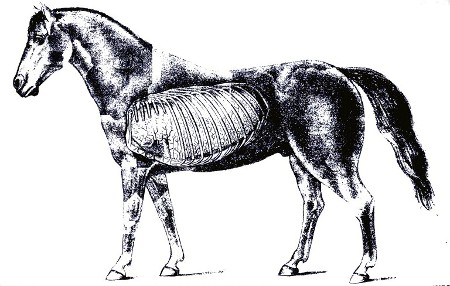
The lungs (see Pl. VII) are the essential organs of respiration. They consist of two (right and left) spongy masses, commonly called the "lights," situated entirely within the thoracic cavity. On account of the space taken up by the heart, the left lung is the smaller. Externally, they are completely covered by the pleura. The structure of the lung consists of a light, soft, but very strong and remarkably elastic tissue, which can be torn only with difficulty. Each lung is divided into a certain number of lobes, which are subdivided into numberless lobules (little lobes). A little bronchial tube terminates in every one of these lobules. The little tube then divides into minute branches which open into the air cells (pulmonary vesicles) of the lungs. The air cells are little sacs having a diameter varying from one-seventieth to one two-hundredth of an inch; they have but one opening, the communication with the branches of the little bronchial tubes. Small blood vessels ramify in the walls of the air cells. The air cells are the consummation of the intricate structures forming the respiratory apparatus. They are of prime importance, all the rest being complementary. It is here that the exchange of gases takes place. As before stated, the walls of the cells are very thin; so, also, are the walls of the blood vessels. Through these walls escapes from the blood the carbonic acid gas that has been absorbed by the blood in its circulation through the different parts of the body; through these walls also the oxygen gas, which is the life-giving element of the atmosphere, is absorbed by the blood from the air in the air cells.
CONGESTION OF THE LUNGS.
Congestion is essentially an excess of blood in the vessels of the parts affected. Congestion of the lungs in the horse, when it exists as an independent affection, is generally caused by overexertion when the animal is not in a fit condition to undergo more than moderate exercise. Very often what is recognized as congestion of the lungs is but a symptom of exhaustion or dilatation of the heart.
The methods practiced by the trainers of running and trotting horses will give an idea of what is termed "putting a horse in condition" to stand severe exertion. The animal at first gets walking exercises, then after some time he is made to go faster and farther each day; the amount of work is daily increased until he is said to be "in condition." An animal so prepared runs no risk of being affected with congestion of the lungs, if he is otherwise healthy. On the other hand, if the horse is kept in the stable for the purpose of laying on fat or for want of something to do, the muscular system becomes soft, and the horse is not in condition to stand the severe exertion of going fast or far, no matter how healthy he may be in other respects. If such a horse be given a hard ride or drive, he may start off in high spirits, but soon becomes exhausted, and if he is pushed he will slacken his pace, show a desire to stop, and may stagger or even fall. Examination will show the nostrils dilated, the flanks heaving, the countenance haggard, and the appearance of suffocation. The heart and muscles were not accustomed to the sudden and severe strain put upon them; the heart became unable to perform its work; the blood accumulated in the vessels of the lungs, which eventually became engorged with the stagnated blood, constituting congestion of the lungs.
The animal, after having undergone severe exertion, may not exhibit alarming symptoms until returned to the stable; then he will be noticed standing with his head down, legs spread out, the eyes wildly staring or dull and sunken. The breathing is very rapid and almost gasping; in most cases the body is covered with perspiration, which, however, may soon evaporate, leaving the surface of the body and the legs and ears cold; the breathing is both abdominal and thoracic; the chest rises and falls and the flanks are powerfully brought into action. If the pulse can be felt at all it will be found beating very frequently, one hundred or so to a minute. The heart may be felt tumultuously thumping if the hand is placed against the chest behind the left elbow, or it may be scarcely perceptible. The animal may tremble all over. If the ear is placed against the side of the chest a loud murmur will be heard and perhaps a fine, crackling sound.
One can scarcely fail to recognize a case of congestion of the lungs when brought on by overexertion, as the history of the case indicates the nature of the ailment. In all cases of suffocation the lungs are congested. It is also seen in connection with other diseases.
Treatment.—If the animal is attacked by the disease while on the road, stop him immediately. Do not attempt to return to the stables. If he is in the stable, make arrangements at once to insure an unlimited supply of pure air. If the weather is warm, out in the open air is the best place, but if too cold let him stand with head to the door. Let him stand still; he has all he can do, if he obtains sufficient pure air to sustain life. If he is encumbered with harness or saddle, remove it at once and rub the body with cloths or wisps of hay or straw. This stimulates the circulation in the skin, and thus aids in relieving the lungs of the extra quantity of blood that is stagnated there. If you have three or four assistants, let them rub the body and legs well until the skin feels natural; rub the legs until they are warm, if possible. When the circulation is reestablished, put bandages on the legs from the hoofs up as far as possible. Throw a blanket over the body and let the rubbing be done under the blanket. Diffusible stimulants are the medicines indicated—brandy, whisky (or even ale or beer if nothing else is at hand), ether, and aromatic spirits of ammonia. A drench of 2 ounces each of spirits of nitrous ether and alcohol, diluted with a pint of water, every hour until relief is afforded, is among the best remedies. Or, give a quarter of a pint of whisky in a pint of water every hour, or the same quantity of brandy as often, or a quart of ale every hour, or 1 ounce of tincture of arnica in a pint of water every hour until five or six doses have been given. If none of these remedies are at hand, 2 ounces of oil of turpentine, shaken with a half pint of milk, may be given once, but not repeated. The animal may be bled from the jugular vein. Do not take more than 5 or 6 quarts from the vein, and do not repeat the bleeding. The blood thus drawn will have a tarry appearance.
PLATE VII. POSITION OF THE LEFT LUNG.
When the alarming symptoms have subsided active measures may be stopped, but care must be used in the general treatment of the animal for several days, for it must be remembered that congestion may be followed by pneumonia. The animal should have a comfortable stall, where he will not be subjected to drafts or sudden changes of temperature; he should be blanketed and the legs kept bandaged. The air should be pure, a plentiful supply of fresh, cold water always in the stall; and a diet composed principally of bran mashes, scalded oats, and, if in season, grass. When ready for use again the horse should at first receive only moderate exercise, which may be daily increased until he may safely be put to regular work.
PNEUMONIA, OR LUNG FEVER.
Pneumonia is inflammation of the lungs. The chief varieties of pneumonia are catarrhal—later discussed in connection with bronchitis, under the name of broncho-pneumonia—and the fibrinous or croupous variety. The latter form receives its names from the fact that the air spaces are choked with coagulated fibrin thrown out from the blood. This causes the diseased portions of the lungs to become as firm as liver, in which condition they are said to be hepatized. As air is excluded by the inflammatory product, the diseased lung will not float in water.
The inflammation usually begins in the lower part of the lung and extends upward. The first stage of the disease consists of congestion, or engorgement, of the blood vessels, followed by leakage of serum containing fibrin from the blood vessels into the air passages. The fluids thus escaping into the air cells and in the minute branches of the little bronchial tubes become coagulated.
The pleura covering the affected parts may be more or less inflamed. A continuance of the foregoing phenomena is marked by a further escape of the constituents of the blood, and a change in the membrane of the cells, which becomes swollen. The exudate that fills the air cells and minute bronchial branches undergoes disintegration and softening when healing commences.
The favorable termination of pneumonia is in resolution that is, a restoration to health. This is gradually brought about by the exuded material contained in the air cells and lung tissues being broken down and softened and absorbed or expectorated through the nostrils. The blood vessels return to their natural state, and the blood circulates in them as before. In the cases that do not terminate so happily the lung may become gangrenous (or mortified), an abscess may form, or the disease may be merged into the chronic variety.
Pneumonia may be directly induced by any of the influences named as general causes for diseases of the organs of respiration, but in many instances it is from neglect. A common cold or sore throat may be followed by pneumonia if neglected or improperly treated. An animal may be debilitated by a cold, and when in this weakened state may be compelled to undergo exertion beyond his strength; or he may be kept in a badly ventilated stable, where the foul gases are shut in and the pure air is shut out; or the stable may be so open that parts of the body are exposed to drafts of cold air. An animal is predisposed to pneumonia when debilitated by any constitutional disease, and especially during convalescence if exposed to any of the exciting causes. Foreign bodies, such as feed accidentally getting into the lungs by way of the windpipe, as well as the inhalation of irritating gases and smoke, ofttimes produce fatal attacks of inflammation of the lung and bronchial tubes. Pneumonia is frequently seen in connection with other diseases, such as influenza, purpura hemorrhagica, strangles, glanders, etc. Pneumonia and pleurisy are most common during cold, damp weather, and especially during the prevalence of the cold north or northeasterly winds. Wounds puncturing the thoracic cavity may cause pneumonia.
Symptoms.—Pneumonia, when a primary disease, is ushered in by a chill, more or less prolonged, which in many cases is seen neither by the owner nor the attendant, but is overlooked. The breathing becomes accelerated, and the animal hangs its head and has a very dull appearance. The mouth is hot and has a sticky feeling to the touch; the heat conveyed to the finger in the mouth demonstrates a fever; if the thermometer is placed in the rectum the temperature will be found to have risen to 103° F. or higher. The pulse is frequent, beating from fifty or sixty to eighty or more a minute. There is usually a dry cough from the beginning, which, however, changes in character as the disease advances; for instance, it may become moist, or if pleurisy sets in, the cough will be peculiar to the latter affection; that is, cut short in the endeavor to suppress it. In some cases the discharge from the nostrils is tinged with blood, while in other cases it has the appearance of muco-pus. The appetite is lost to a greater or less extent, but the desire for water is increased, particularly during the onset of the fever. The membrane within the nostrils is red and at first dry, but sooner or later becomes moist. The legs are cold. The bowels are more or less constipated, and what dung is passed is usually covered with a slimy mucus. The urine is passed in smaller quantities than usual and is of a darker color.
The animal prefers to have the head where the freshest air can be obtained. When affected with pneumonia a horse does not lie down, but persists in standing from the beginning of the attack. If pneumonia is complicated with pleurisy, however, the horse may appear restless and lie down for a few moments to gain relief from the pleuritic pains, but he soon rises. In pneumonia the breathing is rapid and difficult, but when the pneumonia is complicated with pleurisy the ribs are kept as still as possible and the breathing is abdominal; that is, the abdominal muscles are now made to do as much of the work as they can perform. If pleurisy is not present there is little pain. To the ordinary observer the animal may not appear dangerously ill, as he does not show the seriousness of the ailment by violence, as in colic, but a careful observer will discover at a glance that the trouble is something more serious than a cold. By percussion it will be shown that some portions of the chest are less resonant than in health, indicating exclusion of air. If the air is wholly excluded the percussion is quite dull, like that elicited by percussion over the thigh.
By auscultation important information may be gained. When the ear is placed against the chest of a healthy horse, the respiratory murmur is heard more or less distinctly, according to the part of the chest that is beneath the ear. In the very first stage of pneumonia this murmur is louder and hoarser; also, there is a fine, crackling sound something similar to that produced when salt is thrown in a fire. After the affected part becomes solid there is an absence of sound over that particular part. After absorption begins one may again hear sounds that are of a more or less moist character and resemble bubbling or gurgling, which gradually change until the natural sound is heard announcing return to health.
When a fatal termination is approaching all the symptoms become intensified. The breathing becomes still more rapid and difficult; the flanks heave; the animal stares wildly about as if seeking aid to drive off the feeling of suffocation; the body is bathed with sweat; the horse staggers, but quickly recovers his balance; he may now, for the first time during the attack, lie down; he does so, however, in the hope of relief, which he fails to find, and with difficulty struggles to his feet; he pants; the nostrils flap; he staggers and sways from side to side and backward and forward, but still tries to retain the standing position, even by propping himself against the stall. It is no use, as after an exhausting fight for breath he goes down; the limbs stretch out and become rigid. In fatal cases death usually occurs in from 10 to 20 days after the beginning of the attack. On the other hand, when the disease is terminating favorably the signs are obvious. The fever abates and the animal gradually improves in appetite; he takes more notice of things around him; his spirits improve; he has a general appearance of returning health, and he lies down and rests. In the majority of cases pneumonia, if properly treated, terminates in recovery.
Treatment.—The comfort and surroundings of the patient must be attended to first. The quarters should be the best that can be provided. Pure air is essential. Avoid placing the animal in a stall where he may be exposed to drafts of cold air and sudden changes of temperature. It is much better for the animal if the air is cold and pure than if it is warm and foul. It is better to make the animal comfortable with warm clothing than to make the stable warm by shutting off the ventilation. From the start the animal should have an unlimited supply of fresh, cold drinking water. Blanket the body. Rub the legs until they are warm and then put bandages on them from the hoofs up to the knees and hocks. If warmth can not be reestablished in the legs by hand rubbing alone, apply dry, ground mustard and rub well in. The bandages should be removed once or twice every day, the legs well rubbed, and the bandages replaced. Much harm is often done by clipping off hair and rubbing in powerful blistering compounds. They do positive injury and retard recovery, and should not be allowed. Much benefit may be derived from hot application to the sides of the chest if the facilities are at hand to apply them. If the weather is not too cold, and if the animal is in a comfortable stable, the following method may be tried: Have a tub of hot water handy to the stable door; soak a woolen blanket in the water, then quickly wring as much water as possible out of it and wrap it around the chest. See that it fits closely to the skin; do not allow it to sag so that air may get between it and the skin. Now wrap a dry blanket over the wet hot one and hold in place with three girths. The hot blanket should be renewed every half hour, and while it is off being wetted and wrung the dry one should remain over the wet part of the chest to prevent reaction. The hot applications should be kept up for three or four hours, and when stopped the skin should be quickly rubbed as dry as possible, an application of alcohol rubbed over the wet part, and a dry blanket snugly fitted over the animal. If the hot applications appear to benefit, they may be tried on three or four consecutive days. Unless every facility and circumstance favors the application of heat in the foregoing manner, it should not be attempted. If the weather is very cold or any of the details are omitted, more harm than good may result. Mustard may be applied by making a paste with a pound of freshly ground mustard mixed with warm water. This is to be spread evenly over the sides back of the shoulder blades and down to the median line below the chest. Care should be taken to avoid rubbing the mustard upon the thin skin immediately back of the elbow. The mustard-covered area should be covered with a paper and this with a blanket passed up from below and fastened over the back. The blanket and paper should be removed in from one to two hours. When pneumonia follows another disease, the system is always more of less debilitated and requires the careful use of stimulants from the beginning. To weaken the animal still further by bleeding him is one of the most effectual methods of retarding recovery, even if it does not hasten a fatal termination.
Another and oftentimes fatal mistake made by the nonprofessional is the indiscriminate and reckless use of aconite. This drug is one of the most active poisons, and should not be handled by anyone who does not thoroughly understand its action and uses. It is only less active than prussic acid in its poisonous effects. It is a common opinion, often expressed by nonprofessionals, that aconite is a stimulant. Nothing could be more erroneous; in fact, it is just the reverse. It is one of the most powerful sedatives used in the practice of medicine. In fatal doses it kills by paralyzing the very muscles used in breathing; it weakens the action of the heart, and should not be used. Do not give purgative medicines. If constipation exists, overcome it by an allowance of laxative diet, such as scalded oats, bran, and linseed mashes; also, grass, if in season. If the costiveness is not relieved by the laxative diet, give an enema of about a quart of warm water three or four times a day.
A diet consisting principally of bran mashes, scalded oats, and, when in season, grass or corn fodder is preferable if the animal retains an appetite; but if no desire is evinced for feed of this particular description, then the animal must be allowed to eat anything that will be taken spontaneously. Hay tea, made by pouring boiling water over good hay in a large bucket and allowing it to stand until cool, then straining off the liquid, will sometimes create a desire for feed. The animal may be allowed to drink as much of it as he desires. Corn on the cob is often eaten when everything else is refused. Bread may be tried; also apples or carrots. If the animal can be persuaded to drink milk, it may be supported by it for days. Three or four gallons of sweet milk may be given during the day, in which may be stirred three or four fresh eggs to each gallon. Some horses will drink milk, while others will refuse to touch it. It should be borne in mind that all feed must be taken by the horse as he desires it; none should be forced down him. If he will not eat, you will only have to wait until a desire is shown for feed. All kinds may be offered, first one thing and then another, but feed should not be allowed to remain long in trough or manger; the very fact of its constantly being before him will cause him to loathe it. When the animal has no appetite for anything the stomach is not in a proper state to digest food, and if it is poured or drenched into him it will only cause indigestion and aggravate the case. It is a good practice to do nothing when there is nothing to be done that will benefit. This refers to medicine as well as feed. Nothing is well done that is overdone.
There are many valuable medicines used for the different stages and different types of pneumonia, but in the opinion of the writer it is useless to refer to them here, as this work is intended for the use of those who are not sufficiently acquainted with the disease to recognize its various types and stages; therefore they would only confuse. If you can administer a ball or capsule, or have anyone at hand who is capable of doing it, a dram of sulphate of quinin in a capsule, or made into a ball, with sufficient linseed meal and molasses, given every three hours during the height of the fever, will do good in many cases. The ball of carbonate of ammonia, as advised in the treatment of bronchitis, may be tried if the animal is hard to drench. The heart should be kept strong by administering digitalis in doses of 2 drams of the tincture every three hours, or strychnia 1 grain, made into a pill with licorice powder, three times daily.
If the horse becomes very much debilitated, stimulants of a more pronounced character are required. The following drench is useful: Rectified spirits, 3 ounces; spirits of nitrous ether, 2 ounces; water, 1 pint. This may be repeated every four or five hours if it seems to benefit; or 6 ounces of good whisky diluted with a pint of water may be given as often, instead of the foregoing.
During the period of convalescence good nutritive feed should be allowed in a moderate quantity. Tonic medicines should be substituted for those used during the fever. The same medicines advised for the convalescing period of bronchitis are equally efficient in this case, especially the iodid of potash; likewise, the same general instructions apply here.
The chief causes of death in pneumonia are heart failure from exhaustion, suffocation, or blood poisoning from death (gangrene) of lung tissue. The greater the area of lung tissue diseased the greater the danger; hence double pneumonia is more fatal than pneumonia of one lung.
THE WINDPIPE.
The windpipe, or trachea as it is technically called, is the flexible tube that extends from, the larynx, which it succeeds at the throat, to above the base of the heart in the chest, where it terminates by dividing into the right and left bronchi—the tubes going to the right and left lung, respectively. The windpipe is composed of about fifty incomplete rings of cartilage united by ligaments. A muscular layer is situated on the superior surface of the rings. Internally the tube is lined with a continuation of the mucous membrane that lines the entire respiratory tract, which here has very little sensibility in contrast to that lining the larynx, which is endowed with exquisite sensitiveness.
The windpipe is not subject to any special disease, but is more or less affected during laryngitis (sore throat), influenza, bronchitis, etc., and requires no special treatment. The membrane may be left in a thickened condition after these attacks. One or more of the rings may be accidentally fractured, or the tube may be distorted or malformed as the result of violent injury. After the operation of tracheotomy it is not uncommon to find a tumor or malformation as a result, or sequel, of the operation. In passing over this section attention is merely called to these defects, as they require no particular attention in the way of treatment. It may be stated, however, that any one of the before-mentioned conditions may constitute one of the causes of noisy respiration described as "thick wind."
GUTTURAL POUCHES.
These two sacs are situated above the throat, and communicate with the pharynx, as well as with the cavity of the tympanum of the ear. They are peculiar to solipeds. Normally, they contain air. Their function is unknown.
One or both guttural pouches may contain pus. The symptoms are as follows: Swelling on the side below the ear and an intermittent discharge of matter from one or both nostrils, especially when the head is depressed.
The swelling is soft, and, if pressed upon, matter will escape from the nose if the head is depressed. As before mentioned, these pouches communicate with the pharynx, and through this small opening matter may escape. A recovery is probable if the animal is turned out to graze, or if he is fed from the ground, as the dependent position of the head favors the escape of matter from the pouches. In addition to this, give the tonics recommended for nasal gleet. If this treatment fails, an operation must be performed, which should not be attempted by any one unacquainted with the anatomy of the part.
BRONCHITIS AND BRONCHO-PNEUMONIA.
Bronchitis is an inflammation of the bronchial tubes. When this inflammation extends to the air sacs at the termini of the smallest branches of the bronchial tubes, the disease is broncho-pneumonia. Bronchitis affecting the larger tubes is less serious than when the smaller are involved. The disease may be either acute or chronic. The causes are generally much the same as for other diseases of the respiratory organs, noticed in the beginning of this article. The special causes are these: The inhalation of irritating gases and smoke and fluids or solids gaining access to the parts. Bronchitis is occasionally associated with influenza and other specific fevers. It also supervenes on common cold or sore throat.
Symptoms.—The animal appears dull; the appetite is partially or wholly lost; the head hangs; the breathing is quickened; the cough, at first dry, and having somewhat the character of a "barking cough," is succeeded in a few days by a moist, rattling cough; the mouth is hot; the visible membranes in the nose are red; the pulse is frequent, and during the first stage is hard and quick, but as the disease advances becomes smaller and more frequent. There is a discharge from the nostrils that is at first whitish, but later becomes creamy or frothy, still later it is sometimes tinged with blood, and occasionally it may be of a brownish or rusty color. By auscultation, or placing the ear to the sides of the chest, unnatural sounds can now be heard. The air passing through the diseased tubes causes a wheezing sound when the small tubes are affected, and a hoarse, cooing, or snoring sound when the larger tubes are involved. After one or two days the dry stage of the disease is succeeded by a moist state of the membrane. The ear now detects a different sounds caused by the bursting of the bubbles as the air passes through the fluid, which is the exudate of inflammation and the augmented mucous secretions of the membrane. The mucus may be secreted in great abundance, which, by blocking up the tubes, may cause a collapse of a large extent of breathing surface. Usually the mucus is expectorated; that is, discharged through the nose. The matter is coughed up, and when it reaches the larynx much of it may be swallowed, and some is discharged from the nostrils. The horse can not spit, like the human being, nor does the matter coughed up gain access to the mouth. If in serious cases all the symptoms become aggravated, the breathing is labored, short, and quick, it usually indicates that the inflammation has reached the breathing cells and that catarrhal pneumonia is established. In this case the ribs rise and fall much more than natural. This fact alone is enough to exclude the idea that the animal may be affected with pleurisy, because in that disease the ribs are as nearly fixed as it is in the power of the animal to do so, and the breathing is accomplished to a great extent by aid of the abdominal muscles. The horse persists in standing throughout the attack. He prefers to stand with head to a door or window to gain all the fresh air possible, but if not tied may occasionally wander listlessly about the stall. The bowels most likely are constipated; the dung is covered with slimy mucus. The urine is decreased in quantity and darker in color than usual. The animal shows more or less thirst; in some cases the mouth is full of saliva. The discharge from the nose increases in quantity as the disease advances and inflammation subsides. This is rather a good symptom, as it shows that one stage has passed. The discharge then gradually decreases, the cough becomes less rasping, but of more frequent occurrence, until it gradually disappears with the return of health.
Bronchitis, affecting the smaller tubes, is one of the most fatal diseases, while that of the larger tubes is never very serious. It must be stated, however, that it is an exceedingly difficult matter for a nonexpert to discriminate between the two forms, and, further, it may as well be said here that he will have difficulty in discriminating between bronchitis and pneumonia.
Treatment.—The matter of first importance is to insure pure air to breathe, and next to make the patient's quarters as comfortable as possible. A well-ventilated box stall serves best for all purposes. Cover the body with a blanket, light or heavy, as the season of the year demands. Hand-rub the legs until they are warm, then wrap them in cotton and apply flannel or Derby bandages from the hoofs to the knees and hocks. If the legs can not be made warm with hand rubbing alone, apply dry mustard. Rub in thoroughly and then put the bandages on; also rub mustard paste well over the side of the chest, covering the space beginning immediately behind the shoulder blade and running back about eighteen inches, and from the median line beneath the breast to within ten inches of the ridge of the backbone. Repeat the application to the side of the chest about three days after the first one is applied.
Compel the animal to inhale steam from a bucketful of boiling water containing a tablespoonful of oil of turpentine and spirits of camphor, as advised for cold in the head. In serious cases the steam should be inhaled every hour, and in any case the oftener it is done the greater will be the beneficial results. Three times a day administer an electuary containing acetate of potash (2 drams), with licorice and molasses or honey. It is well to keep a bucketful of cold water before the animal all the time. If the horse is prostrated and has no appetite, give the following drench: Spirits of nitrous ether, 2 ounces; rectified spirits, 3 ounces; water, 1 pint. Repeat the dose every four or five hours if it appears to benefit. When the horse is hard to drench, give the following: Pulverized carbonate of ammonia, 3 drams; linseed meal and molasses sufficient to make the whole into a stiff mass; wrap it with a small piece of tissue paper and give as a ball. This ball may be repeated every four or five hours. When giving the ball care should be taken to prevent its breaking in the mouth, as in case of such accident it will make the mouth sore and prevent the animal from eating. If the bowels are constipated, give enemas of warm water. Do not give purgative medicines. Do not bleed the animal.
If the animal retains an appetite, a soft diet is preferable, such as scalded oats, bran mashes, and grass, if in season. If he refuses cooked feed, allow in small quantities anything he will eat. Hay, cob corn, oats, bread, apples, and carrots may be tried in turn. Some horses will drink sweet milk when they refuse all other kinds of feed, and especially is this the case if the drinking water is withheld for a while. One or 2 gallons at a time, four or five times a day, will support life. Bear in mind that when the disease is established recovery can not occur in less than two or three weeks, and more time may be necessary. Good nursing and patience are required.
When the symptoms have abated and nothing remains of the disease except the cough and a white discharge from the nostrils, all other medicines should be discontinued and a course of tonic treatment pursued. Give the following mixture: Reduced iron, 3 ounces; powdered gentian, 8 ounces; mix well together and divide into sixteen powders. Give a powder every night and morning mixed with bran and oats, if the animal will eat it, or shaken with about a pint of flaxseed tea and administered as a drench.
If the cough remains after the horse is apparently well, give 1 dram of iodid of potassium dissolved in a bucketful of drinking water one hour before each meal for two or three weeks if necessary. Do not put the animal to work too soon after recovery. Allow ample time to regain strength. This disease is prone to become chronic and may run into an incurable case of thick wind.
PLEURISY.
The thoracic cavity is divided into two lateral compartments, each containing one lung and a part of the heart. Each lung has its separate pleural membrane, or covering. The pleura is the thin, glistening membrane that covers the lung and also completely covers the internal walls of the chest. It is very thin, and to the ordinary observer appears to be part of the lung, which, in fact, it is for all practical purposes. The smooth, shiny surface of the lung, as well as the smooth, shiny surface so familiar on the rib, is the pleura. In health this surface is always moist. A fluid is thrown off by the pleura, which causes the surface to be constantly moist. This is to prevent the effects of friction between the lungs and the walls of the chest and other contiguous parts which come in contact. It must be remembered that the lungs are dilating each time a breath is taken in, and contracting each time a breath of air is expelled. It may be readily seen that if it were not for the moistened state of the surface of the pleura the continual dilatation and contraction and the consequent rubbing of the parts against each other would cause serious friction.
Inflammation of this membrane is called pleurisy. Being so closely united with the lung, it can not always escape participation in the disease when the latter is inflamed. Pleurisy may be due to the same predisposing and exciting causes as mentioned in the beginning of this work as general causes for diseases of the organs of respiration, such as exposure to sudden changes of temperature, confinement in damp stables, etc. It may be caused also by wounds that penetrate the chest, for it must be remembered that such wounds must necessarily pierce the pleura. A fractured rib may involve the pleura. The inflammation following such wounds may be circumscribed; that is, confined to a small area surrounding the wound, or it may spread from the wound and involve a large portion of the pleura. The pleura may be involved secondarily when the heart or its membrane is the primary seat of the disease. It may occur in conjunction with bronchitis, influenza, and other diseases. Diseased growths that interfere with the pleura may induce pleurisy. The most frequent cause of pleurisy is an extension of inflammation from adjacent diseased lung. It is a common complication of pneumonia. Pleurisy will be described here as an independent affection, although it should be remembered that it is very often associated with the foregoing diseases.
The first lesion of pleurisy is overfilling of the blood vessels that ramify in this membrane and dryness of the surface. This is followed by the formation of a coating of coagulated fibrin on the diseased pleura and the transudation of serum which collects in the chest. This serum may contain flakes of fibrin and it may be straw colored or red from an admixture of blood. The quantity of this accumulation may amount to several gallons.
Symptoms.—When the disease exists as an independent affection it is ushered in by a chill, but this is usually overlooked. About the first thing noticed is the disinclination of the animal to move or turn around. When made to do so he grunts or groans with pain. He stands stiff; the ribs are fixed—that is, they move very little in the act of breathing—but the abdomen works more than natural; both the fore feet and elbows may be turned out; during the onset of the attack the animal may be restless and act as if he had a slight colic; he may even lie down, but does not remain long down, for when he finds no relief he soon gets up. After effusion begins these signs of restlessness disappear. Every movement of the chest causes pain; therefore the cough is peculiar; it is short and suppressed and comes as near being no cough as the animal can make it in his desire to suppress it. The breathing is hurried, the mouth is hot, the temperature being elevated from 102° or 103° to 105° F. Symptoms that usually accompany fever are present, such as costiveness, scanty, dark-colored urine, etc. The pulse is frequent, perhaps 70 or more a minute, and is hard and wiry. The legs and ears are cold.
Percussion is of valuable service in this affection. After effusion occurs the sound produced by percussing over the lower part of the chest is dull. By striking different parts one may come to a spot of greater or less extent where the blows cause much pain to be evinced. The animal may grunt or groan every time it is struck. Another method of detecting the affected part is to press the fingers between the ribs, each space in succession, beginning behind the elbow, until a place where the pressure causes more flinching than at any other part is reached. Auscultation is also useful. In the first stage, when the surfaces are dry and rough, one may hear, immediately under the ear, a distinct sound very much like that produced by rubbing two pieces of coarse paper together. No such friction sound occurs when the membrane is healthy, as the natural moisture, heretofore mentioned, prevents the friction. In many cases this friction is so pronounced that it may be felt by placing the hand over the affected part. When the dry stage is succeeded by the exudation of fluid this friction sound disappears. After the effusion into the cavity takes place sometimes there is heard a tinkling or metallic sound, due to dropping of the exudate from above into the collected fluid in the bottom of the cavity, as the collected fluid more of less separates the lung from the chest walls.
Within two or three days the urgent symptoms may abate owing to the exudation of the fluid, and the subsidence of the pain. The fluid may now undergo absorption, and the case may terminate favorably within a week or 10 days.
If the quantity of the effusion is large its own volume retards the process of absorption to a great extent, and consequently convalescence is delayed. In severe cases the pulse becomes more frequent, the breathing more hurried and labored, the flanks work like bellows, the nostrils flap, the eyes stare wildly, the countenance expresses much anxiety, and general signs of dissolution are plain. After a time swellings appear under the chest and abdomen and down the legs. The accumulation in the chest is called hydrothorax, or dropsy of the chest. When this fluid contains pus the case usually proves fatal. The condition of pus within the cavity is called empyema.
Pleurisy may affect only a small area of one side or it may affect both sides. It is oftener confined to the right side.
Treatment.—The instructions in regard to the general management of bronchitis and pneumonia must be adhered to in the treatment of pleurisy. Comfortable quarters, pure air, warm clothing to the body and bandages to the legs, a plentiful supply of pure cold water, the laxative feed, etc., in this case are equally necessary and efficacious. The hot applications applied to the chest, as directed in the treatment of pneumonia, are very beneficial in pleurisy, and should be kept up while the symptoms show the animal to be in pain.
During the first few days, when pain is manifested by restlessness, apply hot packs to the sides diligently. After four or five days, when the symptoms show that the acute stage has somewhat subsided, mustard may be applied as recommended for pneumonia. From the beginning the following drench may be given every six hours, if the horse takes it kindly: Solution of the acetate of ammonia, 3 ounces; spirits of nitrous ether, 1 ounce; bicarbonate of potassium, 3 drams; water, 1 pint.
If the patient becomes debilitated, the stimulants as prescribed for pneumonia should be used according to the same directions. The same attention should be given to the diet. If the animal will partake of the bran mashes, scalded oats, and grass, it is the best; but if he refuses the laxative diet, then he should be tried with different kinds of feed and allowed whichever kind he desires.
In the beginning of the attack, if the pain is severe, causing the animal to lie down or paw, morphin may be given by the mouth in 5-grain doses, or the fluid extract of Cannabis indica may be used in doses of 2 to 4 drams.
If the case is not progressing favorable in ten or twelve days after the beginning of the attack, convalescence is delayed by the fluid in the chest failing to be absorbed. The animal becomes dull and weak and evinces little or no desire for feed. The breathing becomes still more rapid and difficult. An effort must now be made to excite the absorption of the effusion. An application of liniment or mild blister should be rubbed over the lower part of both sides and the bottom of the chest. The following drench may be given three times a day, for seven or eight days, if it is necessary and appears to benefit: Tincture of the perchlorid of iron, 1 ounce; tincture of gentian, 2 ounces; water, 1 pint. Also give 1 dram of iodid of potassium, dissolved in the drinking water, an hour before feeding every night and morning for a week or two.
Hydrothorax is sometimes difficult to overcome by means of the use of medicines alone, when the operation of tapping the chest is performed to allow an escape for the accumulated fluid. The operation is performed with a combined instrument called the trocar and cannula. The puncture is made in the lower part of the chest, in the space between the eighth and ninth ribs. Wounding of the intercostal artery is avoided by inserting the instrument as near as possible to the anterior edge of the rib. If the operation is of benefit, it is only so when performed before the strength is lowered beyond recovery. The operation merely receives a passing notice here, as it is not presumed that the nonprofessional will attempt it, although in the hands of the expert it is attended with little danger or difficulty.
We have described here bronchitis, pneumonia, and pleurisy mainly as they occur as independent diseases, but it should be remembered that they merge into each other and may occur together at one time. While it is true that much more might have been said in regard to the different stages and types of the affections, and also in regard to the treatment of each stage and each particular type, the plan adopted of advising plain, conservative treatment is considered the wisest on account of simplifying as much as possible a subject of which the reader is supposed to know very little.
PLEUROPNEUMONIA.
This is the state in which an animal is affected with pleurisy and pneumonia combined, which is not infrequently the case. At the beginning of the attack only one of the affections may be present, but the other soon follows. It has already been stated that the pleura is closely adherent to the lung. The pleura on this account is frequently more or less affected by the spreading of the inflammation from the lung tissue. There is a combination of the symptoms of both diseases, but to the ordinary observer the symptoms of pleurisy are the most obvious. The course of treatment to be pursued differs in no manner from that given for the affections when they occur independently. The symptoms will be the guide as to the advisability of giving oil and laudanum for the pain if the pleurisy is very severe. It should not be resorted to unless it is necessary to allay the pain.
BRONCHO-PLEUROPNEUMONIA.
This is the term or terms applied when bronchitis, pleurisy, and pneumonia all exist at once. It is impossible for one who is not an expert to diagnose the state with certainty. The apparent symptoms are the same as when the animal is affected with pleuropneumonia.
SUPPURATION AND ABSCESS IN THE LUNG.
There are instances, and especially when the surroundings of the patient have been bad or the disease is of an especially severe type, when pneumonia terminates in an abscess in the lung. Sometimes, when the inflammation has been extreme, suppuration in a large portion of the lung takes place. Impure air, the result of improper ventilation, is among the most frequent causes of this termination. The symptoms of suppuration in the lung are chronic pneumonia, a solidified area of lung tissue, continued low fever, and, in some cases, offensive smell of the breath, and the discharge of the matter from the nostrils.
MORTIFICATION.
Gangrene, or mortification, means the death of the part affected. Occasionally, owing to the intensity of the inflammation or bad treatment, pneumonia and pleuropneumonia terminate in mortification, which is soon followed by the death of the animal. Perhaps the most common cause of this complication is the presence of a foreign body in the lung, as food particles or medicine. Rough drenching or drenching through the nostrils may cause this serious condition.
HEMOPTYSIS, OR BLEEDING FROM THE LUNGS.
Bleeding from the lungs may occur during the course of congestion of the lungs, bronchitis, pneumonia, influenza, purpura hemorrhagica, or glanders. An accident or exertion may cause a rupture of a vessel. Plethora and hypertrophy of the heart predispose to it. Following the rupture of a vessel the blood may escape into the lung tissue and cause a serious attack of pneumonia, or it may fill up the bronchial tubes and prove fatal by suffocating the animal. When the hemorrhage is from the lung it is accompanied with coughing; the blood is frothy, of a bright red color, and comes from both nostrils; whereas when the bleeding is merely from a rupture of a vessel in some part of the head (heretofore described as bleeding from the nose) the blood is most likely to issue from one nostril only, and the discharge is not accompanied with coughing. The ear may be placed against the windpipe along its course, and if the blood is from the lungs a gurgling or rattling sound will be heard. When it occurs in connection with another disease it seldom requires special treatment. When caused by accident or overexertion the animal should be kept quiet. If the hemorrhage is profuse and continues for several hours, 1 dram of the acetate of lead dissolved in a pint of water may be given as a drench, or 1 ounce of the tincture of the perchlorid of iron, diluted with a pint of water, may be given instead of the lead. It is rare that the hemorrhage is so profuse as to require internal remedies. But hemorrhage into the lung may occur and cause death by suffocation without the least manifestation of it by the discharge of blood from the nose.