DISEASES OF THE GENERATIVE ORGANS. (continued 2)
Reading edition
Fore limb turned back from shoulders.—In this case, on exploration by the side of the head and presenting limb, the shoulder only can be reached at first. (Plate XV, fig 4.) By noosing the head and presenting fore limb, they may be drawn forward into the pelvis, and the oiled hand being carried along the shoulder in the direction of the missing limb is enabled to reach and seize the forearm just below the elbow. The body is now pushed back by the assistants pressing on the head and presenting limb or on a repeller planted in the breast until the knee can be brought up into the pelvis, after which the procedure is the same as described in the last paragraph.
Head bent down between fore limbs.—This may be so that the poll or nape of the neck, with the ears, can be felt far back between the fore limbs, or so that only the upper border of the neck can be reached, head and neck being bent back beneath the body. With the head only bent on the neck, noose the two presenting limbs, then introduce the hand between them until the nose can be seized in the palm of the hand. Next have the assistants push back the presenting limbs, while the nose is strongly lifted upward over the brim of the pelvis. This accomplished, it assumes the natural position and parturition is easy.
When both head and neck are bent downward it may be impossible to reach the nose. If, however, the labor has only commenced, the limbs may be drawn upon until the operator can reach the ear, by dragging on which the head may be so far advanced that the fingers may reach the orbit; traction upon this while the limbs are being pushed back may bring the head up so that it bends on the neck only, and the further procedure will be as described in the last paragraph.
If the labor has been long in progress and the fetus is jammed into the pelvis, the womb emptied of the waters, and firmly contracted on its solid contents, the case is incomparably more difficult. The mare may be chloroformed and turned on her back with hind parts elevated, and the womb may be injected with sweet oil. Then, if the ear can be reached, the correction of the malpresentation may be attempted as above described. Should this fail, one or more sharp hooks may be inserted in the neck as near the head as can be reached, and ropes attached to these may be dragged on, while the body of the foal is pushed back by the fore limbs or by a repeller. Such repulsion should be made in a direction obliquely upward toward the loins of the mother, so as to rotate the fetus in such a way as to bring the head up. As this is accomplished a hold should be secured nearer and nearer to the nose, with hand or hook, until the head can be straightened out on the neck.
All means failing; it becomes necessary to remove the fore limbs (embryotomy) so as to make more space for bringing up the head. If, even then, this can not be accomplished, it may be possible to push the body backward and upward with the repeller until the hind limbs are brought to the passage, when they may be noosed and delivery effected with the posterior presentation.
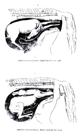
Head turned on shoulders.—In this case the fore feet present, and the oiled hand passed along the fore arms in search of the missing head finds the side of the neck turned to one side, the head being perhaps entirely out of reach. (Plate XVIII, fig. 1.) To bring the head forward it may be desirable to lay the mare on the side opposite to that to which the head is turned, and even to give chloroform or ether. Then the feet being noosed, the body of the fetus is pushed by the hand or repeller forward and to the side opposite to that occupied by the head until the head comes within reach, near the entrance of the pelvis. If such displacement of the fetus is difficult, it may be facilitated by a free use of oil or lard. When the nose can be seized it can be brought into the passage, as when the head is turned down. If it can not be reached, the orbit may be availed of to draw the head forward until the nose can be seized or the lower jaw noosed. In very difficult cases a rope may be passed around the neck by the hand or with the aid of a curved carrier (Plate XIV), and traction may be made upon this while the body is being rotated to the other side. In the same way in bad cases a hook may be fixed in the orbit or even between the bones of the lower jaw to assist in bringing the head up into position. Should all fail, the amputation of the fore limbs may be resorted to, as advised under the last heading.
Head turned upward on back.—This differs from the last malpresentation only in the direction of the head, which has to be sought above rather than at one side, and is to be secured and brought forward in a similar manner. (Plate XVIII, fig. 2.) If a rope can be passed around the neck it will prove most effectual, as it naturally slides nearer to the head as the neck is straightened and ends by bringing the head within easy reach.
Hind feet engaged in pelvis.—In this case fore limbs and head present naturally, but the hind limbs bent forward from the hip and the loins arched allow the hind feet also to enter the passages, and the further labor advances the more firmly does the body of the foal become wedged into the pelvis (Plate XVII, fig. 2.) The condition is to be recognized by introducing the oiled hand along the belly of the fetus, when the hind feet will be felt advancing. An attempt should at once be made to push them back, one after the other, over the brim of the pelvis. Failing in this, the mare may be turned on her back, head downhill, and the attempt renewed. If it is possible to introduce a straight rope carrier, a noose passed through this may be put on the fetlock and the repulsion thereby made more effective. In case of continued failure the anterior presenting part of the body may be skinned and cut off as far back toward the pelvis as possible (see "Embryotomy"); then nooses are placed on the hind fetlocks and traction is made upon these while the quarters are pushed back into the womb. Then the remaining portion is brought away by the posterior presentation.
Anterior presentation with back turned to one side.—The diameter of the axis of the foal, like that of the pelvic passages, is from above downward, and when the fetus enters the pelvis with this greatest diameter engaged transversely or in the narrow diameter of the pelvis, parturition is rendered difficult or impossible. In such a case the pasterns and head may be noosed, and the passages and engaged portion of the foal freely lubricated with lard, the limbs may be crossed over each other and the head, and a movement of rotation effected in the fetus until its face and back are turned up toward the croup of the mother; then parturition becomes natural.
Back of foal turned to floor of pelvis.—In a roomy mare this is not an insuperable obstacle to parturition, yet it may seriously impede it, by reason of the curvature of the body of the foal being opposite to that of the passages, and the head and withers being liable to arrest against the border of the pelvis. Lubrication of the passage with lard and traction of the limbs and head will usually suffice with or without the turning of the mare on her back.
In obstinate cases two other resorts are open: First, to turn the foal, pushing back the fore parts and bringing up the hind so as to make a posterior presentation, and, second, the amputation of the fore limbs, after which extraction will usually be easy.
PLATE XV. ABNORMAL PRESENTATIONS.
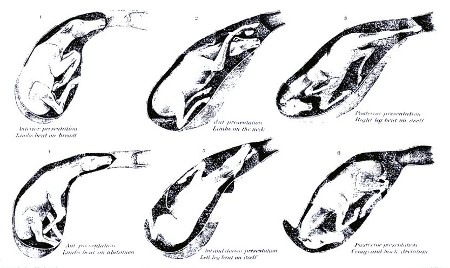
PLATE XVI. ABNORMAL PRESENTATIONS.
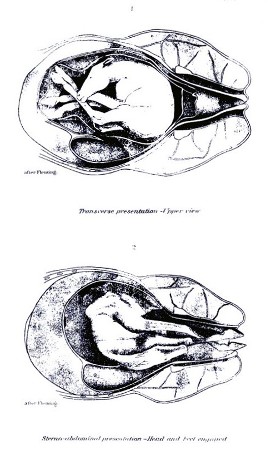
PLATE XVII. ABNORMAL PRESENTATIONS.
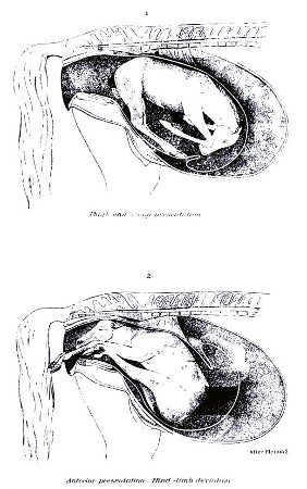
PLATE XVIII. ABNORMAL PRESENTATIONS.
Hind presentation with leg bent at hock.—In this form the quarters of the foal with the hind legs bent up beneath them present, but can not advance through the pelvis by reason of their bulk. (Plate XV, fig. 3.) The oiled hand introduced can recognize the outline of the buttocks, with the tail and anus in the center and the sharp points of the hocks beneath. First pass a rope around each limb at the hock, then with hand or repeller push the buttocks backward and upward, until the feet can be brought up into the passages. To this the great length of the shank and pastern in the foal is a serious obstacle, and in all cases the foot should be protected in the palm of the hand while being brought up over the brim of the pelvis; otherwise the womb may be torn. When the pains are too violent and constant to allow effective manipulation, some respite may be obtained by the use of chloroform or morphin and by turning the mare on her back, but too often the operator fails and the foal must be sacrificed. Two courses are still open: First, to cut through the cords behind and above the hock and extend the upper part of the limb, leaving the hock bent, and extract in this way, and, second, to amputate the hind limbs at the hip joint and remove them separately, after which the body may be extracted.
Hind presentation with legs bent forward from hip.—This is merely an aggravated form of the presentation last described. (Plate XVII, fig. 1.) If the mare is roomy, a rope may be passed around each thigh and the body pushed upward and forward, so as to bring the hocks and heels upward. If this can be accomplished, nooses are placed on the limb further and further down until the fetlock is reached and brought into position. If failure is met with, then amputation at the hips is the last resort.
Hind presentations with back turned sideways or downward.—These are the counterparts of similar anterior presentations and are to be managed in the same way.
Presentation of the back.—This is rare, yet not unknown, the foal being bent upon itself with the back, recognizable by its sharp row of spines, presented at the entrance of the pelvis and the head and all four feet turned back into the womb. (Plate XVI, fig. 1.) The body of the fetus may be extended across the opening transversely, so that the head corresponds to one side (right or left), or it may be vertical, with the head above or below.
In any such position the object should be to push the body of the fetus forward and upward or to one side, as may best promise to bring up the fore or hind extremities, and bring the latter into the passage so as to constitute a normal anterior or posterior presentation. This turning of the fetus may be favored by a given position of the mother, by the free use of oil or lard on the surface of the fetus, and by the use of a propeller.
Presentation of breast and abdomen.—This is the reverse of the back presentation, the foal being extended across in front of the pelvic opening, but with the belly turned toward the passages and with all four feet engaged in the passage. (Plate XVI, fig. 2.) The most promising course is to secure the hind feet with nooses and then push the fore feet forward into the womb. As soon as the fore feet are pushed forward clear of the brim of the pelvis, traction is made on the hind feet so as to bring the thighs into the passage and prevent the reentrance of the fore limbs. If it proves difficult to push the fore limbs back, a noose may be passed around the fetlock of each and the cord drawn through the eye of a rope carrier, by means of which the members may be easily pushed back.
EMBRYOTOMY.
Embryotomy consists in the dissection of the fetus, so as to reduce its bulk and allow of its exit through the pelvis. The indications for its adoption have been furnished in the foregoing pages. The operation will vary in different cases according to the necessity for the removal of one or more parts in order to secure the requisite reduction in size. Thus it may be needful to remove head and neck, one fore limb or both, one hind limb or both, to remove different parts of the trunk, or to remove superfluous (monstrous) parts. Some of the simplest operations in embryotomy (incision of the head in hydrocephalus, incision of the belly in dropsy) have already been described. It remains to notice the more difficult procedures which can be best undertaken by the skilled anatomist.
Amputation of the head.—This is easy when both fore limbs are turned back and the head alone has made its exit in part. It is more difficult when the head is still retained in the passages or womb, as in double-headed monsters. The head is secured by a hook in the lower jaw, or in the orbit, or by a halter, and the skin is divided circularly around the lower part of the face or at the front of the ears, according to the amount of head protruding. Then an incision is made backward along the line of the throat, and the skin dissected from the neck as far back as possible. Then the muscles and other soft parts of the neck are cut across, and the bodies of two vertebra (neck bones) are severed by cutting completely across the cartilage of the joint. The bulging of the ends of the bones will serve to indicate the seat of the joint. The head and detached portion of the neck may now be removed by steady pulling. If there is still an obstacle, the knife may be again used to sever any obstinate connections. In the case of a double-headed monster, the whole of the second neck must be removed with the head. When the head has been detached, a rope should be passed through the eyeholes, or through an artificial opening in the skin, and tied firmly around the skin, to be employed as a means of traction when the missing limbs or the second head have been brought up into position.
Amputation of the hind limb.—This may be required when there are extra hind limbs or when the hind limbs are bent forward at hock or hip joint. In the former condition the procedure resembles that for removal of a fore limb, but requires more anatomical knowledge. Having noosed the pastern, a circular incision is made through the skin around the fetlock, and a longitudinal one from that up to the groin, and the skin is dissected from the limb as high up as can be reached, over the croup, if possible. Then cut through the muscles around the hip joint, and, if possible, the two interarticular ligaments of the joint (pubofemoral and round), and extract the limb by strong dragging.
Amputation of the fore limbs.—This may usually be begun on the fetlock of the limb projecting from the vulva. An embryotomy knife is desirable. This knife consists of a blade with a sharp, slightly hooked point, and one or two rings in the back of the blade large enough to fit on the middle finger, while the blade is protected in the palm of the hand. (See Plate XIII, fig. 4.) Another form has the blade inserted in a mortise in the handle, from which it is pushed out by a movable button when wanted. First place a noose around the fetlock of the limb to be amputated, cut the skin circularly entirely around the fetlock, then make an incision on the inner side of the limb from the fetlock up to the breastbone. Next dissect the skin from the limb, from the fetlock up to the breastbone on the inner side, and as far up on the shoulder blade as possible on the outer side. Finally, cut through the muscles attaching the limb to the breastbone, and employ strong traction on the limb, so as to drag out the whole limb, shoulder blade included. The muscles around the upper part of the shoulder blade are easily torn through and need not be cut, even if that were possible. In no case should the fore limb be removed unless the shoulder blade is taken with it, as that furnishes the greatest obstruction to delivery, above all when it is no longer advanced by the extension of the fore limb, but is pressed back so as to increase the already thickest posterior portion of the chest. The preservation of the skin from the whole limb is advantageous in various ways; it is easier to cut it circularly at the fetlock than at the shoulder; it covers the hand and knife in making the needful incisions, thus acting as a protection to the womb; and it affords a means of traction on the body after the limb has been removed. In dissecting the skin from the limb the knife is not needful at all points; much of it may be stripped off with the fingers or knuckles, or by a blunt, iron spud, pushed up inside the hide, which is meanwhile held tense to render the spud effective.
In case the limb is bent forward at the hock, a rope is passed round that and pulled so as to bring the point of the hock between the lips of the vulva. The hamstring and the lateral ligaments of the hock are now cut through, and the limbs extended by a rope tied round the lower end of the long bone above (tibia). In case it is still needful to remove the upper part of the limb, the further procedure is the same as described in the last paragraph.
In case the limb is turned forward from the hip, and the fetus so wedged into the passage that turning is impossible, the case is very difficult. I have repeatedly succeeded by cutting in on the hip joint and disarticulating it, then dissecting the muscles back from the upper end of the thigh bone. A noose was placed around the neck of the bone and pulled on forcibly, while any unduly resisting structures were cut with the knife.
Cartwright recommends to make free incisions round the hip joints and tear through the muscles when they can not be cut; then with cords round the pelvic bones, and hooks inserted in the openings in the floor of the pelvis to drag out the pelvic bones; then put cords around the heads of the thigh bones and extract them; then remove the intestines; finally, by means of the loose, detached skin, draw out the body with the remainder of the hind limbs bent forward beneath it.
Reuff cuts his way into the pelvis of the foal, and with a knife separates the pelvic bones from the loins, then skinning the quarter draws out these pelvic bones by means of ropes and hooks, and along with them the hind limbs.
The hind limbs having been removed by one or the other of these procedures, the loose skin detached from the pelvis is used as a means of traction and delivery is effected. In case of a monstrosity with extra hind limbs, it may be possible to bring these up into the passage and utilize them for traction.
Removal of the abdominal viscera.—In case the belly is unduly large, from decomposition, tumors, or otherwise, it may be needful to lay it open with the knife and cut or tear out the contents.
Removal of the thoracic viscera.—To diminish the bulk of the chest it has been found advisable to cut out the breastbone, remove the heart and lungs, and allow the ribs to collapse with the lower free ends overlapping each other.
Dissection of the trunk.—In case it becomes necessary to remove other portions of the trunk, we should follow the general rule of preserving the skin so that all manipulations can be made inside this as a protector, that it may remain available as a means of exercising traction on the remaining part of the body, and as a covering to protect the vaginal walls against injuries from bones while such part is passing.
FLOODING, OR BLEEDING FROM THE WOMB.
This is rare in the mare, but not unknown, in connection with a failure of the womb to contract on itself after parturition, or with eversion of the womb (casting the withers), and congestion or laceration. If the blood accumulates in the flaccid womb, the condition may be suspected only by reason of the rapidly advancing weakness, swaying, unsteady gait, hanging head, paleness of the eyes and other mucous membranes, and weak, small, failing pulse. The hand introduced into the womb detects the presence of the blood partly clotted. If the blood escapes by the vulva, the condition is evident.
Treatment consists in evacuating the womb of its blood clots, giving a large dose of powdered ergot of rye, and in the application of cold water or ice to the loins and external generative organs. Besides this, a sponge impregnated with a strong solution of alum, or, still better, with tincture of muriate of iron, may be introduced into the womb and squeezed so as to bring the liquid in contact with the walls generally.
EVERSION OF THE WOMB.
If the womb fails to contract after difficult parturition, the after-pains will sometimes lead to the fundus passing into the body of the organ and passing through that and the vagina until the whole inverted organ appears externally and hangs down on the thighs. The result is rapid engorgement and swelling of the organ, impaction of the rectum with feces, and distention of the bladder with urine, all of which conditions seriously interfere with the return of the mass. In returning the womb the standing is preferable to the recumbent position, as the abdomen is more pendent and there is less obstruction to the return. It may, however, be necessary to put hobbles on the hind limbs to prevent the mare from kicking. A clean sheet should be held beneath the womb, and all filth, straw, and foreign bodies washed from its surface. Then with a broad, elastic (india-rubber) band, or in default of that a long strip of calico 4 or 5 inches wide, wind the womb as tightly as possible, beginning at its most dependent part (the extremity of the horn). This serves two good ends. It squeezes out into the general circulation the enormous mass of blood which engorged and enlarged the organ, and it furnishes a strong protective covering for the now delicate, friable organ, through which it may be safely manipulated without danger of laceration. The next step may be the pressure on the general mass while those portions next the vulva are gradually pushed in with the hands; or the extreme lowest point (the end of the horn) may be turned within itself and pushed forward into the vagina by the closed fist, the return being assisted by manipulations by the other hand, and even by those of assistants. By either mode the manipulations may be made with almost perfect safety so long as the organ is closely wrapped in the bandage. Once a portion has been introduced into the vagina the rest will usually follow with increasing ease, and the operation should be completed with the hand and arm extended the full length within the womb and moved from point to point so as to straighten out all parts of the organ and insure that no portion still remain inverted within another portion. Should any such partial inversion be left it will give rise to straining, under the force of which it will gradually increase until the whole mass will be protruded as before. The next step is to apply a truss as an effectual mechanical barrier to further escape of the womb through the vulva. The simplest is made with two 1-inch ropes, each about 18 feet long, each doubled and interwoven at the bend, as seen in Plate XIV, figure 4. The ring formed by the interlacing of the two ropes is adjusted around the vulva, the two ends of the one rope are carried up on the right and left of the tail and along the spine, being wound around each other in their course, and are finally tied to the upper part of the collar encircling the neck. The remaining two ends, belonging to the other rope, are carried downward and forward between the thighs and thence forward and upward on the sides of the belly and chest to be attached to the right and left sides of the collar. These ropes are drawn tightly enough to keep closely applied to the opening without chafing, and will fit still more securely when the mare raises her back to strain. It is desirable to tie the mare short so that she may be unable to lie down for a day or two, and she should be kept in a stall with the hind parts higher than the fore. Violent straining may be checked by full doses of opium (one-half dram), and any costiveness or diarrhea should be obviated by a suitable laxative or binding diet.
In some mares the contractions are too violent to allow of the return of the womb, and full doses of opium one-half dram, laudanum 2 ounces, or chloral hydrate 1 ounce, may be demanded, or the mare must be rendered insensible by ether or chloroform.
RUPTURE, OR LACERATION, OF THE WOMB.
This may occur from the feet of the foal during parturition, or from ill-directed efforts to assist, but it is especially liable to take place in the everted, congested, and friable organ. The resultant dangers are bleeding from the wound, escape of the bowels through the opening and their fatal injury by the mare's feet or otherwise, and peritonitis from the extension of inflammation from the wound and from the poisonous action of the septic liquids of the womb escaping into the abdominal cavity. The first object is to close the wound, but unless in eversion of the womb this is practically impossible. In the last-named condition the wound must be carefully and accurately sewed up before the womb is returned. After its return, the womb must be injected daily with an antiseptic solution (borax, one-half ounce, or carbolic acid, 3 drams to a quart of tepid water). If inflammation threatens, the abdomen may be bathed continuously with hot water by means of a heavy woolen rag, and large doses of opium (one-half dram) may be given twice or thrice daily.
RUPTURES OF THE VAGINA.
These are attended with dangers similar to those belonging to rupture of the womb, and in addition by the risk of protrusion of the bladder, which appears through the lips of the vulva as a red, pyriform mass. Sometimes such lacerations extend downward into the bladder, and in others upward into the terminal gut (rectum). In still other cases the anus is torn so that it forms one common orifice with the vulva.
Too often such cases prove fatal, or at least a recovery is not attained, and urine or feces or both escape freely into the vagina. The simple laceration of the anus is easily sewed up, but the ends of the muscular fibers do not reunite and the control over the lower bowel is never fully reacquired. The successful stitching up of the wound communicating with the bladder or the rectum requires unusual skill and care, and though I have succeeded in a case of the latter kind, I can not advise the attempt by unprofessional persons.
BLOOD CLOTS IN THE WALLS OF THE VAGINA.
(See "Effusion of blood in the vaginal walls," p. 190.)
LAMINITIS, OR FOUNDER, FOLLOWING PARTURITION.
This sometimes follows on inflammation of the womb, as it frequently does on disorder of the stomach. Its symptoms agree with those of the common form of founder, and treatment need not differ.
INFLAMMATION OF THE WOMB AND PERITONEUM.
These may result from injuries sustained by the womb during or after parturition, from exposure to cold or wet, or from the irritant infective action of putrid products within the womb. Under the inflammation the womb remains dilated and flaccid, and decomposition of its secretions almost always occurs, so that the inflammation tends to assume a putrid character and general septic infection is likely to occur.
Symptoms.—The symptoms are ushered in by shivering, staring coat, small, rapid pulse, elevated temperature, accelerated breathing, loss of appetite, with arched back, stiff movement of the body, looking back at the flanks, and uneasy motions of the hind limbs, discharge from the vulva of a liquid at first watery, reddish, or yellowish, and later it may be whitish or glairy, and fetid or not in different cases. Tenderness of the abdomen shown on pressure is especially characteristic of cases affecting the peritoneum or lining of the belly, and is more marked lower down. If the animal survives, the inflammation tends to become chronic and attended by a whitish mucopurulent discharge. If, on the contrary, it proves fatal, death is preceded by extreme prostration and weakness from the general septic poisoning.
Treatment.—In treatment the first thing to be sought is the removal of all offensive and irritant matters from the womb through a caoutchouc tube introduced into the womb, and into which a funnel is fitted. Warm water should be passed until it comes away clear. To insure that all the womb has been washed out, the oiled hand may be introduced to carry the end of the tube into the two horns successively. When the offensive contents have been thus removed, the womb should be injected with a quart of water holding in solution 1 dram permanganate of potash, or, in the absence of the latter, 2 teaspoonfuls of carbolic acid, twice daily. Fomentation of the abdomen, or the application of a warm flaxseed poultice, may greatly relieve. Acetanilid, in doses of half an ounce, twice or thrice a day, or sulphate of quinia in doses of one-third ounce, may be employed to reduce the fever. If the great prostration indicates septic poisoning, large doses (one-half ounce) bisulphite of soda, or salicylate of soda, or sulphate of quinin may be resorted to.
LEUCORRHEA.
This is a white, glutinous, chronic discharge, the result of a continued, subacute inflammation of the mucous membrane of the womb. Like the discharge of acute inflammation, it contains many forms of bacteria, by some of which it is manifestly inoculable on the penis of the stallion, producing ulcers and a specific, gonorrheal discharge.
Treatment may consist in the internal use of tonics (sulphate of iron, 3 drams, daily) and the washing out of the womb, as described under the last heading, followed by an astringent antiseptic injection (carbolic acid, 2 teaspoonfuls; tannic acid, 1/2 dram; water, 1 quart). This may be given two or three times a day.
DISEASES OF THE UDDER AND TEATS.
CONGESTION AND INFLAMMATION OF THE UDDER.
This is comparatively rare in the mare, though in some cases the udder becomes painfully engorged before parturition, and a doughy swelling, pitting on pressure, extends forward on the lower surface of the abdomen. When this goes on to active inflammation, one or both of the glands becomes enlarged, hot, tense, and painful; the milk is dried up or replaced by a watery or reddish, serous fluid, which at times becomes fetid; the animal walks lame, loses appetite, and shows general disorder and fever. The condition may end in recovery, in abscess, induration, or gangrene, and, in some cases, may lay the foundation for a tumor of the gland.
Treatment.—The treatment is simple so long as there is only congestion. Active rubbing with lard or oil, or, better, camphorated oil, and the frequent drawing off of the milk, by the foal or with the hand, will usually bring about a rapid improvement. When active inflammation is present, fomentation with warm water may be kept up for an hour and followed by the application of the camphorated oil, to which has been added some carbonate of soda and extract of belladonna. A dose of laxative medicine (4 drams Barbados aloes) will be of service in reducing fever, and one-half ounce saltpeter daily will serve a similar end. In case the milk coagulates in the udder and can not be withdrawn, or when the liquid becomes fetid, a solution of 20 grains carbonate of soda and 10 drops carbolic acid dissolved in an ounce of water should be injected into the teat. In doing this it must be noted that the mare has three separate ducts opening on the summit of each teat and each must be carefully injected. To draw off the fetid product it may be needful to use a small milking tube, or spring teat dilator designed by the writer. (Plate XIV, figs. 2 and 3.) When pus forms and points externally and can not find a free escape by the teat, the spot where it fluctuates must be opened freely with the knife and the cavity injected daily with the carbolic-acid lotion. When the gland becomes hard and indolent, it may be rubbed daily with iodin ointment 1 part, vaseline 6 parts.
TUMORS OF THE UDDER.
As the result of inflammation of the udder it may become the seat of an indurated diseased growth, which may go on growing and seriously interfere with the movement of the hind limbs. If such swellings do not give way in their early stages to treatment by iodin, the only resort is to cut them out with a knife. As the gland is often implicated and has to be removed, such mares can not in the future suckle their colts and therefore should not be bred.
SORE TEATS, SCABS, CRACKS, WARTS.
By the act of sucking, especially in cold weather, the teats are subject to abrasions, cracks, and scabs, and as the result of such irritation, or independently, warts sometimes grow and prove troublesome. The warts should be clipped off with sharp scissors and their roots burned with a solid pencil of lunar caustic. This is best done before parturition to secure healing before suckling begins. For sore teats use an ointment of vaseline 1 ounce, balsam of tolu 5 grains, and sulphate of zinc 5 grains.