DISEASES OF THE FETLOCK, ANKLE, AND FOOT.
Reading edition
By A. A. Holcombe, D. V. S.,
Veterinary Inspector, Bureau of Animal Industry.
ANATOMICAL REVIEW OF THE FOOT.
In a description of the foot of the horse it is customary to include only the hoof and its contents, yet, from a zoological standpoint, the foot includes all the leg from the knee and the hock down.
The foot of the horse is undoubtedly the most important part of the animal, so far as veterinary surgery is concerned, for the reason that it is subject to so many injuries and diseases which in part or in whole render the patient unfit for the labor demanded of him. The old aphorism "no foot no horse" is as true to-day as when first expressed; in fact, domestication, coupled with the multiplied uses to which the animal is put, and the constant reproduction of hereditary defects and tendencies, has largely transformed the ancient "companion of the wind" into a very common piece of machinery which is often out of repair, and at best is but shortlived in its usefulness.
Since the value of the horse depends largely or even entirely upon his ability to labor, it is essential that his organs of locomotion be kept sound. To accomplish this end it is necessary not only to know how to cure all diseases to which these organs are liable but, better still, how to prevent them.
An important prerequisite to the detection and cure of disease is a knowledge of the construction and function of the parts which may be involved in the diseased process. Hence, first of all, the anatomical structures must be understood. (See also p. 583.)
The bones of the fetlock and foot constitute the skeleton on which the other structures are built and comprise the lower end of the cannon bone (the metacarpus in the fore leg, the metatarsus in the hind leg), the two sesamoids, the large pastern or os suffraginis, the small pastern or coronet, the small sesamoid or navicular bone, and the coffin bone or os pedis. (Plate XXXIV, fig. 3.)
The cannon bone extends from the knee or hock to the fetlock, is cylindrical in shape, and stands nearly or quite perpendicular.
The sesamoids occur in pairs, are small, shaped like a three-faced pyramid, and are set behind the fetlock joint, at the upper end of the large pastern, with the base of the pyramid down.
The large pastern is a very compact bone, set in an oblique direction downward and forward, and extends from the cannon bone to the coronet.
The coronet is a short, cube-shaped bone, set between the large pastern and coffin bone, in the same oblique direction.
The navicular bone is short, flattened above and below, and is attached to the coffin bone behind.
The coffin bone forms the end of the foot and is shaped like the horny box in which it is inclosed.
All these bones are covered on the surfaces which go to make up the joints with a cartilage of incrustation, while the portions between are covered with a fibrous membrane called the periosteum.
The joints of the legs are of especial importance, since any interference with their function very largely impairs the value of the animal for most purposes. As the joints of the foot and ankle are at the point of greatest concussion they are the ones most subject to injury and disease.
There are three of these joints—the fetlock, pastern, and coffin. They are made by the union of two or more bones, held together by ligaments of fibrous tissue, and are lubricated by a thick, viscid fluid, called synovia, which is secreted by a special membrane inclosing the joints.
The fetlock joint is made by the union of the lower end of the cannon and the upper end of the large pastern bones, supplemented by the two sesamoids, so placed behind the upper end of the pastern that the joint is capable of a very extensive motion. These bones are held together by ligaments, only one of which—the suspensory—demands special mention.
The suspensory ligament of the fetlock starts from the knee, extends down behind the cannon, lying behind the two splint bones, until near the fetlock, where it divides and sends a branch on each side of the joint, downward and forward, to become attached on the sides of the extensor tendon at the lower end of the pastern bone. As it crosses the sesamoids, on the posterior borders of the fetlock, it throws out fibers which hold it fast to these bones. (Plate XXXIV, fig. 2.)
The pastern joint is made by the union of the two pastern bones.
The coffin joint is made by the union of the small pastern, coffin, and small sesamoid, or navicular bones, the latter being set behind and beneath the joint surface of the coffin bone in such way as to receive largely the weight of the small pastern.
Three tendons serve to move the bones of the foot one on another. Two of these flex, or bend, the joints, while the other extends, or straightens, the column of bones. (Plate XXXIII, fig. 5.)
The flexor pedis perforans, or deep flexor of the foot, passes down behind the cannon bone, lying against the suspensory ligament in front, crosses the fetlock joint in the groove made by the union of the two sesamoids, and is attached to the bottom on the coffin bone, after covering the navicular, by a wide expansion of its fibers. It is the function of this tendon to flex the coffin bone and, with it, the horny box.
The flexor pedis perforatus, or superficial flexor of the foot, follows the course of the preceding tendon and is attached to the middle of the ankle. The function of this tendon is to flex the foot at the fetlock.
The extensor pedis runs down in front of the leg, is attached on the most prominent point of the coffin bone, and has for function the straightening of the bones of the ankle and foot.
The bones, ligaments, and tendons are covered by a loose connective tissue, which gives a symmetry to the parts by filling up and rounding off, and all are protected by the skin and hoof.
The skin of the fetlock and ankle is generally characterized by its thickness and the length of its hairs, especially around the hind parts of the fetlock joint in certain breeds of horses. The most important part of this envelope is that known as the coronary band.
The coronary band is that portion of the skin which secretes the horn of which the wall of the hoof is made. This horn much resembles the nail which grows on the fingers and toes of man. It is composed of cylindrical tubes, which are held together by a tenacious, opaque matter. The horn extends from the coronary band to the lower border of the hoof. (Plate XXXII, fig. 1.)
The hoof is a box of horn, consisting of a wall, sole, and frog, and contains, besides the coffin, navicular, and part of the small pastern bones, the sensitive laminæ, plantar cushion, and the lateral cartilages. (Plate XXXIII, fig. 4.)
The sole of the foot incloses the box on the ground surface, is shaped like the circumference of the foot, except that a V-shaped opening is left behind for the reception of the frog, and is concave on the lower surface. The sole is produced by the velvety tissue, a thin membrane covering the plantar cushion and other soft tissues beneath the coffin bone. The horn of the sole differs from the horn of the wall in that its tubes are not straight and from the fact that it scales off in pieces over the whole surface.
The frog is a triangular-shaped body, divided into two equal parts by a deep fissure, extending from its apex in front to the base. It fills the triangular space in the sole, to which it is intimately attached by its borders. The horn of the frog is produced in the same manner as the sole; but it differs from both the wall and sole in that the horn is soft, moist, and elastic to a remarkable degree. It is the function of the frog to destroy shock and to prevent slipping.
The sensitive laminæ are thin plates of soft tissue covering the entire anterior surface of the coffin bone. They are present in great numbers, and by fitting into corresponding grooves on the inner surface of the horn of the wall the union of the soft and horny tissues is made complete. (Plate XXXII, fig. 1.)
The plantar cushion is a thick pad of fibrous tissue placed behind and under the navicular and coffin bones and resting on the sole and frog, for the purpose of receiving the downward pressure of the column of bones and to destroy shock. (Plate XXXII, fig. 4.)
The lateral cartilages are attached, one on each side, to the wings of the coffin bone by their inferior borders. They are thin plates of fibro-cartilage, and their function is to assist the frog and adjacent structures to regain their proper position after having been displaced by the weight of the body while the foot rested on the ground. (Plate XXXII, fig. 2.)
FAULTS OF CONFORMATION.
A large percentage of horses have feet which are not perfect in conformation, and as a consequence they are especially predisposed to certain injuries and diseases.
Flatfoot is that condition in which the sole has little or no convexity. It is a peculiarity common to some breeds, especially heavy, lymphatic animals raised on low, marshy soils. It is confined to the fore feet, which are generally broad, low-heeled, and with a wall less upright than is seen in the perfect foot.
In flatfoot there can be little or no elasticity in the sole, for the reason that it has no arch, and the weight of the animal is received on he entire plantar surface, as it rests upon the ground instead of on the wall. For these reasons such feet are particularly liable to bruises of the sole, corns, pumiced sole, and excessive suppuration when the process is once established. Horses with flatfoot should be shod with a shoe having a wide web, pressing on the wall only, while the heels and frog are never to be pared. Flatfoot generally has weak walls, and as a consequence the nails of the shoe are readily loosened and the shoe cast.
Clubfoot is a term applied to such feet as have the walls set nearly perpendicular. When this condition is present the heels are high, the fetlock joint is thrown forward, or knuckles, and the weight of the animal is received on the toes. Many mules are clubfooted, especially behind, where it seems to cause little or no inconvenience. Severe cases of clubfoot may be cured by cutting the tendons, but as a rule special shoeing is the only measure of relief that can be adopted. The toe should not be pared, but the heels are to be lowered as much as possible and a shoe put on with a long, projecting toe piece, slightly turned up, while the heels of the shoe are to be made thin.
Crookedfoot is that condition in which one side of the wall is higher than the other. If the inside wall is the higher, the ankle is thrown outward, so that the fetlock joints are abnormally wide apart and the toes close together. Animals with this deformity are "pigeon-toed," and are prone to interfere, the inside toe striking the opposite fetlock. If but one foot is affected, the liability to interfere is still greater, for the reason that the fetlock of the perfect leg is nearer the center plane.
When the outside heel is the higher the ankle is thrown in and the toe turns out. Horses with such feet interfere with the heel. If but one foot is so affected, the liability to interfere is less than when both feet are affected, for the reason that the ankle of the perfect leg is not so near to the center plane. Such animals are especially liable to stumbling and to lameness from injury to the ligaments of the fetlock joints. This deformity is to be overcome by such shoeing as will equalize the disparity in length of walls, and by proper boots to protect the fetlocks from interfering.
INTERFERING.
An animal is said to interfere when one foot strikes the opposite leg, as it passes by, during locomotion. The inner surface of the fetlock joint is the part most subject to this injury, although, under certain conditions, it may happen to any part of the ankle. It is seen more often in the hind than in the fore legs. Interfering causes a bruise of the skin and deeper tissues, generally accompanied with an abrasion of the surface. It may cause lameness, dangerous tripping, and thickening of the injured parts. (See also p. 387.)
Causes.—Faulty conformation is the most prolific cause of interfering. When the bones of the leg are so united that the toe of the foot turns in (pigeon-toed), or when the fetlock joints are close together and the toe turns out, when the leg is so deformed that the whole foot and ankle turn either in or out, interfering is almost sure to follow. It may happen, also, when the feet grow too long, from defective shoeing, rough or slippery roads, from the exhaustion of labor or sickness, swelling of the leg, high knee action, fast work, and because the chest or hips are too narrow.
Symptoms.—Generally, the evidences of interfering are easily detected, for the parts are tender, swollen, and the skin broken. But very often, especially in trotters, the flat surface of the hoof strikes the fetlock without evident injury, and attention is directed to these parts only by the occasional tripping and unsteady gait. In such cases proof of the cause may be had by walking and trotting the animal, after first painting the inside toe and quarter of the suspected foot with a thin coating of chalk, charcoal, mud, or paint.
Treatment.—When the trouble is due to deformity or faulty conformation it may not be possible to overcome the defect.
In such cases, and as well in those due to exhaustion or fatigue, the fetlock or ankle boot must be used. In many instances interfering may be prevented by proper shoeing. The outside heel and quarter of the foot on the injured leg should be lowered sufficiently to change the relative position of the fetlock joint by bringing it farther away from the center plane of the body, thereby permitting the other foot to pass by without striking.
A very slight change is often sufficient to effect this result. At the same time the offending foot should be so shod that the shoe may set well under the hoof at the point responsible for the injury. The shoe should be reset every three or four weeks.
When the cause has been removed, cold-water bandages to the injured parts will soon remove the soreness and swelling, especially in recent cases. If, however, the fetlock has become calloused from long-continued bruising, a Spanish-fly blister over the parts, repeated in two or three weeks if necessary, will aid in reducing the leg to its natural condition.
KNUCKLING, OR COCKED ANKLES.
Knuckling is a partial dislocation of the fetlock joint, in which the relative position of the pastern bone to the cannon and coronet bones is changed, the pastern becoming more nearly perpendicular, with the lower end of the cannon bone resting behind the center line of the large pastern, while the lower end of this bone rests behind the center line of the coronet. While knuckling is not always an unsoundness, it nevertheless predisposes to stumbling and to fracture of the pastern.
Causes.—Young foals are quite subject to this condition, but in the great majority of cases it is only temporary. It is largely due to the fact that before birth the legs were flexed, and time is required after birth for the ligaments, tendons, and muscles to adapt themselves to the function of sustaining the weight of the body.
As they grow old, horses with erect pasterns are very prone to knuckle, especially in the hind legs. All kinds of heavy work, particularly in hilly districts, and fast work on hard race tracks or roads are exciting causes of knuckling. It is also commonly seen as an accompaniment of that faulty conformation called clubfoot, in which the toe of the wall is perpendicular and short, and the heels high—a condition most often seen in the mule, especially in the hind feet.
PLATE XXXII. Anatomy of Foot.
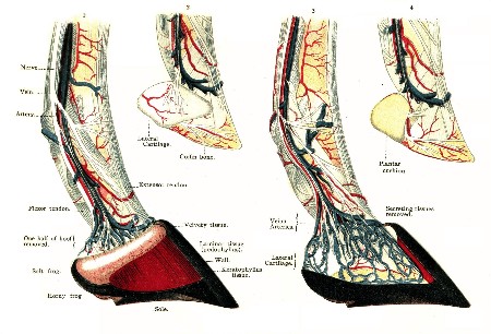
PLATE XXXIII. Anatomy of Foot.
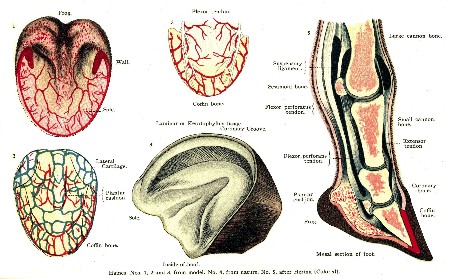
PLATE XXXIV. ANATOMY AND DISEASES OF THE FOOT.
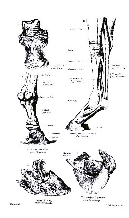
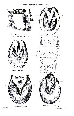
PLATE XXXV. SOUND AND CONTRACTED FEET.
Lastly, knuckling is produced by disease of the suspensory ligament or of the flexor tendons, whereby they are shortened, and by disease of the fetlock joints. (See p. 372.)
Treatment.—In young foals no treatment is necessary, unless there is some deformity present, since the legs straighten up without interference in the course of a few weeks. When knuckling has commenced, the indications are to relieve the tendons and ligaments by proper shoeing. The foot is to be prepared for the shoe by shortening the toe as much as possible, leaving the heels high; or if the foot is prepared in the usual way the shoe should be thin in front, with thick heels or high calks. For the hind feet a long-heeled shoe with calks seems to do best. Of course, when possible, the causes of knuckling are to be removed; since this can not always be done, however, the time may come when the patient can no longer perform any service, particularly in those cases in which both fore legs are affected, and it becomes necessary either to destroy the animal or obtain relief by surgical interference. In such cases the tendons between the fetlock and knee may be divided for the purpose of obtaining temporary relief. Firing and blistering the parts responsible for the knuckling may, in some instances, effect a cure; but a consideration of these measures belongs properly to the treatment of the disease in which knuckling appears simply as a sequel.
WINDGALL.
Joints and tendons are furnished with sacs containing a lubricating fluid called synovia. When these sacs are overdistended by reason of an excessive secretion of synovia, they are called windgalls. They form a soft, puffy tumor about the size of a hickory nut, and are most often found in the fore leg, at the upper part of the fetlock joint, between the tendon and the shin bone. When they develop in the hind leg it is not unusual to see them reach the size of a walnut. Occasionally they appear in front of the fetlock on the border of the tendon. The majority of horses are not subject to them after colt-hood has passed. (See also p. 355.)
Causes.—Windgalls are often seen in young, overgrown horses, in which the body seems to have outgrown the ability of the joints to sustain the weight. In cart and other horses used to hard work, in trotters with excessive knee action, in hurdle racers and hunters, and in most cow ponies there is a predisposition to windgalls. Street-car horses and others used to start heavy loads on slippery streets are the ones most liable to develop windgalls in the hind legs.
Symptoms.—The tumor is more or less firm and tense when the foot is on the ground, but is soft and compressible when the foot is off the ground. In old horses windgalls generally develop slowly and cause no inconvenience. If they are caused by excessive tension of the joint the tumor develops rapidly, is tense, hot, and painful, and the animal is exceedingly lame. The patient stands with the joint flexed, and walks with short steps, the toe only being placed on the ground. When the tumor is large and situated upon the inside of the leg it may be injured by interfering, causing stumbling and inflammation of the sac. Rest generally causes the tumor to diminish in size, only to fill up again after renewed labor. In old cases the tumors are hardened, and may become converted into bone by a deposit of the lime salts.
Treatment.—The large, puffy joints of suckling colts, as a rule, require no treatment, for as the animal grows older the parts clean up and after a time the swelling entirely disappears.
When the trouble is from an injury, complete rest is to be obtained by the use of slings and a high-heeled shoe. Cold-water douches should be used once or twice a day, followed by cold-water bandages, until the fever has subsided and the soreness is largely removed, when a blister is to be applied.
In old windgalls, which cause more or less stiffness, some relief may be had by the use of cold-compress bandages, elastic boots, or the red iodid of mercury blisters. Opening the sacs, as recommended by some authors, is of doubtful utility, and should be adopted only by the surgeon capable of treating the wound he has made. Enforced rest until complete recovery is effected should always be insisted upon, since a too early return to work is sure to be followed by relapse.
SPRAIN OF THE FETLOCK.
Sprain of the fetlock joint is most common in the fore legs, and, as a rule, affects but one at a time. Horses doing fast work, as trotters, runners, steeplechasers, hunters, cow ponies, and those that interfere, are particularly liable to this injury.
Causes.—Horses knuckling at the fetlock, and all those with diseases which impair the powers of locomotion, such as navicular disease, contracted heels, sidebones, chronic laminitis, etc., are predisposed to sprains of the fetlock. It generally happens from a misstep, stumbling, or slipping, which results in the joint being extended or flexed to excess. The same result may happen where the foot is caught in a rut, a hole in a bridge, or in a car track, and the animal falls or struggles violently. Direct blows and punctured wounds may also set up inflammation of the joint.
Symptoms.—The symptoms of sprain of the fetlock vary with the severity of the injury. If slight, there may be no lameness, but simply a little soreness, especially when the foot strikes on uneven ground and the joint is twisted a little. In more severe cases the joint swells, is hot and puffy, and the lameness may be so intense as to compel the animal to hobble on three legs. While at rest the leg is flexed at the joint affected, and the toe rests on the ground.
Treatment.—If the injury is slight, cold-water bandages and a few days' rest are sufficient to effect recovery. When there is an intense lameness, swelling, etc., the leg should be placed under a constant stream of cold water, as described in the treatment for quittor. When the inflammation has subsided, a blister to the joint should be applied.
In some cases, especially in old horses long accustomed to fast work, the ligaments of the joints are ruptured, in whole or in part, and the lameness may last a long time. In these cases the joint should be kept completely at rest; this condition is best obtained by the application of plaster of Paris bandages, as in cases of fracture. As a rule, patients take kindly to this bandage, and, while wearing it, may be given the freedom of a roomy box or yard. If they are disposed to tear it off, or if sufficient rest can not otherwise be obtained, the patient must be kept in slings.
In the majority of instances the plaster bandage should remain on from two to four weeks. If the lameness returns when the bandage is removed, a new one should be put on. The swellings which always remains after the other evidences of the disease have disappeared, may be largely dissipated and the joint strengthened by the rise of the firing iron and blisters.
A joint once injured by a severe sprain never entirely regains its original strength, and is ever after particularly liable to a repetition of the injury.
RUPTURE OF THE SUSPENSORY LIGAMENT.
Sprain with or without rupture of the suspensory ligament may happen in either the fore or hind legs, and is occasionally seen in horses of all classes and at all ages. Old animals, however, and especially hunters, runners, and trotters, are the most subject to this injury, and with them the seat of the trouble is nearly always in one or both the fore legs. Horses used for heavy draft are more liable to have the ligament of the hind legs affected.
When the strain upon the suspensory ligament becomes too great, one or both of the branches may be torn from the sesamoid bones, one or both of the branches may be torn completely across, or the ligament may rupture above the point of division.
Symptoms.—The most common injury to the suspensory ligament is sprain of the internal branch in one of the fore legs. The trouble is indicated by lameness, heat, swelling, and tenderness of the affected branch, beginning just above the sesamoid bone and extending obliquely downward and forward to the front of the ankle. If the whole ligament is involved, the swelling comes on gradually, and is found above the fetlock and in front of the flexor tendons. The patient stands or walks upon the toe as much as possible, keeping the fetlock joint flexed so as to relieve the ligament of tension.
When both branches are torn from their attachments to the sesamoids, or both are torn across, the lameness comes on suddenly and is most intense; the fetlock descends, the toe turns up, and, as the animal attempts to walk, the leg has the appearance of being broken off at the fetlock. These symptoms, followed by heat, pain, and swelling of the parts at the point of injury, will enable anyone to make a diagnosis.
Treatment.—Sprain of the suspensory ligament, no matter how mild it may be, should always be treated by enforced rest of at least a month, and the application of cold douches and cold-water bandages, firmly applied until the fever has subsided, when a cantharides blister should be put on and repeated in two or three weeks if necessary. When rupture has taken place the patient should be put into slings and a constant stream of cold water allowed to trickle over the seat of injury until the fever is reduced. In the course of a week or ten days a plaster of Paris splint, such as is used in fractures, is to be applied and left on for a month or six weeks. When this is taken off, blisters may be used to remove the remaining soreness; but it is useless to expect a removal of all the thickening, for, in the process of repair, new tissue has been formed which will always remain.
In old cases of sprain the firing iron may often be used with good results. As a rule, severe injuries to the suspensory ligament incapacitate the subject for anything but slow, light work.
OVERREACH.
When the shoe of the hind foot strikes and injures the heel or quarter of the forefoot the horse is said to overreach. It rarely happens except when the animal is going fast; hence is most common in trotting and running horses. In trotters the accident generally happens when the animal breaks from a trot to a run. The outside heels and quarters are most liable to the injury.
Symptoms.—The coronet at the heel or quarter is bruised or cut, the injury in some instances involving the horn as well. When the hind foot strikes well back on the heel of the fore foot—an accident known among horsemen as "grabbing"—the shoe may be torn from the forefoot or the animal may fall to its knees. Horses accustomed to overreaching are often "bad breakers," for the reason that the pain of the injury so excites them that they can not readily be brought back to the trotting gait.
Treatment.—If the injury is but a slight bruise, cold-water bandages applied for a few days will remove all the soreness. If the parts are deeply cut, more or less suppuration will follow, and, as a rule, it is well to poultice the parts for a day or two, after which cold baths may be used, or the wounds dressed with tincture of aloes, oakum, and a roller bandage.
When an animal is known to be subject to overreaching, he should never be driven fast without quarter boots, which are specially made for the protection of the heels and quarters.
If there is a disposition to "grab" the forward shoes, the trouble may be remedied by having the heels of these shoes made as short as possible, while the toe of the hind foot should project well over the shoe. When circumstances permit of their use, the fore feet may be shod with the "tips" instead of the common shoe, as described in treatment for contracted heels.
CALK WOUNDS.
Horses wearing shoes with sharp calks are liable to wounds of the coronary region, either from trampling on themselves or on each other. These injuries are most common in heavy draft horses, especially on rough roads and slippery streets. The fore feet are more liable than the hind ones, and the seat of injury is commonly on the quarters. In the hind feet the wound often results from the animal resting with the heel of one foot set directly over the front of the other. In these cases the injury is generally close to the horn, and often involves the coronary band, the sensitive laminæ, the extensor tendon, and even the coffin bone.
Treatment.—Preventive measures include the use of boots to protect the coronet of the hind foot and of a blunt calk on the outside heel of the fore shoe, since this is generally the offending instrument when the fore feet are injured. If the wound is not deep and the soreness slight, cold-water bandages and a light protective dressing, such as carbolized cosmoline, will be all that is needed. When the injury is deep, followed by inflammation and suppuration of the coronary band, lateral cartilages, sensitive laminæ, etc., active measures must be resorted to. Cold, astringent baths, made by adding 2 ounces of sulphate of iron to 1 gallon of water, should be used, followed by poultices, if it is necessary to hasten the cleansing of the wound by stimulating the sloughing process. If the wound is deep between the horn and the skin, especially over the anterior tendon, the horn should be cut away so that the injured tissues may be exposed. The subsequent treatment in these cases should follow the directions given in the article on toe cracks.
FROSTBITES.
Excepting the ears, the feet and legs are about the only parts of the horse liable to become frostbitten. The cases most commonly seen are found in cities, especially among car horses, where salt is used for the purpose of melting the snow on curves and switches. This mixture of snow and salt is splashed over the feet and legs, rapidly lowering the temperature of the parts to the freezing point. In mountainous districts, where the snowfall is heavy and the cold often intense, frostbites are not uncommon, even among animals running at large.
Symptoms.—When the frosting is slight the skin becomes pale and bloodless, followed soon after by intense redness, heat, pain, and swelling. In these cases the hair may fall out and the epidermis peel off, but the inflammation soon subsides, the swelling disappears, and only an increased sensitiveness to cold remains.
In more severe cases irregular patches of skin are destroyed and after a few days slough away, leaving slow-healing ulcers behind. If produced by low temperatures and deep snow, the coronary band is the part most often affected.
In many instances there is no destruction of the skin, but simply a temporary suspension of the horn-producing function of the coronary band. The fore feet are more often affected than the hind ones, and the heels and quarters are less often involved than the front part of the foot. The coronary band becomes hot, swollen, and painful, and after two or three days the horn separates from the band and slight suppuration follows. For a few days the animal is lame, but as the suppuration disappears the lameness subsides. New horn, often of an inferior quality, is produced by the coronary band, and in time the cleft is grown off and complete recovery is effected. The frog is occasionally frostbitten and may slough off, exposing the soft tissues beneath and causing severe lameness for a time.
Treatment.—Simple frostbites are best treated by cold fomentations followed by applications of a 5 per cent solution of carbolized oil. When portions of the skin are destroyed, their early separation should be hastened by warm fomentations and poultices. Ulcers are to be treated by the application of stimulating dressings, such as carbolized oil, a 1 per cent solution of nitrate of silver or of chlorid of zinc, with pads of oakum and flannel bandages. In many of these cases recovery is exceedingly slow. The new tissue by which the destroyed skin is replaced always shrinks in healing, and, as a consequence, unsightly scars are unavoidable. When the coronary band is involved it is generally advisable to blister the coronet over the seat of injury as soon as the suppuration ceases, for the purpose of stimulating the growth of new horn. Where a crevasse is formed between the old and the new horn no serious trouble is liable to be met with until the cleft is nearly grown out, when the soft tissues may be exposed by a breaking off of the partly detached horn. But even if this accident happens final recovery is effected by poulticing the foot until a sufficient growth of horn protects the parts from injury.
QUITTOR.
Quittor is a term applied to various affections of the foot wherein the tissues which are involved undergo a process of degeneration that results in the formation of a slough followed by the elimination of the diseased structures by means of a more or less extensive suppuration.
For convenience of consideration quittors may be divided into four classes, as suggested by Girard: (1) Cutaneous quittor, which is known also as simple quittor, skin quittor, and carbuncle of the coronet; (2) tendinous quittor; (3) subhorny quittor; and (4) cartilaginous quittor.
CUTANEOUS QUITTOR.
Simple quittor consists in a local inflammation of the skin and subcutaneous connective tissue on some part of the coronet, followed by a slough and the formation of an ulcer which heals by suppuration.
It is an extremely painful disease, owing to the dense character of the tissues involved; for in all dense structures the swelling which accompanies inflammation always produces intense pressure. This pressure not only adds to the patient's suffering but may at the same time endanger the life of the affected parts by strangulating the blood vessels. It is held by some writers that simple quittor is most often met with in the hind feet, but in my experience more than two-thirds of the cases have developed in the fore feet. While any part of the coronet may become the seat of attack, the heels and quarters are undoubtedly most liable.
Causes.—Bruises and other wounds of the coronet are often the cause of cutaneous quittor, yet there can be no question that in the great majority of cases the disease develops without any known cause. For some reason not yet satisfactorily explained most cases happen in the fall of the year. One explanation of this fact has been attempted in the statement that the disease is due to the injurious action of cold and mud. This claim, however, seems to lose force when it is remembered that in many parts of this country the most mud, accompanied with freezing and thawing weather, is seen in the early springtime without a corresponding increase of quittor. Furthermore, the serious outbreaks of this disease in the mountainous regions of Colorado, Wyoming, and Montana are seen in the fall and winter seasons, when the weather is the driest. It may be claimed, and perhaps with justice, that during these seasons, when the water is low, animals are compelled to wade through more mud to drink from lakes and pools than is necessary at other seasons of the year, when these lakes and pools are full. Add to these conditions the further fact that much of this mud is impregnated with alkaline salts which, like the mineral substances always found in the mud of cities, are more or less irritating, and it seems fair to conclude that under certain circumstances mud may become an important factor in the production of quittor.[3]
While this disease attacks any and all classes of horses, it is the large, common breeds, with thick skins, heavy coats, and coarse legs that are most often affected. Horses well groomed and cared for in stables seem to be less liable to the disease than those running at large or than those which are kept and worked under adverse circumstances.
Symptoms.—Lameness, lasting from one to three or four days, nearly always precedes the development of the strictly local evidences of quittor. The next sign is the appearance of a small, tense, hot, and painful tumor in the skin of the coronary region. If the skin of the affected foot is white, the inflamed portion will present a dark-red or even a purplish appearance near the center. Within a few hours the ankle, or even the whole leg as high as the knee or hock, becomes much swollen. The lameness is now so great that the patient refuses to use the foot at all, but carries it if compelled to move. As a consequence, the opposite leg is required to do the work of both, and if the animal persists in standing a greater part of the time it, too, becomes swollen. In many of these cases the suffering is so intense during the first few days as to cause general fever, dullness, loss of appetite, and increased thirst. Generally the tumor shows signs of suppuration within 48 to 72 hours after its first appearance; the summit softens, a fluctuating fluid is felt beneath the skin, which soon ulcerates completely through, causing the discharge of a thick, yellow, bloody pus, containing shreds of dead tissue which have sloughed away. The sore is now converted into an open ulcer, generally deep, nearly or quite circular in outline, and with hardened base and edges. In exceptional cases large patches of skin, varying from 1 to 2-1/2 inches in diameter, slough away at once, leaving an ugly superficial ulcer. These sores, especially when deep, suppurate freely, and if there are no complications they tend to heal rapidly as soon as the degenerated tissue has softened and is entirely removed. When suppuration is fully established, the lameness and general symptoms subside. When but a single tumor and abscess form, the disease progresses rapidly, and recovery, under proper treatment, may be effected in from two to three weeks; but when two or more tumors are developed at once, or if the formation of one tumor is rapidly succeeded by another for an indefinite time, the sufferings of the patient are greatly increased, the case is more difficult to treat, and recovery is more slow and less certain.
This form of quittor is often complicated with the tendinous and subhorny quittors by an extension of the sloughing process.
Treatment.—The first step in the treatment of an outbreak of quittor should be the removal of all exciting causes. Crowding animals into small corrals and stables, where injuries to the coronet are likely to happen from trampling, especially among unbroken range horses, must be avoided as much as possible.
Watering places accessible without having to wade through mud should be provided. In towns, where the mud or dust is largely impregnated with mineral products, it is not possible to adopt complete preventive measures. Much can be done, however, by careful cleansing of the feet and legs as soon as the animal returns from work. Warm water should be used to remove the mud and dirt, after which the parts are to be thoroughly dried with soft cloths.
The means which are to be adopted for the cure of cutaneous quittor vary with the stage of the disease at the time the case is presented for treatment. If the case is seen early—that is, before any of the signs of suppuration have developed—the affected foot is to be placed under a constant stream of cold water, with the object of arresting a further extension of the inflammatory process. To accomplish this, put the patient in slings in a narrow stall having a slat or open floor. Bandage the foot and leg to the knee or hock, as the case may be, with flannel bandages loosely applied. Set a tub or barrel filled with cold water above the patient, and by the use of a small rubber hose of sufficient length make a siphon which will carry the water from the bottom of the tub to the leg at the top of the bandage. The stream of water should be quite small, and is to be continued until the inflammation has entirety subsided or until the presence of pus can be detected in the tumor. When suppuration has commenced, the process should be aided by the use of warm baths and poultices of lineseed meal or boiled turnips. If the tumor is of rapid growth, accompanied with intense pain, relief is obtained and sloughing largely limited by a free incision of the parts. The incision should be vertical and deep into the tumor, care being taken not to divide the coronary band entirely. If the tumor is large, more than one incision may be necessary.
The foot should now be placed in a warm bath for half an hour or longer and then poulticed. The hemorrhage produced by the cutting and encouraged by the warm bath is generally very copious and soon gives relief to the overtension of the parts.
In other cases it will be found that suppuration is well under way, so that the center of the tumor is soft when the patient is first presented for treatment. It is always good surgery to relieve the tumor of pus whenever its presence can be detected; hence, in these cases a free incision must be made into the softened parts, the pus evacuated, and the foot poulticed.
By surgical interference the tumor is now converted into an open sore or ulcer, which, after it has been well cleaned by warm baths and poultices applied for two or three days, needs to be protected by proper dressings. The best of all protective dressings is made of small balls, or pledgets, of oakum, carefully packed into the wound and held in place by a roller bandage 4 yards long, from 3 to 4 inches wide, made of common bedticking and skillfully applied.
The remedies which may be used to stimulate the healing process are many, and, as a rule, they are applied in the form of solutions or tinctures.
In my own practice I prefer a solution of bichlorid of mercury 1 part, water 500 parts, with a few drops of muriatic acid or a few grains of muriate of ammonia added to dissolve the mercury. The balls of oakum are wet with this solution before they are applied to the wound.
Among the other remedies which may be used, and perhaps with equally as good results, will be noted the sulphate of copper, iron, and zinc, 5 grains of either to the ounce of water; chlorid of zinc, 5 grains to the ounce; carbolic acid, 20 drops dissolved in an equal quantity of glycerin and added to 1 ounce of water; and nitrate of silver, 10 grains to the ounce of water.
If the wound is slow to heal, it will be found of advantage to change the remedies every few days.
If the wound is pale in color, the granulations transparent and glistening, the tincture of aloes, tincture of gentian, or the spirits of camphor may do best.
When the sore is red in color and healing rapidly, an ointment made of 1 part of carbolic acid to 40 parts of cosmoline or vaseline is all that is needed.
If the granulations continue to grow until a tumor is formed which projects beyond the surrounding skin, it should be cut off with a sharp, clean knife, and the foot poulticed for twenty-four hours, after which the wound is to be well cauterized daily with lunar caustic and the bandages applied with great firmness.
The question as to how often the dressings should be renewed must be determined by the condition of the wound, etc. If the sore is suppurating freely, it will be necessary to renew the dressing every 24 or 48 hours; if the discharge is small in quantity and the patient comfortable, the dressing may be left on for several days; in fact, the less often the wound is disturbed, the better, so long as the healing process is healthy. When the sore commences to "skin over," the edges should be lightly touched with lunar caustic at each dressing. The patient may now be given a little exercise daily, but the bandages must be kept on until the wound is entirely healed.
TENDINOUS QUITTOR.
This form of quittor differs from the cutaneous in that it not only affects the skin and subcutaneous tissues, but involves also the tendons of the leg, the ligaments of the joints, and, in many cases, the bones of the foot as well.
Fortunately, this form of quittor is less common than the preceding, yet any case beginning as simple cutaneous quittor may at any time during its course become complicated by the death of some part of the tendons, by gangrene of the ligaments, sloughing of the coronary band, caries of the bones, or inflammation and suppuration of the synovial sacs and joints, thereby converting a simple quittor into one which will, in all probability, either destroy the patient's life or maim him for all time.
Causes.—Tendinous quittor is caused by the same injuries and influences that produce the simple form. Zundel believes it to be a not infrequent accompaniment of distemper. In my own experience I have seen nothing to verify this belief, but I am convinced that young animals are more liable to have tendinous quittor than older ones, and that they are much more likely to make a good recovery.
Symptoms.—When a case of simple quittor is transformed into the tendinous variety the change is announced by a sudden increase in the severity of all the symptoms. On the other hand, if the attack primarily is one of tendinous quittor, the earliest symptom seen is a well-marked lameness. In those cases due to causes other than injuries this lameness is at first very slight, and the animal limps no more in trotting than in walking; later on, generally during the next 48 hours, the lameness increases to such extent that the patient often refuses to use the leg at all. An examination made during the first two days rarely discloses any cause for this lameness; it may not be possible even to say with certainty that the foot is the seat of the trouble. On the third or fourth day, sometimes as late as the fifth, a doughy-feeling tumor will be found forming on the heel or quarter. This tumor grows rapidly, feels hot to the touch, and is extremely painful. As the tumor develops, all the other symptoms increase in intensity; the pulse is rapid and hard; the breathing quick; the temperature elevated 3° or 4°; the appetite is gone; thirst increased; and the lameness so great that the foot is carried if locomotion is attempted. At this stage of the disease the patient generally seeks relief by lying upon the broad side, with outstretched legs; the coat is bedewed with a clammy sweat, and every respiration is accompanied with a moan. The leg soon swells to the fetlock; later this swelling gradually extends to the knee or hock, and in some cases reaches the body. As a rule, several days elapse before the disease develops a well-defined abscess, for, owing to the dense structure of the bones, ligaments, and tendons, the suppurative process is a slow one, and the pus is prevented from readily collecting in a mass.
I made a post-mortem examination on a typical case of this disease, in which the animal had died on the fourth day after being found on the range slightly lame. The suffering had been intense, yet the only external evidences of the disease consisted in the shedding of the hoof from the right fore foot and a limited swelling of the leg to the knee. The sloughing of the hoof took place two or three hours before death, and was accompanied with but little suppuration and no hemorrhage. The skin from the knee to the foot was thickened from watery infiltration (edema), and on the inside quarter three holes, each about one-half inch in diameter, were found. All had ragged edges, while but one had gone deep enough to perforate the coronary band. The loose connective tissue beneath the skin was distended, with a gelatinous infiltration over the whole course of the flexor tendons and to the fetlock joint over the tendon in front. The soft tissues covering the coffin bone were loosened in patches by collections of pus which had formed beneath the sensitive laminæ. The coffin and pastern joints were inflamed, as were also the coffin, navicular, and coronet bones, while the outside toe of the coffin bone had become softened from suppuration until it readily crumbled between the fingers. The coronary band was largely destroyed and completely separated from the other tissues of the foot. The inner lateral cartilage was gangrenous, as was also a small spot on the extensor tendon near its point of attachment on the coffin bone. Several small collections of pus were found deep in the connective tissue of the coronary region; along the course of the sesamoid ligaments; in the sheath of the flexor tendons; under the tendon just below the fetlock joint in front; and in the coffin joint.
But all cases of tendinous quittor are by no means so complicated as this one was. In rare instances the swelling is slight, and after a few days the lameness and other symptoms subside, without any discharge of pus from an external opening. In most cases, however, from one to half a dozen or more soft points arise on the skin of the coronet, open, and discharge slowly a thick, yellow, fetid, and bloody matter. In other cases the suppurative process is largely confined to the sensitive laminæ and plantar cushion, when the suffering is intense until the pus finds an avenue of escape by separating the hoof from the coronary band, at or near the heels, without causing a loss of the whole horny box. When the flexor tendon is involved deep in the foot, the discharge of pus usually takes place from an opening in the follow of the heel; if the sesamoid ligament or the sheath of the flexors are affected, the opening is nearer the fetlock joint, although in most of these cases the suppuration spreads along the course of the tendons until the navicular joint is involved, and extensive sloughing of the deeper parts follows.
Treatment.—The treatment of tendinous quittor is to be directed toward the saving of the foot. First of all an effort must be made to prevent suppuration; if the patient is seen at the beginning, cold irrigation, recommended in the treatment for cutaneous quittor, is to be resorted to. Later, when the tumor is forming on the coronet, the knife must be used, and a free and deep incision made into the swelling. Whenever openings appear, from which pus escapes, they should be carefully probed; in all instances these fistulous tracts lead down to dead tissue which nature is trying to remove by the process of sloughing. If a counter opening can be made, which will enable a more ready escape of the pus, it should be done at once; for instance, if the probe shows that the discharge originates from the bottom of the foot, the sole must be pared through over the seat of trouble. Whenever suppuration has commenced the process is to be stimulated by the use of warm baths and poultices. The pus which accumulates in the deeper parts, especially along the tendons, around the joints, and in the hoof, is to be removed by pressure and injections made with a small syringe, repeated two or three times a day. As soon as the discharge assumes a healthy character and diminishes in quantity, stimulating solutions are to be injected into the open wounds. When the tendons, ligaments, and other deeper parts are affected, a strong solution of carbolic acid—1 to 4—should be used at first; or strong solutions of tincture of iodin, sulphate of iron, sulphate of copper, bichlorid of mercury, etc., may be used in place of the carbolic; after this the remedies and dressings directed for use in simple quittor are to be used. In those cases in which the fistulous tracts refuse to heal it is often necessary to burn them out with a saturated solution of caustic soda, equal parts of muriatic acid and water, or, better still, with a long, thin iron, heated white hot.
But no matter what treatment is adopted, a large percentage of the cases of tendinous quittor fail to make good recoveries. If the entire hoof sloughs away, the growth of a new, but soft and imperfect hoof may be obtained by carefully protecting the exposed tissues with proper bandages. When the joints are opened by deep sloughing, recovery may eventually take place, but the joint remains immovable ever after. If caries of a small part of the coffin bone takes place, it may be removed by an operation; but if much of the bone is affected, or if the navicular and coronet bones are involved in the carious process, the only hope for a cure is in the amputation of the foot. This operation is advisable only when the animal is valuable for breeding purposes. In all other cases in which there is no hope for recovery the patient's suffering should be relieved by death. In tendinous quittor much thickening of the coronary region, and sometimes of the ankle and fetlock, remains after suppuration has ceased and the fistulous tracts have healed. To stimulate the reabsorption of this new and unnecessary tissue, the parts should be fired with the hot iron, or, in its absence, repeated blistering with the biniodid of mercury ointment may largely accomplish the same results.
SUBHORNY QUITTOR.
This is the most common form of the disease. It is generally seen in but one foot at a time, and more often in the fore than in the hind feet. It nearly always attacks the inside quarter, but may affect the outside, the band in front, or the heel, where it is of but little consequence. It consists in the inflammation of a small part of the coronary band and adjacent skin, followed by sloughing and suppuration, which in most cases extends to the neighboring sensitive laminæ.
Causes.—Injuries to the coronet, such as bruises, overreaching, and calk wounds, are considered as the common causes of this disease. Still, cases occur in which there appears to be no existing cause, just as in the other forms of quittor, and it seems fair to conclude that subhorny quittor may also be produced by internal causes.
Symptoms.—At the outset the lameness is always severe, and the patient often refuses to use the affected foot. Swelling of the coronet close to the top of the hoof causes the quarter to protrude beyond the wall. This tumor is extremely sensitive, and the whole foot is hot and painful. After a few days a small spot in the skin, over the most elevated part of the tumor, softens and opens or the hoof separates from the coronary band at the quarter or well back toward the heel. From this opening, wherever it may be, a thin, watery, often dark, offensive discharge escapes, at times mixed with blood and always containing a considerable percentage of pus.
Probing will now disclose a fistulous tract leading to the bottom of the diseased tissues. If the opening is small, there is a tendency upon the part of the suppurative process to spread downward; the pus gradually separates the hoof from the sensitive laminæ until the sole is reached, and even a portion of this may be undermined.
As a rule, the slough in this form of quittor is not deep, and if the case receives early and proper treatment complications are generally avoided; but if the case is neglected, and, occasionally, even in spite of the best treatment, the disease spreads until the tendon in front, the lateral cartilage, or the coffin bone and joint as well are involved.
In all cases of subhorny quittor much relief is experienced when the slough comes away, and rapid recovery is made. If, however, after the lapse of a few days, the lameness remains and the wound continues to discharge a thin, unhealthy matter, the probabilities are that the disease is spreading, and pus collecting in the deeper parts of the foot. In Zundel's opinion, if the use of the probe now detects a pus cavity below the opening, a cartilaginous quittor is in the course of development.
Treatment.—Hot baths and poultices are to be used until the presence of pus can be determined, when the tumor is to be opened with a knife or sharp-pointed iron heated white hot. The hot baths and poultices are now continued for a few days or until the entire slough has come away and the discharge is diminished, when dressings recommended in the treatment for cutaneous quittor are to be used until recovery is completed. In cases in which the discharge comes from a cleft between the upper border of the hoof and the coronary band, always pare away the loosened horn, so that the soft tissues beneath are fully exposed, care being taken not to injure the healthy parts. This operation permits of a thorough inspection of the diseased parts, the easy removal of all gangrenous tissue, and a better application of the necessary remedies and dressings. The only objection to the operation is that the patient is prevented from being early returned to work.
When the probe shows that pus has collected under the coffin bone the sole must be pared through, and, if caries of the bone is present, the dead parts cut away. After either of these operations the wound is to be dressed with the oakum balls, saturated in the bichlorid of mercury solution, as previously directed, and the bandages tightly applied. Generally the discharge for the first two or three days is so great that the dressings need to be changed every 24 hours; but when the discharge diminishes, the dressing may be left on from one to two weeks. Before the patient is returned to work, a bar shoe should be applied, since the removed quarter or heel can only be made perfect again by a new growth from the coronary band.
Tendinous or cartilaginous complications are to be treated as directed under those headings.
CARTILAGINOUS QUITTOR.
This form of quittor may commence as a primary inflammation of the lateral cartilage, but in the great majority of cases it appears as a sequel to cutaneous or subhorny quittor. It may affect either the fore or hind feet, but is most commonly seen in the former. As a rule, it attacks but one foot at a time, and but one of the cartilages, generally the inner one. It is always a serious affection for the reason that, in many cases, it can only be cured by a surgical operation, requiring a thorough knowledge of the anatomy of the parts involved, and much surgical skill.
Causes.—Direct injuries to the coronet, such as trampling, pricks, burns, and the blow of some heavy falling object which may puncture, bruise, or crush the cartilage, are the common direct causes of cartilaginous quittor. Besides being a sequel to the other forms of quittor, it sometimes develops as a complication in suppurative corn, canker, grease, laminitis, and punctured wounds of the foot. Animals used for heavy draft, and those with flat feet and low heels, are more liable to the disease than others, for the reason that they are more exposed to injury. Rough roads also predispose to the disease by increasing liability to injury.
Symptoms.—When the disease commences as a primary inflammation of the cartilage, lameness develops with the formation of a swelling on the side of the coronet over the quarter. The severity of this lameness depends largely upon the part of the cartilage which is diseased, for if the disease is situated in that part of the cartilage nearest the heel, where the surrounding tissues are soft and spongy, the lameness may be very slight, especially if the patient is required to go no faster than a walk; but when the middle and anterior parts of the cartilage are diseased, the pain and consequent lameness are much greater, for the tissues are less elastic and the coffin joint is more liable to become affected.
Except in the cases to be noted hereafter, one or more fistulous openings finally appear in the tumor on the coronet. These openings are surrounded by a small mass of granulations which are elevated above the adjacent skin and bleed readily if handled. A probe shows these fistulous tracts to be more or less sinuous, but always leading to one point—the gangrenous cartilage. When cartilaginous quittor happens as a complication of suppurative corn, or from punctured wounds of the foot, the fistulous tract may open alone at the point of injury on the sole.
The discharge in this form of quittor is generally thin, watery, and contains pus enough to give it a pale-yellow color; it is offensive to the sense of smell, due to the detachment of small flakes of cartilage which have become gangrenous and are seen in the discharge as small, greenish-colored particles. In old cases it is not unusual to find some of the fistulous openings heal at the surface; this is followed by the gradual collection of pus in the deeper parts, forming an abscess, which in a short time opens at a new point. The wall of the hoof, over the affected quarter and heel, in very old cases becomes rough and wrinkled like the horn of a ram, and generally it is thicker than the corresponding quarter, owing to the stimulating effect which the disease has upon the coronary band.
Complications may arise by an extension of the disease to the lateral ligament of the coffin joint, to the joint itself, to the plantar cushion, and by caries of the coffin bone.
Treatment.—Before recovery can take place all the dead cartilage must be removed. In rare instances this is effected by nature without assistance. Usually, however, the disease does not tend to recovery, and active curative measures must be adopted. The best and simplest treatment, in a majority of cases, is the injection of strong caustic solutions, which destroy the diseased cartilage and cause its discharge, along with the other products of suppuration. In favorable cases these injections will secure a healing of the wound in from two to three weeks. While the saturated solution of sulphate of copper, or a solution of 10 parts of bichlorid of mercury to 100 parts of water, has given the best results in my hands, equally as favorable success has been secured by others from the use of caustic soda, nitrate of silver, sulphate of zinc, tincture of iodin, etc. No matter which one of these remedies may be selected, however, it must be used at least twice a day for a time. The solution is injected into the various openings with force enough to drive it to the bottom of the wound, after which the foot is to be dressed with a pad of oakum, held in place by a roller bandage tightly applied. While it is not always necessary, it is often of advantage to relieve the pressure on the parts by rasping away the hoof over the seat of the cartilage; the coronary band and laminæ should not be injured in the operation.
If the caustic injections prove successful, the discharge will become healthy and gradually diminish, so that by the end of the second week the fistulous tracts are closing up and the injections are made with much difficulty.
If, on the other hand, there is but little or no improvement after this treatment has been used for three weeks, it may reasonably be concluded that the operation for the removal of the lateral cartilage must be resorted to for the cure of the trouble. As this operation can be safely undertaken only by an expert surgeon, it will not be described in this connection.
THRUSH.
Thrush is characterized by an excessive secretion of unhealthy matter from the cleft of the frog. While all classes of horses are liable to this affection, it is more often seen in the common draft horse than in any other breed, owing to the conditions of servitude and not to the fault of the breed. Country horses are much less subject to the disease, except in wet, marshy districts, than are the horses used in cities and towns.
Causes.—The most common cause of thrush is the filthy condition of the stable in which the animal is kept. Mares are more liable to contract the disease in the hind feet when filth is the cause, while the gelding and stallion are more liable to develop it in the fore feet. Hard work on rough and stony roads may also induce the disease, as may a change from dryness to excessive moisture. The latter cause is often seen to operate in old track horses, whose feet are constantly soaked in the bathtub for the purpose of relieving soreness. Muddy streets and roads, especially where mineral substances are plentiful, excite this abnormal condition of the frog. Contracted heels, scratches, and navicular disease predispose to thrush, while by some a constitutional tendency is believed to exist among certain animals which otherwise present a perfect frog.
Symptoms.—At first there is simply an increased moisture in the cleft of the frog, accompanied with an offensive smell. After a time a considerable discharge takes place—thin, watery, and highly offensive, changing gradually to a thicker puriform matter, which rapidly destroys the horn of the frog. Only in old and severe cases is the patient lame and the foot feverish—cases in which the whole frog is involved in the diseased process.
Treatment.—Thrushes are to be treated by cleanliness, the removal of all exciting causes, and a return of the frog to its normal condition. As a rule, the diseased and ragged portions of horn are to be pared away and the foot poulticed for a day or two with boiled turnips, to which may be added a few drops of carbolic acid or a handful of powdered charcoal to destroy the offensive smell. The cleft of the frog and the grooves on its edges are then to be cleaned and well filled with dry calomel and the foot dressed with oakum and a roller bandage. If the discharge is profuse, the dressing should be changed daily; otherwise it may be left on two or three days. Where a constitutional taint is supposed to exist, with swelling of the legs, grease, etc., a purgative, followed by dram doses of sulphate of iron, repeated daily, may be prescribed. In cases where the growth of horn seems too slow a Spanish-fly blister applied to the heels is often followed by good results. Feet in which the disease is readily induced may be protected in the stable with a leather boot. If the thrush is but a sequel to other disease, a permanent cure may not be possible.
CANKER.
Canker of the foot is due to the rapid reproduction of a vegetable parasite. It not only destroys the sole and frog, but, by setting up a chronic inflammation in the deeper tissues, prevents the growth of a healthy horn by which the injury may be repaired. Heavy cart horses are more often affected than those of any other class.
Causes.—The essential element in the production of canker is the parasite; consequently the disease may be called contagious. As in all other diseases due to specific causes, however, the seeds of the disorder must find a suitable soil in which to grow before they are reproduced. It may be said, then, that the conditions which favor the preparation of the tissues for a reception of the seeds of this disease are simply predisposing causes.
The condition most favorable to the development of canker is dampness—in fact, dampness seems indispensable to the existence and growth of the parasite; the disease is rarely, if ever, seen in high, dry districts, and is much more common in rainy than in dry seasons. Filthy stables and muddy roads have been classed among the causes of canker; but it is very doubtful whether these conditions can do more than favor a preparation of the foot for the reception of the disease germ.
All injuries to the feet, by exposing the soft tissues, may render the animal susceptible to infection; but neither the injury nor the irritation and inflammation of the tissues which follow are sufficient to induce the disease.
For some unknown reason horses with lymphatic temperaments—thick skins, flat feet, fleshy frogs, heavy hair, and particularly with white feet and legs—are especially liable to canker.
Symptoms.—Usually, canker is confined to one foot; but it may attack two, three, or all of the feet at once; or, as is more commonly seen, the disease attacks first one then another, until all may have been successively affected. When the disease follows an injury which has exposed the soft tissues of the foot, the wound shows no tendency to heal, but instead there is secreted from the inflamed parts a profuse, thin, fetid, watery discharge, which gradually undermines and destroys the surrounding horn, until a large part of the sole and frog is diseased. The living tissues are swollen, dark colored, and covered at certain points with particles of new, soft, yellowish, thready horn, which are constantly undergoing maceration in the abundant liquid secretion by which they are immersed. As this secretion escapes to the surrounding parts, it dries and forms small, cheesy masses composed of partly dried horny matter, exceedingly offensive to the sense of smell. When the disease originates independently of an injury, the first evidences of the trouble are the offensive odor of the foot, the liquid secretion from the cleft and sides of the frog, and the rotting away of the horn of the frog and sole.
In the earlier stages there is no interference with locomotion, but later the foot becomes sensitive, particularly if the animal is used on rough roads, and, finally, when the sole and frog are largely destroyed the lameness is severe.
Treatment.—Since canker does not destroy the power of the tissues to produce horn, but rather excites them to an excessive production of an imperfect horn, the indications for treatment are to restore the parts to a normal condition, when healthy horn may again be secreted. In my experience, limited though it has been, the old practice of stripping off the entire sole and deep cauterization, with either the hot iron or strong acids, is not attended with uniformly good results.
I am of the opinion that recovery can generally be effected as surely and as speedily with measures which are less heroic and much less painful. True, the treatment of canker is likely to exhaust the patience, and sometimes the resources, of the attendant; but after all success depends more on the persistent application of simple remedies and great cleanliness than on the special virtues of any particular drug.
First, then, clean the foot with warm baths and apply a poultice containing powdered charcoal or carbolic acid. A handful of the charcoal or a tablespoonful of the acid mixed with the poultice serves to destroy much of the offensive odor. The diseased portions of horn are to be carefully removed with sharp instruments, until only healthy horn borders the affected parts. The edges of the sound horn are to be pared thin, so that the swollen soft tissues may not overlap their borders. With sharp scissors cut off all the prominent points on the soft tissues, shorten the walls of the foot, and nail on a broad, plain shoe. The foot is now ready for the dressings, and any of the many stimulating and drying remedies may be used; but it will be necessary to change frequently from one to another, until finally all may be tried.
The list from which a selection may be made comprises wood tar, gas tar, petroleum, creosote, phenic acid; sulphates of iron, copper, and zinc; chlorid of zinc, bichlorid of mercury, calomel, caustic soda, nitrate of silver, chlorid of lime; carbolic, nitric, and sulphuric acids.
In practice I prefer to give the newly shod foot a bath for an hour or two in a solution of the sulphate of iron made by adding 2 ounces of the powdered sulphate to a gallon of cold water. When the foot is removed from the bath it is dressed with oakum balls dipped in a mixture made of Barbados tar 1 part, oil of turpentine 8 parts, to which is slowly added 2 parts of sulphuric acid, and the mixture well stirred and cooled. The diseased parts being well covered with the balls, a pad of oakum sufficiently thick to cause considerable pressure is placed over them, and all are held in place by pieces of heavy tin fitted to slip under the shoe. The whole foot is now incased in a boot or folded gunny sack and the patient turned into a loose, dry box. The dressings are to be changed daily or even twice a day at first. When they are removed, all pieces of new horny matter which are now firmly adherent must be rubbed off with the finger or a tent of oakum. As the secretion diminishes, dry powders, such as calomel, sulphates of iron, copper, etc., may prove of most advantage. The sulphates should not be used pure, but are to be mixed with powdered animal charcoal in the proportion of one of the former to eight or ten of the latter. When the soft tissues are all horned over, the dressings should be continued for a time, weak solutions being used to prevent a recurrence of the disease. If the patient is run down in condition, bitter tonics, such as gentian, may be given in 2-dram doses twice a day and a liberal diet of grain allowed.
CORNS.
A corn is an injury to the living horn of the foot, involving the soft tissues beneath, whereby the capillary blood vessels are ruptured and a small quantity of blood escapes which, by permeating the horn in the immediate neighborhood, stains it a dark color. If the injury is continuously repeated, the horn becomes altered in character and the soft tissues may suppurate or a horny tumor develop. Corns always appear in the sole in the angle between the bar and the outside wall of the hoof. In many cases the laminæ of the bar, of the wall, or of both, are involved at the same time.
Three kinds of corns are commonly recognized—the dry, the moist, and the suppurative—a division based solely on the character of the conditions which follow the primary injury.
The fore feet are almost exclusively the subjects of the disease, for two reasons: First, because they support a greater part of the body; secondly, because the heel of the fore foot during progression is first placed upon the ground, whereby it receives much more concussion than the heel of the hind foot, in which the toe first strikes the ground.
Causes.—It may be said that all feet are exposed to corns, and that even the best feet may suffer from them when conditions necessary to the production of the peculiar injury are present. The heavier breeds of horses generally used for heavy work on rough roads and streets seem to be most liable to this trouble. Mules rarely have corns.
Among the causes and conditions which predispose to corns may be named high heels, which change the natural relative position of the bones of the foot and thereby increase the concussion to which these parts are subject; contracted heels, which in part destroy the elasticity of the foot, increase the pressure upon the soft tissues of the heel, and render lacerations more easy; long feet, which by removing the frog and heels too far from the ground deprive them of necessary moisture; this, in turn, reduces the elastic properties of the horn and diminishes the transverse diameter of the heels; weak feet, or those in which the horn of the wall is too thin to resist the tendency to spread, whereby the soft tissues are easily lacerated. Wide feet with low heels are always accompanied with a flat sole whose posterior wings either rest upon the ground or the shoe, and as a consequence are easily bruised; at the same time the arch of the sole is so broad and flat that it can not support the weight of the body, and in the displacement which happens when the foot is rested upon the ground the soft tissues are liable to become bruised or torn.
It is universally conceded that shoeing, either as a direct or predisposing cause, is most prolific in producing corns. One of the most serious as well as the most common of the errors in shoeing is to be found in the preparation of the foot. Instead of seeking to maintain the integrity of the arch, the first thing done is to weaken it by freely paring away the sole; nor does the mutilation end here, for the frog, which is nature's main support to the branches of the sole and the heels, is also largely cut away. This not only permits of an excessive downward movement of the contents of the horny box, but it at the same time removes the one great means by which concussion of the foot is destroyed. As adjuncts to the foregoing errors must be added the faults of construction in the shoe and in the way it is adjusted to the foot. An excess of concavity in the shoe, extending it too far back on the heels, high calks, thin heels which permit the shoe to spring, short heels with a calk set under the foot, and a shoe too light for the animal wearing it or for the work required of him, are all to be avoided as causes of corns. A shoe so set so as to press upon the sole or one that has been on so long that the hoof has overgrown it until the heels rest upon the sole and bars becomes a direct cause of corns. Indirectly the shoe becomes the cause of corns when small stones, hard, dry earth, or other objects collect between the sole and shoe. Lastly, a rapid gait and excessive knee action, especially on hard roads, predispose to this disease of the feet.
Symptoms.—Ordinarily a corn induces sufficient pain to cause lameness. It may be intense, as seen in suppurative corn, or it may be but a slight soreness, such as that which accompanies dry corn. It is by no means unusual in chronic corns to see old horses apparently so accustomed to the slight pain which they suffer as not to limp at all. But they are generally very restless. They paw their bedding behind them at night and often refuse to lie down for a long rest. The lameness of this disease, however, can hardly be said to be characteristic, for the reason that it varies so greatly in intensity; but the position of the leg while the patient is at rest is generally the same in all cases. The foot is so advanced that it is relieved of all weight, and the fetlock is flexed until all pressure by the contents of the hoof is removed from the heels. In suppurative corn the lameness subsides or entirely disappears as soon as the abscess opens. When the injured tissues are much inflamed, as may happen in severe and recent cases, the heel of the affected side, or even the whole foot, is hot and tender to pressure. In dry corn and in most chronic cases all evidences of local fever are often wanting. It is in these cases that the patient goes well when newly shod, for the smith cuts away the sole over the seat of injury until all pressure by the shoe is removed and lowers the heels so that concussion is reduced to a minimum.
If a corn is suspected, the foot should be examined for increased sensibility of the inside heel. Tapping the heel of the shoe with a hammer and grasping the wall and bar between the jaws of pincers with moderate pressure will cause more or less flinching if the disease is present. For further evidence the shoe is removed and the heel cut away with the drawing knife. As the horn is pared out, not only the sole in the angle is found discolored, but in many instances the insensible laminæ of the bar and wall adjacent are also stained with the escaped blood. In moist and suppurative corns this discoloration is less marked than in dry corn and even may be entirely wanting. In these cases the horn is soft, often white, and stringy or mealy, as seen in pumiced sole resulting from founder. When the whole thickness of the sole is discolored and the horn dry and brittle it is generally evidence that the corn is an old one and that the exciting cause has existed continuously. A moist corn differs from the dry one in that the injury is more severe. The parts affected are more or less inflamed, and the horn of the sole in the angle is undermined by a citron-colored fluid, which often permeates the injured sole and laminæ, causing the horn to become somewhat spongy.
A suppurative corn differs from others in that the inflammation ends in suppuration. The pus collects at the point of injury and finally escapes by working its way between the sensitive and insensible laminæ to the top of the hoof, where an opening is made between the wall and coronary band at or near the heels. This is the most serious form of corns, for the reason that it may induce gangrene of the plantar cushion, cartilaginous quittor, or caries of the coffin bone.
Treatment.—Since a diversity of opinion exists as to what measures must be adopted for the radical cure of corns, the author will advise the use of those which have proved most efficient in his hands.
As in all other troubles, the cause must be discovered, if possible, and removed. In the great majority of cases the shoeing is at fault. While sudden changes in the method of shoeing are not advisable, it may be said that all errors, either in the preparation of the foot, in the construction of the shoe, or in its application may very properly be corrected at any time. Circumstances may at times make it imperative that shoes be worn which are not free from objections; as, for instance, the shoe with a high calk; but in such cases it is considered that the injuries liable to result from the use of calks are less serious than those which are sure to happen for the want of them.
For a sound foot perfectly formed, a flat shoe, with heels less thick than the toe, and which rests evenly on the wall proper, is the best. In flat feet it is often necessary to concave the shoe as much as possible on the upper surface, so that the sole may not be pressed upon. If the heels are very low the heels of the shoe may be made thicker. If the foot is very broad and the wall light toward the heels, a bar shoe resting upon the frog will aid to prevent excessive tension upon the soft tissues when the foot receives the weight of the body. A piece of leather placed between the foot and shoe serves largely to destroy concussion, and its use is absolutely necessary on some animals to enable them to work.
Last among the preventive measures may be mentioned those which serve to maintain the suppleness of the hoof. The dead horn upon the surface of the sole not only retains moisture for a long time, but protects the living horn beneath from the effects of evaporation; for this reason the sole should be pared as little as possible. Stuffing the feet with flaxseed meal, wet clay, or other like substances, or damp dirt floors or damp bedding of tanbark, greasy hoof ointments, etc., are all means which may be used to keep the feet from becoming too dry and hard.
As to the curative measures which are to be adopted much will depend upon the extent of the injury. If the case is one of chronic dry corn, with but slight lameness, the foot should be poulticed for a day or two and the discolored horn pared out, care being taken not to injure the soft tissues. The heel on the affected side is to be lowered until all pressure is removed and, if the patient's labor is required, the foot must be shod with a bar shoe or with one having stiff heels. Care must be taken to reset the shoe before the foot has grown too long, else the shoe will no longer rest on the wall, but on the sole and bar.
I believe in cutting moist corns out. If there is inflammation, cold baths and poultices should be used; when the horn is well softened and the fever allayed, pare out the diseased horn, lightly cauterize the soft tissues beneath, and poultice the foot for two or three days. When the granulations look red, dress the wound with oakum balls saturated in a weak solution of tincture of aloes or spirits of camphor and apply a roller bandage. Change the dressing every two or three days until a firm, healthy layer of new horn covers the wound, when the shoe may be put on, as in dry corn, and the patient returned to work.
In suppurative corns the loosened horn must be removed, so that the pus may freely escape. If the pus has worked a passage to the coronary band and escapes from an opening between the band and hoof, an opening must be made on the sole, and cold baths made astringent with a little sulphate of iron or copper are to be used for a day or two. When the discharge becomes healthy, the fistulous tracts may be injected daily with a weak solution of bichlorid of mercury, nitrate of silver, etc., and the foot dressed as after operation for moist corns. When complications arise, the treatment must be varied to meet the indications; if gangrene of the lateral cartilage takes place it must be treated as directed under the head of cartilaginous quittor; if the velvety tissue is gangrenous, it must be cut away; if the coffin bone is necrosed, it must be scraped, and the resulting wounds treated on general principles. After any of the operations for corns have been performed, in which the soft tissues have been laid bare, it is best to protect the foot by a sole of soft leather set beneath the shoe when the animal is returned to work. Only in rare instances are the complications of corns so serious as to destroy the life or usefulness of the patient. It is the wide, flat foot with low heels and thin wall which is most liable to resist all efforts toward effecting a complete cure.
BRUISE OF THE FROG.
When the frog is severely bruised the injury is followed by suppuration beneath the horn, and at times by partial gangrene of the plantar cushion.
Causes.—A bruise of the frog generally happens from stepping on a rough stone or other hard object. It is more liable to take place when trotting, running, or jumping than when at a slower pace. A stone wedged in the shoe and pressing on the frog or between the sides of the frog and the shoe, if it remains for a time, produces the same results. A cut through the horny frog with some sharp instrument or a punctured wound by a blunt-pointed instrument may also cause suppuration and gangrene of the plantar cushion. Broad, flat feet with low heels and a fleshy frog are most liable to these injuries.
Symptoms.—Lameness, severe in proportion to the extent of the bruise and the consequent suppuration, is always an early symptom. When the animal moves, the toe only is placed to the ground or the foot is carried and the patient hobbles along on three legs. When he is at rest, the foot is set forward with the toe on the ground and the leg flexed at the fetlock joint. As soon as the pus finds its way to the surface the lameness improves. If the frog is examined early the injured spot may usually be found; later, if no opening exists, the pus may be discovered working its way toward the heels. The horn is loosened from the deeper tissues, and, if pared through, a thin, yellow, watery and offensive pus escapes. In other cases a ragged opening is found in the frog, leading down to a mass of dead, sloughing tissues, which are pale green in color if gangrene of the plantar cushion has set in. In rare cases the coffin bone may be involved in the injury and a small portion of it may become carious.
Treatment.—If the injury is seen at once, the foot should be placed in a bath of cold water to prevent suppuration. If suppuration has already set in, the horn of the frog, and of the bars and branches of the sole, if necessary, is to be pared thin so that all possible pressure may be removed, and the foot poulticed. When the pus has loosened the horn, all the detached portions are to be cut away. If the pus is discharging from an opening near the hair, the whole frog, or one-half of it, will generally be found separated from the plantar cushion, and is to be removed with the knife. After a few days the gangrenous portion of the cushion will slough off from the effects of the poultice; under rare circumstances only should the dead parts be removed by surgical interference. When the slough is all detached, the remaining wound is to be treated with simple stimulating dressings, such as tincture of aloes or turpentine, oakum balls, and bandages as directed in punctured wounds. When the lameness has subsided, and a thin layer of new horn has covered the exposed parts, the foot may be shod. Cover the frog with a thick pad of oakum, held in place by pieces of tin fitted to slide under the shoe, and return to slow work. Where caries of the coffin bone, etc., follow the injury the treatment recommended for these complications in punctured wounds of the foot must be resorted to.
PUNCTURED WOUNDS OF THE FOOT.
Of all the injuries to which the foot of the horse is liable, none are more common than punctured wounds, and none are more serious than these may be when involving the more important organs within the hoof. A nail is the most common instrument by which the injury is inflicted, yet wounds may happen from glass, wire, knives, sharp pieces of rock, etc.
A wound of the foot is more serious when made by a blunt-pointed instrument than when the point is sharp, and the nearer the injury is to the center of the foot the more liable are disastrous results to follow. Wounds in the heel and in the posterior parts of the frog are attended with but little danger, unless they are so deep as to injure the lateral cartilages, when quittor may follow. Punctured wounds of the anterior parts of the sole are more dangerous, for the reason that the coffin bone may be injured, and the suppuration, even when the wound is not deep, tends to spread and always gives rise to intense suffering. The most serious of the punctured wounds are those which happen to the center of the foot, and which, in proportion to their depth, involve the plantar cushion, the plantar aponeurosis, the sesamoid sheath, the navicular bone, or the coffin joint.
Punctured wounds are more liable to be deep in flat or convex feet than in well-made feet, and as a rule, recovery is neither so rapid nor so certain. These wounds are less serious in animals used for heavy draft than in those required to do faster work; for the former may be useful, even if complete recovery is not effected. Lastly, punctured wounds of the fore feet are more serious than of the hind feet, for the reason that in the former the instrument is liable to enter the foot in a nearly perpendicular line, and, consequently, is more liable to injure the deeper structures of the foot; in the hind foot, the injury is generally near the heels and the wound oblique and less deep.
Symptoms.—A nail or other sharp instrument may penetrate the frog and remain for several days without causing lameness; in fact, in many cases of punctured wound of the frog the first evidence of the injury is the finding of the nail or the appearance of an opening where the skin and frog unite, from which more or less pus escapes. Even when the sole is perforated, if the injury is not too deep, no lameness develops until suppuration is established. In all cases of foot lameness, especially if the cause is obscure, the foot should be examined for evidence of injury.
The lameness from punctured wounds, accompanied with suppuration, is generally severe, the patient often refusing to use the affected member at all. The pain being lancinating in character, he stands with the injured foot at rest or constantly moves it back and forth. In other cases the patient lies down most of the time with the feet outstretched; the breathing is rapid, the pulse fast, the temperature elevated, and the body covered with patches of sweat.
When the plantar aponeurosis is injured, the pus escapes with difficulty and the wound shows no signs of healing; the whole foot is hot and very painful. If the puncture involves the sesamoid sheath, the synovial fluid escapes. At first this fluid is pure, like joint water, but later becomes mixed with the products of suppuration and loses its clear, amber color. Suppuration generally extends up the course of the flexor tendon, an abscess forms in the hollow of the heel, and finally opens somewhere below the fetlock joint. The whole coronet is more or less swollen, the discharge is profuse and often mixed with blood, yet the suffering is greatly relieved from the moment the abscess opens.