Volume 2 — DISEASES OF THE DIGESTIVE SYSTEM.
Reading edition
DISEASES OF THE MOUTH AND TONGUE. CHOLERA MORBUS.
DISEASES OF THE TONSILS. INTESTINAL AFFECTIONS OF CHILDREN IN HOT WEATHER.
DISEASES OF THE PHARYNX. PSEUDO-MEMBRANOUS ENTERITIS.
DISEASES OF THE OESOPHAGUS. DYSENTERY.
FUNCTIONAL AND INFLAMMATORY DISEASES OF THE STOMACH. TYPHLITIS, PERITYPHLITIS, AND PARATYPHLITIS.
SIMPLE ULCER OF THE STOMACH. INTESTINAL ULCER.
CANCER OF THE STOMACH. HEMORRHAGE OF THE BOWELS.
HEMORRHAGE FROM THE STOMACH. INTESTINAL OBSTRUCTION.
DILATATION OF THE STOMACH. CANCER AND LARDACEOUS DEGENERATION OF THE INTESTINES.
MINOR ORGANIC AFFECTIONS OF THE STOMACH. DISEASES OF THE RECTUM AND ANUS.
INTESTINAL INDIGESTION. INTESTINAL WORMS.
CONSTIPATION. DISEASES OF THE LIVER.
ENTERALGIA (INTESTINAL COLIC). DISEASES OF THE PANCREAS.
ACUTE INTESTINAL CATARRH (DUODENITIS, JEJUNITIS, ILEITIS, COLITIS, PROCTITIS). PERITONITIS.
CHRONIC INTESTINAL CATARRH. DISEASES OF THE ABDOMINAL GLANDS (TABES MESENTERICA).
DISEASES OF THE MOUTH AND TONGUE.
BY J. SOLIS COHEN, M.D.
Stomatitis.
DEFINITION.—Inflammation of the interior of the mouth.
The term Stomatitis is used to designate inflammatory affections of the mucous membranes of the structures of the interior of the mouth, including thus the mucous membrane of the lips, gums, tongue, cheek, palate, and anatomical adnexes. Inflammatory affections of the mucous membrane of the palate, palatine folds, and tonsils are usually described more particularly under the heads of angina, sore throat, and tonsillitis.
Stomatitis occurs idiopathically, deuteropathically, and traumatically.
Several varieties of stomatitis occur, sufficiently characteristic to require separate description: viz. erythematous or catarrhal, aphthous or vesicular, folliculous or glandular, pseudo-membranous or diphtheritic, ulcerous, gangrenous, cryptogamous or parasitic, and toxic.
Stomatitis Catarrhalis.
Simple, superficial, erythematous, or catarrhal stomatitis; pultaceous stomatitis.
DEFINITION.—A simple inflammation or erythema, general or partial, of the mucous membrane of the interior of the mouth.
It occurs both in adults and in children, and may be primary or secondary, acute or chronic. In adults and adolescents it accompanies catarrhal and ulcerous affections of the throat, and is described, therefore, to a certain extent, in connection with these affections.
SYNONYMS.—Ordinary or common diffuse Inflammation of the mouth; Erythema of the mouth; Oral catarrh.
ETIOLOGY.—In many cases of catarrhal stomatitis, both in adults and in children, the affection is of obscure origin and the cause eludes detection. In the great majority of instances the cause lies in some irritation of the alimentary tract, whether local or at a distance.
The local causes, which are by far the more frequent, include every variety of topical irritation to which the oral mucous membrane is in itself liable or to which it may be subjected. Thus, irritating foreign substances taken into the mouth; unduly heated, unduly iced, or unduly spiced food and drink; the excessive use or abuse of tobacco and of stimulants; contact of acrid and corrosive acid and alkaline mixtures; the constitutional action of certain medicines, particularly mercury, but likewise bromine, iodine, arsenic, antimony, and, to a slighter extent, other medicinal substances also; inspiration of irritating dust, gases, vapors, steam, and smoke; even hare-lip, cleft palate, and congenital or acquired deformities of the mouth generally,—may all be included in this category.
In the newly-born a special hyperæmia of the mucous membrane has been cited (Billard) as the cause.
Morbid dentition is the most frequent local cause of catarrhal stomatitis in children, but it is an occasional cause in adults likewise. Hence it is frequent from the sixth to the thirtieth month of life; again, between the ages of six and fifteen years, the period of second dentition; and likewise between the eighteenth and twenty-second years, the period for the eruption of the last molars. Deformed, carious, and broken teeth, improper dentistry, wounds and ulcerations of the gums, negligence in cleansing the teeth,—all these contribute their quota as exciting causes. Nurslings occasionally contract the affection from the sore nipples of their nurses. In some instances they acquire it by protracted sucking at an exhausted breast. Protracted crying, from whatever cause, sometimes induces catarrhal stomatitis, not only in nursing children, but in older ones. Prolonged or too frequent use of the voice, whether in talking, reading, singing, or shouting, may be the exciting cause.
Distant irritations of the alimentary tract, exciting catarrhal stomatitis, include stomachic and intestinal derangements of all sorts. Poor food and lack of hygiene on the one hand, and over-feeding, excess of spices, alcohol, and tobacco on the other, are not infrequent exciting causes. Undue excitement, excessive mental emotion, unrestrained passion, deranged menstruation, normal and abnormal pregnancy and lactation, sometimes incite the affection. Slight colds from cold feet or wet clothing give rise to catarrhal stomatitis. It likewise presents as an extension from coryza, sore throat, glossitis, tonsillitis, pharyngitis, and laryngitis.
Deuteropathic or secondary catarrhal stomatitis occurs in various febrile diseases, especially the acute exanthemata—measles, scarlet fever, small-pox; in syphilis, in pulmonary tuberculosis, and in long-continued chronic pneumonia.
Infantile stomatitis is most frequent between the ages of two and twelve months; the stomatitis of adolescents at the periods of dentition; and that of adults when local sources of irritation predominate.
SYMPTOMATOLOGY.—The symptoms in catarrhal stomatitis vary in severity with the intensity and extent of the inflammatory processes.
In the infant the subjective symptoms usually commence with restlessness, fretfulness, and crying. Unwillingness to nurse or inability to do so soon becomes manifest. The child may seize the nipple eagerly with a firm grasp of the lips, but at the first suction lets it drop away with a cry of pain and disappointment. The cause of the pain is made evident on inspection and palpation of the interior of the mouth. The parts are dry, glazed, hot, and tender. So hot is the mouth at times that its heat, conveyed to the nipple in suckling, is sometimes the first intimation of the existence of the malady. Similar conditions often prompt an older child to refuse the teaspoon. This sensitiveness is observed in the tongue and on the inner surface of the cheeks. It increases during movements of the tongue and jaw. Deglutition becomes painful, especially when the food tendered is rather hot or rather cold. There is a grayish-white accumulation of partially detached epithelium on the tongue, sometimes in longitudinal strips, sometimes in a continuous layer. Should the stomatitis be due to dentition, the affected gums will be swollen, hot, and painful. There is usually an augmentation of the secretions in the mouth. Sometimes they flow from the mouth in great quantity, inflaming the lips. These secretions acquire an increased viscidity, so that they become adherent in clammy masses to the tongue, the gums, and the lips. Taste thus becomes impaired, while decomposition of these masses in sitû imparts fetor to the breath; the odor being especially pronounced when the child awakens from a night's sleep, the secretions having accumulated meanwhile more rapidly than they could be discharged. When the secretions of the mouth are not excessive there may be merely a faint mawkish odor to the breath, sweetish in some instances, sour in others. Loss of appetite is usual. Diarrhoea sometimes exists to a moderate degree, attended at times by gaseous distension of the intestines.
In severe cases dependent on morbid dentition swelling of the submaxillary glands and infiltration of the connective tissue may take place. More or less pyrexia becomes evident. In some instances convulsions supervene; either directly from cerebral hyperæmia, or in reflex manner from irritation of the sensitive gingival nerves.
In the adult impairment of taste is one of the earliest subjective symptoms. This symptom is usually accompanied or else closely followed by peculiar viscid and sticky sensations about the tongue, gums, and palate—sensations that excite vermicular motions of the lips and tongue to get rid of the foreign material by expectoration or by deglutition. The taste is usually a bitter one, and the viscid sensations are usually due to accumulations of desquamated epithelium upon the tongue and other structures. An unpleasant odor is sometimes exhaled, the result of decomposition of the excessive secretions.
In the chronic form of the affection, especially as it occurs in the adult, the alterations of taste, the saburral coatings of the tongue, and the fetor of the breath are more marked than in the acute form.
The mucus accumulating during sleep often awakens the patient in efforts at hawking and spitting to detach and expectorate it. These movements are occasionally so violent as to provoke emesis. The disagreeable odor from the mouth is almost continuous.
In uncomplicated cases there is no loss of appetite or impairment of digestion. The presence of these symptoms is presumptively indicative of gastric disease, usually ulcerous or carcinomatous.
The course of the disease varies according to the causes which have given rise to it. When these subside, the stomatitis soon ceases; when they are irremediable, the stomatitis remains incurable. No special period can be mentioned, therefore, for its duration. It terminates, when cured, in complete restoration of the parts to their normal condition. There are no special complications or sequelæ.
PATHOLOGY AND MORBID ANATOMY.—The hyperæmia of the tissues, physiological during the entire process of dentition, is readily provoked into a pathological hyperæmia. Whatever the origin, however, acute catarrhal stomatitis begins, usually, with congestion and tumefaction of the oral mucous membrane. The congestion is sometimes preceded by pallor, as though anæmia from constriction of the capillaries were the initial step in the phenomena. The congestion and swelling are more rarely diffuse than circumscribed; i.e. confined to certain portions of the tissues, especially the gums, which become swollen and painful to contact. The surface is dry and glistening, and the secretion diminished. The mucous membrane is raised in patches here and there where the submucous tissues are the most lax. These patches, irregular in size and configuration, are seen on the tips and edges of the tongue, on the inner surface of the cheeks, at the gingival junctions of the jaws, around the dental margins of the gums, about the angle of the mouth, and on the palate. Sometimes the patches coalesce—to such an extent in rare instances as to cover the entire mucous membrane even of the palate and the gums. Their margins are bright red, their centres yellowish. These elevated patches are due to local accumulation of new-formed cellular elements, perhaps determined by the distribution of capillaries or lymphatics. Intensification of the inflammatory process around or upon them, giving rise to a more abundant cell-proliferation, sometimes occurs; the results presenting macroscopically in ridges or welts of a vivid red, surrounding the patches or traversing them.
The tongue undergoes engorgement, and becomes increased in bulk; exhibiting dentated facets along its edges and around its tip, due to the pressure sustained from the adjoining teeth. Opposite the lines of junction of the two rows of teeth the impression is double. The dividing lines separating the facets project a little, and are opalescent, grayish, or whitish, owing to increased proliferation of epithelium. Similar dentate impressions from a like cause may be seen on the inner surfaces of the cheeks.
The hyperæmia of the parts is soon followed by excessive production of new cellular elements, rendering the now increased secretions turbid; so that the surfaces of the tongue and cheeks become moist again, and covered with a grayish-white, pultaceous form of desquamated epithelium, but slightly adherent, and therefore readily detached by movements of the tongue, lips, and cheeks. In some instances the epithelium becomes raised into minute vesicles, and chiefly on the edges of the tongue, thus presenting a sort of lingual herpes. Excoriations, and even shallow ulcerations, may follow.
Isolated lesions occur. There may be congestion of the palate without tumefaction, its epithelium undergoing detachment in shreds. The congested patches at the dental margins of the gums may become overlaid by opalescent masses of desquamated epithelium, followed by their actual ulceration, and even by detachment of the teeth.
In children the lips may be swollen and excoriated or surrounded by an eruption of herpes. Profuse salivation may occur in a child a few months old when the affection becomes protracted. Febrile movement is rare before the fifth or sixth month.
In chronic stomatitis the tumefaction is usually greater, with distension of the capillaries and hypertrophy of some of the mucous follicles, especially those upon the cheeks and palate. There is also hypertrophy of the lingual papillæ, especially those at the tip of the tongue. Adherent to the gums and the tongue is a yellowish tenacious mucus, composed of squamous epithelia, fat-globules, bacteria, and the usual débris of disorganization. The saliva is secreted in unusual quantities, and sometimes dribbles more or less continuously.
DIAGNOSIS.—Recognition of the conditions described under the head of Pathology and Morbid Anatomy, in the presence of the symptoms described under Symptomatology, renders the diagnosis easy.
Chronic stomatitis may be mistaken for mere indication of gastric catarrh, which is likewise attended with loss of appetite, fetor of breath, and coating of the tongue.
PROGNOSIS.—The prognosis is favorable in almost every instance, recovery being almost universal in the acute form. Stomatitis of dentition subsides with the physiological completion of that process; stomatitis of exanthematic origin ceases with the evolution of the eruptive disorder. In the chronic form ultimate recovery will depend upon the permanency of the existing cause and the extent of the inflammatory new formations.
TREATMENT.—The first indication, as a matter of course, is to obviate the cause, whatever that may be. This, when practicable, usually suffices to bring the malady promptly to a favorable termination.
Intestinal disturbances, whether causative or incidental, must be duly corrected, and the administration of a saline purge is almost always desirable. In addition, resort is made to frequent ablutions with fresh water, warm or tepid, in sprays, gargles, or washes, as may be most convenient or practicable. Emollients (gum-water, barley-water, quinceseed-water), astringents (alum, tannin), and detergents (borax, sodium bicarbonate), may be added, with opiates to relieve pain if need be.
Frequent or continuous suction of fragments of ice usually affords prompt relief to local pain and heat. The anæsthetic properties of salicylic acid have been utilized,1 one part to two hundred and fifty of water containing sufficient alcohol for its solution.
1 Berthold, cited by Ringer, Handbook of Therapeutics, 10th ed., London, 1883, p. 612.
Aphthous Stomatitis.
DEFINITION.—Inflammation of the mucous membrane of the interior of the mouth, characterized by small superficial ulcers. These ulcers are irregularly circular or oval, are not depressed below the general surface of the mucous membrane, and support a creamy sebum or exudation. They occupy positions known to be normally supplied with mucous glands.
The classical description of this affection includes the initial eruption of vesicles or groups of vesicles which rupture within a day or two of their appearance, leaving, upon discharge of their contents, the little superficial characteristic ulcers. Modern investigation, however, casts some doubt upon the vesicular character of the initial lesion, and renders it extremely probable that the reiterated expression of this opinion has been a simple deference by writer after writer to the descriptions given by his predecessors. This subject will receive further elucidation more appropriately in describing the pathology and morbid anatomy of the disease.
Aphthous stomatitis may be either idiopathic or symptomatic, discrete or confluent. It is often recurrent, and is sometimes epidemic.
SYNONYMS.—Aphthæ; Vesicular stomatitis; Follicular stomatitis (Billard); Canker sore mouth.
ETIOLOGY.—Aphthous stomatitis occurs at all ages, and is most prevalent during summer heat. In children it is most frequent from the period of the commencement of dentition to the completion of the eruption of the temporary teeth. It is infrequent during the fourth year of life, and is rare after the fifth. It is most apt to appear in pale, delicate, and scrofulous children, especially in such as are predisposed to catarrhal and cutaneous diseases (Billard, Barthez and Rilliet). Sometimes it seems to be hereditary (Barthez). Some individuals are subject to frequent recurrences. Poor food, insufficient clothing, want of due ventilation, lack of cleanliness, and similar deprivations act as predisposing causes. Hence the disease is apt to occur in the crowded wards of hospitals and asylums for children.
Anything that exhausts the physical forces of the adult, such as excessive heat, overwork, anxiety, hardship and privation as in shipwreck, and the drains of menstruation, pregnancy, and lactation, excessive sexual intercourse, etc., may predispose to the disease.
Long-continued debility from severe constitutional maladies, with chronic febrile conditions, such as chronic phthisis, chronic syphilis, chronic enteritis, chronic gastritis, and from diabetes and carcinoma, likewise acts as a predisposing cause, giving rise, during the final stages of the systemic disease, to symptomatic aphthæ, often of the confluent variety. Aphthous stomatitis sometimes accompanies certain of the continued fevers, exanthematous and non-exanthematous.
As exciting causes the following may be cited: gingivitis, from morbid dentition in children, and from neglect of the teeth, dental caries, and dental necrosis in adults; tobacco-smoking; the local contact of acrid substances in food or otherwise; acute gastro-intestinal disorder from improper or tainted food. Excessive humidity of the atmosphere is assigned as a prominent exciting cause of the disease in some countries. This is especially the case in Holland, where it often exists epidemically. The confluent form at these times is said to attack parturient women principally (Ketslaer). Inundations, not only in Holland, but in Hayti, Porto Rico, and in the United States, are sometimes followed by an endemic of aphthous stomatitis. It is believed that the emanations from decayed animal and vegetable matters left ashore on the reflux of the water, produce the morbid conditions which constitute the predisposing cause under such circumstances.
The use of certain drugs—preparations of antimony, for example—sometimes produces a vesicular stomatitis sufficiently analogous to aphthæ to be mentioned in this connection, and only to be distinguished therefrom by the history of the case.
PATHOLOGY AND MORBID ANATOMY.—As has been intimated, the morbid anatomy of aphthæ has long been described as a series of initial vesicles2 upon the buccal, labial, gingival, or lingual mucous membrane. Their variance from analogous cutaneous vesicles—herpes, for instance—is attributed to anatomical differences in the constitution of the mucous membrane and the skin. The rarity of their detection has been accounted for by the rapid maceration of the epithelium.
2 Tardieu, Hardy and Behier, Barthez and Rilliet, Meigs and Pepper, and many others.
The general opinion at present, however, is that the apparent vesicle is an inflamed mucous follicle.3 Some observers contend that it is an inflammation of the mucous membrane pure and simple (Taupin); others consider it an inflammation, sometimes in a follicle, sometimes in the mucous membrane (Grisolle); others, a fibrinous exudation in the uppermost layer of the mucous membrane (Henoch). Some have described it as the analogue of a miliary eruption (Van Swieten, Sauvage, Willan and Bateman); others, of herpes (Gubler, Simonet, Hardy and Behier); others, of ecthyma (Trousseau) and of acne (Worms).
3 Bichat, Callisen and Plenck, Billard, Worms, and others.
The vesicle of the primary stage, though generally vouched for, is rarely seen by the practitioner, so rapid is the metamorphosis into the aphthous ulcer. Its very existence is positively denied by several authorities (Vogel, Henoch), and Vogel states that he has never, even upon the most careful examination, discovered a real vesicle upon the mucous membrane of the mouth—one which, upon puncture, discharged thin fluid contents and then collapsed.
Beginning in a few instances, only, in a simple stomatitis, the initial anatomical lesion presents as a red, hemispherical elevation of epithelium one to two millimeters in diameter, and barely perceptible to the touch of the finger, though described by the patient as positively appreciable to the touch of the tongue. Believed to have been transparent or semi-transparent at first, its summit is usually opaque when first seen by the medical attendant, appearing as a little white papule. Billard describes a central dark spot or depression—the orifice of the duct of the inflamed follicle, as he considers it. Worms and others, however, who likewise attribute the little tumor to an inflamed follicle, have failed to recognize any such central depression. There may be but four or five of these papules; rarely are there more than twenty. Diffuse inflammation between them is rare. A few new papules are seen on the second day, perhaps a few fresh ones on the third day. Eventually, contiguous desquamations coalesce into an irregular excoriated or ulcerated surface. These appearances and processes may be summed up as hyperæmia, increased cell-proliferation into circumscribed portions of the mucous structures, with distension of the epithelium (dropsical degeneration?), rupture, and ulceration.
This is the stage at which the local lesion usually comes under professional notice as a superficial circular or ovoidal ulceration or patch, with irregularly rounded edges and an undermined border of shreddy epithelium. It is level with the surface or but slightly tumefied, and is usually surrounded by an inflammatory areola that gives it a slightly excavated aspect. Sometimes this is a narrow red rim, and sometimes it is a delicate radiating arborescence of several millimeters. Adjacent ulcerations coalesce and produce irregularly elongated losses of substance. The floor of the ulcer is covered with an adherent semi-opaque or opaque lardaceous mass, sometimes grayish-white, sometimes creamy or yellowish-white when unadmixed with other matters; the color depending more or less upon the number of oil-globules present, the result of fatty degeneration of the epithelium.
For a few days, three to five or more, the surface of the ulcer increases slightly by detachment of its ragged edges, eventually leaving a clean-cut sore, gradually reddening in color, with an inflammatory margin indicative of the reparative process. Repair steadily progresses by the reproduction of healthy epithelium from periphery to centre, so that within a day or two the size of the ulcer becomes diminished to that of a pinhead; and this is promptly covered over, leaving a red spot to mark its site, until, in a few days more, the color fades in its turn, and no trace of the lesion remains. The period of ulceration is prolonged to one or more weeks in some subjects, chiefly those of depraved constitution.
It was the uniform configuration of the initial lesions, their invariable seat, and the central depression which he detected, that led Billard to the opinion that the so-called eruption or vesicle was an inflamed mucous follicle. This view was further supported by the fact that the disease does not occur in the new-born subject, in whom the lymphatic glands and follicles of the digestive tract are barely developed, while it does occur after the fifth or sixth month of life, up to which time these structures are growing rapidly, and thus predisposing the infant to this peculiar disease by reason of the physiological nutritive hyperæmia.
Discrete aphthæ are found principally in the sides of the frenum and on the tip and sides of the tongue; on the internal face of the lips, the lower lip particularly, near their junction with the gums; on the internal face of the cheeks, far back, near the ramus of the jaw; upon the sides of the gums, externally and internally; on the summit of the gums of edentulous children (Billard); exceptionally upon the soft palate; in rare instances upon the pharynx.
Confluent aphthæ appear in the same localities as are mentioned above, and are much more frequent in the pharynx and oesophagus than are discrete aphthæ. They are said to be found occasionally in the stomach and in the intestinal canal.
In the confluent form of the disease the aphthæ are much more numerous, and the individual ulcerations run into each other; coalescing into elongated ulcers, especially upon the lower lip and at the tip of the tongue.
SYMPTOMATOLOGY, COURSE, DURATION, TERMINATIONS, COMPLICATIONS, AND SEQUELÆ.—The discrete form of the affection is rarely attended by constitutional disturbance of any gravity, and such disturbance, slight as it may be, is much more frequent in children than in adults. The local manifestation gradually wanes from periphery to centre in from eight to ten days, the patches changing in color from grayish to yellow, becoming translucent, and losing their red areola, until nothing but dark-red spots remain to mark their site. These spots fade in time, removing all trace of lesion.
Aphthous stomatitis of secondary origin attends conditions of serious constitutional disturbance—circumstances under which it is incidental and not causal.
The confluent form, unless exceedingly mild, is attended by symptoms of gastric or intestinal derangement—viz. coated tongue, thirst, salivation, acid or acrid eructations, nausea, perhaps vomiting, indigestion, and constipation or diarrhoea, as may be. The vomiting in these instances is usually attributed to the presence of aphthæ in the oesophagus and stomach, and the diarrhoea to their presence in the intestines.
Severer cases present, in addition, febrile phenomena, restlessness, loss of appetite, and unhealthy fecal discharges.
The constitutional symptoms precede the local manifestations in some instances by a number of days.
Confluent epidemic aphthous stomatitis, as it occurs in parturient women, is described (Guersant) as commencing with rigors, headache, and fever. The local symptoms are very severe. Pustules form upon the palate and pharynx. Deglutition becomes painful and difficult. Vomiting and painful diarrhoea occur, indicating extension of the disease to the stomach and the intestines. Typhoid conditions may supervene, and continue as long as three weeks, even terminating fatally.
The earliest local symptoms consist in some degree of discomfort and heat, to which severe smarting becomes added at the period of ulceration. The little sores, no matter how minute they may be, are exceedingly painful to the touch, even to the contact of the tongue. Mastication thus becomes painful, and even impracticable, in the adult; and suction at the breast or the bottle difficult and painful in the infant. The mouth of the infant is so hot that its heat is imparted to the nipple of the nurse, whose sensations in nursing sometimes furnish the earliest indication of the disease. Indeed, the heat of the child's mouth at this time, and the acridity of the buccal secretions, are often sufficient to irritate and inflame the nipple, and even to produce superficial excoriation. The general mucous secretions of the mouth are usually augmented. Hypersalivation is much less frequent.
The course of the disease is mild as a rule. The chief inconvenience is the difficulty in alimentation consequent on the pain in mastication and in swallowing.
The duration of the affection in idiopathic cases varies, as the rule, from four to seven days, counting from the first appearance of the local lesion to the complete repair of the succeeding ulceration. Individual cases are often more protracted. Successive crops of aphthæ may prolong the disease for many days. In confluent aphthæ the course is slower and the disease less amenable to treatment; ulceration often continuing longer than a week, and recovery requiring twelve or fifteen days. The duration in consecutive cases varies with the nature of the underlying malady. In individuals seriously debilitated by protracted constitutional disease, as in the subjects of phthisis, the affection may continue, with intermissions and exacerbations, as long as the patient lives. The termination of the individual ulcerations is in repair.
There are no special complications. The accompanying stomatitis is usually a gingivitis simply, and is apt to be circumscribed when more extensive.
There are no sequelæ. Sometimes labial herpes or similar ulcerations follow, which are likewise sore and painful.
DIAGNOSIS.—The isolated patches of the discrete form are usually sufficiently characteristic to establish the diagnosis.
In children the gums are usually seen to be congested, swollen, moist, and glistening. Sometimes they are even sanious. This condition is deemed of great importance in cases of small, solitary aphthæ concealed in the sinus between gums and lips (Rilliet).
Confluent aphthæ may be mistaken for ulcerative or ulcero-membranous stomatitis, especially when the emanations from a coated tongue exhale a disagreeable or fetorous odor.
From thrush—with which it is most frequently confounded—it is to be discriminated by the absence, upon naked-eye inspection, of the peculiar curdy-like exudations to be described under the appropriate section, and under microscopic inspection by the lack of the peculiar thrush-fungus (Oïdium albicans).
PROGNOSIS.—Recovery is usually prompt in discrete cases, but relapses are not infrequent. In confluent cases recovery is dependent upon the character of the constitutional disorder by which the local disease has been caused or with which it is associated, and is therefore much slower.
The disease is grave in certain epidemic confluent forms, such as are described as occurring in Holland and elsewhere under conditions alluded to. Parturient women under such circumstances occasionally succumb to the typhoid condition into which they are thrown. When following measles there is some danger of laryngitis, and the case becomes grave. Oedema of the larynx is sometimes produced.
TREATMENT.—Very simple treatment suffices in the discrete form of the disease. A mild antacid, or even an emetic, may be indicated when there is gastric derangement or disturbance; or a mild laxative when the patient is costive. Castor oil, rhubarb, or magnesia may be given, followed, if need be, by an astringent if diarrhoea should occur. A little opium may be administered if requisite. The diet should be quite simple and unirritating. Cold milk is often the very best diet, especially while the mouth remains quite sore.
Topical treatment in the milder cases may be limited to simple ablutions, by rinsing or by spray, with water, cold or tepid as may be most agreeable to the patient. A little opium may be added when the parts are painful or tender. In severer cases an antiseptic wash may be substituted, as the sodium sulphite or hyposulphite, thirty grains to the ounce, creasote-water, or the like.
Demulcent washes of elm, sassafras-pith, or flaxseed are often more soothing than simple water. Pellets of ice from time to time are quite refreshing and agreeable. Occasional topical use of borax or alum, applied several times a day by means of a hair pencil, soft cotton wad, or the like, is often useful, care being taken to touch the sores lightly, and not to rub them. If the course toward repair is retarded, the parts may be touched lightly with silver nitrate in stick or in strong solution (60 grains), or washed more freely, two or three times a day, with a weaker solution, five or ten grains to the ounce of distilled water. Cupric sulphate, ten grains to the ounce, zinc sulphate, twenty grains to the ounce, mercuric chloride, one grain to the ounce, or potassium chlorate, twenty grains to the ounce, may be used as local applications, repeated at intervals of four or five hours. Iodoform has been highly recommended of late.
The confluent variety requires constitutional treatment adapted to the underlying malady. Nutritious diet is often demanded, together with tonics, such as iron and quinia, or even stimulants, wine or brandy. Topically, cauterization with silver nitrate is more apt to be indicated, and to be indicated more promptly than in the discrete form. Potassium chlorate in doses of one or more grains may often be given with advantage, at intervals of from four to two hours.
Stomatitis Parasitica.
DEFINITION.—An exudative inflammation of the interior of the mouth, due to the development upon the mucous membrane of a parasitic vegetable confervoid growth, the Oïdium albicans (Robin).
SYNONYMS.—Stomatitis cremosa; Stomatitis pseudo-membranosa; Thrush; Muguet of the French; Schwämmchen of the Germans.
HISTORY.—Thrush was long regarded as a pseudo-membranous variety of stomatitis, and was likewise confounded with other varieties of stomatitis, especially aphthæ, its differentiation from which will be rendered apparent by a study of its etiology and morbid anatomy.
The microscopic researches of Berg4 of Stockholm upon the minute structure of the supposed pseudo-membrane developed the fact that it was largely composed of certain cryptogams. This growth was named Oïdium albicans by Prof. Ch. Robin,5 by whom it had been subjected to minute study.
4 Ueber die Schwämmchen bei Kindern, 1842—Van der Busch's translation from the Swedish, Bremen, 1848.
5 Histoire naturelle des Végétaux parasites, Paris, 1853.
Later observers consider the oïdia in general simply transitional forms in the life-history of fungi otherwise classified. According to Grawitz, the O. albicans is a stage of the Mycoderma vini, his experiments having shown that on cultivation the filaments germinate like Torula and Mycoderma, and that the latter can be grown in the epithelium of the mucous membrane.6
6 Ziegler, A Text-book of Pathological Anatomy and Pathogenesis, translated by Macalister, vol. i. p. 319, London, 1883.
FIG. 13.
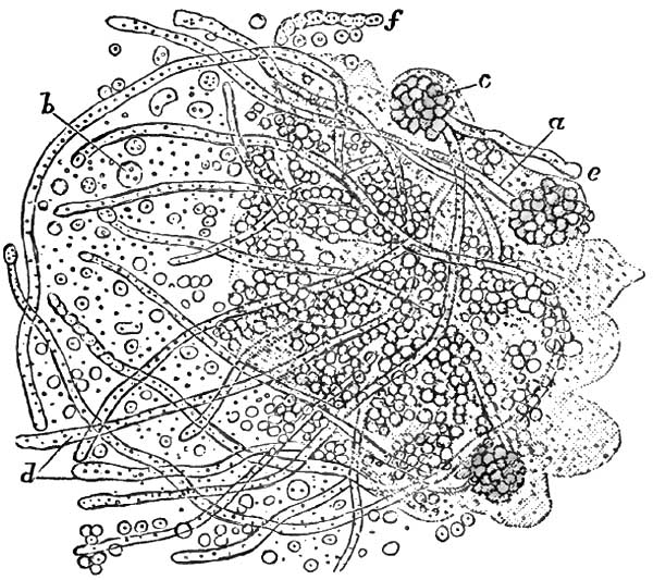
Oïdium albicans, from the Mouth in a case of Thrush (Küchenmeister). a, fragment of a separated thrush-layer implanted in a mass of epithelium; b, spores; d, thallus-threads with partition walls; e, free end of a thallus somewhat swollen; f, thallus with constriction, without partition walls.
ETIOLOGY.—Thrush is usually a symptomatic disease, secondary to an acid condition of the fluids of the mouth. Athrepsia (Parrot, Meigs and Pepper), or innutrition, is the presumable predisposing cause. Negligence in maintaining cleanliness of the mouth and of the articles which are placed in it is regarded as the main exciting cause. It occurs both in the adult and in the infant, but it is much more frequent in infancy and in early childhood. It is most frequently encountered in asylums and hospitals for children, being often transmitted from child to child by the nurse or by means of the feeding-bottle. The poor health of the child seeming less accountable for the disease than the unsanitary condition of the wards, buildings, and surroundings, it is consequently much less frequent in private than in public practice. It is more frequent in the first two weeks of life than later. Seux observed it within the first eight days in 394 cases out of 402 (Simon). It is much more frequent during summer than at any other season, more than half the cases (Valleix) occurring at that portion of the year.
In senile subjects, in adults, and in children more than two years of age it is cachectic, and observed chiefly toward the close of some fatal and exhausting disease, such as diabetes, carcinoma, tuberculosis, chronic pneumonia, enteric fever, puerperal fever, erysipelas, chronic entero-colitis and recto-colitis, and pseudo-membranous sore throat. It is sometimes observed in the early stage of enteric fever.
Meigs and Pepper, apparently following Parrot, deem the central cause to lie in a certain failure of nutrition under which the general vitality slowly ebbs away. They are inclined7 to recognize a causal factor in a deficiency in the supply of water in much of the artificial food administered to young subjects. The normal acidity of the fluids of the mouth of the newly-born (Guillot, Seux) is not sufficiently counteracted until saliva becomes abundant. Premature weaning, entailing, as it often does, the use of improper foods, renders the child liable to gastro-intestinal disorders. To this add want of care of the bottle and nipples, of the teaspoon or pap-boat, and of the mouth itself, and the conditions are fulfilled in fermentations of remnants of milk taking place without and within, which produce the acid condition of the fluids and secretions of the mouth said always to accompany and precede the development of the disease (Gubler).
7 A Practical Treatise on the Diseases of Children, 7th ed., Philada., 1882.
The theory of contagiousness seems established (Guillot, Berg, Gubler, Robin, Trousseau). This has been further demonstrated by experiments upon sheep (Delafeud), in which thrush has been implanted whenever the animals were unhealthy, but not otherwise.
PATHOLOGY AND MORBID ANATOMY.—The mucous membrane of the mouth within a few hours after its invasion by thrush is seen to be covered to some extent by minute masses of a granular curdy substance adherent to the tissues, which often bleed slightly when the substance is forcibly removed.
In children much reduced by inanition or severe disease, much of the deposit soon coalesces into a membraniform product, grayish or yellowish from rarefaction by the air, or even brownish from admixture of blood. By the same time the general congestion of the mucous membrane will have subsided into the pallor of anæmia. Though tolerably adherent when fresh, the deposit when older often becomes loosened spontaneously, so that it may be removed by the finger in large flakes without producing any hemorrhage whatever.
The characteristic masses present both as delicate roundish flakes, isolated, not larger than a pinhead, and as confluent patches several times as large and more irregular in outline. These masses under microscopic inspection are seen to be composed of the filaments and spores of a confervoid parasitic plant, the Oïdium albicans, enclosing altered epithelia in various conditions. This parasitic growth does not become developed upon healthy mucous membrane with normal secretory products. Acidity of the fluids and exuberance of epithelium are the requisites for its production, whatever be the cause. The acidity of the fluids irritates the mucous membrane upon which they lie. This irritation induces abnormal proliferation of epithelium, upon which the spores of the cryptogam then germinate. Dissociated epithelial cells become proliferated at the surface of the mucous membrane, between which and upon which both free and agglutinated spores accumulate. From these spores sprout out simple and ramified filaments in compartments containing moving granular elements. (For the minute detailed anatomy of these filaments and spores the reader is best referred to Robin's work on Vegetable Parasites.)
It may suffice here to mention that the filaments are sharply-defined tubercles, slightly amber-tinted, of a mean diameter of between four and three millimeters, simple while immature and branched when fully developed. These tubules are filled with link-like groups of elongated cells in compartments, giving them an appearance of regular constriction at the junctions of adjoining groups of cells. Surrounding these tubules are groups of spheroid or slightly ovoid spores from five to four millimeters in diameter. Each spore contains one or two granules and a quantity of fine dust. This cryptogamic growth is developed in the proliferated cells of epithelium. The filaments in their further growth separate the epithelia, and even penetrate them. Thence they penetrate the mucous membrane and the submucosa (Parrot).
The mucous membrane beneath the growth is red, smooth, and glistening. Papillæ are sometimes prominent. It is not excoriated unless the growth has been removed with some violence, when, as noted, it may bleed slightly. Duguet and Damaschino have recently encountered cases associated with a special ulceration of one of the palatine folds; the former in enteric fever, the latter in a primitive case. The growth is quickly reproduced after removal—even within a few minutes when the secretions are very acid.
The glossal mucous membrane is usually the tissue first involved, the specks being more numerous at the tip and edges of the tongue than at its central portion. The glands at the base of the tongue may become invaded. From the tongue extension takes place to the lips, the cheeks, the gums, and the palate, hard and soft. The growth is especially prolific in the folds between lips and gums and between cheeks and gums. Sometimes the parts mentioned become involved successively without actual extension. In several recently reported instances occurring during enteric fever,8 the affection began on the soft palate, tonsils, and pharynx, and then progressed anteriorly toward the tongue, the cheeks, and the lips.
8 Duguet, Soc. Méd. des. Hôp., Mai 11, 1883; Rev. mens., Juin 1, 1883, p. 187.
But there is no limitation of the disease to these structures. The growth may cover the entire mucous membrane of the mouth. From the mouth it may reach the lateral walls of the pharynx, and in rare instances the posterior wall of the pharynx. The product is said to be more adherent on the pharynx (Reubold) than in the mouth. From the pharynx it may reach the epiglottis, and even the larynx (Lelut), in which organ it has been seen upon the vocal bands (Parrot). It has never been observed in the posterior nares or at the pharyngeal orifice of the Eustachian tube. It flourishes best, therefore, upon squamous epithelium. In infants much reduced, Parrot has seen ulceration in the neighborhood of the pterygoid apophyses, but attributable to the cachectic state of the child, and not to the disease in the mouth.
In many cases—in as large a proportion as two-thirds, according to some observers—the oesophagus becomes invaded, either in irregular longitudinal strips or in rings, in all instances (Simon) terminating a little above the cardia. In exceptional cases the entire mucous surface of the oesophagus may be covered with the product (Seux). It has been seen in the stomach (Lelut, Valleix), and is even said to be developed there (Parrot), presenting as little yellow projections, isolated or contiguous, from the size of millet-seeds to that of peas, and usually located along the curvatures, especially the smaller curvature and cardia (Simon).
In instances still more rare it is found in the intestinal canal (Seux), even at the anus (Bouchut, Robin), and thence upon the genitalia. In a child thirteen days old, Parrot found it in the pulmonary parenchyma at the summit of the right lung, where it had probably been drawn by efforts of inspiration.
The nipple of the nurse often becomes covered with the growth (Gubler, Robin, Trousseau, Simon).
SYMPTOMATOLOGY.—In infants the earliest symptom is distress during nursing, the nipple being seized repeatedly, and as frequently released with cries of pain and disappointment. This cry is hoarse when the vocal bands are involved.
The constitutional symptoms depend upon the underlying malady, and may of course vary with its character. Thus we may have the symptoms of simple diarrhoea, gastro-enteritis, or entero-colitis on the one hand, and of tuberculosis and other diseases elsewhere enumerated on the other. Cachectic children, especially in asylum and hospital practice, lose flesh, and their skins become harsh, dry, and inelastic from loss of fluids (Meigs and Pepper). The genitalia, the anus, and the adjacent parts become eroded by the acridity of the discharges, and then become covered with the growth.
The disease rarely lasts longer than eight days in strong children that can be well cared for. It may continue indefinitely, on the other hand, in cachectic children; that is to say, for several months or until the patient succumbs, as may be. Death occurs usually from the causal disease, and not as a result of the morbid condition of the mouth.
DIAGNOSIS.—In the Infant.—Examination of its mouth to detect the cause of the child's inability to nurse reveals congestion of the mucous membrane, intense and often livid in severe cases. It is first noticed at the extremity of the tongue. When the congestion is general it is darkest in the tongue. This livid congestion may extend over the entire visible mucous membrane, save upon the hard palate, where it is tightly adherent to the periosteum, and upon the gums, where it is rendered tense by the approach of erupting teeth. The papillæ at the tip and sides of the tongue are very prominent. Sometimes the organ is quite dry, even sanious, while it is painful to the touch. The reaction of the secretions of the mouth is acid instead of alkaline, and the parts are hot and very sensitive.
Two or three days later the circular milky-white or curdy spots or slightly prominent and irregularly-shaped flakes or patches may be seen on the upper surface of the tongue toward the tip and inside the lips and the cheeks, especially in the grooves connecting gums and lips and gums and cheeks. The surrounding mucous membrane is unaltered in mild cases, and there is no evidence of other local disorder or of any constitutional involvement. In severe cases the entire mucous membrane is dry and deeply congested.
The affection can be positively discriminated from all others by microscopic examination of the deposit, which reveals the presence of the cryptogam described.
TREATMENT.—In infants, artificial nourishment, whether with milk of the lower animals or prepared food of whatever composition, should be given up, if possible, and a wet-nurse be supplied. If this procedure be impracticable, the least objectionable mode of preparation of cow's milk should be employed (and this will vary with the practice of the physician), and the utmost circumspection should be maintained in securing the cleanliness of the vessels in which it is prepared, the bottle from which it is given, and the nipple which is placed in the child's mouth. Should the sugar and casein in the milk appear to keep up the disease, weak soups may be substituted for the milk diet until it has subsided. Weiderhofer advises artificial nourishment, by way of a funnel inserted in the nasal passages, in case the child should refuse to swallow. Deglutition is excited in a reflex manner when the milk or other fluid reaches the pharynx.9
9 Journ. de Méd. Bordeaux, Juin 10, 1883.
The local treatment should consist in careful removal of the patches from time to time—say every two or three hours—with a moistened soft rag. This must be done without roughness of manipulation. In addition to this, the parts may be washed or painted every hour or so with an alkaline solution for the purpose of neutralizing the acidity of the fluids of the mouth. For this purpose borax is most generally used, in the proportion of twenty grains to the ounce of water or the half ounce of glycerin. Sodium bicarbonate or sodium salicylate may be substituted for the sodium borate. The use of honey in connection with the drug is calculated to promote acidity by fermentation of its glucose, and is therefore, theoretically, contraindicated.
Adults may use washes, gargles, or sprays of solutions of sodium borate or of sodium bicarbonate.
The constitutional treatment in each case must be adapted to the nature of the underlying malady which has favored the local disease, with resort in addition to the use of quinia, iron, wine, spirit, and beef-essence. The hygienic surroundings should be made as sanitary as possible.
Stomatitis Ulcerosa.
DEFINITION.—Inflammation of the interior of the mouth, usually unilateral, eventuating in multiple ulcerations of the mucous membrane.
SYNONYMS.—Fetid stomatitis, Phlegmonous stomatitis, Putrid sore mouth, Stomacace, are synonymous terms for idiopathic ulcerous stomatitis. Ulcero-membranous stomatitis, Mercurial stomatitis (Vogel), are synonymous terms for the deuteropathic variety of the disease.
ETIOLOGY.—The principal predisposing cause of the disease is to be found in ochlesis; the contaminating atmosphere of crowded dwellings and apartments insufficiently ventilated; uncleanliness; insufficiency of proper clothing; unhealthy food, and the like. It prevails epidemically in crowded tenements, schools, prisons, asylums, and hospitals; in garrisons and in camps; in transports and men-of-war. It is often propagated by contagion, but whether by infection or actual inoculation seems undetermined. Measles is an active predisposing cause. Feeble individuals are the most liable to the disease. It occurs at all ages. In civil life it is most frequent between the ages of four and ten years. Sometimes more girls are affected than boys (Meigs), and sometimes it is the more prevalent among boys (Squarrey). Autumn is the season of greatest prevalence.
Carious teeth, fracture and necrosis of the jaw (Meigs), and protracted catarrhal stomatitis are among the chief exciting causes. Irregular dentition is sometimes the exciting cause; and this may occur at the first and second dentition or at the period of eruption of the last molars.
PATHOLOGY.—The anatomical lesion is the destructive inflammation of portions of the mucous membrane of the mouth, leaving ulceration on detachment of the eschars. It usually commences as a gingivitis. At two periods of life—namely, from the fourth to the eighth year of life, and from the eighteenth to the twenty-fifth year—it is apt to be ulcero-membranous, a condition asserted to be altogether exceptional at other periods (Chauffard).
A diffuse fibro-purulent infiltration of the lymph-spaces of the mucosa is regarded as the first step in the pathological process. This infiltration is sufficiently abundant to compress the capillary vessels of the tissues, and thus arrest the circulation (Cornil et Ranvier). All those localized portions of mucous membrane from which the circulation is cut off perish and are discharged in fragments. The ulcers thus left are grayish, granular, and sanious, with thin, irregularly dentated borders a little undermined, through which pus can be expressed on pressure. The usual cryptogams of the oral cavity, in various stages of development, are in great abundance in the grayish detritus, which likewise contains altered red and white blood-corpuscles.
According to some observers (Caffort, Bergeron), the first evidence of the disease is an intensely congested erythematous patch, upon which one or more pustules present, point, and rupture promptly, leaving the characteristic ulcerations.
For some indeterminate reason, the ulcerations are mostly unilateral, and occur much the more frequently on the left side. The principal primal points of ulceration are upon the external borders of the gums, more frequently those of the lower jaw, and upon the corresponding surface of the cheek and lip—the cheek much oftener than the lip. Thence ulceration may extend to the tongue, less frequently to the palate. The ulcerative process follows the outline of the gums, baring the bases of the teeth to a variable extent, so that they seem elongated. On the cheek the patch of inflammation is generally oval, the longest diameter being antero-posterior, and the most frequent position is opposite to the last molar.
Each ulcer is surrounded by an intensely red areola, beyond which the tissues are succulent and tumid from collateral inflammatory oedema, often giving the ulcers an appearance of great depth; but when the detritus is discharged they are seen to have been superficial. Detachment of the necrosed segments of mucous membrane takes place by gradual exfoliation from periphery to centre. Sometimes detachment occurs in mass, usually in consequence of friction or suction. The ulcers, gingival and buccal, bleed easily when disturbed. They may remain separate, or may coalesce by confluence of interposing ulcerations extending across the furrow between gum and cheek or lip. The adjoining side of the tongue sometimes undergoes similar ulceration from behind forward, inoculated, most likely, by contact with adjoining ulceration. In rare instances, neglected cases most probably, the ulceration may extend to the palatine folds, the tonsils, and the soft palate.
SYMPTOMATOLOGY.—The affection usually begins without any constitutional symptoms. Young infants sometimes present slight febrile symptoms, with impairment of appetite and general languor. Fetid breath, salivation, and difficulty in deglutition are usually the first manifestations of the disease to attract attention. The mouth will be found to be hot, painful, and sensitive to the contact of food. Infants often refuse food altogether, though usually they can be coaxed to take liquid aliment. Larger children and adults complain of scalding sensations. They find mastication painful, and cannot chew at all on the affected side. The salivation is excessive, the saliva bloody and often extremely fetid. When swallowed, this fetid saliva causes diarrhoea. The cheeks sometimes become swollen, and the submaxillary connective tissue oedematous. Adenitis takes place in the submaxillary, retro-maxillary, and sublingual glands of the affected side. Sometimes the other side becomes affected likewise, but to a less extent. The glands do not suppurate, but the adenitis may remain as a chronic manifestation in scrofulous subjects.
The disease, left to itself, will often continue for a number of weeks, or even months as may be, unmodified even by intercurrent maladies (Bergeron). Long continuance may result in partial or complete disruption of the teeth, or in local gangrene, or even in necrosis of the alveoli (Damaschino). Properly managed, the ulcers become cleansed of their detritus, and within a few days heal by granulation, their position long remaining marked by delicate red cicatrices upon a hard and thickened substratum. Repeated recurrences are sometimes observed.
DIAGNOSIS.—The appearances of the gums and adjoining structures described under the head of Pathology establish the diagnosis. The usually unilateral manifestation and the peculiar fetid odor distinguish it from severe forms of catarrhal stomatitis. From cancrum oris it is distinguished by the absence of induration of the skin of the cheek over the swollen membrane, and by the succulence and diffuseness of the tumefaction. From mercurial stomatitis it is discriminated by the history, and by the absence of the peculiar manifestations to be discussed under the head of that disease.
PROGNOSIS.—The prognosis is good, the disease being susceptible of cure in from eight to ten days in ordinary cases. When due nutrition is prevented by the pain in mastication and deglutition, and in much-reduced subjects, the disease may continue for several weeks. It is in these cases that detachment of the teeth takes place, with periostitis and necrosis of the alveoli. Protracted suppuration and failure in nutrition may lead to a fatal result, but such a termination is uncommon.
TREATMENT.—Fresh air, unirritating and easily digestible food, the best hygienic surroundings practicable, attention to secretions from skin and bowels by moderate and judicious use of ablutions, diaphoretics, and laxatives, with the internal administration of cinchona or its derivatives, with iron and cod-liver oil, comprise the indications for constitutional treatment.
Locally, demulcent mouth-washes are called for, containing astringents, detergents, or antiseptics. Acidulated washes are more agreeable in some instances. For antiseptic purposes, however, sprays and douches may be used of solutions of potassium permanganate, boric acid, carbolic acid, or salicylic acid. Gargles of potassium chlorate, ten or twenty grains to the ounce, are highly recommended, as well as the internal administration of the same salt in doses of from two to five grains three times a day for children, and of ten to twenty grains for adults.
If the sores are slow to heal, the ulcerated surfaces may be touched once or twice daily with some astringent, such as solution of silver nitrate (ten grains to the ounce), or, if that be objectionable, with alum, tincture of iodine, or iodoform.
Prompt extraction of loose teeth and of loose fragments of necrosed bone is requisite.
Stomatitis Gangrenosa.
DEFINITION.—A non-contagious, deuteropathic inflammation of the interior of the mouth, almost invariably unilateral, and characterized by a peculiar gangrenous destruction of all the tissues of the cheek from within outward.
SYNONYMS.—Gangrenous stomatitis; Gangrena oris; Grangrenopsis; Cancrum oris; Stomato-necrosis; Necrosis infantilis; Gangrene of the mouth; Gangrenous erosion of the cheek; Noma; Buccal anthrax; Aquatic cancer; Water cancer; Scorbutic cancer; Sloughing phagedæna of the mouth.
HISTORY.—The most important work upon the subject was published in 1828, from the pen of Dr. A. L. Richter,10 whose accurate historical account of the disease was in great part reproduced, with additions thereto, by Barthez and Rilliet in their Treatise on the Diseases of Infants, Paris, 1843, and quoted by nearly all subsequent writers on the theme. From these records it appears that the first accurate description of the affection was given in 1620 by Dr. Battus, a Dutch physician, in his Manual of Surgery. The term aquatic cancer, water-kanker, bestowed on it by van de Voorde, has been generally followed by the physicians of Holland, although van Swieten (1699) properly designated it as gangrene. J. van Lil termed it noma, as well as stomacace and water-kanker, and cited a number of Dutch physicians who had observed its epidemic prevalence. The majority of more recent observers, however, deny its epidemic character.
10 Der Wasserkrebs der Kinder, Berlin, 1828; further, Beiträg zur Lehre vom Wasserkrebs, Berlin, 1832; Bemerkungen über den Brand der Kinder, Berlin, 1834.
Of Swedish writers, Lund described it as gangrene of the mouth; Leutin, under the name of ulocace. In England, Boot was the first to write of gangrene of the mouth, and was followed by Underwood, Symmonds, Pearson, S. Cooper, West, and others.
In France it has received great attention. Berthe11 described it as gangrenous scorbutis of the gums; Sauvages (1816) as necrosis infantilis. Baron in 1816 published12 a short but excellent account of a gangrenous affection of the mouth peculiar to children; and Isnard presented in 1818 his inaugural thesis on a gangrenous affection peculiar to children, in which he described, simultaneously, gangrene of the mouth and gangrene of the vulva. Then followed Rey, Destrees (1821), Billard (1833), Murdoch, Taupin (1839), and others, until we reach the admirable description by Barthez et Rilliet, from which the present historical record has been chiefly abstracted.
11 Mémoires de l'Académie royale de Chirurgie, Paris, 1774, t. v. p. 381.
12 Bullétins de la Faculté de Médecine de Paris, 1816, t. v. p. 161.
German physicians likewise have largely studied the subject. De Hilden, A. G. Richter, C. F. Fischer, Seibert, and many others preceded A. L. Richter, whose important contribution to the literature and description of the disease has been so highly extolled by Barthez and Rilliet.
In America the disease has been best described by Coates, Gerhard, and Meigs and Pepper, all of Philadelphia.
(For extensive bibliographies the following sources should be consulted in addition to those cited: J. Tourdes, Du Noma ou du Sphacèle de la Bouche chez les Enfants, Thèse, Strasbourg, 1848: A. Le Dentu, Nouveau Dictionnaire de Médecine et de Chirurgie pratique, article "Face," Paris, 1871.)
ETIOLOGY.—Almost exclusively a disease of childhood, gangrenous stomatitis is exceedingly rare in private practice, and very infrequent at the present day even in hospital and dispensary practice. Lack of hygienic essentials of various kinds, impoverishment, long illnesses, and debilitating maladies in general are the predisposing causes. It is sometimes endemic in hospitals and public institutions, but rarely, if at all, epidemic. It is not generally deemed contagious, though so considered by some writers. It appears to have been more frequent in Holland than elsewhere, to be more frequent in Europe generally than in the United States, and now much less frequent in the United States than formerly. To recognition of the predisposing causes and to their abolition and avoidance may probably be attributed its diminished frequency all over the world. Though attacking children only as a rule, it has been observed in adults (Barthez et Rilliet, Tourdes, Vogel). Nurslings are not liable to the disease. Though occurring occasionally earlier in life, the greatest period of prevalence is from the third to the fifth or sixth year of age, and thence, with diminishing frequency, to the twelfth and thirteenth years. It is probably equally frequent in the two sexes, though the majority of authors have described it as more frequent in females.
Healthy children are not attacked. Even in delicate children it is so rarely idiopathic that this character is utterly denied it by many observers. The disease which it follows, or with which it becomes associated, may be acute or chronic. According to most writers, it occurs with greatest frequency after measles. It follows scarlatina and variola much less often. It is observed likewise after whooping cough, typhus fever, malarial fever, entero-colitis, pneumonitis, and tuberculosis. Excessive administration of mercury has been recognized as an exciting cause, some cases of mercurial stomatitis progressing to gangrene.
According to Barthez et Rilliet, acute pulmonary diseases, and especially pneumonia, are the most frequent concomitant affections, and are usually consecutive.
SYMPTOMATOLOGY, COURSE, DURATION, TERMINATIONS, COMPLICATIONS, AND SEQUELÆ.—The disease usually becoming manifested during other disease, acute or chronic, or during convalescence therefrom, there are no special constitutional symptoms indicating its onset. Hence considerable progress may be made before its detection. The earliest local characteristic symptom distinguishing gangrenous stomatitis is a tense tumefaction of one cheek, usually in proximity to the mouth. The lower lip is generally involved, thus rendering it a matter of difficulty to open the mouth. This tumefaction in some instances progresses over the entire side of the face up to the nose, the lower eyelid, and even out to the ear in one direction, and down to the chin, and even to the neck, in the other. Before the parts become swollen externally, ulceration will have taken place to some extent in the mucous membrane, but usually without having attracted special attention, the subjective symptoms having been slight. A gangrenous odor from the mouth, however, is almost always constant. Its presence, therefore, should lead to careful investigation as to its seat and cause. The gums opposite the internal ulcer become similarly affected in most instances, and undergo destruction, so that the teeth may become denuded and loosened, and even detached, exposing their alveoli. The bodies of the maxillary bones suffer in addition in some instances, and undergo partial necrosis and exfoliation.
It is maintained (Löschner, Henoch) that in some instances there is no involvement of the mucous membrane until the ulcerative process has reached it from the exterior.
The tumefied portions of the check and lip are pale, hard, unctuous, and glistening. They are rarely very painful, and often painless. On palpation a hard and rounded nodule one or two centimeters in diameter can be detected deep in the central portion of the swollen cheek.
From the third to the sixth day a small, black, dry eschar, circular or oval, becomes formed at the most prominent and most livid portion of the swelling, whether cheek or lip. This gradually extends in circumference for a few days or for a fortnight, sometimes taking in almost the entire side of the face or even extending down to the neck. As it enlarges the tissues around become circumscribed with a zone intensely red. The internal eschar extends equally with the external one. Eventually, the eschar separates, in part or in whole, and becomes detached, leaving a hole in the cheek through which are seen the loosened teeth and their denuded and blackened sockets.
During this time the patient's strength remains tolerably well maintained, as a rule, until the gangrene has become well advanced. Intelligence usually remains good. Many children sit up in bed and manifest interest in their surroundings. Others lie indifferent to efforts made for their amusement. Some exhibit insomnia and delirium. The pulse is small and moderately frequent, rarely exceeding 120 beats to the minute until near the fatal close, when it often becomes imperceptible. Appetite is often well preserved, unless pneumonia or other complications supervene, but thirst is often intense, even though the tongue remain moist. The desire for food sometimes continues until within a few hours of death. Toward the last the skin becomes dry and cold, diarrhoea sets in, emaciation proceeds rapidly, collapse ensues and death.
Death usually occurs during the second week, often before the complete detachment of the eschar—in many instances by pneumonia, pulmonary gangrene, or entero-colitis. Some die in collapse, which is sometimes preceded by convulsions. When the eschars have become detached, suppuration exhausts the forces of the patient, and death takes place by asthenia.
The complication most frequent is pneumonia, and the next entero-colitis. Gangrene of the lungs, of the palate, pharynx, or oesophagus, of the anus, and of the vulva, may supervene. Hemorrhage from the facial artery or its branches has been noted as an exceptional mode of death (Hueber), the rule being that the arteries in the gangrenous area become plugged by thrombi, and thus prevent hemorrhage.
Recovery may take place before the local disease has penetrated the cheek—indeed, while the mucous membrane alone is involved. In recent instances, however, the disease does not subside until after the loss of considerable portions of the cheek, and the child recovers with great deformity, not only from loss of tissue in the cheek and nose, but from adhesions between the jaws and the cheek.
PATHOLOGY AND MORBID ANATOMY.—Gangrenous stomatitis always involves the cheek, almost always that portion in proximity to the mouth. It is almost invariably unilateral. Either side seems to be equally liable. Both sides suffer only, it is contended, when the gangrene is limited in extent, confined to the mucous membrane, and occupies the sides of the frenums of the lips (Barthez et Rilliet). It usually if not invariably begins in the mucous membrane, as a phlyctenular inflammation, which undergoes ulceration, followed by gangrene, immediately or not for several days, and then becomes covered with a more or less brownish-gray eschar. The ulceration of the mucous membrane is occasionally preceded by an oedematous condition of the cheek externally, similar to that sometimes observed in ordinary ulcerous stomatitis; but this is not the characteristic circumscribed, tense infiltration observed later. This ulceration is situated most frequently opposite the junction of the upper and lower teeth. Sometimes it proceeds from the gingivo-buccal sulcus of the lower jaw, sometimes from the alveolar border of the gums. It extends in all directions, and often reaches the lower lip. From three to sixteen days may be consumed in these extensions. The surrounding mucous membrane becomes oedematous. The ulceration soon becomes followed by gangrene, sometimes within twenty-four hours, sometimes not for two or three days, and exceptionally not for several days. The ulcerated surfaces bleed readily, change from gray to black, and become covered with a semi-liquid or liquid putrescent detritus. They are sometimes surrounded by a projecting livid areola, which soon becomes gangrenous in its turn. The shreds of mortified membrane, though clinging a while to the sound tissues, are easily detached, and often drop spontaneously into the mouth. Meanwhile, there is abundant salivation, the products of which pour from the mouth, at first sanguinolent, and subsequently dark and putrescent and mixed with detritus of the tissues. Large portions of the gums, and even of the mucous membrane of the palate, may undergo destruction within a few (three to six) days. The gangrenous destruction of the gums soon exposes the teeth, which become loose and are sometimes spontaneously detached. Thence the periosteum and bone become implicated and undergo partial denudation and necrosis, and portions of necrosed bone become detached if the patient survives. The characteristic implication of the exterior of the cheek becomes manifest from the first to the third day, but occasionally not until a day or two later. A hard, circumscribed swelling of the cheek or cheek and lip occurs, sometimes preceded, as already intimated, by general oedematous infiltration. The surface is tense and unctuous, often discolored. In its central portion is an especially hard nucleus, one to two centimeters or more in diameter. Gangrene often takes place at this point from within outward at a period varying from the third to the seventh day or later. The skin becomes livid, then black; a pustule is formed at the summit of the swelling, which bursts and discloses a blackened gangrenous eschar from less than a line in thickness to the entire thickness of the cheek beneath. The area of gangrene gradually extends. The dead tissues become detached, and a perforation is left right through the cheek, through which are discharged saliva and detritus. Meanwhile, the submaxillary glands become swollen and the surrounding connective tissue becomes oedematous. In some instances, however, no change is noticeable in these glands.
Examinations after death have shown that thrombosis exists for some distance around the gangrenous mass. Hence the rarity of hemorrhage during the detachment of the eschar.
DIAGNOSIS.—In the early stage of the disease the main point of differential diagnosis rests in the locality of the primitive lesion, the mucous membrane of the inside of one cheek. Subsequently there is the gangrenous odor from the mouth; the rapid peripheric extension of the local lesion, which acquires a peculiar grayish-black color; its rapid extension toward the exterior of the cheek or lip; the tumefaction of the cheek, discolored, greasy, hard, surrounded by oedematous infiltration, and presenting a central nodule of especial hardness; then the profuse salivation, soon sanguinolent, subsequently purulent and mingled with detritus of the mortified tissues. Finally, the eschar on the exterior of the swollen cheek or lip leaves no doubt as to the character of the lesion. From malignant pustule it is distinguished by not beginning on the exterior, as that lesion always does (Baron).
PROGNOSIS.—The prognosis is bad unless the lesion be quite limited and complications absent. At least three-fourths of those attacked perish; according to some authorities fully five-sixths die. The objective symptoms of the local disease are much more important in estimating the prognosis than are the constitutional manifestations, the vigor of the patient, and the hygienic surroundings, although, as a matter of course, the better these latter the more favorable the prognosis. Prognosis would be more favorable in private practice than in hospital or asylum service.
TREATMENT.—Active treatment is required, both locally and constitutionally. Local treatment is of paramount importance, and alone capable of arresting the extension of the process of mortification. The topical measure in greatest repute is energetic cauterization with the most powerful agents, chemical and mechanical—hydrochloric acid, nitric acid, acid solution of mercuric nitrate, and the actual cautery, whether hot iron, thermo-, or electric cautery. The application of acids is usually made with a firm wad or piece of sponge upon a stick or quill, care being taken to protect the healthy tissues as far as practicable with a spoon or spatula. After the application the mouth is to be thoroughly syringed with water to remove or dilute the superfluous acid. Hydrochloric acid has been preferred by most observers.
As these cauterizations must be energetic to prove effective, anæsthesia ought to be induced. Should ether be employed for this purpose, hydrochloric acid or the acid solution of mercuric nitrate would be selected of course.
In the early stages these agents are to be applied to the inside of the cheek, so as to destroy all the tissue diseased, if practicable, and expose a healthy surface for granulation. Should the exterior of the cheek become implicated before cauterization has been performed or in spite of it, it is customary to destroy the tissues from the exterior, including a zone of apparently healthy surrounding tissue. As the gangrene extends, the cauterization is to be repeated twice daily or even more frequently. After cauterization the parts are dressed with antiseptic lotions, and antiseptic injections or douches are to be used frequently during day and night to wash out the mouth and keep it as clear as possible from detritus.
Meigs and Pepper report beneficial results from the topical use of undiluted carbolic acid, followed by a solution of the same, one part in fifty of water, frequently employed as a mouth-wash. The progress of the sloughing was checked and the putridity of the unseparated dead tissue completely destroyed in the two cases mentioned by them, one of which recovered quickly without perforation of the cheek. Gerhard preferred undiluted tincture of the chloride of iron; Condie, cupric sulphate, thirty grains to the ounce. Bismuth subnitrate has recently been lauded as a topical remedial agent.13
13 Maguire, Medical Record N.Y., Feb. 3, 1883.
The mouth should be frequently cleansed by syringing, douching, spraying, or washing with disinfectant solutions, such as chlorinated soda liquor, one part to ten; carbolic acid, one to twenty. Lemon-juice is sometimes an agreeable application, as in some other varieties of stomatitis. Constitutionally, tonic and supporting treatment is demanded, even in those instances where the appetite is well maintained and the general health apparently well conserved. Soups, milk, semi-solid food, egg-nog, egg and wine, wine whey, milk punch, finely-minced meat, should be administered as freely as the state of the digestive functions will permit. If necessary, resort should be had to nutritive enemata. Quinia and tincture of chloride of iron are the medicines indicated. When sufficient alcohol cannot be given with the food, it should be freely exhibited in the most available form by the mouth or by the rectum. The apartment should be well ventilated, the linen frequently changed, the discharges promptly removed.
Toxic Stomatitis.
DEFINITION.—An inflammation of the interior of the mouth due to poisoning, especially by drugs, and chiefly by mercury, copper, and phosphorus.
Mercurial Stomatitis.
DEFINITION.—An inflammation of the mucous membrane of the mouth, eventually ulcerating, the result of systemic poisoning by the absorption of mercury.
SYNONYMS.—Stomatitis mercurialis; Mercurial ptyalism, Ptyalismus mercurialis; Mercurial salivation, Salivatio mercurialis.
ETIOLOGY—Predisposing and Exciting Causes.—Special vulnerability to the toxic influence of mercury, and special proclivity to inflammatory affections of the mouth and the organs contained therein, are the predisposing causes of mercurial stomatitis. The exciting cause is the absorption of mercury into the tissues of the organism. The susceptibility of healthy adults is much greater than that of healthy children. The susceptibility of adults varies very greatly. Constitutions deteriorated by prolonged disease, undue exposure, and the like are much more promptly influenced in consequence. Tuberculous subjects do not bear mercury well.
Idiosyncratic susceptibility to toxæmia by mercurial preparations is now and then encountered in practice, and instances have been published14 in which fatal results have ensued, after prolonged suffering, from the incautious administration of a single moderate dose of a mercurial drug.
14 For example, see in Watson's Practice of Physic a case of furious salivation following one administration of two grains of calomel as a purgative, the patient dying at the end of two years, worn out by the effects of the mercury and having lost portions of the jaw-bone by necrosis.
Until comparatively recent years the most common cause of mercurial poisoning was the excessive employment of mercurial medicines, whether by ingestion, inunction, or vapor bath. Topical cauterization with acid solution of mercuric nitrate is likewise an infrequent, and usually an accidental, cause of the affection. Elimination of the mercury by way of the mucous glands of mouth and the salivary glands proper excites the stomatitis in these instances. An entirely different series of cases occur in artisans exposed to handling the metal and its preparations or to breathing its vapor or its dust. In these instances the poison may gain entrance into the absorbent system by the skin, the mucous membranes of the nose, mouth, and throat, the stomach, or the lungs. No matter what care may be exercised in cleansing the hands, it is often impossible to prevent occasional transference of the noxious material from fingers to throat, or to thoroughly free the finger-tips under the nails. The avocations entailing the risks of mercurial stomatitis comprise quicksilver-mining, ore-separating, barometer- and thermometer-making, gilding, hat-making, manufacturing of chemicals, and exhausting the globes employed in certain forms of electric illumination.15 The slow absorption of mercury into the bodies of artisans induces in addition serious constitutional nervous disturbances—tremors, palsy, etc.
15 Med. and Surg. Reporter, Philada., Dec. 30, 1883, p. 734.
SYMPTOMATOLOGY, COURSE, DURATION, TERMINATIONS, COMPLICATIONS, AND SEQUELÆ.—The principal subjective symptoms of mercurial stomatitis are—characteristic fetor of the breath, sore gums and mouth, continuous nauseous metallic brassy or coppery taste, and profuse salivation.
At first the mouth feels parched and painful, the gums tender, the teeth, the lower incisors especially, set on edge. Soon the gums become swollen, and when touched with the tongue seem to have receded from the necks of the teeth, which thereby appear to be longer than usual. The gums feel quite sore when pressed upon with the finger or when put on the stretch by clashing the rows of teeth against each other. This sort of soreness is often watched for in the therapeutic administration of mercurials purposely given to "touch the gums," as an indication that the system is under the influence of the drug. It is, therefore, one of the earliest indications of mercurial poisoning, but if not sought for it may elude attention until after the mouth has become sore a little later. The pain in the mouth is augmented by efforts of mastication and expectoration, and may be associated with pains at the angle of the lower jaw or extending along the domain of the third or of the third and second divisions of the distribution of the fifth cerebral nerve. Mastication of solid food is often unendurable. Constitutional manifestations become evident about this time in increased heat of skin, acceleration of pulse, furred tongue, dry mouth, great thirst, and loss of appetite. The dryness of the mouth does not last long, but is soon followed by hypersalivation, one of the characteristic phenomena of the disorder. The saliva secreted, often acid in reaction, varies greatly in quantity, which is usually proportionate to the severity of the case. It is secreted night and day, sometimes to the amount of several pints in the twenty-four hours—in moderately severe cases to the amount of from one to two pints in that space of time. It is limpid or grayish, mawkish or somewhat fetid, and reacts readily to the simplest tests for mercury. The salivation is almost continuous, sometimes quite so. The patient soon becomes unable to endure the fatigue of constant expectoration, and the fluid then dribbles from his mouth or runs off in an unimpeded slobber. When excessive, the patient's strength becomes rapidly exhausted—in part by impoverishment of the fluids, in great measure from the lack of refreshing sleep.
Meanwhile, the local inflammatory process extends from the gums to the floor of the mouth and to the lips, and thence to the tongue and the cheeks. The salivary glands are in a state of inflammation likewise, but rather in consequence of direct irritation in the elimination of the poison through their channels than by extension of the stomatitis along their ducts. The lymphatic glands of the lower jaw become engorged and tender. Mastication, deglutition, and articulation all become impeded mechanically by tumefaction of the tissues.
In some instances the glossitis is so great that the tongue protrudes, thereby impeding respiration and even threatening suffocation. In some cases oedema of the larynx has been noted, threatening suffocation from that cause. Should the inflammatory process extend along the pharynx to the Eustachian tubes, deafness and pains in the ears will become additional symptoms.
The subsequent progress of unarrested mercurial stomatitis is that of ulcerous stomatitis.
Should gangrene of the mucous membrane take place, there will be great fetor from the mouth, and some danger of hemorrhage on detachment of the sloughs should the process be taking place in the direction of vessels of some calibre. Necrosis of the inferior maxilla entails continuance of the disagreeable local symptoms until the discharge in fragments or in mass of the dead portions of bone.
In the earlier stages of the attack the constitutional symptoms may be sthenic. Fever, cephalalgia, and the usual concomitants of pyrexia, however, soon give way to the opposite condition of asthenia. Exhausted by the excessive salivation, and unable to repair waste by eating or sleeping, the sufferer soon passes into a condition of hopeless cachexia. Those who survive remain cachectic and feeble for a long time—some of them disfigured for life by various cicatrices between cheeks and jaw, by loss of teeth or of portions of the jaw-bone.
The duration of mercurial stomatitis varies with the susceptibility of the patient, the intensity of the toxæmia, and the character of the treatment. Mild cases may get well in a week or two; severe cases may continue for weeks, and even months; extreme cases have persisted for years. Even moderate cases occasionally resist treatment for weeks.
Under the improved therapeutics of the present day mercurial stomatitis almost always terminates in recovery, especially if it receive early and prompt attention. Neglected or improperly managed, it may terminate in serious losses of tissue in gums, cheeks, teeth, and bone, leaving the parts much deformed and the patient in a permanently enfeebled condition.
Erysipelas, metastatic abscesses, inflammations, pyæmia, or colliquative diarrhoea may be mentioned as complications which may prove sufficiently serious to produce death, independently of the virulence of the primary stomatitis.
PATHOLOGY AND MORBID ANATOMY.—Mercurial stomatitis is an ulcerative process attended with an excessive flow of saliva containing mercury. It has a tendency to terminate in destruction and exfoliation of the mucous membrane of the gums and other tissues attacked, and eventually in necrosis of the jaw-bone. The detritus is found, microscopically, to consist of granular masses of broken-down tissue, swarming with bacteria and micrococci, and containing some blood-cells and many pus-cells. In some instances micrococci have been detected in the blood.
The disease usually begins in the gums of the lower incisors, and extends backward, often being confined to one side of the jaw. The gums, first swollen and then livid, become separated from the necks of the teeth. Their edges undergo ulceration. The ulcers are surrounded by fungous margins, pale or red, which bleed on the slightest contact, and some become covered with grayish-yellow detritus. The ulceration extends in depth, destroying the supports of the teeth, so that they become loosened and even detached. The inflammatory process extends to the lips, the cheek, and the tongue, which undergo tumefaction and exhibit the impressions of the teeth in grayish opalescent lines or festoons of thickened epithelium at the points of pressure. The glossitis may become intense. It is almost always present, to some extent, as a superficial or mucous glossitis. Occasionally acute oedematous glossitis has ensued, and such cases sometimes terminate fatally. Ulceration takes place in these structures similar to that which has taken place in the gums. If not arrested, gangrenous destruction ensues, not only in these tissues, but beneath them. Thus, the teeth become loosened, and even detached; the jaw-bones themselves may become bared, necrosed, and in part exfoliated; and the cheeks undergo partial destruction by gangrene. Sometimes the inflammation descends to the larynx, and this may produce oedematous infiltration of the loose connective tissue of that structure. Sometimes it mounts the pharynx and reaches the orifices of the Eustachian tubes. The salivary glands become swollen and discharge great quantities of fluid, as detailed under Symptomatology. The retro-maxillary and submaxillary lymphatic glands become enlarged by inflammatory action.
DIAGNOSIS.—In the earliest stages the inflammation of the gums in mercurial stomatitis cannot be distinguished from that which takes place in other forms of ulcerative stomatitis. The fetor of the breath, however, the profuse salivation, and the chemical reaction of the saliva, together with the history of exposure to mercury, soon place the nature of the case beyond doubt. Similar results following poisonings by copper salts and by phosphorus are differentiated by the history of the special exposure.
PROGNOSIS.—In mild cases the prognosis is favorable, provided further exposure to the cause can be avoided. This holds good almost invariably in cases due to over-medication with mercurials, but is far less applicable to cases in artisans, the result of prolonged exposure to the poisonous influences of mercury and its slow absorption. On the whole, the affection is much less serious than formerly, both because it can, in great measure, be guarded against by proper prophylaxis in risky vocations, and because its treatment has been made much more efficient. In severe cases serious results may ensue despite the most judicious treatment, and convalescence is usually very slow, weeks often elapsing before solid food can be chewed without pain or without injury to the gums.
When death ensues, it may be by asthenia, erysipelas, pneumonia, pyæmia, or colliquative diarrhoea.
TREATMENT.—Mercurial stomatitis may sometimes be prevented by the administration of potassium chlorate during exposure. Mild cases following the administration of mercurials often subside upon mere withdrawal of the drug. Should spontaneous subsidence not take place, the administration of potassium chlorate every few hours, in doses of from thirty to sixty grains or more in the twenty-four hours, soon effects amelioration, which promptly terminates in recovery. The characteristic fetor often ceases within twelve hours' use of this drug. Should the inflammatory manifestations be severe, a few leeches applied beneath the edge of the lower jaw, followed by a poultice enveloping the neck to promote further flow of blood, often affords prompt relief (Watson). Lead acetate (ten grains to the ounce of water) and iodine (half a fluidrachm of the compound tincture to the ounce of water) are useful as gargles and washes. When the result of slow poisoning, elimination of the mercury by sulphur vapor baths and the administration of small doses of potassium iodide are recommended.
Cauterization of the ulcerated surfaces is sometimes serviceable, silver nitrate or hydrochloric acid (Ricord), or chromic acid 1:5 (Butlin, Canquil), being used for the purpose.
Opium in decided doses is indicated for the relief of pain. It may be added with advantage to detergent and disinfective mouth-washes (potassium chlorate, sodium borate, creasote-water, saponified emulsion of coal-tar, tincture of cinchona, tincture of myrrh, etc.), the use of which should form an important part of the treatment. Watson highly recommended a wash of gargle of brandy and water, 1:4 or 5. In severe cases difficulty is encountered in maintaining effective alimentation. When mastication is not impracticable, soft-boiled egg and finely-chopped raw beef may be given. When the patient cannot chew at all, resort is confined to milk, soups, and the juice of beef. Nourishing enemata should be administered, as in all affections where it becomes impracticable to sustain the patient by way of the mouth. Tonics and stimulants are indicated to avoid debility from the excessive salivation and its sequelæ—quinia, coffee, wine, and alcohol, the first, if required, by hypodermatic injection, all of them by enema if necessary.
Glossitis and oedema of the larynx may require the surgical procedures often necessary when they occur under other circumstances.
Other forms of toxic stomatitis hardly require special elucidation.
Abnormalities and Vices of Conformation of the Tongue.
Apart from the anomalies presented in monsters, there are a few congenital abnormalities of the tongue with which it becomes the accoucheur at least to be familiar, as their presence may interfere materially with the nutrition of the infant, whether nursed or spoon-fed.
CONGENITAL DEFICIENCY OF THE TONGUE.—A considerable portion of the tongue may be wanting anteriorly, comprising, in some instances, the entire free portion of the organ. The stump then presents as a single or a bifid protuberance of variable size. In some instances considerable power of movement exists, and even conservation of taste. Suction and deglutition are both practicable. When the child grows it can speak, though with a certain amount of difficulty. A few cases are on record, however, of ability to speak without any evidence of a tongue above the floor of the mouth.
An instance of lateral deficiency has been observed by Chollet,16 the deficient half being represented merely by the two layers of the lingual mucous membrane, without any intervening muscular substance.
16 Demarquay, Dict. de Méd. et de Chir. prat., xx. p. 130.
BIFID TONGUE, separate investment of the two sides, has been occasionally observed in connection with similar arrest of development in the lower jaw and other organs.
Ankyloglossia.
DEFINITION.—An abnormal attachment or adhesion of some portion of the tongue to some portion of the surrounding structures of the mouth.
SYNONYM.—Tongue-tie.
PATHOLOGY AND MORBID ANATOMY.—The ordinary form of tongue-tie consists in an abnormal development of the frenum of the tongue, the anterior vertical portion of the duplicature of mucous membrane which connects the lower surface of the raphé of the tongue with the floor of the mouth. The tongue cannot be extended beyond the lips. Suction is interfered with in some cases. If not remedied spontaneously or by surgical interference, mastication and articulation may become seriously impeded.
Other forms of ankyloglossia, congenital and acquired, possess special interest from surgical points of view mainly.
DIAGNOSIS.—Inspection and digital exploration readily reveal the nature of the restriction in the movements of the tongue and the size of the frenum.
PROGNOSIS.—The prognosis is good, the difficulty being susceptible of relief by division of a portion of the constricting frenum. Accidents have been reported following the operation, the occasional occurrence of which should be borne in mind. These are hemorrhage, which is not dangerous except in the prolonged absence of some one competent to restrain it should it be extreme; and retroversion of the tongue, an accident which has been known to prove fatal by occluding the orifice of the larynx (Petit).
TREATMENT.—Slight cases rarely need operation; but when the movements of the tongue are restricted by a very short and deep frenum its division becomes necessary. The operation is usually performed with scissors, the ranine arteries being protected by means of a fissured plate of metal (Petit), such as has long been used as a handle to the ordinary grooved director of the physician's pocket-case. The cut should be more extensive in the lateral directions of the fold than antero-posteriorly. After-treatment is rarely necessary, unless annoying hemorrhage is produced by movements of suction. Compression between the fingers, maintained for a number of minutes, suffices to restrain the hemorrhage in most instances. When this fails, recourse may be had to cauterization with the point of a heated iron or some other form of actual cautery.
Macroglossia.
DEFINITION.—Hypertrophy of the tongue.
SYNONYMS.—Megaloglossia, Glossoptosis, Prolapsus linguæ, Lingua propendula, Chronic prolapse of the tongue, Chronic intumescence of the tongue.
HISTORY.—This rare affection has been long known, the first cases on record being in the works of Galen. Other cases have been recorded by Celsus and Avicenna. Among more modern recorders may be mentioned Scaliger (1570), Bartholin (1680), Benedict and Pencer; among recent recorders, Lassus,17 Percy,18 Harris,19 Humphrey,20 Gayraud,21 W. Fairlie Clarke,22 Bryant,23 and the French dictionaries in present process of publication; to all of which the reader is referred for bibliographic, descriptive, and illustrative details.
17 Mémoire de l'Institut National, 18—, an VI. t. i.
18 Dict. Sci. Méd., t. xxvii.
19 Am. Journ. Med. Sci., vol. vii., 1830, p. 17; vol. xx., 1837, p. 15—both illustrated.
20 Trans. Med.-Chir. Soc. London, 1853, p. 113.
21 Thèse de Montpellier, No. 68, 1865.
22 Diseases of the Tongue, London, 1873.
23 "Surgical Affections of the Tongue," Guy's Hosp. Reports, 1883, p. 102 et seq.
ETIOLOGY.—This affection is usually congenital, at least to a certain extent, and augments with the growth of the child. It has been attributed, on apparently insufficient grounds, to injury received during parturition. It is probably intra-uterine in origin. Though encountered in both sexes, the majority of recorded cases have been in females. In summing up these observations, it appears that the affection often attracts little or no attention until dentition is in progress. The hypertrophy begins to augment rapidly during the second or third year of age, or a year or two later in some cases.
The gradual increase of the congenital deformity during infancy has been attributed to hypernutrition from local irritation produced by habits of sucking on the organ, induced, in some subjects, by forcible efforts at suction from a short nipple. Similarly, the rapid augmentation of volume noted as occurring during the period of dentition or a little later has been attributed to hypernutrition excited by irritation suffered by the protruding organ from the lower row of teeth. Cases commencing at this age have been supposed to be due exclusively to tongue-sucking. In some instances, due to this cause apparently, the deformity is associated with idiocy (Lawson24). Convulsions, epileptic seizures, and whooping cough have been regarded by some writers as occasional causes of the deformity. Indeed, idiocy and cretinism are not infrequent co-associates with the deformity (Parrot25). It has been observed likewise in anencephalous monsters (Brissot, idem).
24 Trans. Clin. Soc. London, vol. v. p. 158.
25 Gaz. Méd. Paris, Dec. 10 and 17, 1881; Lond. Med. Record, Mar. 15, 1882, p. 113.
SYMPTOMS, COURSE, DURATION, TERMINATIONS, COMPLICATIONS, AND SEQUELÆ.—The prominent symptom of macroglossia is the enlarged tongue protruding beyond the mouth. The resemblance of the protruding tongue of a child with macroglossia to the tongue hanging from the mouth of a calf gave rise to the name lingua vitulina by which it has sometimes been designated. In some instances, where the enlargement is but moderate, the organ can be retained within the mouth. When bilateral, the enlargement may be symmetrical, or may interest one side of the tongue more than the other. When the enlargement is confined to the free portion of the tongue, it interferes little with respiration and with movements of suction. When occupying the base of the organ, it may seriously embarrass respiration, and even produce suffocation in some instances (Clarke). The mouth being maintained open, saliva dribbles away constantly except during alimentation. Thirst is often very great in consequence of this, and of the desiccation of the tongue and of the walls of the mouth by the unmodified air to which they are continuously exposed. The tongue is usually free from pain.
In some subjects, although the tongue, left to itself, protruded considerably, it has been found quite practicable to maintain it within the cavity of the mouth by means of bandages or other appliances secured to the back and top of the head. These bandages are removed from time to time to give relief from the restraint and to permit food and drink to be taken. Systematic compression, indeed, has been induced in this way in some instances, and has produced considerable diminution in the size of the organ—sufficient to maintain its concealment without the aid of an appliance. When the tongue cannot be retained within the mouth the patient becomes unable to close the jaws. Hence saliva dribbles constantly, save when food or drink is being taken. The protruded portion of the tongue undergoes a livid discoloration, sometimes diffuse, sometimes disseminated. Though sometimes remaining comparatively soft in texture, it usually becomes hard, dry, rough, fissured, ulcerated and sanious, covered with desiccating layers of mucus and epithelium, and marked by indentations made by the edges of the teeth, which sometimes seem almost to strangle it. Mastication, deglutition, and articulation often become very difficult, and respiration also, but less frequently. The lower lip becomes much everted. The larynx and hyoid bone become drawn upward and forward by the weight of the organ. The configuration of the lower jaw undergoes considerable change, and the teeth become pressed out of position. Dislocation of the jaw from this cause has been noticed (Chalk26).
26 Trans. Path. Soc. London, vol. viii. p. 305.
These symptoms undergo aggravation with the growth of the subject, and, while presenting general features of resemblance in all cases, vary considerably in individual instances. Great difficulty is encountered, as a rule, in taking food, and mastication has to be performed very slowly. In some instances mastication can be performed satisfactorily by the molars, owing to a compensatory curvature of the lower jaw, even though the anterior portions of the jaw may remain permanently separated (Harris). Some patients get along by using their fingers to push the bolus far enough back to permit of its deglutition. Some have used a cup with a long tube slightly curved to convey fluids to the back part of the mouth for a similar purpose. Some have been systematically fed by means of a catheter passed through a nasal passage and thus on into the oesophagus. The difficulties in nourishing patients reduce some of them to extreme emaciation.
Notwithstanding all these drawbacks, quite a number of cases are on record where the patients have reached well into adult life before being submitted to radical measures for relief. One patient is recorded as having reached the age of eighty, having worn for some sixty-five years a silver shield to conceal her deformity (Clarke).
PATHOLOGY AND PATHOLOGICAL ANATOMY.—The hypertrophy may involve all the structures of the tongue, but usually implicates the muscular tissue especially (Sédillot, Paget, Bouisson). In a case published by W. Fairlie Clarke it was found on microscopic examination that the papillæ as well as the mucous and submucous tissues were somewhat enlarged and thickened, while the bundles of muscular fibre were slightly coarser than natural. Maas reports a unilateral case of macroglossia in a male child two months of age associated with hyperdevelopment of the entire left side of the body.27 In some instances the blood-vessels and lymphatics are chiefly involved (Virchow, Billroth, Maas), two cases of which have been described by Virchow as cavernous lymphatic tumors.
27 Arch. klin. Chir., p. 413, Bd. xiii. Heft 3.
Hilliard reports28 a congenital case from vicious growth, removed at fourteen months of age. Microscopic sections showed the large lacunæ filled with corpuscles, blood-pigment in different stages of degeneration, and the papillæ much hypertrophied. Winiwarter29 reports a congenital macroglossia associated with congenital cysts of the neck.
28 Brit. Med. Journ., Nov. 26, 1870, p. 591.
29 Arch. klin. Chir., 1874, Bd. xvi. Heft 3.
Sometimes there is very little true muscular hypertrophy, as in a case quoted by Bryant which was presented to the Pathological Society of London in 1872 by M. H. Arnott. In this specimen the epithelial covering was very thick and the papillæ enlarged. The blood-vessels were larger than usual, and there were large irregular spaces, thin-walled and filled with blood or clear fluid. "A few vesicular bodies which may have been enlarged lymphatics were also present"—probably cross-sections of lymphatic vessels.
The size that may be attained even in young children seems incredible, three and four inches protruding from the mouth in some instances. The free portion is more bulky than the intra-oral portion. One case reported "as thick as an arm" probably refers to the arm of the child. As a rule, both sides of the tongue are involved; exceptionally, the affection is unilateral.