Volume 2 — DISEASES OF THE DIGESTIVE SYSTEM. (continued 10)
Reading edition
Atony of the muscular walls of the stomach may be a part of general muscular weakness and impaired nutrition. Here belong cases of adynamic dilatation of the stomach secondary to typhoid fever, cholera, tuberculosis, anæmia, chlorosis, cachexia, senile marasmus, neurasthenia.
Whether primary paralysis of the stomach can occur or not is wholly uncertain. We have no positive knowledge as to the occurrence of paresis of the stomach in consequence of organic or functional changes in the peripheral or central nervous system. Nor does our meagre information as to the relation between the nervous system and the muscular movements of the stomach justify the construction of any hypotheses as to this point.
For the sake of clearness the various causes of dilatation of the stomach according to the foregoing classification may be recapitulated as follows. Some of the more doubtful and of the rarer causes are omitted:
A. Stenosis of the Pylorus or of the Duodenum. 1. Cancerous; 2. Cicatricial; 3. Hypertrophic (of pylorus); 4. From external pressure; 5. Congenital (of pylorus)?; 6. From torsion of duodenum?
B. Abnormalities in Contents of Stomach. 1. Ingesta: a. Excessive; b. Imperfectly masticated; c. Indigestible. 2. Stagnation and fermentation in consequence of chemical insufficiency of the stomach, as in chronic catarrhal gastritis and functional dyspepsia.
C. Impairment of Muscular Force of Stomach. 1. Organic changes in muscular coat: a. Partial destruction by ulcers and cancers; b. Inflammation, as in chronic catarrhal gastritis and peritonitis; c. Degenerations (fatty, colloid, amyloid); d. Oedema?; e. Cirrhosis of stomach. 2. Mechanical Restraint: a. By adhesions; b. By weight of herniæ. 3. Impaired Nutrition and General Muscular Weakness, Adynamic dilatation from typhoid fever, tuberculosis, anæmia, etc. 4. Paresis from neuropathic causes?
As a rule, not a single one, but several, of the above-mentioned causes are operative in the production of dilatation of the stomach, and it is often impossible to say which is the primary cause. The various gastric functions are so dependent upon each other that if one is disturbed the others also suffer. If, for instance, atony of the muscular coat of the stomach exists, then in consequence of enfeebled peristalsis the secretion of gastric juice is insufficient, the food is not thoroughly mingled with the gastric juice, and the absorption of the products of digestion in the stomach is interfered with; in consequence of which the accumulating peptones still further hinder the digestive process. The pylorus remains contracted for an abnormal length of time, as it naturally is closed until the process of chymification in the stomach is far advanced, and this process is now delayed. The stagnating contents of the stomach readily ferment, and the irritating products of fermentation induce a chronic catarrhal gastritis, which further impairs the functions of the mucous and muscular coats of the stomach. Thus, in a vicious circle one cause of dilatation induces another. To assign to each cause its appropriate share in the production of the final result is a matter of difficulty, and often of impossibility. From this point of view the dispute as to whether in atonic dilatation the most important factor in causation is chemical insufficiency of the stomach (impaired secretion of gastric juice, fermentations) or mechanical insufficiency (weakened muscular action, stagnation), appears of little practical importance.
Of the causes of non-stenotic dilatation of the stomach, the first place is to be assigned to chronic catarrhal gastritis and to atonic dyspepsia, as this term is understood by most English and American writers.
As regards frequency, gastric dilatation is a common result of cancer of the pylorus. It is less frequently caused by simple ulcer. Other forms of pyloric stenosis than the cancerous and the cicatricial are rare.
Opinions differ as to the frequency of non-stenotic or atonic dilatation of the stomach according to the manner in which one interprets the cases. Non-stenotic dilatations which are comparable in degree to those produced by stenosis are rare. The lesser grades of atonic dilatation, however, are not rare; but here arises the difficulty of distinguishing these cases from mere chemical or mechanical insufficiency of the stomach, which often represents the early stage of the process. Hence it has been proposed to discard altogether the term dilatation, and to substitute that of insufficiency of the stomach. But this latter term is applicable to many affections of the stomach other than dilatation. A typical case of atonic dilatation of the stomach is a well-defined disease, and because it is difficult to diagnosticate its early stages is not sufficient reason for discarding altogether the designation.
Gastrectasia may develop at any age. It is most frequent in middle and advanced life. The largest number of cases of atonic dilatation is met with between thirty and forty years of age. The disease is rare in childhood.11 The disease occurs in all classes of life. Atonic dilatation seems to be comparatively more frequent in private practice and among the favorably situated than in hospitals and among the poor. Kussmaul says that the largest contingent of patients is furnished by persons who lead a sedentary life and eat and drink a great deal.
11 Kundrat and Widerhofer mention no case of stenotic dilatation of the stomach in children. They say, however, that atonic dilatation due to over-feeding, and particularly to rachitis, is not infrequent in children. Widerhofer reports a case of very large dilatation of the stomach in a girl twelve years old. The cause of the dilatation was not apparent, and the clinical history was imperfect (Gerhardt's Handb. d. Kinderkrankh., Bd. iv. Abth. 2). Lafage (Thèse, Paris, 1881) reports a case of gastric dilatation at ten years, and another at sixteen years of age. R. Demme (abstract in Berl. kl. Wochenschr., 1883, No. 1) reports a case of large dilatation of the stomach in a boy six and a half years old. Cicatricial stenosis was suspected. Pauli (De Ventriculi Dilatatione, Frankfurt, 1839) reports an enormous dilatation of the stomach, believed to be due to congenital stenosis.
SYMPTOMATOLOGY.—Inasmuch as dilatation of the stomach is usually secondary to some other disease, the symptoms of the primary disease have often existed a long time before those of dilatation appear.
The subjective symptoms of gastric dilatation are for the most part directly referable to disturbances of the functions of the stomach. These subjective symptoms alone do not suffice for a positive diagnosis of the disease. Of the greatest diagnostic importance are an examination of the vomit and a careful physical exploration of the stomach.
The appetite with dilatation of the stomach may be normal, diminished, increased, or perverted. In the majority of cases the appetite is diminished, and there may be complete anorexia. Sometimes the appetite is increased even to voracity, which is explicable by the small amount of nutriment which is absorbed. Polyphagia may therefore be a result as well as a cause of dilatation of the stomach.
Often there is excessive thirst in consequence of the small quantity of fluid absorbed.
Dilatation of the stomach in itself does not usually cause sharp epigastric pain, although it is often associated with painful diseases of the stomach.
There is usually in the region of the stomach a sense of fulness and weight, which is often distressing and may be accompanied with dull pain.
Heartburn and eructations of gas and of bitter or of acid fluids are frequently present. The gas is often odorless, but sometimes it is very offensive. It may contain sulphuretted hydrogen. In a number of cases—which, however, are exceptional—the gas has been found inflammable, burning usually with a colorless flame (hydrogen), but rarely, as in a case from Frerichs' clinic, with a bright yellowish-white flame (hydrocarbons). Detonation upon setting fire to the gas has been noted. The analysis of the inflammable gas has shown oxygen and nitrogen in approximately the same proportion as in the atmosphere, in addition to large quantities of carbonic acid and of hydrogen, also marsh gas, and in Frerichs' case olefiant gas in small amount.12 The oxygen and nitrogen are doubtless simply swallowed, but the carbonic acid and hydrogen are the result of abnormal fermentations in the stomach. The origin of the hydrocarbons in the gas is not clear, but they are probably also produced by fermentation within the stomach.
12 One of the analyses in Frerichs' case gave carbonic acid, 17.40; hydrogen, 21.52; marsh gas, 2.71; olefiant gas, traces; oxygen, 11.91; nitrogen, 46.44. In another analysis were found marsh gas, 10.75, and olefiant gas, 0.20. Sulphuretted hydrogen was also present (Ewald, in Reichert und Du Bois-Reymond's Archiv, 1874, p. 222).
One of the most frequent symptoms, although not a constant one, of dilatation of the stomach is vomiting. This symptom often presents characters which, if not pathognomonic of dilatation, at least raise a strong presumption in favor of its presence. The act of vomiting is sometimes accomplished with such ease that it is hardly more than regurgitation; at other times the act is accompanied with violent and exhausting retching. A feature particularly characteristic of dilatation of the stomach is the abundance of the vomited material. In no other disease is such an enormous quantity evacuated from the stomach at one time. Blumenthal relates a case in which the vomited material amounted to sixteen pounds. Such large quantities can accumulate in the stomach of course only when a considerable time intervenes between the acts of vomiting. The vomiting of gastric dilatation does not generally occur until some hours after a meal. It often presents a certain periodicity, occurring, for instance, at intervals of two or three days, and followed usually by temporary relief. It is often observed that as the stomach becomes larger and larger the vomiting becomes less and less frequent, but at the same time more abundant. Especially toward a fatal termination of the disease the walls of the stomach may become so paralyzed and insensible, and the patient so feeble, that the vomiting ceases altogether. Another valuable diagnostic sign furnished by the vomit is the presence of undigested food which has been taken a considerable time, it may be many days, previously.13 If the morning vomit habitually contains undigested food which has been eaten the previous day, gastric dilatation either exists or is almost sure to develop.
13 Ritter relates the case of a man who vomited cherry-pits, although he had not eaten cherries for over a year (Canstatt's Jahresbericht, 1851, iii. p. 260)!
The vomited matter is almost always in a condition of fermentation. If the vomit be allowed to stand in a vessel, it will separate into three layers—an upper, frothy; a middle, of turbid fluid, usually yellowish or brownish in color; and a lower layer, composed of solid particles, mostly alimentary débris. The vomit often emits an extremely offensive odor. The reaction is nearly always acid. Different kinds of fermentation—alcoholic, acetous, lactic acid, and butyric acid—are present, usually in combination with each other. The microscope reveals, besides undigested and partly-digested food, crystals of fatty acids, sarcinæ ventriculi, fungus-spores, and various forms of bacteria, particularly rod-shaped ones. The connection between sarcinæ and fermentative processes is not understood. There is no evidence that sarcinæ are capable of causing fermentation. Of greater importance is the recognition by the microscope of the spores of the yeast-fungus (Torula cerevisiæ). These spores are rarely absent, and their constant presence is evidence that fermentation is in progress. Fermentation often exists in undilated stomachs, but, as has already been mentioned, it is an important factor in the production of dilatation, so that its early recognition, if followed by proper treatment (washing out the stomach especially), may ward off the development of dilatation. The article on GASTRIC CANCER is to be consulted with reference to the habitual absence of free hydrochloric acid from the stomach in cases of cancerous dilatation. If cancer or ulcer of the stomach exists, blood is frequently present in the vomit, but even in the absence of ulcer or cancer or other demonstrable source of hemorrhage the vomit in cases of dilatation of the stomach may exceptionally contain blood, even for a considerable length of time. If the dilatation be due to pyloric stenosis, bile is not often found in the vomited material.
It has already been mentioned that vomiting is not a constant symptom of dilatation of the stomach. It remains to add that vomiting may be present without any of the distinctive features which have been described. Gastric dilatation, especially in its early stages, is often accompanied by attacks of acute indigestion (embarras gastrique) after some indiscretion in diet.
Constipation is an almost constant symptom of dilatation of the stomach. This is naturally to be expected when so little substance passes from the stomach into the intestine. The constipation is also to be explained in part by the absence of the usual reflex stimulus which the stomach during digestion normally exerts upon intestinal peristalsis, for the constipation is usually much relieved when the overweighted stomach is systematically washed out.
Occasionally, attacks of diarrhoea occur in cases of dilatation of the stomach. The diarrhoea may perhaps be explained by the sudden discharge of a large quantity of fermenting material from the stomach into the intestine.
With marked dilatation of the stomach, especially when there is profuse vomiting, the urine is often considerably diminished in quantity. Particularly in cases treated by systematic washing out of the stomach, but also in other cases, especially with abundant vomiting, the acidity of the urine is often much reduced. The reaction may be even continuously alkaline (Quincke). Crystals of phosphate of magnesium have been occasionally found in the alkaline urine of gastrectasia (Ebstein). The urine is prone to deposit abundant sediments. It often contains an excess of indican.
The patient may suffer from attacks of dyspnoea and of palpitation of the heart in consequence of flatulent distension of the stomach.
The general condition of the patient will of course depend chiefly upon the character of the primary disease and upon the severity of the gastric symptoms. A moderate degree of dilatation may exist without much disturbance of the general health of the patient. But as the disease progresses and the food stagnates more and more in the stomach, finally to be rejected by vomiting, the patient cannot fail to lose flesh and strength. In extreme cases of gastrectasia, even without organic obstruction, the patient may be reduced to a degree of emaciation and of cachexia indistinguishable from that of cancer. As in so many other gastric diseases, the patient is usually mentally depressed and hypochondriacal. His sleep is disturbed. He suffers much from headache and vertigo. He feels incapable of physical or mental exertion. The skin is dry and harsh; the extremities are cold. Toward the last, cachectic oedema about the ankles can often be recognized.
Kussmaul was the first to call attention to the occurrence of tetanic spasms in cases of dilatation of the stomach.14 This symptom has been observed almost exclusively in an advanced stage of the disease when the patient has become anæmic and weak. The spasms come on chiefly after attacks of profuse vomiting or after evacuating large quantities by the stomach-tube. The spasms may be preceded by a sense of pain or distress in the region of the stomach, by dyspnoea, by numbness of the extremities, or by great prostration. The tetanic spasms affect especially the flexor muscles of the hand and forearm, the muscles of the calves of the legs, and the abdominal muscles. The spasm may be confined to one or more of these groups of muscles, or there may be general tetanic contraction of the muscles of the body. Sometimes typical epileptiform convulsions with loss of consciousness occur. With general tetanic spasms the pupils are usually contracted, and often irresponsive to light. Sometimes there is abnormal sensitiveness upon pressure over the contracted muscles. The spasms may last for only a few minutes, or they may continue for several hours, or even for days. After their disappearance the patient is left extremely prostrated. Although tetanic spasms increase the gravity of the prognosis, they are not necessarily fatal.
14 Deutsches Arch. f. kl. Med., Bd. vi. p. 481.
Kussmaul considers that these spasms are analogous to those occurring in cholera, and are referable to abnormal dryness of the tissues in consequence of the extraction of fluid. This view is supported by the usual occurrence of the spasms after profuse vomiting or after washing out the stomach. Another explanation, which is perhaps more applicable to the epileptiform attacks, refers the convulsions to auto-infection by toxic substances produced in the stomach by abnormal fermentative and putrefactive changes (Bouchard).15
15 Laprevotte, Des Accidents tétaniformes dans la Dilatation de l'Estomac, Thèse, Paris, 1884, p. 48.
Coma, with or without the peculiar dyspnoea of diabetic coma, is a rare occurrence in gastrectasia. (For a description of this form of coma see page 205.)
The temperature in gastric dilatation is generally unaffected. Penzoldt, however, saw two cases with moderate rise of temperature in the evening, which could not be explained by any complication. On the other hand, abnormally low temperature with slow pulse has been observed (Wagner).
Essential to the diagnosis of gastric dilatation is the physical examination of the stomach.
If the stomach be markedly dilated, inspection may reveal an abnormal prominence of the abdominal walls in the epigastric region and extending a variable distance below the level of the umbilicus. This prominence is most marked on the left side. When the abdominal walls are sufficiently thin and relaxed, sometimes the outline of the greater curvature between the umbilicus and pubes, less frequently that of the lesser curvature, can be made out. Sometimes the peristaltic waves of the stomach can be perceived through the thin abdominal walls. By pressure or by passing the hand across the abdomen gastric peristalsis may sometimes be excited. The peristaltic movements of the stomach, however, are rarely perceived except when the dilatation is due to stenosis and the muscular coat of the stomach is hypertrophied. The peristaltic waves generally pass from left to right, rarely in the opposite direction as well. Careful attention to the situation, direction, and extent of these waves is necessary to distinguish them from similar peristaltic movements of the intestine.16 The diminution in size of the abdominal prominence caused by a dilated stomach after profuse vomiting or after washing out the stomach may aid in the diagnosis.
16 Kussmaul says that vigorous peristaltic movements of the stomach may be perceptible through the abdominal walls even when there is no dilatation of the stomach. Under these circumstances he attributes the peristaltic commotion to an independent neurosis of the stomach ("Die Peristaltische Unruhe des Magens," Volkmann's Samml. klin. Vorträge, No. 181).
An important aid in bringing out the contours of the stomach is the artificial distension of the organ by the generation within it of carbonic acid gas, as first suggested by Frerichs. For this purpose 20-30 grains of bicarbonate of sodium and 15-20 grains of tartaric acid, each dissolved in a little lukewarm water, may be given one after the other. If the stomach be much dilated and relaxed, it may be necessary to give much larger quantities of the powders (up to 2-2½ drachms of the soda and a corresponding quantity of the acid). It is well to have a stomach-tube at hand in order to withdraw the gas in case unpleasant symptoms develop. Sometimes the stomach fails to become distended by this procedure. This is due in some cases to the escape of the gas through the pyloric orifice into the intestine—a condition designated by Ebstein as incontinence of the pylorus. It may be that sometimes the gas produces such firm contraction of the gastric walls that the stomach does not expand by the pressure of the gas. Instead of generating gas, a similar result may be produced by simply injecting air into the stomach through a stomach-tube, as recommended by Oser and by Runeberg. The air may, however, escape along the side of the tube.
Palpation of the distended stomach yields an elastic resistance which has been compared to that of an air-cushion. By palpating carefully from above downward the greater curvature can sometimes be appreciated, but in general it is difficult to distinguish it from the transverse colon. Peristaltic movements may perhaps be appreciated by palpation when they are not evident on inspection. If a dilated stomach contains considerable fluid and the abdominal walls are yielding, fluctuation may be perceived by palpation. The lowest level at which this fluctuation can be felt may afford a clue as to the position of the lower border of the stomach, but not much dependence can be placed upon this sense of fluctuation unless many sources of error in its interpretation are excluded.
By pressing gently and repeatedly against the abdominal walls a splashing sound can usually be heard in cases of well-marked dilatation of the stomach (bruit de clapotement). This sound can best be brought out when the patient is in the recumbent position, with relaxed abdominal walls, by palpating near the left border of the ribs. The same sound can be produced by shaking the patient (bruit de glou-glou). This sound may be heard at a considerable distance from the patient, to whom it may be a source of much annoyance. In itself this splashing sound is without diagnostic significance, as the condition for its production—namely, the simultaneous presence of gas and liquid in the stomach—exists often in healthy persons. By paying attention, however, to the time at which this sound can be produced after eating or drinking, and to its greater or less constancy, some diagnostic importance can be attached to this sign. If the splashing sound can be usually produced two hours or longer after the ingestion of liquid or six hours after an ordinary meal, it generally indicates that dilatation of the stomach exists.17 It is necessary to exclude somewhat similar sounds which may be produced in the intestines, particularly in the transverse colon and cæcum. The series of gurgling sounds which may sometimes be produced in the intestine by palpation are not likely to be confounded with the single splashing sound produced in the stomach, but under certain circumstances a splashing sound may be produced in the transverse colon which cannot be distinguished from the gastric sound. If a dilated stomach contains a very large quantity of fluid, the splashing sound cannot readily be produced.
17 Baradat, Étude sur le Bruit de Clapotement stomacal, Thèse, Paris, 1884. Baradat says that this bruit is also diagnostic of dilatation when it can be produced by palpating below a horizontal line passing through the junction of the ninth and tenth ribs, but it is evident that motion might be transmitted to the stomach even when its greater curvature is above this line by palpating below the level of the umbilicus.
Leube has pointed out that the end of the stomach-tube (of course the hard tube), after it has been inserted into the stomach, can often be felt through the abdominal walls.18 He says that if the tube can be felt below a horizontal line passing through the anterior superior spinous processes of the ileum, dilatation of the stomach may be positively diagnosed, and the existence of dilatation is probable if the tube can be felt much below the umbilicus. No force should be used in trying to make the end of the tube palpable. Of course if the abdominal walls are thick or very tense the tube cannot be felt. Leube's method is simple and convenient, and applicable to many cases. As will be explained hereafter, a position of the lower border of the stomach even quite as low as Leube's rule demands cannot be considered by itself positive evidence of dilatation.19
18 Deutsches Arch. f. kl. Med., Bd. xv. p. 394.
19 According to Oser, there is a possible source of error in Leube's method—namely, that the sound may slide along the greater curvature of the stomach, and even reach the pylorus, so that the end may be felt higher than the lowest point of the stomach. Considerable objection has been made to Leube's method on the ground of its danger, but this objection is based on the assumption that greater energy is employed in pressing forward the sound than Leube recommends (Oser, article "Magenerweiterung" in Eulenburg's Real Encyclopädie, Bd. viii., 1881).
The length to which the stomach-tube can be inserted before meeting resistance may evidently give some idea as to the size of the stomach. The attempt, however, to establish any general law with reference to this point has not proven successful.
In many cases valuable information as to the size of the stomach is afforded by percussion. Over the greater part of the stomach the percussion note is tympanitic, sometimes with a metallic quality. Over the most dependent part of the stomach a dull sound is produced on percussion in case a sufficient quantity of fluid or solid material is contained in the viscus. In the upright position, therefore, in percussing over the stomach from above downward, the tympanitic note gives place to a dull sound, and this zone of dulness will change with the position of the patient, so that in the recumbent posture the dulness may entirely disappear. In order to prove that the region of dulness belongs to the stomach, Piorry caused the patient to drink a large quantity of water (a pint to a quart). In a healthy empty stomach this quantity of water suffices to produce a zone of dulness which does not descend below the level of the umbilicus. If, however, the dulness be produced below the level of the umbilicus, it is inferred that dilatation exists. Penzoldt's modification of Piorry's method gives more certain results.20 By withdrawing the fluid from the stomach by the stomach-tube the dulness may be made to disappear, and by injecting more fluid the dulness may be made to reappear at will. By noting the lower limit of the dulness thus produced the position of the lower border of the stomach may be determined. The farther this lies below the umbilicus the greater, presumptively, is the degree of dilatation. The artificial distension of the stomach with gas may also aid in determining its limits by percussion. It must be said that in general the separation of the lower limits of the stomach from the transverse colon by means of percussion is a matter of great difficulty, and sometimes is impossible.
20 Penzoldt, op. cit., p. 48.
Upon auscultation over a dilated stomach sometimes a fine crackling or sizzling sound, like that audible upon first uncorking a bottle of soda-water, can be heard.21 This is referable to the fermentation which is in progress in the stomach. Eichhorst says that a similar but finer crackling sound can be heard over a stomach in which carbonic acid gas is being artificially generated, and that this sound abruptly ceases when the ear passes below the limits of the greater curvature.22
21 Pauli was the first to record this phenomenon (De Ventriculi Dilatatione, Frankfurt, 1839).
22 Handb. d. spec. Path. u. Ther., Bd. i. p. 750, 1883.
The displacement of neighboring organs by a dilated stomach does not generally give rise to important physical signs. As the tendency of a dilated stomach is to sink down in the abdomen, there is not usually much displacement of the thoracic viscera. If, however, the fundus of the stomach be distended with gas, the heart may be pushed upward, and, being pressed against the chest-wall, its apex-beat may be more forcible and diffused than normal. The tympanitic stomach may impart a metallic quality to the cardiac sounds.
MORBID ANATOMY.—Considerable degrees of dilatation of the stomach are easily recognized by post-mortem examination. In extreme cases the stomach occupies all of the anterior region of the abdomen, covering over the intestines and extending down to the pubes or even into the true pelvis. Many cases are recorded in which the stomach was capable of holding six to twelve pints. Godon23 describes under the name ventriculi hydrops a hardly credible case in which it is said that the stomach contained ninety pounds of fluid! In the ordinary cases of gastrectasia the lower border of the stomach is found somewhere between the umbilicus and the pubes, frequently about a hand's breadth below the level of the umbilicus.
23 Diss. de Hydrops Ventriculi, London, 1646. This celebrated case is described with much detail. For three years the abdomen was enormously distended, but the patient, a woman, never vomited. The affection was supposed to be dropsy of the peritoneum. Death occurred in a condition of extreme marasmus. The pylorus was the seat of a hydatid cyst which extended into the duodenum. The stomach, which was enormously distended, contained ninety pounds of fluid, in which floated a great number of hydatid cysts, some of which were ruptured. The anterior wall of the stomach was adherent to the parietal peritoneum. The two orifices of the stomach were drawn close to each other. The length of the stomach equalled a Paris ell.
The fundus, being the most dilatable part of the stomach, is in most cases disproportionately dilated in comparison with the pyloric region. This excessive dilatation of the fundus is most noticeable in gastrectasia due to stenosis. In most cases of dilatation the pylorus sinks down somewhat in the abdomen, but in consequence of the distension of the lower segment of the stomach the long axis of the organ is more nearly transverse than normal. If the pylorus be fixed, the lesser curvature may be drawn down in its middle so as to acquire a hooked shape. The lesser curvature, which should be covered by the liver, may be found considerably below its normal level. The dilated fundus may extend from the left hypochondrium into the left iliac region.24
24 Fogt reports a case in which an enormously dilated stomach occupied a scrotal hernia of the left side. He refers to two other similar cases (Aerztl. Intelligenzbl., 1884, No. 26).
More or less dilatation of the oesophagus is associated with marked dilatation of the stomach. If dilatation of the stomach be due to obstruction in the upper part of the intestine, then the pyloric orifice and the intestine on the proximal side of the obstruction will be found dilated.
The walls of a dilated stomach may be hypertrophied, and such cases are called hypertrophic dilatation; or the walls may be of normal thickness or may be thinned, and these cases are called atrophic or atonic dilatation. In general, the thickness of the gastric walls in gastrectasia depends upon that of the muscular coat. As a rule, in cases of pyloric stenosis the muscular coat of the stomach is hypertrophied. This hypertrophy affects chiefly the muscle of the pyloric region. The gastric walls in stenotic dilatation may, however, be of normal thickness or even atrophied. In non-stenotic dilatation the muscular coat may be either hypertrophied or atrophied, but it rarely attains the thickness observed in cases of gastrectasia due to obstruction. Maier and others have repeatedly observed fatty and colloid degeneration of the muscular fibres of dilated stomachs.25 More frequently, however, no degenerative change has been found in the muscle.
25 Deutsches Archiv f. klin. Med., Bd. vi. p. 480; Landerer, op. cit. Maier designates as colloid degeneration a peculiar homogeneous, glistening appearance of the muscular fibres. This change is not such as would usually be called colloid, but this term is loosely used to designate a great variety of pathological changes. The form of muscular hypertrophy in gastrectasia is chiefly the numerical.
The mucous membrane in dilatation of the stomach is usually in the condition of chronic catarrhal gastritis. Although there are various statements as to atrophy of the gastric tubules and degeneration of the epithelial cells in the tubes in cases of gastric dilatation, satisfactory histological investigations of the mucous membrane of the stomach in this disease are wanting.26
26 For satisfactory studies of this nature it is desirable that alcohol or some preservative fluid should be injected into the stomach immediately after death.
Atrophy of various abdominal viscera—particularly of the spleen, which is usually small in this disease—has been attributed to the pressure of a dilated stomach. This atrophy, however, is probably in many cases only a part of the general emaciation and anæmia.
While well-marked cases of dilatation of the stomach cannot be mistaken on post-mortem examination, it is important to add that the pathological anatomist cannot always decide whether or not dilatation of the stomach exists in the clinical sense. The following considerations will make this evident: In the first place, the stomach is a very variable organ as regards its size, so that it is impossible to set definite limits, and say that a stomach exceeding these is necessarily dilated, while a stomach not exceeding these limits is normal. In the second place, it belongs to the clinical definition of dilatation of the stomach that the organ is insufficient for the performance of its normal functions. This insufficiency cannot be determined at the post-mortem table. To determine, therefore, whether stomachs which fall within certain not easily definable limits of size are pathologically dilated or not, it is necessary to correct and complete the results of the post-mortem examination by a knowledge of the clinical history.27
27 To deny all value to post-mortem examination in the determination of dilatation of the stomach, as has been done, is absurd. In the majority of cases this examination affords satisfactory evidence, but for some cases a reservation like that in the text must be made. Rosenbach in an able article shows the error of regarding dilatation of the stomach too exclusively from the anatomical point of view ("Der Mechanismus und die Diagnose der Mageninsufficienz," Volkmann's Samml. klin. Vorträge, No. 153).
DIAGNOSIS.—A considerable degree of dilatation of the stomach can generally be diagnosticated without difficulty by means of the symptoms and physical signs which have been described. The most important diagnostic features relate to the character of the vomiting and to the physical signs, together with the information afforded by the use of the stomach-tube. The diagnostic characters of the vomiting are the large quantity rejected, its occurrence several hours after a meal, its periodicity with long intervals, the temporary relief afforded, the presence of undigested food taken a considerable time previously, and the existence of fermentation. Washing out the stomach will also afford evidence of stagnation of food. The time generally occupied in the digestion of an ordinary meal is not over six to seven hours, so that in health the contents of the stomach removed by the stomach-tube at the end of this time should usually be free from undigested food. There are of course individual idiosyncrasies with reference to the time occupied in digestion, so that implicit reliance cannot be placed on this diagnostic test. Delayed digestion is in itself no evidence of the existence of dilatation, but the establishment of the presence of this symptom may confirm other points in the diagnosis.
Simple inspection, palpation, and percussion of the abdomen are sometimes, although rarely, sufficient for the diagnosis of dilatation of the stomach. Various devices have already been described which aid in the physical examination of the stomach, such as the administration of effervescing powders, the introduction of the stomach-tube, and Piorry's and Penzoldt's methods of determining the lower border of the stomach.28 It is not necessary to repeat here the diagnostic evidence afforded by physical examination. Excellent service as these devices often perform, it must be confessed that they do not always answer the purpose intended. The artificial distension of the stomach with gas does not enable us always to distinguish intestine from stomach. If the abdominal walls are thick or very rigid, this method, like most of the others, is of little or no assistance. Then, as already mentioned, the administration of the powders may fail to produce any distension of the stomach, and may possibly mislead by causing distension of intestine. Moreover, the artificial tympanites may cause the patient much discomfort. The method of determining the lower border of the stomach by Piorry's or Penzoldt's method is not always conclusive. If the stomach be much dilated, it may take a very large quantity of water to produce an appreciable zone of dulness. If the transverse colon be distended with feces, it will not be easy to separate the dulness of the stomach from that of the colon. Moreover, loops of intestine containing feces or gas may lie over the anterior surface of the stomach. The use of the stomach-tube simply for diagnostic purposes is, for various reasons, not always practicable. With due recognition of the important additions during the last few years to our means of exploring the stomach, it must be admitted that we are still far from any positive and universally applicable method of determining the size and position of this organ during life. This admission is the more necessary in view of the extravagant claims which have been made for various more or less complicated contrivances for physical exploration of the stomach.
28 Several other methods have been suggested for determining the size and position of the stomach, but they have not found general acceptance. Thus, Schreiber attaches a soft rubber balloon to the end of a stomach-tube, and after its insertion in the stomach inflates it (Deutsches Arch. f. kl. Med., Bd. 19). In Neubauer's method the long end of a syphon-tube communicating with the stomach is bent upward, and a glass tube is placed in the bent portion. The fluid will evidently stand at the same level in the tube as in the stomach in case the atmospheric pressure in both is the same. The atmospheric pressure in the stomach is produced by using a double tube or by passing a second tube into the stomach (Prager med. Wochenschr., 1879). Purjesz attached a manometer to a stomach-tube, and thought that he could fix the position of the cardia by noting the moment when in the passage downward of the tube the negative pressure changed to positive, but Schreiber has shown that the manometer may indicate negative pressure even after the tube has entered the stomach (Deutsches Arch. f. kl. Med., Bd. 33, p. 425). It has been asserted that by means of auscultatory percussion of a stomach artificially distended with gas the boundaries of the organ can be determined. Leichtenstern considers the metallic quality of the tone heard over the stomach under these circumstances more or less characteristic, while Skamper compares the characteristic tone to that produced by tapping with the finger on the dorsal surface of the hand of which the valar surface is placed against the external ear (Inaug. Diss., Berlin, 1879, p. 30). It has been claimed that the sound as of water dropping into a large cavity, which can be heard when the patient is drinking, can no longer be heard when the auscultating ear passes beyond the greater curvature (V. Bamberger). Wunderlich suggests the possibility of feeling the arteries of the greater curvature through the abdominal walls. Ferber calls attention in cases of gastrectasia to a strip of dulness, with absent vocal and respiratory sounds, corresponding to the posterior inferior border of the left lung. This dulness, which is produced by material in the most dependent part of the dilated stomach, disappears when the patient assumes the knee-elbow position (Deutsche Zeitschr. f. prakt. Med., 1876, No. 42). When it is impossible by other methods to distinguish the lower portion of the stomach from the transverse colon, it has been proposed to distend the colon with water, with gas, or with air injected through a tube passed into the rectum. Penzoldt (op. cit.) found that the length of a tube (hard) or bougie reaching the bottom of the stomach, estimating from the upper incisor teeth, should be in a normal stomach at least 5 cm. less than that of the vertebral column (occiput to coccyx), and at the most not much more than one-third of the length of the body (1:2.8-1:3.3). In three cases of gastrectasia he found the length of the tube inserted into the stomach considerably more than one-third of the length of the body (1:2.4); in one of the cases this length even exceeded that of the vertebral column, and in the others it nearly equalled the length of the vertebral column. Rosenbach's method of determining the elastic and contractile power of the stomach is ingenious, but hardly of practical utility. By injecting air into the stomach through a bulb apparatus attached to the end of a stomach-tube, he is able to tell when the point of the tube passes beneath the surface of fluid in the stomach by hearing on auscultation a characteristic moist bubbling sound. Elevation or depression of the level of the fluid can be determined by withdrawing or by pushing forward the tube. That quantity of fluid which, introduced into an empty stomach, causes no elevation, or perhaps causes a depression, of the level of the food in the stomach, indicates the utmost limit of the elastic and contractile forces of the stomach (Rosenbach, Volkmann's Samml. klin. Vorträge, No. 153).
The determination of the position of the lower border of the stomach does not in itself enable us to infer positively the size of the organ. It may be taken as a general rule that if the lower border of the stomach be found persistently below the level of the umbilicus, the stomach is dilated; but there are many exceptions to this rule. Sometimes an otherwise normal stomach preserves in adult life the vertical position which it had in the foetus, so that its lowest point may be below the umbilicus. According to Kussmaul, a vertical position of the stomach is a predisposing cause of dilatation. Occasionally a stomach has a looped shape, so that without any dilatation of the organ the lowest point may fall below the level of the umbilicus. It is, moreover, a clinical fact established by the experience of many observers that the lower border of the stomach may be found below the level of the umbilicus without the existence of any symptoms of dilatation.
The uncertainty of the anatomical diagnosis of dilatation of the stomach in some cases makes it all the more necessary, as has been repeatedly urged in the course of this article, to make a careful study of the evidences of disordered gastric functions. The symptoms of most importance in determining whether the condition called insufficiency of the stomach is present or not are fermentation of the gastric contents and the persistent presence of undigested food in the stomach beyond the limits of normal digestion. It is true that these symptoms may be present without any dilatation of the stomach, but they are likely to lead to dilatation if unchecked, and, what is of practical importance, they require essentially the same treatment as dilatation.
A differential diagnosis between chronic catarrhal gastritis and atonic dyspepsia on the one hand, and the early stages of dilatation of the stomach on the other, cannot be made with any positiveness.
Of course, with our present means of diagnosis the confounding of dilated stomachs with ascites, ovarian cysts, pregnancy, hydatid cysts (of each of these errors there are recorded instances), is inexcusable.
PROGNOSIS AND COURSE.—The prognosis of dilatation of the stomach depends first of all upon the nature of the primary disease causing the dilatation. The prognosis of cancerous dilatation is as unfavorable as possible. In dilatation due to non-cancerous stenosis the prognosis is in general more favorable. Life may be prolonged sometimes for many years, and the patient's condition greatly benefited by proper treatment. A permanent cure of stenotic dilatation is not impossible, but it is rarely to be expected. Even if temporarily relieved, the symptoms of dilatation are likely sooner or later to return and to lead to a fatal termination. The progress of the disease depends upon the degree and the stationary or advancing character of the stenosis. In the article on GASTRIC ULCER mention has been made of the cure of a few cases of desperate gastric dilatation due to cicatricial pyloric stenosis by means of resection of the diseased pylorus.
In general, the prognosis is more favorable in dilatation without stenosis. If the degree of dilatation be only moderate, a permanent cure may often be effected by proper treatment. If, however, the dilatation be considerable, while the symptoms may be relieved or even made to disappear for a time, relapses are prone to occur, and a permanent cure is rarely obtained. Undoubtedly, Kussmaul's publication in 1869, in giving to us a most valuable method of treatment, at the same time raised extravagant expectations of the frequency with which dilatation of the stomach can be cured. Too often the treatment with the stomach-tube proves only palliative and not curative.
The course of dilatation of the stomach is chronic. The mode of death is usually by inanition, very rarely from rupture of the stomach.
TREATMENT.—Reference to the causation of dilatation of the stomach will show that there is considerable scope for the prophylactic treatment of dilatation not referable to stenosis or incurable organic disease. Thus, the correction of the habits of eating or drinking inordinate quantities, or of imperfectly masticating the food in consequence of haste or bad teeth or vicious custom, may avert the development of gastric dilatation. Of especial importance is the timely treatment of cases of dyspepsia or of chronic catarrhal gastritis which are accompanied with fermentation or delayed digestion—conditions in which the stomach-tube is of great service.
Of the means at our disposal for meeting the causal and the symptomatic indications of dilatation of the stomach, the most important by far is the use of the stomach-tube for the purpose of emptying and of washing out the stomach. The introduction of this procedure by Kussmaul in 1867 marked a new era in the treatment of gastric disorders.
By washing out the stomach we accomplish three important things: first, we remove the weight which helps to distend the organ; secondly, we remove mucus and stagnating and fermenting material which irritates and often inflames the stomach and impedes digestion; and, thirdly, we cleanse the inner surface of the stomach and obtain the beneficial influence of the direct application of water, to which various medicinal substances can be added. It is probable that in removing the fermenting contents of the stomach we also remove a possible source of self-infection of the system (see page 596).
By accomplishing these things we may possibly also enable the stomach to regain its lost elasticity and muscular contractility. But unless the normal elastic and contractile powers of the stomach are restored, the treatment with the stomach-tube, indispensable as it is for the relief of symptoms, is only palliative and not curative. Whether or not this restoration of the stomach to its normal functional activity is to be expected depends chiefly upon the cause and the degree of the dilatation. Unfortunately, as has already been stated under Prognosis, the permanent cure of dilatation of the stomach due to organic stenosis, although possible, is not to be expected, and the number of cases in which largely dilated stomachs can be restored to their normal volume or made to perform permanently their normal functions is small. There remains, however, a considerable number of curable cases—to be sure, not always easily diagnosticated—in which the muscular coat of the stomach has not been seriously damaged and in which the dilatation is generally only moderate. Furthermore, excellent results are obtained by the use of the stomach-tube in the cases which have been designated insufficiency of the stomach, and which are closely allied to dilatation—in fact, often represent its early stage. As has already been mentioned, the most important criteria of this so-called insufficiency are the fermentation of the contents of the stomach and the presence therein of undigested food after the period required for normal digestion (six to seven hours for an ordinary meal).
There are two principal methods of washing out the stomach—one by the stomach-pump, the other by the siphon process. The stomach-pump is the older method, and still has its advocates. The pump used by Kussmaul is the Wyman pump, described by Bowditch in the American Journal of Medical Sciences, vol. xxiii. p. 320, 1852. This (which is also called the Weiss pump), as well as other forms of stomach-pump, consists in principle simply of an aspirating syringe having at its anterior extremity two openings communicating with the barrel of the syringe. These openings can be alternately opened and closed by means of an arrangement of valves. Through one opening, which is made to communicate with an incompressible tube inserted into the stomach (the other opening being now closed), the gastric contents are drawn into the barrel of the syringe. This opening is now closed, and through the other opening the contents of the syringe are discharged through a tube externally. In a similar way fluid can be drawn into the syringe and pumped into the stomach.
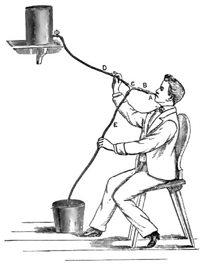
In the siphon process the outer end of the tube inserted into the stomach is connected with a piece of elastic tubing about three and a half feet long, in the free end of which is inserted the extremity of a medium-sized glass funnel. A single elastic tube about six feet long may also be used. When the funnel is elevated, water which has been poured into it will run into the stomach. If now, before the water has all run out, the funnel be depressed below the level of the stomach, the fluid contents of the stomach will flow out through the tube according to the principle of the siphon. Figs. 20 and 21 will make clear the mode of operation of this process. (The tube shown in these figures is the Faucher tube, commonly used in France, and consisting, with the funnel, of one piece. A longer tube than that shown in the figure should be used.)
FIG. 20.29 FIG. 21.29


29 From Souligoux, De la Dilatation de l'Estomac, Paris, 1883.
Another convenient but somewhat more complicated method of employing the siphon process is according to Rosenthal's principle, and is represented in Fig. 22. To the outer end of the stomach-tube is attached a Y-shaped glass tube, one arm of which is connected with an elastic tube running to an irrigator, while the other arm is connected with the discharging tube. Through the irrigating tube water runs into the stomach, the discharging tube being compressed. If the discharging tube be opened while the fluid is flowing from the irrigator, and if then, after the establishment of a column of water in the discharging tube, the irrigating tube be compressed or the stopcock of the irrigator be closed, a siphon communicating with the stomach is formed and empties this organ of its fluid contents.
FIG. 22.30
30 From Leube, in Ziemssen's Handb. d. spec. Path. u. Therap., Bd. vii.
In the siphon process the tube inserted into the stomach may be an incompressible hard-rubber tube like that employed with the stomach-pump, but by far the simplest, most convenient, and safest form of stomach-tube is the soft, flexible, red rubber tube, resembling the Jacques catheter, but of course larger and longer.31 This soft tube can inflict no injury, and in most cases it is readily introduced. Generally, the patient himself can best manipulate the introduction of the tube. After the tube is introduced into the pharynx, the patient, who should be in a sitting posture, makes repeated acts of swallowing, by means of which, accompanied by directing and gently pushing the tube with the fingers, the tube passes along the oesophagus into the stomach. Often at first the nervousness and inexperience of the patient occasion some trouble, but after a little practice he generally succeeds in introducing the tube without discomfort or difficulty. Before its introduction the tube should be anointed with a little vaseline or some similar substance. In an adult the tube is introduced for a length of at least 20 to 25 inches, and in cases of dilatation of the stomach of course for a greater distance. Whatever form of stomach-tube be used, it is important that the tube should be at least 30 inches long, and should be provided with one, and preferably with two, large eyes at its distal extremity.
31 Such a tube (marked 19 A) is made by Tieman & Co. of New York, and is to be had of most surgical instrument-makers. (For a fuller description of the tube and the mode of its employment see article by W. B. Platt, "The Mechanical Treatment of Diseases of the Stomach," Maryland Medical Journal, March 8, 1884.)
Oser's tube is 2 meters long, and is made of mineralized rubber. There are two sizes. The smaller has a lumen of 8 mm. The thickness of the wall is 2½ mm. In the larger tube the lumen is 10 mm., and the thickness of the wall 3 mm. He generally uses the smaller tube.
Faucher's tube is 1½ meters long. The external diameter of the tube is 10 to 12 mm. The walls are of such thickness that the tube can be bent without effacing its lumen. At one extremity is a lateral eye with two orifices. To the other extremity is adapted a funnel with a capacity of about 500 grammes.
Although the stomach-pump has the advantage of more completely evacuating the stomach and of removing coarser solid particles than is possible with the siphon, nevertheless its disadvantages—namely, the possibility of inflicting injury to the mucous membrane of the stomach,32 the expense and greater complexity of the instrument, and the circumstance that it should be used only by the physician—in contrast with the advantages of the siphon—namely, its cheapness, simplicity, safety, and possible employment by the patient or his attendants—have led to the general adoption of the latter process. Only the soft-rubber stomach-tube should be left to the employment of the patient.
32 A number of cases have been recorded in which pieces of the mucous membrane of the stomach have been detached by the stomach-pump. Although as yet no serious effects have followed this accident, the possibility of its occurrence can certainly not be regarded with equanimity.
Sometimes the flow through the siphon is interrupted by occlusion of the eye of the stomach-tube by a solid mass or by some cause not always clear. As already mentioned, it is desirable that there should be two openings at the gastric extremity of the tube. When the flow is interrupted the position of the tube in the stomach may be changed, or the patient may be directed to cough or to exert the pressure of the abdominal muscles, or more water may be allowed to run into the stomach in order to displace an occluding mass in the tube. It is, however, well for such cases to have, if possible, a stomach-pump and an incompressible tube in reserve. Moreover, as is apparent from the foregoing statement of the advantages of the stomach-pump, there are cases in which this instrument is much more useful than the siphon, so that one cannot decide unconditionally in favor of one instrument over the other.
The stomach-tube should be secured so that there can be no possibility of its being swallowed entirely. A string may be attached to the distal end of the tube. Leube33 has reported an instance in which the whole tube disappeared into the stomach, and Jackson34 has also narrated a case in which an insane patient swallowed the stomach-tube. In both cases the tube was subsequently rejected by vomiting.
33 Deutsches Arch. f. klin. Med., Bd. 33, p. 6.
34 Extracts from the Records of the Boston Society for Medical Improvement, vol. vi. p. 261.
For washing out the stomach after the greater part of the contents have been withdrawn, about a pint of tepid fluid is allowed to slowly run into the stomach, and is then siphoned out. This process is to be repeated several times. In general, tepid water suffices for washing out the stomach, but it is often better to use, at least a part of the time, a 1 to 2 per cent. solution of bicarbonate of sodium, which facilitates the removal of mucus. The artificial and the natural Vichy and Carlsbad waters are also excellent for this purpose. Various additions are also made to the water with the view of counteracting fermentative changes in the stomach. For this purpose perhaps the best agents are salicylate of sodium (1 per cent. solution) or resorcin (2 per cent. solution). Other substances which have also been recommended are carbolic acid, permanganate of potassium, hyposulphite of sodium, creasote, benzine. Simple water, however, accomplishes about all that is possible, and many are satisfied to use it without any medication.
As regards the frequency with which the stomach is to be washed out, one is to be guided by the symptoms and the effect obtained by the use of the stomach-tube. As a general rule, it suffices to wash out the stomach once a day, and often the process need be repeated only every second or third day.
Opinions are divided as to the best time of day to select for washing out the stomach. Kussmaul recommends the morning before breakfast, and the majority have followed his advice; others prefer the evening. There is much, however, in favor of washing out the stomach about half an hour before the principal meal of the day. The best opportunity has been offered for the digestion and absorption of the food taken at the previous main meal, and the stomach is placed in the best possible condition for the reception of more food.
The habitual washing out of the stomach is not without its drawbacks. We often remove, as has been pointed out especially by Leube, not only noxious substances from the stomach, but also the completed products of digestion. To withdraw from the nourishment of the body this chyme which the stomach has laboriously manufactured cannot be a matter of indifference. Still, with the weakened absorptive powers of the stomach, and its inability to properly propel its contents into the intestine, it is a question how much of this chyme would eventually be utilized for nutrition. Another point is worthy of attention. The relief which the patient experiences when his overloaded stomach is freed of its burden, and the knowledge that this method of relief is always at hand, may make him careless in the observance of the dietetic rules which are of great importance in the treatment of this disease. It is well, therefore, not to wash out the stomach oftener than is necessary, nor to continue the habitual use of the stomach-tube longer than is required.
There are contraindications to the use of the stomach-tube. In very rare instances the attempt to introduce the tube causes the patient so much distress, produces such violent spasm of the pharyngeal and adjacent muscles, or induces so much retching and vomiting, or is attended with such prostration or even syncope, that this method of treatment has to be abandoned. Great weakness, recent gastric hemorrhage, ulcer of the stomach in most cases (see page 523), often cancer of the cardia or of the oesophagus, and aneurism of the aorta, are contraindications to the use of the stomach-tube.
If we group together the results obtained by the use of the stomach-tube in gastric dilatation, we shall find cases in which no benefit results; cases which are benefited, but are obliged to continue the use of the stomach-tube throughout life; cases in which recovery is slow and gradual; cases with more or less speedy relief or apparent cure, but followed by relapses; and cases of prompt relief and permanent cure.
The regulation of the diet is never to be neglected in cases of dilatation of the stomach. Here the guiding principles are that little fluid should be taken, and that the food should be small in bulk, nutritious, easily digestible, and not readily undergoing fermentation. The patient should drink as little water as possible, and should therefore avoid whatever occasions thirst. It is hardly practicable to carry out the plan of giving water mostly by the rectum, as has been proposed. In most cases milk is useful, but an exclusively milk diet is not generally well borne on account of the quantity of fluid required. Leube's beef-solution is often serviceable. Soft-boiled eggs and tender meats are to be allowed, particularly the white meat of fowl and rare beefsteak, especially that prepared from scraped and finely-chopped beef, as recommended in the treatment of gastric ulcer (page 521). Fatty, saccharine, and amylaceous articles of food—hence most vegetables and fruits—are to be avoided on account of their tendency to undergo fermentation in the stomach. Alcohol in any form is usually detrimental. If gastric symptoms, particularly vomiting, be very urgent, or if food introduced into the stomach affords little or no nourishment, as in some cases of tight pyloric stricture, then rectal alimentation is to be resorted to.
An important indication is to restore the tone and contractile power of the muscular coat of the stomach. For this purpose electricity, in the form both of the constant and of the faradic current, has been beneficially employed. The best results are reported from the use of the faradic current. Both poles may be applied over the region of the stomach. The application of electricity to the inside of the stomach by means of electrodes attached to stomach-tubes or bougies is a more difficult procedure, but has its advocates. Uniformly good results are not obtained by the use of electricity in gastric dilatation, but there can be no doubt that in some cases decided benefit follows this method of treatment.
Nux vomica, particularly its alkaloid strychnia, has been much employed with the view of stimulating the muscular power of the stomach. Strychnia is given either internally or hypodermically. Hypodermic injections of ergotin have also been used for the same purpose. It has been hoped to increase the contraction of the stomach by cold applications to the abdomen, as by ice-bags applied immediately after washing out the stomach. The benefit derived from these various attempts to increase the tonicity of the gastric muscle is not very apparent.
A belt or bandage around the abdomen in order to support the stomach sometimes makes the patient feel more comfortable; in other cases it aggravates the symptoms.
In many cases digestion is promoted by giving dilute hydrochloric acid with or without pepsin. About ten drops of dilute hydrochloric acid may be given half an hour to an hour after each meal.
When the stomach is systematically washed out, the individual symptoms of dilatation of the stomach will rarely require special treatment. The sensation of fulness and weight in the stomach, the eructations, the vomiting, and the constipation are generally relieved, at least temporarily, by washing out the stomach. The appetite is improved, and an increase in weight is usually soon noticeable.
If heartburn and eructations of gas continue troublesome, an antacid, such as bicarbonate of sodium or prepared chalk, will be found useful.
Leube, in order to relieve constipation and to increase the peristalsis of the stomach, administers Carlsbad water (see page 522). Not more than five or six ounces of the water need be given, and this should be taken slowly in divided doses. A laxative pill containing rhubarb may be given occasionally.
If anæmia be the cause or a prominent accompaniment of dilatation of the stomach, iron may be administered in a form as little disturbing the digestion as possible, as the effervescing citrate or the lactate, or arsenic in the form of Fowler's solution may be tried. In general, however, all drugs which impair the appetite or digestion are to be withheld. The digestion and the general condition of the patient are often benefited by massage.
Resection of the pylorus in cases of cancerous and of cicatricial stenosis of this orifice has been performed in several instances. The subject, as regards its medical in distinction from its surgical bearings, has already been discussed in connection with cancer of the stomach (see page 577). Here it may be added that the propriety of resection is less open for dispute in cases of non-cancerous pyloric stenosis than it is in cancer of the pylorus.
Remarkable results have been reported by Loreta in cases of cicatricial stenosis of the pylorus. After performing gastrotomy he inserts his fingers through the constricted pyloric orifice and forcibly dilates the stricture.35 To judge from experience in divulsing strictures in other parts of the body, it does not seem probable that a permanent cure can be often effected by this bold and dangerous procedure.
35 Loreta has performed this operation successfully no less than nine times (The Lancet, April 26, 1884).
Acute Dilatation of the Stomach.
Under the name acute dilatation of the stomach36 have been described cases in which it has been supposed that a more or less suddenly developed paralysis of the muscular coat of the stomach exists. But the propriety of the term acute dilatation, and the very existence of an acute paralysis of the stomach, are, to say the least, questionable.
36 The literature pertaining to the subject of acute dilatation of the stomach is to be found in Poensgen, Die Motorischen Verrichtungen des Menschlichen Magens, Strasburg, 1882, p. 95.
As causes of this so-called acute dilatation of the stomach have been assigned injuries, particularly those affecting the abdomen, surgical operations involving the peritoneum, acute inflammations of the mucous and of the peritoneal coats of the stomach, acute fevers, especially during convalescence, and overloading the stomach with food or with liquids.
The symptoms which have been chiefly emphasized are severe abdominal pain, tympanitic distension of the stomach, and absence or cessation of vomiting if this has previously existed. It will be noted that inability to vomit under these circumstances implies not only paralysis of the stomach, but also that of the abdominal muscles.
The prognosis depends on the character of the primary disease causing the alleged paralysis.
If there be acute distension of the stomach with inability of the organ to expel its contents either externally or into the intestine, the stomach-tube may be employed to evacuate the gas and other material present.
In a case described by Hilton Fagge37 as acute dilatation of the stomach the symptoms of dilatation appeared suddenly and ran an acute course, but the autopsy showed that the dilatation was doubtless of much longer development than the symptoms indicated. In a case reported by Nauwerk38 of extreme dilatation in consequence of hypertrophic stenosis of the pylorus, after ten months of insignificant dyspeptic symptoms there suddenly appeared, after excess in eating, symptoms of dilatation of great severity, which continued until a fatal termination at the end of three months. Thus it appears that chronic dilatation of the stomach may cause little disturbance for a considerable time and then run a rapid course.
37 "On Acute Dilatation of the Stomach," Guy's Hosp. Rep., xviii. p. 4, 1873.
38 Deutsches Arch. f. kl. Med., Bd. xxi. p. 573.
MINOR ORGANIC AFFECTIONS OF THE STOMACH.
(CIRRHOSIS; HYPERTROPHIC STENOSIS OF PYLORUS; ATROPHY; ANOMALIES IN THE FORM AND THE POSITION OF THE STOMACH; RUPTURE; GASTROMALACIA.)
BY W. H. WELCH, M.D.
CIRRHOSIS OF THE STOMACH.
DEFINITION.—Cirrhosis of the stomach is characterized by thickening of the walls of the greater part or of the whole of the stomach in consequence of a new growth of fibrous tissue, combined usually with hypertrophy of the muscular layers of the stomach. The cavity of the stomach is usually contracted, but sometimes it is of normal size or even dilated.
SYNONYMS.—Fibroid induration of the stomach; Hypertrophy of the walls of the stomach; Chronic interstitial gastritis; Sclerosis of the stomach; Plastic linitis.
HISTORY.—The writings of the seventeenth and eighteenth centuries contain many records of extremely contracted stomachs with uniformly and greatly thickened walls (Butzen, Löseke, Storck, Portal, Lieutaud, Pohl, etc.). In the works of Lieutaud and of Voigtel may be found references to many such cases.1 Doubtless, some of these cases were examples of cirrhosis of the stomach, but in the absence of microscopical examination it is not possible to separate these from cancer.
1 Lieutaud, Historia anat.-med., t. i. p. 8, Venet., 1779; Voigtel, Handb. d. Path. Anat., Bd. ii. p. 450, Halle, 1804. Here it may be mentioned that Diemerbroeck's case, which is so often quoted to prove that polyphagia instead of causing gastric dilatation may produce hypertrophy of the muscular coat of the stomach, with contraction, was probably an instance of cirrhosis of the stomach.
Andral2 was the first to describe fully and systematically hypertrophy of the walls of the stomach. He attributed the lesion to chronic inflammation. He erroneously supposed that scirrhus of the stomach was only hypertrophy of the gastric walls. Cruveilhier3 distinguished between scirrhous induration and hypertrophy, which he considered to be a final result of the irritation accompanying chronic diseases of the stomach. Rokitansky's4 description of fibroid induration of the stomach, although brief, is accurate. He says that the process usually involves the whole stomach, and that it originates in an inflammation of the submucous connective tissue. This inflammation may occur either independently or in combination with gastritis mucosa. Rokitansky emphasizes the error of confounding the disease with scirrhous cancer. Bruch5 made an elaborate study of hypertrophy of the walls of the stomach, which he considered to be the final stage of various chronic diseases of the stomach. Fibrous or scirrhous cancer he considered to be nothing but this hypertrophy.
2 Précis d'Anat. path., Paris, 1829.
3 Anatomie pathologique, Paris, 1830-42.
4 Lehrb. d. Path. Anat., Wien, 1855-61.
5 Zeitschr. f. rat. Med., Bd. vii., 1849.
The best descriptions of cirrhosis of the stomach have been furnished by English writers, by most of whom it is properly regarded as an independent disease. Brinton6 first employed the names cirrhosis of the stomach and plastic linitis. Excellent descriptions of the disease have been given by Hodgkin, Budd, Brinton, Habershon, H. Jones, Wilks, Quain, and Smith.
6 Diseases of the Stomach.
While in former times cirrhosis of the stomach was confounded with cancer, in recent times it has not been separated by many from chronic catarrhal gastritis. In German systematic works the disease receives, as a rule, only passing mention in connection with chronic catarrhal gastritis.
ETIOLOGY.—Cirrhosis of the stomach is rare, but it is not so exceptional as to be without any clinical importance. I have met with three cases at post-mortem examination.
The disease is more frequent in men than in women. A considerable number of cases have occurred between thirty and forty years of age, but the greatest frequency is after forty. At an earlier age than twenty the disease is very rare.
The causation of cirrhosis of the stomach is obscure. Nearly all writers upon the subject have emphasized the abuse of alcohol as an important cause in this as in other diseases of the stomach. Intemperance cannot, however, be the only cause; and here, as elsewhere, it is not easy to say what importance is to be attached to it as an etiological factor. In only one of the three cases which I examined post-mortem could it be determined that the patient was an immoderate drinker, and in one case intemperance could be positively excluded. Other cases have been recorded in which the abuse of spirits could be positively excluded. In one of my cases syphilis existed, as was established by the presence of gummata in the liver. In some cases the disease has been attributed to cicatrization of a gastric ulcer. In a case reported by Snellen the disease followed an injury to the epigastric region.7
7 Canstatt's Jahresbericht, 1856, iii. 302.
Cirrhosis of the stomach, as well as cancer, ulcer, and most other chronic structural diseases of this organ, is usually associated with chronic catarrhal gastritis. There is, however, no proof of the prevalent idea that chronic catarrhal gastritis is the cause of the enormous new growth of fibrous tissue which characterizes typical cases of this disease.
SYMPTOMATOLOGY AND DIAGNOSIS.—The symptoms of cirrhosis of the stomach are not sufficiently characteristic to warrant a positive diagnosis. Sometimes the disease pursues a latent course. Like cancer of the stomach, it may put on various disguises. Thus, in a case of cirrhosis of the stomach reported by Nothnägel8 the symptoms were typically those of progressive pernicious anæmia. Association with ascites or with chronic peritonitis may lead to a false diagnosis. Thus, in one of the cases which I examined after death, and in which there was chronic peritonitis with abundant fluid exudation, the disease during life was diagnosticated as cirrhosis of the liver. Most frequently, however, cirrhosis of the stomach is mistaken for gastric cancer, from which, in fact, it can rarely be positively diagnosticated.
8 Deutsches Arch. f. kl. Med., Bd. 24, p. 353.
The symptoms are usually those of chronic dyspepsia, which sooner or later assumes a severity which leads to the diagnosis of some grave structural disease of the stomach, usually of cancer.
Indigestion, loss of appetite, oppression in the epigastrium, vomiting, are the common but in no way characteristic symptoms of cirrhosis of the stomach. There may be severe gastralgia, but in general the disease is less painful than either ulcer or cancer of the stomach. The inability to take more than a small quantity of food or of drink at a time, with the sense of fulness which even this small quantity occasions, has been considered somewhat characteristic of cirrhosis of the stomach, but this symptom is too inconstant, and occurs in too many other affections of the stomach, to be of much service in diagnosis. The symptoms of dyspepsia are often of much longer duration than in cancer, existing sometimes for many years (up to fifteen years), but on the other hand there have been cases in which the clinical history of gastric cirrhosis was as rapid in its progress as cancer. Moreover, cancer may be preceded by dyspeptic symptoms of long duration, but long duration is the exception with cancer and the rule with cirrhosis of the stomach.
As the disease progresses the patient loses flesh and strength, and usually dies in a condition of marasmus. Blood is rarely present in the vomit, but in a few cases the vomiting of coffee-ground material has been noted.
By physical examination sometimes a tumor in the region of the stomach can be felt. Under favorable circumstances it can sometimes be determined that this tumor is smooth, elastic, tympanitic on percussion, and presents more or less distinctly the contours of the stomach. By administering effervescing powder it may be possible to obtain further evidence that the tumor corresponds in its form to the stomach. The diagnosis of contraction of the cavity of the stomach is not easy. Some information may be afforded by noting the length to which the inflexible stomach-tube can be passed. The quantity of water which can be poured into the stomach until it begins to run out of the stomach-tube may also bring some confirmatory evidence as to the existence of contraction of the stomach.
Even should the physical signs suffice to determine that the tumor is the thickened and contracted stomach, still cancer cannot be excluded, for this also may grow diffusely in the gastric walls and may cause contraction of the cavity of the stomach. With our present means of diagnosis, therefore, the most which can be said is, that a special combination of favorable circumstances may render probable the diagnosis of cirrhosis of the stomach, but a positive diagnosis is impossible.
MORBID ANATOMY.—In most cases of cirrhosis of the stomach the stomach is contracted. The cavity of the stomach has been found not larger than would suffice to contain a hen's egg, but such extreme contraction is very rare. When the stomach in this disease is found dilated, either the thickening involves only or chiefly the walls of the pyloric portion, or the morbid process probably began there and was followed by dilatation.
In typical cases the walls of the entire stomach are thickened, but frequently the thickening is most marked in the pyloric region. The walls may measure an inch and even more in thickness. The thickened walls are dense and firm, so that often upon incision the stomach does not collapse.
Upon transverse section the different coats of the stomach can be distinguished. The mucous membrane is least affected, being sometimes thickened, sometimes normal or atrophied. The muscularis mucosæ is hypertrophied, and is evident to the naked eye as a grayish band. The submucous coat is of all the layers the most thickened, being sometimes ten to fifteen times thicker than normal. It appears as a dense white mass of fibrous tissue. The main muscular coat is also, as a rule, greatly hypertrophied; the grayish, translucent muscular tissue is pervaded with streaks of white fibrous tissue prolonged from the submucous and subserous coats. This last coat resembles in appearance the submucous coat, which, however, it does not equal in thickness, although it is, proportionately to its normal thickness, much hypertrophied. The free peritoneal surface usually appears opaque and dense.
To the naked eye it is apparent that the new growth of fibrous tissue is most extensive in the submucous coat, which it is probably correct to regard as the starting-point of the disease. The hypertrophy of the muscular layers is also in most cases an important element in the increased thickness of the gastric walls.
Microscopical examination9 shows sometimes a nearly normal mucous membrane. The tubules, however, are usually more or less atrophied. In the case reported by Nothnägel tubules could be found only in the pyloric region of the stomach. The essential lesion is the new growth of fibrillated connective tissue pervading all of the coats of the stomach. In an interesting case reported by Marcy and Griffith,10 which was believed to be caused by an extensive cicatrized ulcer, a new formation of smooth muscular tissue was found not only in the main muscular tunic and the muscularis mucosæ, but also throughout the submucosa. This peculiarity was probably referable to the cicatrization of the ulcer.
9 Microscopical examination is always necessary for a positive diagnosis of cirrhosis of the stomach. In a case which I examined post-mortem of double ovarian cancer, with multiple secondary deposits in the peritoneum and with chronic peritonitis, the stomach presented the typical gross appearances of cirrhosis, but here and there were to be found nests of cancer-cells in the prevailing new growth of fibrous tissue in the walls of the stomach.
10 Am. Journ. of the Med. Sci., July, 1884, p. 182.
Not infrequently adhesions exist between the stomach and surrounding organs. Exceptionally, a diffuse growth of fibrous tissue may invade the greater part of the peritoneum, particularly the visceral layer, and cause a thickening similar to that existing in the stomach. In such cases ascites is usually a marked symptom.
PROGNOSIS.—The prognosis of cirrhosis of the stomach is grave. The disease runs a chronic course, and usually terminates in death by asthenia. There is no reason to believe that the stomach can ever be restored to its normal condition. Still, cases have been reported in which it has been supposed that cirrhosis of the stomach has terminated in recovery.11 The diagnosis, however, in such cases must remain doubtful.
11 Lesser, Cirrhosis Ventriculi, Inaug. Diss., Berlin, 1876; Smith, "Cirrhosis of the Stomach," Edinb. Med. Journ., 1872, p. 521.
TREATMENT.—The treatment is symptomatic, and is to be guided by the general principles developed in previous articles concerning the regulation of the diet and the administration of remedies.
HYPERTROPHIC STENOSIS OF THE PYLORUS.
The various causes of stenosis of the pylorus have already been mentioned under DILATATION OF THE STOMACH, and the most important of these causes have received full consideration in connection with ULCER and with CANCER OF THE STOMACH.
Only one of the varieties of pyloric stenosis can claim consideration as an independent disease. This variety is the so-called hypertrophic stenosis of the pylorus (Lebert) or fibroid degeneration of the pylorus (Habershon12). Under the name of hypertrophic stenosis have been described cases in which the stenosis was due to hypertrophy of only one of the coats of the stomach, usually either the submucous or the muscular coat, sometimes only the mucous coat. In most cases, however, all of the coats of the stomach are involved, and the lesion is similar to that of cirrhosis of the stomach, but it is confined to the pylorus or to the pyloric region. In such cases there is new growth of fibrous tissue, most marked in the submucous coat, and hypertrophy of the muscular coat. The appearance of the pylorus in some instances of hypertrophic stenosis has been not inappropriately compared to that of the cervix uteri.
12 Habershon, On Diseases of the Abdomen, London, 1862; Lebert, Die Krankh. d. Magens, Tübingen, 1878; Nauwerk, Deutsches Arch. f. klin. Med., Bd. 21, 1878.
In the majority of cases the change here described is the result of cicatrization of a gastric ulcer, and some believe that all cases of so-called hypertrophic stenosis or fibroid degeneration of the pylorus are referable to ulcer, although it may be very difficult to discover the cicatrix of the ulcer. It is certainly not always possible to detect either ulcer or cicatrix, so that it seems proper to regard the hypertrophic stenosis in such cases as constituting an independent affection.
The symptoms are those of dilatation of the stomach, sometimes preceded by evidences of chronic catarrhal gastritis. The thickened pylorus can sometimes be felt during life as a small, cylindrical, usually movable tumor, either stationary in progress or of very slow growth.
In most cases the diagnosis of organic stenosis of the pylorus can be made. Cancer may sometimes be excluded by the long duration of the symptoms and the stationary character of the tumor if a tumor can be felt. The exclusion of ulcer is more difficult and hardly possible, for ulcer may have existed without producing characteristic symptoms.
The prognosis and treatment have been considered under DILATATION OF THE STOMACH.
ATROPHY OF THE STOMACH.
Atrophy of the stomach may be the result of stenosis of the cardia or of the oesophagus. The stomach may participate with other organs in the general atrophy attending inanition and marasmus. The walls of a dilated stomach may be very thin.
Especial importance has been attached in recent years to degeneration and atrophy of the gastric tubules. The glands of the stomach may undergo degeneration and atrophy in various diseases of the stomach, such as chronic catarrhal gastritis, phlegmonous gastritis, cirrhosis of the stomach, and cancer of the stomach. Parenchymatous and fatty degeneration of the glandular cells of the stomach occurs in acute infectious diseases, as typhoid fever and yellow fever, also as a result of poisoning with phosphorus, arsenic, and the mineral acids.
It is claimed by Fenwick that atrophy of the stomach may occur not only as a secondary change, but also as a primary disease attended by grave symptoms. Fenwick has described a number of cases in which the gastric tubules were atrophied without thickening of the walls of the stomach and without diminution in the size of the cavity of the stomach—cases, therefore, which cannot be classified with cirrhosis of the stomach.13 He attributes in many cases the atrophy of the tubules to an increase in the connective tissue of the mucous membrane, and draws a comparison between atrophy of the stomach and the atrophic form of chronic Bright's disease.
13 The Lancet, 1877, July 7 et seq.
In 1860, Flint14 called attention to the relation between anæmia and atrophy of the gastric glands. He expressed the opinion that some cases of obscure and profound anæmia are dependent upon degeneration and atrophy of the glands of the stomach. Since Flint's publication cases have been reported by Fenwick, Quincke, Brabazon, and Nothnägel, in which lesions supposed to be due to pernicious anæmia have been found after death associated with atrophy of the gastric tubules.15 Nothnägel's case, which has already been mentioned, was one of cirrhosis of the stomach.
14 A. Flint, American Medical Times, 1860. Further contributions of Flint to this subject are to be found in the New York Medical Journal, March, 1871, and in his Treatise on the Principles and Practice of Medicine, p. 477, Philada., 1881.
15 Fenwick, loc. cit.; Quincke, Volkmann's Samml. klin. Vorträge, No. 100 (case b); Brabazon, British Med. Journ., 1878, July 27 (without microscopical examination!); Nothnägel, Deutsches Arch. f. kl. Med., Bd. 24, p. 353.
The symptoms which have been referred to primary atrophy of the stomach are severe anæmia and disturbances of digestion, such as anorexia, eructations, and vomiting. The digestive disturbances are often not greater than are frequently observed in cases of severe anæmia.
In my opinion, the existence of atrophy of the stomach as a primary and independent disease has not been established. In many cases which have been described as primary atrophy the histological investigation of the stomach has been very defective. Degeneration and atrophy of the gastric tubules secondary to various diseases of the stomach and to certain general diseases is an important lesion when it is extensive, and must seriously impair the digestion, and consequently the nutrition, of the patient.
ANOMALIES IN THE FORM AND IN THE POSITION OF THE STOMACH.
These anomalies, so far as they have not received consideration in previous articles, are of more anatomical than clinical interest, and therefore here require only brief mention.
The stomach may have an hour-glass shape in consequence of a constriction separating the cardiac from the pyloric half of the organ. This constriction is sometimes congenital,16 sometimes caused by cicatrization of a gastric ulcer, and sometimes caused by spasmodic contraction of the muscle, which may persist after death, but disappears when the stomach is artificially distended. Hour-glass shape of the stomach has been diagnosed during life by administering an effervescing powder according to Frerichs' method.
16 A careful study of the congenital form of hour-glass contraction of the stomach has been made by W. R. Williams ("Ten Cases of Congenital Contraction of the Stomach," Journ. of Anat. and Physiology, 1882-83, p. 460).
Foreign substances of hard consistence which have been swallowed sometimes cause diverticula of the stomach.
Sometimes the fundus of the stomach is but little developed, so that the organ is long and narrow like a piece of intestine.
The stomach may be variously distorted by external pressure, as from tumors and by adhesions.
The loop-shaped stomach and vertical position of the stomach have been already considered in connection with DILATATION OF THE STOMACH (page 602).
In transposition of the viscera the stomach is also transposed. In such a case difficulties may arise in the diagnosis of pyloric cancer, as in a case described by Légroux.
The stomach may be found in hernial sacs. Mention has already been made of the presence of dilated stomachs in scrotal hernia. More frequently the stomach is found in umbilical hernias. In diaphragmatic hernia the stomach is found more frequently in the thorax than is any other abdominal viscus. In 266 diaphragmatic hernias collected by Lascher17 the stomach was found either wholly or partly in the thorax in 161 cases. The clinical consideration of diaphragmatic hernia, however, does not belong here.
17 Deutsches Arch. f. kl. Med., Bd. 27.
Furthermore, the stomach may be displaced by tumors, enlargement of neighboring organs, tight-lacing, adhesions, and the weight of hernias. These displacements, however, are generally inconsiderable and of little importance.
In a case described by Mazotti18 the stomach, of which the pyloric portion was fixed by adhesions, was twisted around its long axis. Death was caused by uncontrollable vomiting.
18 Virchow und Hirsch's Jahresbericht, 1874, ii. p. 249.
RUPTURE OF THE STOMACH.
Sufficient attention has already been given to perforation of the stomach in consequence of diseases of its walls, such as ulcer, cancer, abscesses, and toxic gastritis.
A healthy stomach may be ruptured by violent injury to the abdomen even when no external wound is produced. An example of rupture of the stomach from this cause is that sometimes produced when a person has been run over by a heavy vehicle.
It has been claimed that a stomach with healthy walls may burst in consequence of over-distension of the organ with solids or with gas. The older literature is especially rich in reports of so-called spontaneous rupture of the stomach. Most of these cases were examples of perforation of gastric ulcer. In a case of apparently spontaneous rupture of a stomach which had become abnormally distended with gas, Chiari19 found that the rupture was through the cicatrix of a simple ulcer in the lesser curvature. It is hardly conceivable that rupture of the healthy stomach from over-distension can occur so long as the orifices of the organ are unobstructed.
19 Wiener med. Blätter, 1881, No. 3.
Lautschner20 reports a case of spontaneous rupture of the stomach in a woman seventy years old with an enormous umbilical hernia which contained the pyloric portion of the stomach. After drinking eight glasses of water and two cups of tea and eating meat, she was seized with vomiting, during which the stomach burst with a report which was audible to the patient and to those around her. She passed into a state of collapse and died in thirteen hours. A rent several centimeters long was found in the posterior wall of the stomach. Lautschner thinks that the pylorus was bent in the hernial sac so as to be obstructed. In the walls of the stomach he found no evidence of pre-existing disease.
20 Virchow und Hirsch's Jahresbericht, 1881, ii.
There is no satisfactory proof of the possibility of the occurrence of rupture of a stomach with healthy walls except as a result of external violence.
The symptoms and treatment of rupture of the stomach are those of perforation of the stomach, and have already been described. The prognosis is fatal.
GASTROMALACIA.
That the subject of gastromalacia should still occupy so much space in medical works the purpose of which is mainly clinical proves that many physicians still cling to the belief that this process may occur during life. It is, nevertheless, certain that the condition which, according to the ordinary and traditional use of the term, is designated gastromalacia, is always a post-mortem process and is without the slightest clinical significance. So long as the circulation of the blood in the walls of the stomach is undisturbed, self-digestion of this organ cannot occur. No one doubts that parts of the gastric walls in which the circulation has been arrested, and which are exposed to the gastric juice, undergo self-digestion, as has already been set forth in the article on GASTRIC ULCER. To describe cases of this nature under the name of gastromalacia, however, is misleading, and can cause only confusion, for the long-continued discussion as to whether gastromalacia is a vital or a cadaveric process applied certainly to a different conception of the term. In some of the cases which have been published, even in recent years, in support of the vitalistic theory of gastromalacia, and in which it has been proven that perforation of the stomach occurred during life, the solution of continuity took place through parts of the gastric walls in which the circulation had already been obstructed, particularly by extensive hemorrhagic infiltration. Some of these cases are probably also examples of perforation of gastric ulcer or of rupture of cicatrices from over-distension of the stomach, in which post-mortem digestion of the edges of the ulcer or of the cicatrix obscured the real nature of the process. The subject of gastromalacia should be relegated wholly to works on physiology and on pathological anatomy.
INTESTINAL INDIGESTION.
BY W. W. JOHNSTON, M.D.
NATURE.—The term indigestion in its most common meaning refers to gastric indigestion only. This limitation has arisen from the fact that gastric digestion has been more thoroughly understood than intestinal digestion, and because the symptoms, flatulence, acidity, eructations of gas, pyrosis, and vomiting of unaltered food, are readily referred to the stomach as their source. Intestinal digestion has not been well known until within a recent date, and its phenomena in disease have been mistaken for other pathological conditions.
From the important and complex function of the intestinal juices, and the very great share they take in the solution of food, there must be many phases of departure from the normal state. The processes of intestinal digestion are more intricate than those of gastric digestion, of a higher grade, and the chemical reactions are more numerous, depending upon the participation of the bile, the pancreatic juice, and the succus entericus; while intestinal absorption is a more complex act than that of gastric absorption.
A brief review of the physiology of intestinal digestion will be of aid in making clear its pathology.
The object of all digestion is to make such a solution of the ingesta that they may pass through animal membrane and so enter the system. Mechanical disintegration and simple solution do something toward this, but for substances insoluble in water a more thorough change is brought about by ferments which convert insoluble into soluble compounds.
The process of digestion begins in the mouth. Mastication breaks up the masses of food; the saliva softens them, dissolves soluble substances, as salt and sugar, and thus the pleasures of the palate are enhanced. The ferment ptyalin acts upon starch (boiled starch being more rapidly altered than unboiled), and changes it to dextrin and grape-sugar, both of which are diffusible through animal membrane, entering lymph-spaces and blood-vessels. The greater part of the saliva secreted is swallowed with the food or in the intervals of eating. The amount formed in twenty-four hours varies from 1500 gm. (Bidder and Schmidt) to 700 gm. (Tuczek). It must therefore serve some ulterior purpose in the stomach. Ewald1 says that saliva converts starch into sugar in acid as well as in alkaline and neutral solutions. But Langley2 asserts that the ferment of saliva is destroyed by the hydrochloric acid of the gastric juice. The longer food is subjected to mastication and insalivation, the more thorough is the mouth digestion and the better prepared is the mass for the action of the gastric and intestinal juices. It is asserted that fatty matters are emulsified to a certain extent by the alkaline ferments of the saliva.
1 Lectures on Digestion, New York, 1881, p. 37.
2 "On the Destruction of Ferments in the Alimentary Canal," Journal of Physiology, London, Jan., 1882, p. 246.
When the food enters the stomach the nitrogenous (albuminoid) elements are attacked by the gastric juice, the bundles of muscular fibre are broken up, the fibrillæ are reduced to a granular mass, but not completely dissolved (Frerichs), the fat-globules are freed from their envelopes of connective tissue, milk is coagulated, and the casein is dissolved.
"The tangible, practical object of this change is to form out of a little-diffusible body (albumen) one easily diffusible (peptone), which is capable of absorption through animal membrane in a higher degree than ordinary albumen" (Ewald). Peptone is formed out of ordinary albumen, as grape-sugar is formed out of starch, by taking up water; it is therefore the hydrate of albumen.
The more tardy the digestion in the stomach the more highly charged with acid is the gastric juice. According to Wright, the degree of alkalinity of the saliva is in proportion to the acidity of the stomach fluids, and Bence Jones has observed that during the excretion of acid in the stomach the total alkalinity of all alkaline digestive fluids is increased. The lesson is thus learned that a too careful preparation of food, so as to shorten and lessen gastric labor, diminishes the activity of the gastric juice as well as that of all other digestive fluids.