Volume 2 — DISEASES OF THE DIGESTIVE SYSTEM. (continued 2)
Reading edition
In most instances the hypertrophy occupied the free portion of the tongue chiefly, the base of the organ having been implicated in but a few.
FIG. 14.

Chronic Intumescence of the Tongue (Harris).
FIG. 15.
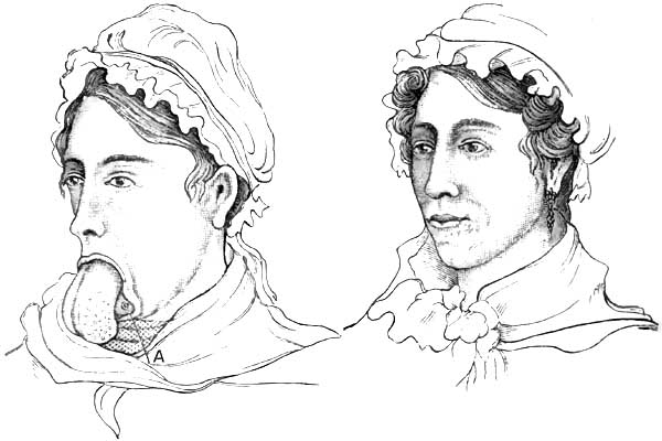
Hypertrophy of the Tongue (Harris), before operation and after. A, odontolith.
DIAGNOSIS.—The presence of the tongue outside of the mouth speaks for itself (Figs. 14 and 15). The age of the patient, usually a young child, the history of the case if it present in the adult, suffice to differentiate macroglossia from the tumefaction of glossitis on the one hand and from certain protruding tumors and malignant diseases on the other. Hypertrophy of the tongue following chronic glossitis, syphilitic or non-specific, must not be confounded with the congenital or idiopathic affection under consideration.
PROGNOSIS.—The prognosis is good as to relief from the deformity, provided the patient is submitted to surgical interference, and the prognosis of the operation depends upon the procedure selected. Sometimes additional operations are requisite to remedy the defects the lower jaw has sustained by prolonged depression. In comparatively young adults restoration of its position, configuration, and function seems likely to result spontaneously after the protruding portion of the tongue has been removed.
TREATMENT.—It has been maintained (Lassus) that the hypertrophy can be overcome by systematic compression of the tongue, by leeching the tongue, bandaging or strapping it, and forcibly maintaining it in the mouth by suitable retentive appliances. While it has been admitted that this plan may prove successful in cases of moderate enlargement of but few years' duration and unaccompanied with change in the shape of the lower jaw, the experience of more recent observers has been recorded as unfavorable, at least in pronounced cases. Clanny30 succeeded in this way with a child five years of age whose tongue protruded three inches. This plan is said to be very painful and irritating. It requires close watching on account of the difficulty of respiration which may ensue from thus blocking up the pharynx. It has been advised as a useful and sometimes an essential preliminary (Syme) to a radical procedure consisting in the excision of a V-shaped segment. This latter operation (Boyer) has been successfully performed by Howe, Harris, Humphry, Syme, and others. Frederici31 extended the incisions to the very base of the tongue. It has been performed both with knife and with scissors, the cut surfaces being united with sutures after ligation or torsion of the bleeding vessels. Re-enlargement ensued in one of Syme's cases operated upon in this way,32 and likewise in a case of Gies,33 requiring subsequent excision of the exuberant portions. Operations with the ligature, though sometimes successful (Fergusson,34 Hodgson35), may be followed by fatal septicæmia during the slough (Liston36), or, failing to strangulate the tongue sufficiently, may require the application of the knife, after all, to affect the separation (Harris37).
30 Edinb. Med. and Surg. Journ., 1805, vol. i., cited by Clarke.
31 Arch. gén. de Méd., 1844; Edinb. Med. and Surg. Journ., p. 528, vol. lxiv., 1845.
32 Edinb. Med. Journ., 1857, vol. ii. p. 1057.
33 Arch. klin. Chir., 1873, p. 640.
34 Practical Surgery, London, 5th ed., p. 518.
35 Trans. Med.-Chir. Soc. London, 1858, p. 129.
36 Elements of Surgery, p. 334, Philada., 1842.
37 Am. Journ. Med. Sci., vol. vii. p. 17.
Excision with the incandescent loop of the galvano-cautery seems to be the most suitable procedure. Valerani38 operated in this manner without the loss of a drop of blood upon a congenital macroglossia in a child seven months of age. Maas39 operated in this way on a child two months of age. Fairlie Clarke, who removed a congenital macroglossia with the écraseur in a child five months of age, recommends operation before dentition begins40—an opinion which appears to be justified by the belief that the pressure of the teeth contributes to the subsequent rapid enlargement of the organ. Nevertheless, the operation may be undertaken at any age. Several of those already cited were performed upon adults, and Stephen O'Sullivan41 excised the hypertrophied tongue of a female sixty-five years of age.
38 Giornale della Reale Accademia di Turino, fasc. 1518; London Med. Record, Sept. 15, 1876, p. 408.
39 Loc. cit.
40 Lancet, March 30, 1872, p. 432.
41 Dublin Journ. Med. Sci., Aug., 1875, p. 178.
Ignipuncture with the thermo-cautery of Paquelin has been successfully used of late by Helferich and by von Bruns of Tübingen.42 In the latter instance the subject was five years of age. Fourteen punctures were made from above downward at intervals of about one centimeter, and five were made transversely. Not a drop of blood was lost. On the third day secondary hemorrhage occurred from the intercommunication of three of the punctures; this was restrained by ferric chloride, and the case went on to a favorable conclusion. Surgical procedures must constitute our sole therapeutic reliance. The temporary subsidence of enlargement under the influence of mercury and the iodides seems sure to be followed, sooner or later, by reproduction of the deformity. It is therefore a waste of time to attempt cures by medication.
42 Centbl. f. Chir.; Med. Times and Gaz., Sept. 23, 1883.
Glossitis.
DEFINITION.—Inflammation of the tongue.
The term glossitis is usually applied to inflammation of the tissues of the tongue as a whole (parenchymatous glossitis), and not to those superficial inflammations which exist associated with the different varieties of stomatitis and with other affections, and which implicate the mucous membrane and its glands and papillæ only (superficial glossitis). Superficial glossitis, however, sometimes terminates in parenchymatous glossitis. Papillæ and glands are usually affected together in superficial glossitis. When the inflammation predominates in the papillæ the disease is often designated as papillary glossitis; when in the glands, it is often termed follicular glossitis. Superficial glossitis, again, is sometimes manifested by the eruption of vesicles on the tongue, under which circumstance it is often denominated vesicular glossitis, sometimes herpetic glossitis. Glossitis is sometimes restricted to a portion of the tongue (circumscribed glossitis), and it sometimes involves the whole of the tongue (diffuse glossitis). Either form may be unilateral (hemiglossitis), though both forms are more frequently bilateral. Either form may be acute or chronic.
Superficial Glossitis.
DEFINITION.—An inflammation of the mucous membrane of the tongue, usually involving likewise both papillæ and glands.
SYNONYMS.—Catarrhal glossitis, Angina lingualis. Varieties: Papillary, follicular, vesicular (herpetic and eczematous), psoriatic, ichthyotic.
ETIOLOGY.—Predisposing and Exciting Causes.—It is rarely idiopathic, is most frequently deuteropathic, and sometimes traumatic. Superficial deuteropathic glossitis usually occurs in connection with gastric and gastro-enteric affections. It occurs likewise in association with stomatitis, tonsillitis, pharyngitis, many febrile affections, scorbutus, tuberculosis, syphilis, so-called psoriasis and ichthyosis of the tongue, carcinoma of the tongue, and the various neoplasms of the organ. Irregular and sharp-cornered or jagged teeth often induce traumatic superficial glossitis. Pungent vapors, such as those of chloride of ammonium, so much used of late years in the treatment of nasal catarrhs, sometimes produce a superficial traumatic glossitis, usually localized on the superior surface of the anterior portion of the tongue. Tobacco-smoking, especially from a short-stemmed pipe, will likewise produce it occasionally at the point where the concentrated smoke strikes the organ. Attempts to drink liquids too hot, too acrid, or too caustic may be mentioned as other occasional causes. Nervous irritation, such as of the chorda-tympani nerve, is attributed as a causal influence of unilateral vesicular glossitis, herpetic or otherwise, and as a probable factor in other varieties of unilateral glossitis. Eczema of the tongue may ensue as a sequel of prolonged cutaneous eczema (De Mussy43).
43 Gaz. hebd., June 22, 1883; Med. News, Aug. 11, 1883, p. 151.
PATHOLOGY AND MORBID ANATOMY.—Superficial glossitis, as indicated, involves the mucous membrane, glands, papillæ, and epithelium. It is hardly necessary to dwell upon the pathological conditions of the lingual mucous membrane and its epithelium in gastro-intestinal and febrile disorders, as these are described in connection with the various diseases. Ordinarily, the epithelium increases in thickness, and when detached, spontaneously or otherwise, exposes a red and swollen membrane with erect papillæ. Sometimes the condensed stratified layer of epithelium becomes dry and very hard. Under some illy-defined conditions, the papillæ of the tongue undergo great hypertrophy. The filiform papillæ become elongated to several times their normal length, and feel and look like so many hairs on the tongue. Like many other lingual affections, this is often unilateral. It is quite marked in some cases of influenza and other febrile disorders, producing great annoyance in the mouth. It occurs likewise in gastro-intestinal disorders and in disorders of the mouth and teeth. It is evidently a deuteropathic phenomenon. In other cases the glands of the tongue, especially at its base, become involved, forming the follicular glossitis of some writers. In another class of cases, most frequently syphilitic or at least quasi-syphilitic, one or more whitish circumscribed patches are seen on the tongue, resembling such as are left after superficial cauterization with nitrate of silver. This condition is described as psoriasis linguæ. It is due to condensation of layers of epithelium, which may become detached in a few days in mass or in fragments, leaving the denuded mucous membrane red and the papillæ erect and somewhat swollen.
When psoriasis of the tongue has existed for a long time, a further change, and a more permanent one, takes place in the papillæ and epithelium. This condition has been denominated ichthyosis linguæ. Superficial ulceration takes place at the psoriatic patches, and the repair eventually excites such a proliferation of epithelium that it becomes quite horny to the sight and to the touch. It spreads over a much larger extent of surface than the original psoriasis, but, like it, leaves the unaffected portions of the tongue in an apparently normal condition. Both affections are usually bilateral, and the patches or series of patches most frequently symmetrical or engaging analogous vascular territory upon the two sides.
In a case reported by Mr. Hulke44 portions of the horny substance were habitually sliced off with a razor. Microscopic examination "showed colossal papillæ; the indurated portion of the mass was altogether epithelial, the lower cells being clear, transparent, and natural, the middle ones granular, and the superficial layer felted together into a dense opaque mass" (Clarke).
44 Medical Times, Nov. 30, 1861, p. 556.
Both of these affections are liable in about one-third of the cases to terminate in epithelioma. Although the opinion generally entertained classes all cases of psoriasis and ichthyosis linguæ in the category of syphilitic affections, there is reason to doubt its accuracy. Sangster45 has drawn up a tabular statement of 44 cases, of which 1 only occurred in a female; 23 occurred in smokers, 12 being inveterate smokers. In but 12 instances (81/3 per cent.) was there positive proof or strong evidence of syphilis; 30 per cent. of the whole number eventuated in epithelioma.
45 Med. Times and Gaz., London, April 8, 1882, p. 370.
Vesicular glossitis, usually unilateral and most frequently right-sided, has been described by Paget,46 Stoker,47 Barker,48 Hill,49 and De Mussy,50 and doubtless by others.
46 Lancet, March 11, 1865; Clarke, op. cit., p. 88.
47 Dub. Journ. Med. Sci., May 1, 1876, p. 401, illustrated.
48 Lancet, Nov. 22, 1879, p. 764.
49 Brit. Med. Journ., Oct. 7, 1882, p. 683.
50 Gaz. hebd., June 22, 1883; Med. News, Aug. 11, 1883, p. 151.
SYMPTOMATOLOGY, COURSE, DURATION, TERMINATIONS, COMPLICATIONS, AND SEQUELÆ.—Dryness of the tongue, stiffness, heat, and pain—the latter especially during movements of the organ in deglutition and in articulation—are the prominent subjective symptoms of acute superficial glossitis. There are rarely any marked symptoms of febrile disturbance unless the disorder is about to undergo extension into parenchymatous glossitis.
DIAGNOSIS.—Redness of the tongue, prominence of the papillæ, slight enlargement, perhaps bearing impressions made by the teeth, and pain or sense of impediment on movement, are the main diagnostic features of superficial glossitis.
Vesicles indicate the vesicular variety of superficial glossitis; irregular whitish patches, the psoriatic variety; and hard, horny patches with intervening fissures, the ichthyotic variety. A superficial circumscribed glossitis attending the local ulcerations of syphilis, tubercle, and epithelioma is differentiated by the clinical history of the case.
TREATMENT.—In ordinary cases the treatment described under catarrhal stomatitis suffices, so far as local measures are concerned. The gastritis or gastro-enteritis requires appropriate attention, as does any systemic malady under which the patient may be laboring. Demulcent and astringent lotions may be applied by douche, spray, or gargle. Local applications of weak solutions of iodine have been recommended. In cases of considerable severity, and especially when there is reason to expect extension into the deeper tissues, superficial scarification of the dorsum of the tongue is advisable.
Ulcers are perhaps best treated locally by touching the edges daily with the pencil of sulphate of copper. Any imperfect teeth in their immediate vicinity to which the ulceration may be attributable should be extracted or put in repair. De Mussy's case of eczema was cured after five months' daily use of a large quantity of water-cress.
Glossitis Parasitica.
DEFINITION.—An inflammation of the tongue said to be due to parasitic vegetation.
SYNONYMS.—Nigrities, Glossophytia, Black tongue.
Under the term black tongue two different affections have been described, the one an epidemic erysipelatous disorder to be mentioned under parenchymatous glossitis, and the other, now to be mentioned, a peculiar black pigmentation due to parasitic disease seated upon and around the hypertrophied filiform papillæ. The ordinary parasitic vegetations found upon the tongue do not produce the affection in question.
HISTORY.—First described by H. Hyde Salter,51 and then by Eulenburg, it has been made the subject of observation by Raynaud,52 Fereol and others,53 Lanceraux,54 Dessois,55 Hirz,56 Pasquier,57 Moure,58 and a few others. Outside of French literature, little had been written of it until very recently.
51 Article "Tongue," Encyclopedia of Anatomy and Physiology, London, 1849-52, vol. iv. pp. 1159, 1160.
52 Gaz. hebd., 1869, No. 14, p. 221.
53 Gaz. des Hôp., June 29, 1875.
54 Union Méd., March 20, 1877.
55 De la Langue noire [Glossophytie], Paris, 1878, 8vo, p. 38, illustrated.
56 Gaz. Méd., Strasbourg, 1879.
57 Bull. Méd. du Nord, 1883.
58 Revue mensuelle de Laryngologie, etc., Sept., 1883, p. 276.
ETIOLOGY.—The affection appears to be due to some fault of nutrition, but the cause has not been determined. The fluids of the mouth always exhibit an acid reaction. It has been seen chiefly in dyspeptics and hypochondriacs, and has seemed in one instance (Moure) to have followed the use of chlorate-of-potash lozenges. A case has been recorded by Solomon Solis Cohen59 in a negro child the subject of congenital syphilis. Mr. George Stoker60 and G. Y. Broatch61 have each reported a case of long duration occurring in a painter.
59 The Polyclinic, Philada., July, 1884, p. 10.
60 Brit. Med. Journ., March 29, 1884, p. 602—said to be first case recorded in England.
61 Ibid., April 19, 1884.
PATHOLOGY AND MORBID ANATOMY.—The disease is characterized by a grayish-black or fully black discoloration on the upper surface of the tongue, which gives it an aspect which recalls the normal appearance of the tongue of the parrot and the giraffe, and an occasional appearance of the organ in the ox, sheep, dog, cat, and some other animals. The filiform papillæ are enormously elongated, so that they closely resemble hairs, and they are described by some writers as lying upon the surface of the tongue in confusion like that of a field of wheat thrown down by the wind. The individual papillæ are surrounded with a parasitic vegetable growth. Raynaud compared the microscopic spores in his case to the microphyte of tinea tonsurans or that of herpes circinatus. According to Malassez, they do not differ from those found in the saburral tongue of the dyspeptic, and he considers that their development is favored by their very arrest by the hypertrophied papillæ. Nevertheless, the subjects of this disease are not all dyspeptics by any means. Dessois made culture-efforts to reproduce the disease upon his own tongue, but failed to inoculate it. For detailed description of the disease we cannot do better than refer the interested reader to Dessois' monograph, from whose observations, chiefly, it appears that the discoloration begins at the central portion of the tongue, increases gradually in extent and intensity for three or four days, and then gradually disappears by desquamation. The tongue is very dry while the affection is at its height. Close examination of the parts and microscopic inspection of papillæ removed for the purpose are said to show that the spores of the cryptogam are first developed at the base of the papillæ, separating them from each other. The irritation produced by the parasite causes longitudinal hypertrophy of the papilla, and the continued growth of the parasite produces a muff-like envelopment of the papilla; the spores at the same time becoming insinuated between the most superficial epithelial cells and dislocating them, so that they maintain their position around the axis of the papilla only by means of the intervening parasitic masses. The papilla continues to elongate and the cryptogam to increase, until finally it invades nearly the entire length of the papilla. This entire parasitic mass soon becomes detached, carrying with it the epithelial cells under which it has become insinuated, and leaves the papilla naked, save for a few cells remaining attached by their superior borders. In the case observed by my brother, as in Mr. Stoker's and Broatch's cases, microscopical examination of the black filaments showed them to be composed of closely-packed epithelial cells, overlapping one another, stained brown, and decreasing in intensity of color from the apex toward the base of the filament. In some cells the nucleus was darker, and in others lighter, than the surrounding protoplasm. The cryptogam, therefore, cannot always be detected. Indeed, there seems reason to believe that the affection may not be parasitic,62 although the prominence given to this feature by French writers apparently warrants its being so considered. There may be two kinds of black tongue—one non-parasitic.
62 Hutchinson, The Medical Press, p. 20, July 11, 1883.
SYMPTOMS.—There are no special subjective symptoms. The objective symptoms are the peculiar dark or black discoloration of the upper surface of the tongue and the excessively elongated filiform papillæ.
DIAGNOSIS.—The chief diagnostic feature is the black discoloration of the tongue which has given the name black tongue to the disease. Discrimination is requisite from discoloration by food or medicine.
PROGNOSIS.—This is favorable, the condition subsiding under treatment, and sometimes spontaneously, though liable to recurrence. In some instances the condition becomes chronic.
TREATMENT.—The indication is to endeavor to favor desquamation of the papilla by means of potassium chloride or sodium borate, and to administer alkalines, so as to render the saliva alkaline and unfavorable for the development of the parasite. It is recommended, in addition, to scrape the tongue with a spatula, and to douche it with a spray of mercuric chloride, 1:500. Attention to the general health is requisite, especially in dyspeptics and hypochondriacs. In the case of the negro child above alluded to the discoloration of the tongue finally disappeared under the systemic use of potassium iodide, without topical treatment, although repeated recurrences took place at varying intervals.
Parenchymatous Glossitis.
DEFINITION.—An inflammation of the tongue involving its substance as well as the mucous membrane.
SYNONYMS.—Idiopathic glossitis. Interstitial glossitis. Erectile glossitis (Salter), Glossomegistus (Sauvages), Paraglossia.
HISTORY.—Albeit a comparatively infrequent disease, especially at the present day, numerous cases and collections of cases are on record from very early times; and the affection seems to have attracted the attention of medical writers ever since. Hippocrates, Galen, Aretæus, Celsus, Aëtius, Avicenna, Forestus, Rivière, Schenkins, Sauvages, Vogel, van Swieten, are referred to by more modern writers as having described the disease. Louis, De la Malle, Lassus, J. P. Frank, Jn. Frank, Fleming,63 Clarke, and Bryant may be cited as the most prominent recent observers.
63 Dublin Journ. Med. Sci., 1850, vol. x.
ETIOLOGY.—Predisposing and Exciting Causes.—Glossitis is sometimes idiopathic, sometimes deuteropathic, and sometimes traumatic. Impaired health from over-fatigue or from exhaustive disease may be regarded as a predisposing cause in the presence of the causes which more frequently give rise to traumatic glossitis. Sudden or prolonged exposure to atmospheric changes, to cold and moisture, as when working in damp and wet localities, is often the apparent determining cause in both the idiopathic and deuteropathic varieties. In many instances occurring in this way it is found that the tongue has long been subjected to mechanical irritation from jagged teeth.
Idiopathic glossitis occurs at all ages, but has been supposed by some observers to be more frequent in scrofulous subjects. It occurs in the apparently healthy only after severe exposure to wet and cold, and in convalescents from acute febrile diseases usually after some moderate exposure to a draught of air or change of temperature. It appears to be more imminent after influenza (Möller, Smee, Graves, Salter) than after other febrile disorders. It has occasionally been caused by chewing acrid plants, some of them food-plants, some of them medicinal. In the list have been included celery, bilberries, Daphne mezereum and Daphne laureola, aconite, and tobacco. It has been known to follow the eating of shellfish (Watson, Salter).
Deuteropathic glossitis has occurred during the course of scarlatina, variola, epidemic erysipelas (black tongue), scorbutus, enteric fever, glanders, septicæmia from various causes, rheumatism, diffuse inflammation of the connective tissue of the cervico-mental region, herpes, syphilis, ptyalism, mercurial and other varieties of toxæmic stomatitis, tonsillitis, pharyngitis, gastritis, and epithelioma of the tongue. It appears to be occasionally endemic (Fleming64), and is occasionally epidemic (Reil65). In the United States it prevailed extensively during an epidemic of erysipelas that overran the country from 1842 to 1846, inclusive, and was frequently reported in the American medical journals of that period under the name of black tongue. In some localities more than half the cases terminated fatally, sometimes within two or three days, more frequently about the eighth or tenth day, and occasionally still later. Traumatic glossitis arises from a number of causes. Among these may be mentioned the irritation of jagged edges of broken and carious teeth; wounds from firearms and other weapons; wounds from splinters of toothpicks, spiculæ of bone, broken pipe-stems, pins, needles, nails, slate-pencils, and other pointed things inadvertently placed in the mouth; wounds from the teeth during epileptic seizures and other convulsive paroxysms; contact of the tongue with cold iron in cold weather; the inspiration of very hot air, as in burning buildings; burns, scalds, scalding beverages; acrid and corrosive substances introduced by design or accident; incautious use of tobacco in bulk, and of ammonia; incautious cauterization; concealed calculi in the tongue; concealed bulbs of teeth; rupture of the lingual frenum; the bites and stings of venomous insects, as the wasp, the hornet, and the bee. For many years writers have referred to a case reported by Dupont to the Parisian Academy of Medicine which followed a young man's attempt to win a wager that he would bite into the body of a living toad, and to two fatal cases reported by Ambrose Paré from drinking a vinous infusion of sage which was subsequently found to have been impregnated with the saliva of the toad.
64 Dub. Journ. Med. Sci., 1850, vol. x. p. 88.
65 Memorabilia Clinica (Dict. Sciences méd.), vol. xviii.
SYMPTOMS, COURSE, DURATION, TERMINATIONS, COMPLICATIONS, AND SEQUELÆ.—In acute parenchymatous glossitis the local symptoms often appear quite suddenly, usually unilaterally, even when they become bilateral subsequently, and they increase in severity with great rapidity. These symptoms are, at first, distinct sensations of heat and tumefaction in the tongue, quickly followed by stiffness and considerable impediment in its movements, as though it were numb and weighted down. In cases where the glossitis is an extension from tonsillitis, these sensations begin in the root of the organ. They commence at the root likewise, in most instances following exposure to severe cold and moisture. In other instances the extremity of the organ is affected first. In cases resulting from local injury the symptoms commence at the injured portion. The local symptoms are sometimes preceded by rigor, followed by fever, cephalalgia, and pains in the neck and occiput. Examined at this time, the tongue is seen to be swollen and studded with indentations due to the pressure sustained from the teeth. At first the surface is punctated and red; subsequently it becomes brownish or decidedly brown. Although the organ may remain moist for several hours, it eventually becomes excessively dry, and supports a thick adhesive coating of mucus and epithelium.
FIG. 16.
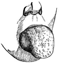
Glossitis (Liston).
In a few hours, sometimes as few as two or three, the entire organ may become involved in the inflammation, enlarging to such an extent as to keep the lower jaw depressed, to fill almost the entire oral cavity or to quite fill it, and to project like a tumor beyond the teeth and the lips (Fig. 16). In exceptional cases the enlargement of the tongue has been so great as to produce dislocation of the lower jaw. The soft palate is lifted up and the epiglottis often pressed down. The latter condition has been known to threaten suffocation. In this condition the patient cannot breathe through the mouth, widely as it may be forced open, and has great difficulty in breathing through the nose. Respiration is therefore laborious. Articulation is impeded or impossible, and deglutition difficult or impracticable. The tumefaction and congestion are often continuous into the floor of the mouth and the parts adjacent. The sublingual and submaxillary glands often become swollen, tense, and painful; and the entire neck is sometimes swollen to such a degree as to exert injurious pressure on the jugular veins. The tongue is very hard to the touch, almost or quite immovable, and is the seat of burning heat and pain. The pain often extends from the root of the tongue along the glosso-pharyngeal folds into the pharynx, and thence by way of the Eustachian tubes into the ears, the folds just named being very much upon the stretch. When the tongue protrudes far out of the mouth it becomes excessively dry, fissured, sanious, and excoriated, or even ulcerated at points where it is subjected to the pressure of the teeth. It is covered with dark viscid secretions, which often extend beyond it and over the entire aperture of the mouth. The epithelial coating often undergoes desquamation, and then the organ becomes exquisitely sensitive to the contact of food, water, or even the air. This desquamation is sometimes in mass, in sheets peeling off like a pseudo-membrane. The general symptoms vary in individual cases. As a rule, the face is turgid and its expression anxious; the conjunctiva suffused, respiration impeded, and sleep disturbed or impracticable. Saliva dribbles externally, often in considerable quantity. The odor from the mouth becomes quite fetid from decomposition of the retained products of secretion. Thirst is usually intense, though immoderate only in some cases. Cough is more or less constant and quite exhausting. This and the dribbling of saliva contribute with the dyspnoea to prevent sleep. Pyrexia is often intense. The pulse is strong and quick at first, 100-120 per minute, and there is often marked throbbing of the temporal and carotid arteries. The skin is hot and dry at first, but cold sweat subsequently accumulates upon the face and neck as the dyspnoea increases. The bowels are constipated. The urine is scanty and high colored. The impediment to the return of blood to the heart from the head causes cerebral congestion, drowsiness, and even threatens asphyxia. In other cases there is intense cephalalgia, nervous irritability, restlessness, and even delirium.
The symptoms sometimes reach their acme in rather less than forty-eight hours, and then gradually subside. More frequently they continue on into the third or fourth day. Occasionally they are protracted as long as the fifth or sixth or even the eighth day, rarely longer. Resolution occasionally takes place within twenty-four hours, however (van Swieten), though more frequently occurring from the fifth to the seventh day. In some instances remittance or intermittence has been noted, the cause therefor not being apparent, although attributed to malaria.
Resolution of the inflammatory process is usually indicated by the gradual return of moisture on the tongue and progressive detumescence of the organ, accompanied by subsidence of the redness, heat, and pain. Increased secretion of saliva, general perspiration, or diuresis sometimes marks the cessation of the pyrexia.
Should the process be going to terminate in suppuration, the local distress increases, markedly about the end of the week. The pains become lancinating, and associated with throbbing of the lingual blood-vessels. The swelling becomes prominent and softer at some one point, although the sense of fluctuation is not very perceptible on palpation, and finally the abscess bursts through the surface, unless previously incised, and discharges a fetid pus. Suppuration always involves a prolonged duration of the attack.
In rare instances glossitis terminates in gangrene of the tongue, circumscribed or diffuse. This result is indicated by adynamic symptoms on the part of the constitution, and by the livid appearance of the parts undergoing mortification. The hemorrhage following extensive sloughs from gangrene has been fatal in some instances.
Fleming66 calls prominent attention to a complication of glossitis, of which he alludes to several examples. This is "an inflammation, circumscribed or diffused, originating in the loose areolar tissue between the genio-hyo-glossi muscles, and first manifesting itself by a train of symptoms identical with those of ordinary glossitis, but soon characterized by peculiar features." These features comprise fulness under the chin like that dubbed double chin, pressure upon which, especially near the hyoid bone, being very painful; and suppuration, which, circumscribed or diffuse, burrows most freely toward the base of the tongue.
66 Loc. cit., p. 91.
Chronic induration of the tongue sometimes remains unilateral, although the acute disease has not been unilateral.
PATHOLOGY AND MORBID ANATOMY.—It has been advanced that in idiopathic glossitis from cold the engorgement of the vessels is probably a consequence of vaso-dilator influence of the glosso-pharyngeal nerve for the base of the organ, and of the chorda-tympani for the anterior portion. An instance of herpetic glossitis from probable irritation of the chorda-tympani nerve by an aural polypus (Berkely Hill67) seems to lend some force to this opinion. However engendered, there is a rapid distension of the organ by blood, followed by infiltration of fibrin and serum into the intermuscular connective tissue and into the planes of the connective tissue separating the muscular fasciculi. In some instances degeneration of muscular fibre has been observed. There is great increase in the thickness of the coats of epithelium, beneath which the mucous membrane is red and its papillæ erect. This coating sometimes peels off like a false membrane. In cases extending from tonsillitis the base of the tongue suffers most.
67 Brit. Med. Journ., Oct. 7, 1882, p. 683.
The disease usually terminates by resolution, although a slight amount of hypertrophy, unilateral or bilateral, sometimes persists, and occasionally to a marked degree (Wells). In instances much less frequent suppuration ensues, usually in debilitated subjects or in cases due to traumatism or in cases inefficiently treated. The suppurative process is usually circumscribed and unilateral, and the abscess points most frequently just beneath the side of the tongue; sometimes, however, the pointing takes place at the dorsum, sometimes at the tip. The pressure of the teeth seems to be the provocative cause of the disposition to point at the edge of the tongue. The pus is usually quite fetid. Sometimes the abscess is gangrenous.
Gangrene is an infrequent result of glossitis. The pressure of the teeth, strangulating the organ at the oral outlet, seems to occasion the failure of nutrition in instances where it occurs. The losses are ordinarily insignificant, though appearing quite extensive while the tongue remains swollen. Sometimes large portions drop off, and fatal hemorrhage has resulted (Frank) in consequence. From the nature of the organ the parts separate more readily than in almost any other instance. Gangrenous abscess, ensuing even from very slight causes, such as a wound with the head of a barleycorn (Ranking68), sometimes proves fatal.
68 Provincial Med. and Surg. Journ., 1844.
In those cases of diffuse inflammation of the interconnective tissue of the genio-hyo-glossi muscles Fleming states that the suppuration—which, whether circumscribed or diffuse, burrows toward the root of the tongue—absolutely dissects its extrinsic muscles and destroys their functions; ultimately injuring the periosteum and laying bare the inside of the inferior maxilla in the vicinity of their attachments. When an incision is made to the parts through the integument, the muscles will be found on palpation flabby and detached, and their interstices filled with purulent matter, sometimes very fetid.
DIAGNOSIS.—These is no difficulty in the diagnosis, except in the early stage of such examples as are attributed to metastatic gout and rheumatism. The subsidence of the peculiar pains elsewhere, and the onset of pain in the tongue, would lead to the inference that a glossitis of this kind was in progress. The acuteness of the tumefaction would distinguish it from hypertrophy of the tongue on the one hand, and from the tumefaction attending malignant disease on the other.
Cystoma of the tongue has sometimes been mistaken for abscess due to glossitis; but even here the history of the case should serve in most instances as a satisfactory factor for the differential diagnosis.
PROGNOSIS.—The prognosis depends upon the gravity of the local symptoms and the activity of the treatment. A case left to itself will be likely to terminate fatally within five or six days. Death, indeed, has been known to take place within forty-eight hours, even in cases submitted to treatment. On the whole, however, the prognosis should be regarded as favorable in the absence of specially lethal complications. Even suppuration adds little gravity to the prognosis, the structure of the organ being but little favorable to accumulations of purulent material. Should an abscess become gangrenous, however, the prognosis becomes grave at once, as it in the presence of gangrene from pressure or other cause. Should the patient survive losses by gangrene, there may be permanent impairment in articulation.
TREATMENT.—Superficial glossitis, as a rule, merely requires active purgation, with the topical use of cold emollient mouth-washes containing mucilage of slippery elm, quince-seed, or the like, to which detergents, such as alum and borax, may be advantageously added in the proportion of five grains to the ounce. In cases resisting this mild treatment topical applications of glycerite of tannin twice or thrice a day are often serviceable.
Parenchymatous glossitis demands the most active antiphlogistic treatment. If the case be seen at an early stage of the process, before the tumefaction of the tongue has become so great as to fill the mouth and interfere with swallowing, a saline purge—say salts and senna—containing some tartar emetic can be advantageously administered to begin with. Following this, tartar emetic may be continued in small doses every two or three hours, associated with small doses of tincture of aconite-root (minim j-iij), according to the condition of the pulse and the effect of medication. Should this treatment fail to produce prompt amelioration in the local symptoms, or should the tongue be considerably swollen when the case comes under care, free leeching should be applied from the hyoid bone to the angle of the jaw on each side, including the region of the hyoid bone (fifteen to twenty-five Spanish leeches). This should be followed by emollient cataplasms, reaching from ear to ear, to favor continuous hemorrhagic oozings from the leech-bites. The internal antiphlogistic treatment is indicated just the same, and if not administrable by the mouth may be administered by the bowel; the nauseant and depressent effects of the tartar emetic and aconite being maintained by hypodermatic injection. Leeching the inflamed tongue itself is said to be often prompter in producing detumescence of the organ than leeching exteriorly, but the leech-bites are apt to add to the local irritation; besides which, the mouth is so filled by the swollen tongue as to leave little more than the tip accessible to the leeches without danger of losing control of them. Venesection from the arm, the jugular vein, or from vessels elsewhere is no longer much in vogue, it being doubtful whether general venesection is more useful than local bleedings. Debility, whether presenting originally or as the result of withdrawal of blood and other antiphlogistic measures, may be met by the systematic use of tincture of chloride of iron and of quinia.
Severe cases demand one or more longitudinal incisions on each side of the raphé of the tongue, deep enough to reach nearly halfway into the substance of the organ and carried from base to tip. Cases are on record in which the patients themselves had in their despair cut into their tongues in order to obtain relief from their local sufferings, and had in this way rescued their own lives by the means most appropriate for the purpose (Camerarius, Lusitanus69). When the mouth is filled by the tongue, it is necessary to insert the knife on the flat until the base of the tongue is reached, and then to turn it and make the cuts as indicated. Copious bleeding usually follows these incisions, often followed by marked diminution in the volume of the organ. Deep as these cuts appear when made, they become quite shallow before the organ has shrunk to its normal volume. Bleeding from the ranular veins, recommended by some practitioners in preference to incisions into the organ, is often impracticable on account of the tumefaction preventing access to them.
69 Dict. Sciences méd., vol. xviii.
If severe hemorrhage takes place from divided vessels, the vessels may be subjected to torsion, which is the preferable mode of management, or to searing with some form of the incandescent cautery (hot iron, electric cautery, Paquelin's thermo-cautery). Astringent and chemical styptics are of little use. The method of searing is open to the objection that secondary hemorrhage may ensue on detachment of the eschars, but this accident is not likely to happen under circumstances at all favorable.
In localized or circumscribed glossitis the incision to be made should interest the swollen portion only.
Should the tongue swell again, the incisions may be repeated. Whether the tongue require incision or not, it is good treatment to have the patient inhale vapor from hot vinegar, alcohol, or cologne spirits to render the parts more comfortable. Washes of weak detergent solutions containing potassium nitrate, sodium borate, or ammonium chloride may be used by syringe or spray to cleanse the parts and promote detachment of the epithelial coatings on the tongue and interior of the mouth, the accumulations of which are sometimes matters of great annoyance. The drug last mentioned exerts in addition a special action on the inflammatory process which is often quite serviceable. I have seen good results follow the prolonged use of sprays of an aqueous solution of ammonium chloride (Stuver), one drachm to the ounce, from the steam-spray apparatus, continued for fifteen to twenty minutes at a time and repeated every two or three hours.
In many instances the patient is unable to take food by the mouth. The best plan under such circumstances is to pass a catheter into the stomach through the larger of the two nasal passages, and retain it in position unless its presence interferes too much with respiration. Milk and stimulus can then be poured into the stomach from time to time with the aid of a funnel passed into the outer opening of the tube, which should be kept corked during the intervals when retained in position. This failing or impracticable, it will be necessary to nourish the patient with enemata.
On the appearance of abscess the same should be freely laid open. In cases of hesitation, the true nature of the presumable abscess can be determined beforehand with the exploring-needle. The pus from an abscess of this kind is rarely copious and is usually fetid. It would be good practice to distend the sac after discharge of pus by injecting into it a solution of carbolic acid.
In resisting or advanced cases of suppurative inflammation of the planes of connective tissue between the muscles at the lower portion of the root of the tongue, Fleming recommends a free incision under the chin in the middle line, through skin and fasciæ and on through the raphé of the muscles themselves.
In cases of gangrene, washes, douches, or sprays of carbolic acid, chlorinated soda, hydrogen peroxide, or potassium permanganate are indicated to relieve fetor; while the most supporting treatment by mouth or otherwise is requisite on general principles.
Chronic Glossitis.
Chronic glossitis, like acute glossitis, may be superficial or parenchymatous.
Chronic Superficial Glossitis.
Superficial chronic glossitis is usually confined to the papillæ of the tongue, territories of which, so to speak, are mapped out on the surface of the tongue, separated by furrows reaching to the basement mucous membrane. In pronounced cases the dividing furrows are quite deep, giving the organ a mamelonnated appearance, and they penetrate into the mucous membrane (dissecting glossitis, Wunderlich), which becomes subjected to great irritation by the retention of articles of food in the fissures. Demarquay70 has recorded a case of this kind in which, to relieve the intense sufferings with which the patient had been plagued for a number of years, he had been forced to amputate the anterior half of the organ—an operation which succeeded thoroughly.
70 Loc. cit., p. 142.
In another group of cases the surface of the swollen tongue is mapped out in small ovoid patches, smooth, red, and glossy, from which the papillæ have become separated without regeneration. Sometimes chronic glossitis presents as an aphthous inflammation. Sometimes superficial ulcers occur upon the dorsum of the tongue, irritable, indolent, and indurated.
ETIOLOGY.—Chronic superficial glossitis is in rare instances a sequel of the acute form of the disease. Usually, however, it is encountered as a chronic affection from the outset, so to speak, generally as a result of long-continued irritation in connection with dyspepsia and other gastric and gastro-intestinal disorders. It is frequently encountered in subjects of chronic alcoholism.
The superficial ulcerations often occur at the sides of the organ, usually in some of the depressions formed by the contact of the teeth.
SYMPTOMATOLOGY.—The symptoms are those due to a consciousness that the tongue is too large, with occasional pain in taking acid and succulent food.
PATHOLOGY.—The pathology does not differ from that of chronic inflammations elsewhere. The apparent obliteration of the papillæ in some cases is due to a filling up of the intervening furrows by permanent deposit of new-formed cells. The excoriations and superficial ulcerations are most frequently due to disturbance of nutrition by pressure.
DIAGNOSIS.—The affection may be confounded with syphilitic disease of the tongue or with papillary epithelioma, but the history of the case, the resistance to antisyphilitic treatment, and the negative results from microscopic examination of fragments of tissue removed for the purpose, serve to establish the diagnosis in cases of doubt. It must not be forgotten, however, that many cases of epithelioma begin in chronic glossitis, non-specific as well as syphilitic.
PROGNOSIS.—Though not threatening to life, the prognosis of the disease itself is bad. It resists treatment, being, in fact, a complication of some obstinate or intractable gastric or gastro-intestinal disorder, or an evidence of constitutional dyscrasia. Cure may be expected in recent cases, following cure of the dyspepsia or other malady upon which the chronic glossitis may be dependent.
TREATMENT.—Care to cleanse the tongue by washes, douches, or wet cloths after each meal, in order to remove particles of food which may have become impacted in the anfractuosities of the organ, is important in order to avoid additional sources of irritation.
Astringents and caustics of various kinds have been extensively employed, carefully applied to the floors of the fissures, but it is very rarely that any benefit ensues. Demarquay71 reports good results in one case of dissecting papillary glossitis from biweekly applications of equal parts of chromic acid and water. Butlin reports good results from chromic acid (1:10).
71 Loc. cit., p. 143.
Careful attention to the gastro-intestinal functions, and a thorough change of diet, such as the adoption of the milk cure and the like, with due attention to bathing and outdoor exercise, comprise the most rational method of constitutional treatment. Should the secretions of the mouth give an acid reaction with litmus-paper, alkalies are strongly indicated, topically and systemically. Avoidance of alcohol in all forms is often absolutely essential.
Chronic Parenchymatous Glossitis.
The chronic parenchymatous form of glossitis is usually circumscribed. When diffuse or general it has usually been a sequel of acute parenchymatous glossitis. It is not a painful disorder, and as a rule is not associated with constitutional manifestations. The circumscribed tumefaction usually presents as an induration upon some portion of the side of the tongue, being most frequently directly or indirectly due to irritation sustained from a jagged tooth. Ordinary sensibility is much diminished, and sometimes the sense of taste likewise. Sometimes the indurated mass is ulcerated superficially. The enlargement of the organ is not sufficient to keep it outside the mouth. Sometimes, indeed, the tongue, as a whole, has undergone atrophy, unilateral or bilateral. Chronic abscess of the tongue sometimes supervenes, chiefly in scrofulous subjects.
PATHOLOGY AND MORBID ANATOMY.—This consists merely in interstitial connective-tissue hyperplasia, with atrophy of muscular fibres from compression.
SYMPTOMS.—In addition to the objective symptoms of induration or circumscribed tumefaction, the subjective symptoms may be summed up as general hypersensitiveness to sapid and acrid substances; diminished tactile sensibility at the part affected; slight stinging sensations while the parts are at rest; occasional or continuous local pains; and a sense of impediment in the movements of the tongue in articulation and even in deglutition.
DIAGNOSIS.—Inspection reveals the swelling, and palpation its induration. In addition, the adjacent source of irritation, a jagged tooth or two, is seen. Abscess is recognized by special prominence at one point of the swelling and by indistinct sense of fluctuation.
Cystic tumor is liable to be mistaken for abscess, but the exploring-needle will solve the difficulty. Circumscribed induration may be confounded with tumor or with epithelioma.
PROGNOSIS.—This is good, provided the source of irritation can be removed or suppressed.
TREATMENT.—The first element in the treatment is the removal or repair of any offending tooth, and next attention to any underlying malady, constitutional or local. Weak solutions of iodine locally are said to be of service. Abscesses require incision and evacuation. Their walls should be distended with solutions of carbolic acid or be touched with solutions of iodine, silver nitrate, or cupric sulphate, to promote reparative inflammation.
Glossanthrax (Carbuncle of the Tongue, Malignant Pustule of the Tongue).
This is a special variety of gangrenous ulcerative glossitis, presenting as an integral phenomenon of a disease peculiar to slaughterers, who become infected from diseased cattle, usually by means of the knife, which they are sometimes in the habit of holding in the mouth (Heyfelder and others). It has been described chiefly by Heyfelder,72 Duhamel, Chavarrien d'Audebert, Felix Plata, Breschet et Finot, and Maisonneuve.73
72 Med. Vereins Zeitung, 1834.
73 Des Tumeurs de la Langue, Paris, 1848, Thèse de Concours.
The period of incubation occupies about one day. The tongue then undergoes rapid tumefaction and becomes the seat of intense pain. The points of inoculation become hard, covered with vesicles containing bloody serum, which blacken, rupture, and leave dark, livid, gangrenous patches of ulceration. Profound cachexia rapidly ensues under typhoid manifestations, and death may result in less than twenty-four hours, though usually not until sixty hours. The prognosis, therefore, is of the gravest character.
The chief treatment consists in thorough cauterization of the inoculated points by means of the actual cautery, followed by deep incisions into the body of the tongue if the glossitis be severe.
Ulceration of the Tongue.
Apart from the ulcerations of the tongue incidentally mentioned in the foregoing pages, there are two forms of ulcer, both of sufficiently frequent occurrence in ordinary practice to require special description: these are the tuberculous ulcer and the syphilitic ulcer.
Tuberculous Ulcer of the Tongue.—Tuberculous ulceration of the tongue occurs in a certain number of cases of advanced tuberculosis of the lungs or of the lungs and larynx. It has even been asserted to precede pulmonary tuberculosis. It is most frequently observed upon the upper surface of one side of the organ, sometimes at the tip, sometimes farther back, and usually on the same side upon which the disease is most advanced in the lungs or the larynx. It is often associated with previous or subsequent tuberculous ulcerations of the palate or of the pharynx or contiguous structures. It gradually extends, and rarely if ever heals. It is characterized by a superficial excavation, and by being covered with a grayish detritus entirely different from the purulent layers seen on other kinds of ulcers. When of long standing its base is indurated, and this may give rise at first to suspicion of squamous-celled carcinoma. Small yellowish elevations are sometimes observed in the reddened mucous membrane around the ulcer—an appearance deemed sometimes characteristic of the tuberculous nature of the lesion (Trélat74).
74 Bull. de l'Acad. de Méd., 1869, or Arch. gén. de Méd., 1870.
PATHOLOGY AND MORBID ANATOMY.—Nodular tuberculous infiltration takes place beneath the mucous membrane, which becomes elevated in small, semiglobular, yellow protuberances of one or more millimeters in diameter, around which the mucous membrane is red and swollen. The epithelium becomes shed without undergoing renewal, and thus a little point of superficial ulceration remains. When several such points are sufficiently contiguous they coalesce into a single ulcer of irregular contour, which gradually spreads without much other change. Practically, it never heals.
SYMPTOMS.—In addition to the superficial ulceration described, and in addition to the constitutional and local symptoms of advanced tuberculosis of the lungs or lungs and larynx, as may be, there are no special symptoms attending the tuberculous ulcer of the tongue. Saliva is sometimes secreted in excess, but that is not characteristic. There is little pain and little impediment to the movements of the tongue until the disease has advanced.
DIAGNOSIS.—The presence in a tuberculous subject of a unilateral, irregular ulcer of the tongue surmounted with grayish detritus and surrounded by reddened edges, should suffice for the recognition of its presumptive tuberculous character. It is most difficult perhaps to differentiate from a small ulcerated squamous-celled carcinoma, and the two indeed sometimes coexist, rendering the discrimination extremely difficult until the advanced progress of the carcinoma places the diagnosis beyond doubt.
In the early stages, however, it is distinguished by lack of the peculiar lancinating pains of carcinoma, which, however, are not invariably attendant, and by lack of secondary involvements of the cervical lymphatic glands. At all times it should be distinguishable from the carcinomatous ulcer by lack of the fungus-like appearance of the bed of the ulcer which is usual in carcinoma.
From syphilitic ulcer it is distinguishable by the history of the case, its tendency to be unilateral, and its failure to respond to antisyphilitic treatment. Syphilitic ulceration of the tongue may represent the primary, the secondary, or the tertiary manifestation of the specific disease. The former will not be discussed in this connection.
Secondary ulcers occur on the upper surface of the tongue, most frequently at the anterior portion, as fissures, usually longitudinal, the floors of which are ulcerated. They occur likewise at the sides, tip, and even lower surface of the organ. They are often associated with secondary ulceration in the mucous membrane of some portion of the mouth. They are quite painful, especially to the contact of pungent articles of food. Some ulcers occur as simple superficial excoriations at some portion of the edge or tip of the tongue, giving little evidence of any specific character.
Tertiary ulcers are usually sequelæ of gummata. They are much deeper than secondary ulcers, sanious at bottom, often serpiginous in configuration, and apt to extend in depth as well as in superficies, sometimes penetrating through and through the organ. They are most frequent in the very central portion of the tongue, or are symmetrically disposed on either side of it.
PROGNOSIS.—The prognosis of tuberculous ulceration is bad, both as regards tongue and patient.
TREATMENT.—The only topical treatment offering any prospect of local cure is the bodily destruction of the ulcer and the surrounding tissue with caustics, the best of which are the incandescent metals, or else the excision, with the incandescent knife, of a portion of the tongue comprising all the affected tissue. In the former case the tuberculous process often reappears about the cicatrix; in the latter, at some more distant point.
Tincture of iodine locally, detergent washes, and the like, often secure a certain amount of comfort as palliatives. The same indications prevail as in simple chronic glossitis, superficial and deep-seated. Iodoform locally is of benefit, inasmuch as it relieves pain and reduces collateral inflammation, but it is powerless to arrest the onward march of the ulcerative process.
Hemorrhage from the Mouth.
DEFINITION.—A loss of blood from the mouth.
SYNONYM.—Stomatorrhagia.
ETIOLOGY.—Hemorrhage from the mouth is usually a symptom of some disease or injury of the mouth, tongue, gums, palate, pharynx, or nose. It may, however, occur as one of the phenomena of scorbutus or of hæmophilia. It is said to occur occasionally as a vicarious menstruation. It may be slight, so as barely to tinge the saliva, or it may be profuse enough to terminate fatally. Between these extremes there is an infinity of gradations. As a result of disease it may be caused by simple hyperæmia of the mucous membrane, by rupture of dilated blood-vessels, by ulceration, by gangrene. As a result of injury it may arise from wounds of various kinds, accidental or self-inflicted.
The gums are the most frequent source of slight hemorrhage from the mouth. The pharynx, probably, is the next most frequent seat. Hemorrhage from the tongue, cheeks, lips, and palate is usually traumatic or the result of ulceration.
SYMPTOMATOLOGY, COURSE, DURATION, COMPLICATIONS, TERMINATIONS, AND SEQUELÆ.—The symptoms of hemorrhage from the mouth are the presence of blood in the saliva or in the mouth itself, or in the expelled products of expectoration, emesis, or catharsis, for sometimes the blood is swallowed, and occasionally inhaled into the air-passages. The course, duration, complications, and terminations of stomatorrhagia depend upon its cause. Prolonged hemorrhage will entail anæmia; profuse hemorrhage may terminate fatally.
DIAGNOSIS.—Careful examination of the mouth, tongue, pharynx, and posterior nares, both by direct and by reflected light, may be necessary to discover the source of the hemorrhage and discriminate it from hæmoptysis and hæmatemesis.
PROGNOSIS.—The prognosis will depend upon the nature of the cause, its susceptibility of arrest, the quantity of blood lost, and the general health of the patient. It is grave, as a rule, in the subjects of hæmophilia, as there is a constitutional malnutrition of the blood-vessel system which cannot be counteracted.
TREATMENT.—Ergot or oil of turpentine internally, astringent mouth-washes, and recumbency constitute the main features in treatment.
Morbid Dentition.
DEFINITION.—Departure from the physiological processes concerned in the eruption of teeth, entailing certain local and systemic disorders.
SYNONYMS.—Dentitio difficilis, Pathological dentition, Odontitis infantum.
The correct comprehension of the subject will be facilitated by considering its etiology, pathology, and symptomatology in connection. Indeed, morbid dentition has been assigned so prominent a part in the etiology of various affections elsewhere discussed that a satisfactory consideration of its own causation would require the repetition of much that belongs more appropriately under other titles. While in some infants the teeth erupt so quietly that the parents are astonished by the accidental discovery of their presence above the gum, few children escape a greater or less amount of local and constitutional disturbance while passing through the process of dentition. So severe may these disturbances become that, according to the mortality-tables of London, as cited by West,75 teething was assigned as the cause of death of 4.8 per cent. of all children dying under one year old, and of 7.3 per cent. of those who died between the ages of twelve months and three years. It is furthermore well recognized that the period of greatest mortality among children is that of the first dentition. Associating these facts, we see, on the one hand, that while disorders of dentition may act a causative part in the production of systemic diseases or aggravate morbid processes due to ordinary causes, on the other hand they may be but one expression of some profound constitutional disturbance; or both aberration in the eruption of the teeth and systemic disease may be dependent upon the influence of dyscrasia. The period is one of active organic processes; the child is becoming fitted for a new manner of existence; and change and development are going on throughout nervous, vascular, respiratory, and alimentary systems. Hence there exists peculiar susceptibility to morbid influences; and any process, physiological or pathological, once started, goes through its stages with excessive energy.
75 Lectures on the Diseases of Infancy and Childhood, Philada., 1860, p. 425.
Although the periods of normal eruption of the deciduous teeth vary within extensive limits, and an invariable order in eruption is not observed in all subjects, it may be stated as a rule that the lower central incisors are cut in quick succession about the seventh month. Some infants get these teeth during the fourth month (Vogel), and others have to wait until the tenth or eleventh month, some even longer. A few weeks after the appearance of these lower incisors—within fourteen days in some subjects, not until nine or more weeks in others—the central incisors of the upper jaw are cut, and its lateral incisors shortly afterward, followed in their turn by the lateral incisors of the lower jaw. In some instances—the majority, according to Vogel—the eruption of the inferior lateral incisors is delayed until the anterior molars are about to become exposed, usually from the twelfth to the fifteenth month. Sometimes the upper molars are cut before the lower, sometimes after them. From the sixteenth to the twentieth, or even the twenty-fourth, month the canine teeth are cut, and the four posterior molars follow between the twentieth and thirtieth months, rarely delayed until the thirty-sixth month,—completing the process of the first dentition.
From this it will be seen that the teeth erupt as a rule in pairs, and that a longer or shorter interval of repose takes place between the eruption of successive pairs.
Variations from the usual order beyond the limits noted above may be considered abnormal. Numerous cases are on record both of precipitate and of tardy dentition. Tanner cites from Haller nineteen examples in which one or more of the central incisors have been found through the gums at birth, and have had to be removed to prevent injury to the mother's nipple; from Crump, a case of full dentition at birth, reported to the Virginia Society of Dentists; and from Ashburner, a case of a child beginning to cut its first tooth, an incisor in the upper jaw, during its twenty-third month, the infant being very delicate, with a large head, tumid abdomen, and peculiarly small-sized extremities. The same author quotes from Serres cases of persons passing through several years of life—in one instance seven—before cutting their first teeth, and mentions on the authority of Tomes that Boxalli and Baumes have each recorded an instance in which the patient reached old age without a single tooth having ever appeared.
Rachitis is often the cause of tardy dentition, and in the subjects of this diathesis not alone are the teeth retarded in development, but they decay early and even fall from their sockets.
The first indication of approaching dentition is the markedly increased production of saliva. For some little time after birth the salivary glands seem to remain wholly inactive, and until the fourth or fifth month of extra-uterine life they furnish very little secretion. At this period a decided change occurs. The mouth is constantly filled with saliva, which dribbles from its corners. To this continual slobbering, wetting the garments covering the chest, has been attributed the bronchial catarrh which attends some infants; and diarrhoea has likewise been referred to the swallowing of large quantities of saliva, acting as a mild laxative by virtue of its saline constituents.
There may be no further manifestation until the seventh month, beyond the broadening of the dental ridge. The exact position of each tooth is usually indicated by greater prominence of the gum above it for some time before it comes through, its entire outline being very distinct in the upper central incisors. As the tooth approaches the surface the gum becomes hot, shining, tense, and tumid, often painful. A slight amount of catarrhal stomatitis is almost invariable. There is some elevation of temperature; flushing of the cheek may occur; the child is restless, peevish, and fretful; its sleep may be broken; it may cry out with pain; its thumb, its fingers, any hard substance it can obtain, are thrust into its mouth to allay the irritation of the gums. Otalgia is not uncommon, and its occurrence may be inferred from the fact that the child pokes its thumb or finger into the auditory canal or firmly presses the tragus down over the external meatus. These may comprise all the disordered manifestations, local or constitutional, or there may be in addition loss of appetite, diarrhoea, vomiting, and the various disturbances of reflex nervous origin to be alluded to later; or, in the not common yet not rare instances already mentioned, there may be absolutely no appreciable disturbance whatever.
Sometimes a disposition exists to the formation of small aphthous ulcerations on the tongue or elsewhere in the mouth, particularly at the duplicature of the lip and the outer surface of the alveoli. Ulceration occurs most frequently at the tip of the tongue, probably occasioned by friction from the new teeth. Usually there is a single flat, round ulcer, its edges somewhat infiltrated, its bed covered with a yellow lardaceous substance. It is extremely painful to the touch, and thus every movement of the tongue occasions distress. It may heal within a few days or continue for weeks. Ulcers in other situations are less obstinate.
Occasionally—and more frequently in debilitated subjects or those exposed to unhygienic surroundings—there is an unusual amount of heat and swelling of the gum, which becomes excessively tender, usually over the summit of a particular tooth—in which case there will be a little tumor-like elevation—or around a tooth which has partially pierced through it. Small sloughy ulcerations form in this situation. There is great pain, and usually high fever and severe gastro-intestinal disorder. This affection, often difficult of cure, is termed by some writers odontitis infantum. So severe is the pain, and so great its tendency to aggravate constitutional disturbances, that life may be placed in jeopardy, and even fatal results ensue.
Less severe than either of the forms just described, and yet far more intense than the mild stomatitis which many authors regard as physiological, is an aggravated form of catarrhal stomatitis sometimes attendant upon morbid dentition, in which there is swelling of the submaxillary glands and infiltration of the adjacent connective tissue. In this case there is usually considerable pyrexia.
The constitutional disturbances of reflex nervous origin occasioned by morbid dentition are of the most varied character, both in their degree of gravity and in the manner and locality of their manifestation. Doubtless the extensive ramifications of the great vagus nerve, and its connections both of origin and distribution with the exquisitely sensitive fifth nerve, as well as with the facial nerve and with the sympathetic system, will explain why the irritation should now be seated in the gastro-intestinal tract, giving rise to vomiting and diarrhoea (gastritis, gastro-enteritis, enteritis, entero-colitis, cholera infantum); now in the respiratory tract, provoking cough more or less severe, or even a well-marked bronchitis; now manifest itself in various cutaneous eruptions (urticaria, eczema, impetigo, lichen, prurigo, herpes); and now accumulate in the cerebro-spinal axis, manifesting its presence by slight spasms (dysuria, muscular twitchings), or discharging with terrific force in some of those convulsive seizures which are the dread of mothers and the cause of much anxiety to physicians.
The mechanical causation of diarrhoea and bronchitis, insisted upon particularly by Vogel, has already been alluded to. While this may be one element, most certainly the nervous factor is too important to be disregarded. Bronchitis, not attributable to ordinary exposure, occurs coincidently with teething even in children who have been protected against wetting of the chest; and the fact that more purely nervous phenomena, and especially the dreaded brain symptoms, are usually absent in children who have an excessive flow of saliva, and particularly if there be also a moderate diarrhoea, would conduce to the belief that nervous irritation, discharging itself in this manner, does not accumulate in the centres.
Doubts have been expressed whether dentition can give rise to convulsions in perfectly healthy children, although its rôle as an exciting cause in predisposed subjects is admitted (Hillier). That dentition alone, in the absence of any other predisposing or exciting influence, will provoke any of the disorders with which it is associated may be doubted in view of the fact already cited, that in some infants there are no untoward occurrences. But there seems to be no valid reason for separating the disturbances purely in the domain of the nervous system from the other pathological processes originated or aggravated by morbid dentition. Doubtless predisposition often determines the direction and severity of the reflected phenomena; and in the same manner reflected irritation may bring an organ within the influence of the ordinary disease-producing cause.
The convulsive phenomena associated with dentition may take the form of general eclampsia or spasms of particular groups of muscles. These latter are very common—according to Vogel, universal—and vary in intensity from that slight contraction of the facial muscles which sends the mother into raptures of delight over the heavenly smile of her sleeping babe to the distressing seizure of laryngismus stridulus. Sometimes the child may sleep with its eyes half open, the eyeballs directed upward, and only the white sclerotic to be seen through the gap between the lids, "producing an appearance which is unnatural and alarming to the laity."
The attacks of general eclampsia are usually sudden. The child has been to all appearances perfectly healthy, when, without warning, there occurs a series of tetanic spasms like a succession of electric shocks. The individual eclamptic shock cannot be distinguished from an epileptic seizure. These convulsions sometimes continue for several days, but frequently they cease after a few minutes. They may pass off and leave nothing to testify to their occurrence; very frequently they occasion permanent distressing lesions. Partial, so-called essential paralyses, squint, or even idiocy, are cited among their sequelæ; infants subject to repeated convulsions while cutting successive teeth have eventually perished from cerebro-spinal meningitis; death has not infrequently been an immediate result. In these graver cases teething is probably but one of the morbid influences at work.
Purulent otitis media follows dentition in some infants, usually, if not invariably, of a scrofulous diathesis. At the clinic of the Jefferson Medical College Hospital fully one-third of all the cases of otorrhoea in children are said to be so occasioned.
Blennorrhoeal conjunctivitis is a rare complication of teething, and when it occurs usually accompanies the eruption of the upper molars and canines (eye teeth). It is attributed to direct extension of the gingival inflammation by continuity through the antrum of Highmore and the nasal passages. By some it is said to occur only in strumous subjects. It is unilateral, and is not contagious, so that there is no cause for alarm concerning the unaffected eye. The lids soon swell enormously and the eyeball is exposed with difficulty. There is considerable pain. The secretion is more mucous, translucent, and stringy than in genuine blennorrhoea. The eyeball always remains intact and the prognosis is always favorable (Vogel). Milder forms of catarrhal conjunctivitis are not very uncommon.
Thus far, we have considered only the process of the first dentition. Before the shedding of any of the deciduous teeth, the first permanent molars inaugurate the second dentition, appearing in position at about the sixth year. Next, displacing their temporary predecessors, come the central incisors, between the sixth and eighth years, the inferior pair generally preceding the superior ones. The lateral incisors are cut between the seventh and ninth years; the anterior bicuspids between the ninth and tenth years; the posterior bicuspids between the tenth and eleventh years; the canines between the eleventh and thirteenth years; the second molars between the twelfth and fourteenth years; the third molars, or wisdom teeth, between the seventeenth and twenty-first years as a rule, occasionally much earlier, sometimes later.
The eruption of the permanent teeth does not usually occasion any very great amount of distress; nevertheless, it sometimes acts both as a predisposing and as an exciting cause of various disorders, local and systemic. The various forms of stomatitis, tonsillitis, sore throat, gastro-intestinal derangements, febrile disturbances, bronchitis, internal rhinitis, diseases of the eye, of the ear, of the skin, chorea, epilepsy, etc., have all been noted as accompanying, if not occasioned by, the second dentition. Ashburner76 records, among other similar instances, that of a lad twelve years old who presented a marked case of chorea, and after three months' continuance of the twitchings fell into a violent epileptic fit, from difficulty in the eruption of the second pair of permanent molars of the upper jaw. The use of the gum lancet relieved the convulsion, and there was no return of the chorea.
76 On Dentition and some Coincident Disorders, London, 1834, cited by Tanner.
Quite frequently, the eruption of the inferior dentes sapientiæ occasions great and protracted suffering, especially when they appear very close to or partially under the coronoid processes. Considerable irritation is occasioned, in which the gums and adjacent tissues participate. Inflammation may result and extend to the fauces; mastication becomes impossible; severe odynphagia is excited. Suppuration may ensue, and then the pus burrows in various directions, finding exit at points more or less remote, internal or external. Among the consequences of the eruption of a wisdom tooth into a crowded arch, White77 cites fistulæ, necrosis, exostosis, ulceration and sloughing of the soft tissues, cystic and other tumors, ankylosis of the jaw, amaurosis, otalgia, otorrhoea, deafness, facial paralysis, hemicrania, oesophagismus, tonsillitis, erysipelas, aphonia, hysteria, neuralgia, chorea, epilepsy, tetanus, death.
77 "Pathological Dentition," extract from annual supplement to the Obstet. Journ. of Great Britain and Ireland, April, 1878.
DIAGNOSIS.—The age of the child and the appearances already described will afford a basis for diagnosis so far as the local manifestations in the mouth are concerned. The diagnosis of local disorders at a distance, or of systemic disturbances of whatever character, can be made out only by careful consideration of all the attending circumstances; and it is always to be borne in mind that while the process of dentition is to be recognized as one of the causative factors, grave injustice might be done the little patient, and its life perhaps endangered, by failure to recognize the presence of other and perhaps more potent morbid influences.
In cases of chorea or epilepsy, of eye or ear troubles, or of any morbid condition not otherwise accounted for, occurring during the period of the second dentition, especially at the sixth, twelfth, and seventeenth years, or until the wisdom teeth are fully erupted, it is well to inspect the mouth and to think of dentition as the possible cause.
PROGNOSIS.—The prognosis will depend upon the character and gravity of the associated symptoms, the presence or absence of diathesis, and the etiological importance attached to dentition. It is impossible to lay down a general law.
TREATMENT.—The treatment of the deuteropathic or associated disorders is to be conducted on the general principles applicable to those diseases; for a consideration of which the reader is referred to the appropriate articles of this work. We have here to consider general prophylaxis and local measures. The proper management of the child during the period of the first dentition is a matter of great importance, and may avert serious complications. The child should be as much as possible in the open air whenever the weather is favorable. The head may be daily sponged with cold water, and caps and warm head-coverings of all kinds should be forbidden.78 Frequent rubbing of the gums with a crust or other hard substance, or with the finger, is advisable; and something for the child to bite on, preferably a silver piece, should be provided. Orris-root, calamus, and other vegetable substances frequently given to children for this purpose are objectionable; their fermentation is apt to lead to thrush. The secretions must be kept active. The diet should be carefully regulated, and cooling drinks be freely given in order that the child may not overload its stomach by too frequent suckling in its efforts to relieve the local heat by moisture. The mother should be warned not to put it too frequently to the breast. Weaned children will often be found unable to digest their ordinary food, and in that case still greater care will be required. Slight diarrhoea does not call for interference, and is often beneficial in relieving nervous tension and thus averting a tendency to convulsions. Indeed, when the bowels are not relaxed gentle aperients should be given, especially in plethoric subjects or in those with cutaneous eruptions (Clarke). Cutaneous eruptions do not call for treatment, and there seems to be ground for the popular fear that they may be driven inward; at least, cases are on record in which their disappearance under treatment, and even spontaneously, has been followed by more or less severe convulsions.
78 Tanner after Clarke.
In cases where bronchitis can be traced wholly or in part to soaking of the clothing, due protection of the chest by an oil-cloth or waterproof bib may be prophylactic against future attacks. In children who have suffered from any special set of morbid manifestations during the eruption of one pair of teeth, similar disturbances may be expected, and should be guarded against, in the future.
Aphthous ulcerations are usually associated with disorders of digestion, the relief of which must be the main object of treatment. Locally, the treatment does not differ from that of aphthous stomatitis in general. Obstinate ulceration of the tongue may require the use of silver nitrate. In that form of ulceration called odontitis infantum, in addition to proper attention to the diet and secretions and mild antiphlogistic medication, local depletion by leeches, preferably at the angle of the jaw, is often beneficial. Some writers advise the application of leeches directly to the gum. Potassium chlorate internally, two grains every four hours to a child twelve months old, is curative in the majority of cases. It may be given dissolved in sweetened water. Solutions of borax, and, in severe cases, of silver nitrate, may be applied locally. The use of the lancet is contraindicated, for the cut surfaces would be liable to ulceration.
FIG. 17.
Incision for a cuspid (White).
FIG. 18.
Incision for a molar (White).
The propriety of resort to the lancet for cure of systemic disturbances by obviating the source of local irritation is one which deserves consideration. It can only be decided upon the indications presented by the individual case. The knife is not a panacea for all the disorders of childhood occurring during dentition, and its indiscriminate use is to be discountenanced. Nevertheless, there can be no doubt that engorged and inflamed gums demand incision for their relief, on the same general principles of surgery applicable to similar conditions elsewhere. Where it is probable that systemic disease, even if not solely caused, is aggravated by the irritation and pain of a tooth unable to make its way to the surface unaided, it is clearly the duty of the physician to give his little patient that modicum of relief, if not of cure, which will be afforded by a proper incision of the gum. It will not do merely to score the gums, but cuts should be made deep enough to reach the presenting surface and extend even beyond its boundaries. The developing enamel cannot be injured unless undue force be exerted. The best instrument to employ is a curved double-edged bistoury, so wrapped as to prevent injury to tongue, cheek, or lips. The child should be firmly held by another person, and in such a position that the parts may be well illuminated. The jaws can be separated by the operator's left hand, and the fingers so disposed as to protect the tongue and lips. Sometimes the insertion of a small cork between the jaws will be of advantage. The cuts should be made with special reference to the form of the presenting tooth. James W. White79 recommends for the incisors and cuspids a division of the gum in the line of the arch; for the molars a crucial incision, thus X, the centre of the crown as near as can be determined indicating the point of decussation. A cuspid partially erupted needs severance of the fibrous ring on the anterior and posterior as well as on the lateral surfaces (Fig. 17). All the cups of a molar may have erupted, and yet strong fibrous bands maintain a decided resistance. In this case White thinks that all the boundaries of the tooth should be traced by the lancet and all such bands completely severed, or else a crucial incision, as in the figure (Fig. 18), should be made so as to ensure perfect release from pressure. The only contraindication to the use of the lancet, except in ulcerative odontitis, as before mentioned, is the existence of a hemorrhagic diathesis.
79 Op. cit.
DISEASES OF THE TONSILS.
BY J. SOLIS COHEN, M.D.
Tonsillitis.
DEFINITION.—An acute inflammation of the tonsil or tonsils; or inflammation of the tonsil or tonsils, with inflammation of the peritonsillar connective tissue and of the palatine folds.
VARIETIES.—When the inflammatory process is confined to the mucous membrane the disease is erythematous, superficial, or catarrhal tonsillitis; when it involves the lacunæ it constitutes lacunal or follicular tonsillitis; when it involves the gland as a whole it constitutes parenchymatous, phlegmonous, or suppurative tonsillitis. The two latter varieties may present in combination. When the superficial inflammatory process is a vesicular one, eventually sheathing the surface of the organ in whole or in part with a membranous envelope, it constitutes herpetic or membranous tonsillitis. This variety may complicate superficial tonsillitis. When the inflammation of the tonsil, usually superficial, is due to the presence of a cryptogamic growth, it is a mycotic or parasitic tonsillitis, benign or malignant (diphtheria), as may be. When the inflammation of the tonsil is due to rheumatism, it is rheumatic or constitutional tonsillitis.
SYNONYMS.—Inflammation of the tonsils, Amygdalitis, Quinsy, Angina tonsillaris, Angina phlegmonosa, Phlegmonous sore throat, Cynanche tonsillaris. Lacunal tonsillitis is more generally known as folliculous tonsillitis (tonsillitis follicularis). Common membranous or pseudo-membranous sore throat (angina membranosa communis) is used as a synonym for herpetic or membranous tonsillitis (tonsillitis herpetica seu membranosa). Mycosis tonsillaris is a synonym for mycotic tonsillitis (tonsillitis mycotica benigna or tonsillitis parasitica). The tonsillitis of diphtheria is sometimes termed tonsillitis diphtheritica, tonsillitis mycotica maligna; that of rheumatism, tonsillitis rheumatica, angina rheumatica, rheumatic sore throat.
HISTORY.—Tonsillitis was described by Hippocrates. Of recent authors, Sauvages, Cullen, Louis for researches on the effects of blood-letting; Bell on the specific value of guaiacum; Velpeau as to the use of powdered alum and nitrate of silver; Bourgeoise on the use of tartar emetic; Maingault on paralytic sequelæ; Hering on mycosis; and the authors of the various encyclopædias and dictionaries,—may be mentioned as chief among the numerous observers whose contributions have been of most value. The bibliographical references appended to the compilations last cited will guide the student in gaining access to the more important special observations of anomalous cases.
ETIOLOGY.—Predisposing and Exciting Causes.—Tonsillitis may be idiopathic, deuteropathic or symptomatic, or traumatic.
The predisposing cause of idiopathic tonsillitis is usually diathetic, and is associated with congenital or inherited vulnerability of the organ. Of diathetic causes, scrofula is undoubtedly the most provocative, but even rheumatism and gout are likewise so considered, though in a far more limited degree. Acute articular rheumatism is, in fact, sometimes preceded by rather a sharp attack of tonsillitis (rheumatic tonsillitis), which subsides spontaneously in a very few days, sometimes within one day, sometimes suddenly and synchronously with the onset of the ordinary manifestations of rheumatism, though the latter are often slight and transient, as if the force of the attack had been spent on the tonsils. Tonsillitis, non-specific in character, is apt to be prevalent during epidemics of scarlatina, diphtheria, rubeola, and variola. Membranous tonsillitis is common before and after epidemics of diphtheria. Epidemics of tonsillitis have been recorded, but in the face of their extreme rarity it becomes questionable whether they were not extensive examples of the proclivity just alluded to.
Tonsillitis is more frequent in individuals with chronically diseased tonsils than in individuals in whom these glands are healthy. Such individuals, too, are more liable to recurrences; and such recurrences often follow very slight provocations.
Idiopathic tonsillitis is rare in infancy. At the period of eruption of the permanent teeth it is much more liable to occur than before that period, and the liability increases progressively until the second dentition has been completed. It is most frequent during the decennium immediately following puberty—that is to say, in adolescents and young adults—or from the fifteenth to the twenty-fifth year. The disposition or predisposition to renewed attacks continues marked during the decennium immediately succeeding; after which attacks are more and more infrequent. Certain anatomical changes occurring in the tonsils, as the rule about the fortieth year, may diminish their proclivity to inflammation. Nevertheless, the disease occasionally occurs in advanced age.1
1 Solomon Solis Cohen, "Abscess of the Tonsil in an Octogenarian," Med. News, Philada., Feb. 16, 1884, p. 186.
Deuteropathic tonsillitis is quite frequent in infancy, being excited by the infection of scarlet fever, diphtheria, measles, and small-pox, as discussed under these headings respectively. Under similar circumstances it occurs in the adolescent and the adult likewise. It is also produced in carcinoma and sarcoma of the tonsil.
Rheumatic tonsillitis, a deuteropathic variety, is most prevalent during atmospheric changes.
Herpetic tonsillitis, often a deuteropathic variety, seems sometimes of nervous origin exclusively. It is sometimes traceable to defective drainage. It is sometimes prevalent during epidemics of diphtheria, when its membranous character renders it extremely liable to be mistaken for the tonsillitis of diphtheria.
Traumatic tonsillitis occurs occasionally. The causes are—inspiration of irritant gases, the deglutition of chemically acrid substances, the accumulation of calcareous concretions in the crypts or in the lacunæ, direct and indirect gunshot and other wounds, the impaction of fish-bones, fragments of toothpicks, cherry-stones, and other foreign bodies, and the like.
Mycotic tonsillitis is due to the development of a cryptogam upon tonsils probably already in a state of catarrhal inflammation in individuals with health impaired by previous disease or unfavorable hygienic influences.
SYMPTOMATOLOGY, COURSE, DURATION, TERMINATIONS, COMPLICATIONS, AND SEQUELÆ.—The onset of tonsillitis, sometimes preceded by headache and general malaise, is often accompanied by a chill, pyrexia following within twenty-four hours. The temperature may reach 105° F., being at its maximum, as a rule, about the third day. It is rarely below 101° F. The pulse is accelerated to 120 beats per minute. Simultaneously with the constitutional symptoms local distress is usually manifested, but either set of disturbances may precede the other by several hours or by an entire day. Heat and soreness of the throat are early complained of, gradually increasing in severity to actual pain. The pain may become intense, especially during deglutition. When the posterior palatine fold is put upon the stretch, additional pain is referred to the ear, for this fold encloses the staphylo-salpingeus muscle, which runs from the palate to the pharyngeal orifice of the Eustachian tube. This pain in the ear, sometimes the principal cause of complaint, is often premonitory of suppuration. Noises in the ears on the one hand, and impairment of hearing on the other, often attend extension of the inflammation in this direction, the enlarged tonsil sometimes pressing the posterior palatine fold against the pharyngeal orifice of the Eustachian tube.
On inspecting the throat early in the disease, one of the tonsils will be seen to be swollen into an irregularly tumid, much-inflamed mass, usually of a vivid red color. Occasionally both tonsils are involved simultaneously, but this is far less frequent than involvement of the second tonsil a few days later or after subsidence of the process in its fellow. In many instances the inflammation affects one tonsil only.
The inflammatory process is seldom confined to the tonsil. All the structures of the throat, even to the base of the tongue, are often involved, and it is rarely indeed that the anterior palatine fold, distended over the surface of the tumefied gland, escapes inflammation. It is this stretching of the anterior palatine fold which occasions much of the exquisite pain that forms so prominent a subjective symptom in severe cases. The soft palate, hanging forward in the cavity of the pharynx, is often inflamed or intensely congested, and the uvula tumefied, elongated, and oedematous. It may be flaccid upon the posterior part of the tongue or hang immediately over the epiglottis or upon it, and induce painful and tiresome efforts at deglutition and expectoration to relieve the consequent titillation. Sometimes it adheres by viscid secretion to the side of the swollen tonsil.
The inflamed tonsil or the tonsil and its coverings project far into the cavity of the pharynx, often as far as the middle line, touching its fellow when both are involved, so that ulceration sometimes ensues at the points of contact. The posterior surface of the anterior palatine fold sometimes becomes unfolded, as it were, in the tumefaction of the gland, and remains stretched over it in a thin continuous layer without any line of demarcation. The swollen palate projects upon the enlarged tonsil like a shelf, from which depends the uvula, the latter being often oedematous, usually anteriorly, but sometimes posteriorly.
Inflammation of the connective tissue about the lower jaw, especially when at or near the articulation, often renders it difficult or even impracticable to open the mouth sufficiently to permit direct inspection of the parts; but it is rarely that sufficient space cannot be made to allow partial protrusion of the tongue on the one hand, and the introduction of a fore finger for exploratory purposes on the other, though both of these acts are sometimes impossible. The tumefaction of the parts impairs the freedom and ease of deglutition, which may become so painful as to prevent the swallowing of the saliva, which then may dribble from the mouth.
The pain experienced in swallowing is often manifested by convulsive action of the muscles of deglutition and of the muscles of the face. The swollen tonsils prevent the soft palate from being applied to the surface of the pharynx, as usual in deglutition; and as the upper or retro-nasal portion of the pharynx thus fails to be shut off from the lower oesophageal portion, liquids are often forced up into the nasal passages posteriorly, and are regurgitated through the nostrils, thus rendering it impracticable, for the time, to slake thirst or to swallow liquid nourishment.
At first sensations of dryness and pastiness in the throat are complained of, but in a few hours these symptoms become relieved by a more copious secretion of mucus or mucus and saliva. This secretion soon becomes viscid, and so adherent to the parts as to be detached only with difficulty, thus causing harassing efforts for its dislodgment by hawking and expectoration, or equally distressing efforts to swallow it. Should the inflammatory process extend to the salivary glands, as is not infrequently the case, secondary ptyalism often results, with increased distress from this source, and the patient lies or sits with his head inclined upon the diseased or most diseased side to favor the uninterrupted flow of saliva from the mouth.
Extension of the inflammatory process to the submaxillary glands, or to the parotid, or to the connective tissue surrounding them, is indicated by tumefaction externally, which is often exquisitively sensitive to pressure.
The timbre or quality of the voice is often impaired in a peculiar manner by the tumefaction of the throat and the immobility of the soft palate. The voice is thick, throaty, or guttural, having a characteristic harsh, rasping aspiration in enunciation, while articulation is much impeded by impairment in the movements of the jaw, palate, tongue, and lips. At times it is also painful. Speech is sometimes indistinguishable or impossible, and the voice may even become suppressed, so that signs and writing remain the sole means of communication.
Impairment of respiration, at least to any considerable degree, does not occur, unless both tonsils are involved and swollen to an intense degree—conditions under which dyspnoea may become pronounced, severe, and even urgent, and suffocation become imminent. Painful respiration is not uncommon in rheumatic tonsillitis.
The fever is sthenic in type. There are often severe aching pains in the limbs. Headache, restlessness, insomnia, nausea, and even vomiting, may occur. The tongue is heavily coated, the breath is fetid, appetite is impaired, and the bowels are constipated. The urine is diminished in quantity, high-colored, and of high specific gravity. It usually shows slight increase of urea and great diminution of chlorides. Albuminuria occurs in rare instances.
The symptoms are proportionate to the severity of the attack. A first attack is usually much severer than subsequent ones, and suppurative cases more severe than those terminating by resolution. Resolution is the usual termination, and the parts are restored to a normal condition at the end of ten to fourteen days, sometimes earlier; in exceptional cases not until three or four weeks. Sometimes permanent hypertrophy of the tonsil remains.
Where the inflammatory process fails to subside, suddenly at the end of five or six days, or a little later, or not until ten days to a fortnight have passed, slight rigors supervene, announcing suppuration, and the local distress is very great, with pulsation and lancinating pains in the tonsils, until all at once the abscess bursts and its contents are discharged with immediate relief. Sometimes the pus or much of it is involuntarily swallowed; sometimes it is expectorated. In exceptional instances the pus has escaped into the larynx and suffocated the patient, usually during sleep.2 In rare instances the abscess, having burrowed beneath the pharyngeal muscles, may open at the external angle of the jaw or behind the sterno-mastoid muscle. It may discharge into the epiglotto-pharyngeal fold, and thence reach and distend the epiglottis. It has been known to descend along the planes of connective tissue into the mediastinum or into the lungs. Even ulceration into the maxillary and carotid arteries has occurred, usually with fatal result, occasionally with an opportunity to save life by ligating the carotid (Erhmann).3
2 Stokes, Med. Times and Gaz., Aug. 29, 1874, p. 251; Littlejohn, Brit. Med. Journ., Jan. 2, 1875, p. 16.
3 Gaz. méd., Paris, 1878, p. 42.
The most frequent point of spontaneous rupture externally is at the upper portion of the gland anteriorly, just beneath the anterior palatine fold. Sometimes internal rupture occurs into the lacunæ.
Termination by gangrene is exceptional, and is confined to individuals with debilitated constitutions. It is much less frequent than formerly—as a result, perhaps, of better methods of treatment.
Metastasis is one of the methods of termination as to joints or muscles in rheumatic tonsillitis—to lungs, brain, or gastro-intestinal tract—as formerly occurred with much more frequency under direct depletory treatment.
In rare cases extension of the inflammation occurs to the epiglottis, even to the larynx, and the laryngitis may be so severe as to threaten life from the occurrence of oedema. Diffuse inflammation of the retro-pharyngeal connective tissue or of the connective tissue of the neck may constitute an unpleasant complication of the disease.
In a few instances paralysis of the palate occurs as a sequel of tonsillitis, and in exceptional cases the paralysis may also affect the arytenoid muscles of the larynx, and even the accommodator muscles of the eyes.
PATHOLOGY AND MORBID ANATOMY.—Tonsillitis is almost always associated with inflammation of the tissues surrounding the tonsil and those contiguous to it, even in the mildest and most frequent manifestations of the affection. Thus, inflammation of the palate (staphyllitis) and uvula, and even of the pharynx (pharyngitis), are anatomically included with tonsillitis in angina or sore throat.
The mildest form of the malady is a catarrhal inflammation of the mucous membrane covering the gland, and does not extend along the lacunæ which dip inward from the surface and divaricate toward the interior of the organ. It is termed catarrhal tonsillitis, and, as has been intimated, is almost always associated with catarrhal sore throat. It is attributed to hyperæmia, with passive engorgement of the vessels, following retrocession of blood from the cutaneous surface after undue exposure to cold and moisture. A severer form of the malady involves the lacunæ in addition—several or all of them. This should be termed lacunar tonsillitis, as suggested by Wagner. Primarily, at least, it does not involve the follicles of the tonsils which open into the lacunæ, and is therefore incorrectly denominated follicular tonsillitis, although it is most generally so described. The lacunæ are involutions of the mucous membrane, and in health furnish a slightly turbid mucoid secretion which serves to lubricate the parts and, as is generally believed, to facilitate deglutition. When the lacunæ are inflamed these products become pent up in them to a certain extent, accumulate, and project in part at their orifices in turbid creamy or curdy masses, plastered over the parts when thin in consistence, or tightly imbedded when thick or desiccated. These masses are usually white, but sometimes, owing to various admixtures, they are more or less yellowish or gray or brown. They consist of epithelium chiefly, with more or less pus and accumulation of cells similar to those of which the follicles are composed—whether from follicles which have become distended by proliferation of their constituents, and have then burst, is not known. This epithelium has often undergone fatty degeneration in part. Cholesterin is an occasional constituent, and swarms of micrococci and bacteria abound when the masses are not recent, especially if the inflammation is occurring in a tonsil long the seat of chronic disease of the lacunæ.
The tonsil itself is moderately swollen and its mucous membrane hyperæmic. If the parenchyma of the tonsil be involved likewise, as often occurs, the swelling will be much greater, so that the gland will project a considerable distance beyond the margins of the palatine folds. When a hypertrophied tonsil is the seat of the inflammation the tumefaction will be much greater than when the inflamed tonsil has been normal.
Acute inflammation of the palatine folds often coexists, especially of the anterior fold. The soft palate may also be engaged in the morbid process, which may involve the uvula likewise. Pharyngitis is an occasional accompaniment, and stomatitis quite a rare one.
Lacunar tonsillitis sometimes subsides by spontaneous evacuation of the pent-up contents of secretion and desquamation, the parts returning to their normal condition. More frequently a desiccation of some of these products ensues, with permanent chronic inflammation. Decomposition then often takes place, fouling the breath by the escape of the gases. Butyric acid has been recognized as one of the most prominent of these fetid gases. Calcareous change occurs in these contents of the lacunæ in some instances. Lacunar or follicular tonsillitis is often associated with severe inflammation of the peritonsillar connective tissue and the contiguous palatine fold, with serous or cellular infiltration into these structures almost always terminating in suppuration. This form of tonsillitis is very frequent, and is often confounded with parenchymatous tonsillitis or with tonsillar abscess. In these cases the abscess is in the peritonsillar tissue or in the posterior leaflet of the anterior palatine fold. It bursts above the tonsil at the angle between the two folds in the greater number of cases. In some instances the suppurative inflammation affects the anterior surface of the posterior palatine fold, sometimes contiguously to the antero-tonsillar abscess, sometimes independently, constituting a retro-tonsillar abscess. The follicles in the posterior palatine fold are sometimes involved, the thickened anterior surface of this structure becoming studded with small projections the size of ordinary pinheads or larger, distended with whitish-yellow contents.