Volume 2 — DISEASES OF THE DIGESTIVE SYSTEM. (continued 4)
Reading edition
The topical treatment consists in the systematic use every few days of aqueous solutions of astringents (alum, tannin, ten to thirty grains to the ounce) or alterants (compound solution of iodine, twenty minims to the ounce) passed gently over the diseased surfaces by means of a piece of soft surgical sponge securely attached to a flexible staff.
Severe pain of rather sudden occurrence is usually attributable to circumscribed ulceration, and is best treated by superficial cauterization, as above, with a dilute solution of silver nitrate (ten grains to the ounce). These remedies may be used in the form of ointments of the same strength smeared upon a rather large flexible bougie. To relieve pain and sense of constriction belladonna or stramonium ointment, applied in the same manner, sometimes fulfils a useful indication. Before making these applications attempts should be made by auscultation to locate the seat of disease or obstruction. After subsidence of the disease, occasional catheterization may be practised at intervals of several weeks, in order to detect any recommencing stenosis.
Ulcerations of the Oesophagus.
DEFINITION.—Circumscribed destruction of portions of the mucous membrane of the oesophagus, the result of inflammatory processes.
ETIOLOGY.—Ulceration of the oesophagus occurs as a result of inflammation of the organ, as discussed in connection with Oesophagitis, and the cause varies with the character of the oesophagitis, whether idiopathic, traumatic, or symptomatic of disease elsewhere. Diseases, constitutional or local, provocative of ulceration of the oesophagus, usually implicate some portion of either the alimentary or the respiratory tract.
SYMPTOMATOLOGY.—The symptoms are in the main those described under Acute Oesophagitis, particularly the expulsion of sanguinolent products or of unmixed blood. Perforation into the trachea is indicated by expectoration of food or drink; perforation into the great vessels, by hæmatemesis, usually fatal; and perforation into the mediastinum, by emphysema and purulent cellulitis. When large or extensive ulcerations have cicatrized they occasion symptoms of organic stricture.
PATHOLOGY AND MORBID ANATOMY.—Referring to the corresponding section under Oesophagitis, attention may be directed here to the liability of deep-seated ulcers of the oesophagus to perforate the gullet and establish fistulæ with the trachea, bronchi, mediastinum, aorta, and carotid artery, according to the locality of the lesion. These lesions are usually necessarily fatal.
DIAGNOSIS.—The presence of blood in matters regurgitated or vomited forms the chief diagnostic indication of ulceration of the oesophagus, taken in connection with the usual symptoms of acute or chronic oesophagitis.
PROGNOSIS.—The prognosis is altogether dependent on the nature of the disease which has given rise to the ulceration.
TREATMENT.—The constitutional treatment will depend on the nature of the disease which has occasioned ulceration. Ergot and turpentine are administered in case of hemorrhage—the former best, perhaps, hypodermatically. Attempts are sometimes made to cauterize the ulcer or ulcers with nitrate-of-silver stick conveyed in a covered slotted canula, to be exposed when the fenestrum reaches the ulcerated locality, previously determined by catheterization, or inferred to be reached by the sensations of the patient. The practice is uncertain in its manipulation and questionable in its results.
Stricture of the Oesophagus.
DEFINITION.—A constriction of the calibre of the oesophagus, whether spasmodic or organic in character. This definition excludes stenosis due to pressure from without.
Spasmodic Stricture of the Oesophagus.
DEFINITION.—A contraction of the muscles of the oesophagus, of variable duration, causing partial or complete stenosis of the gullet and interfering with the passage of food or of food and drink to the stomach.
SYNONYMS.—Oesophagismus, Spasm of the oesophagus, Cramp of the oesophagus, Convulsive dysphagia, Spasmodic dysphagia, Spasmodic stenosis of the oesophagus, Spastic stricture of the oesophagus.
HISTORY.—On this subject there is little of importance in medical annals previous to the observations of Frederick Hoffmann,15 and little of importance subsequently save the observations of Mondière,16 though numerous personal observations are on record, as well as a number of excellent compilations in various monographs, text-books, encyclopædias, and dictionaries.
15 De spasmo gulæ inferioris, Halæ, 1733; De morbis oesophagi spasmodicis, Opera omnia, vol. iii., Geneva, 1761.
16 "Recherches sur l'Oesophagisme ou Spasme de l'Oesophage," Arch. gén. de Méd., April, 1833.
ETIOLOGY.—Spasmodic stricture of the oesophagus is a neurosis often hysterical. It is much more frequent in females than in males, and, although observed in young subjects and less frequently in old ones, is most common between the ages of twenty and fifty. It is sometimes observed in several members of a neurotic family. It is often associated with other evidences of neurosis, but sometimes constitutes the sole manifestation. Sometimes the cause defies detection. Sometimes it can be traced to a fear of strangulation, induced primarily by some accidental impediment to deglutition or the entrance of a foreign body. Strong mental emotion, such as the dread of hydrophobia after having been bitten by a dog, sometimes produces the affection.
It occurs in connection with organic lesions of the oesophagus, organic lesions of the stomach, organic lesions of the larynx and trachea, and organic lesions of the lungs, heart, large blood-vessels, and perioesophageal tissues, but likewise as a reflex disorder, with lesions of distant organs, as the genito-urinary tract, the intestines, the brain and spinal cord. Even pregnancy may produce reflex oesophagismus. It sometimes occurs as a direct or reflex manifestation of gout and of rheumatism. In a few instances it occurs as one of the manifestations of tetanus and of hydrophobia.
SYMPTOMATOLOGY, COURSE, DURATION, ETC.—The spasm may affect the oesophagus only, or may be associated with spasm of the muscles of the pharynx. It is usually manifested in a sudden inability to swallow or to complete the acts of deglutition. This may be transitory or may continue for a number of hours. The relaxation of the spasm is sometimes followed by the discharge of flatus and the copious secretion of pale urine. The spasm may recur at irregular intervals or be more or less distinctly intermittent. Sometimes it precedes every effort at deglutition. In some instances it occurs only upon attempts to swallow certain kinds of food, and the articles of food vary with different patients. Cold viands sometimes produce spasm when warm and hot food is tolerated. Consciousness of a liability to spasm increases the dysphagia for the time being, or brings it on suddenly when this liability had been forgotten. The spasm is sometimes painless and sometimes painful. In some instances it is associated with partial regurgitation of a mass of air (the globus hystericus).
The dysphagia is rarely complete, instances in which no liquids can be swallowed being infrequent. The aliment swallowed usually passes on into the stomach, upon relaxation of the spasm, after a certain period of detention varying from a number of seconds to many minutes. In cases of prolonged or persistent spasm the aliment is usually rejected, either at once or after a time, according as the contraction takes place at the pharyngeal extremity of the oesophagus or lower down. When rejected after some detention in the gullet, the aliments are usually enveloped with mucus or followed by expulsion of mucus and of flatus.
In some subjects the pain in swallowing is severe. Sometimes it is associated with spasm of the diaphragm (hiccough), spasm of the air-passages, palpitation of the heart, and syncope.
The liability to spasm sometimes continues for years. Sometimes it ceases permanently as suddenly and as unexpectedly as it began.
The seat of the spasm is referred by the patient to different regions, which in their totality comprise the entire extent of the oesophagus. In some patients the seat varies on different occasions. The actual seat of any individual spasm is best determined by exploration with the oesophageal bougie or by auscultating the oesophagus during the passage of a bolus. It is most frequent perhaps at the upper extremity, and then perhaps at the cardiac extremity. When habitually low down, there is some liability to permanent distension of the oesophagus from repeated retentions of food at the same place for hours together. In some instances food is regurgitated from the oesophagus after its retention for a day or even longer. When the spasm is high up, the regurgitation may follow the act of deglutition almost immediately.
PATHOLOGY AND MORBID ANATOMY.—The affection being usually a pure neurosis, there is no oesophageal lesion to be cited. In some of the few autopsies recorded, constriction has been noted without lesion of tissue.
DIAGNOSIS.—The diagnosis is based on the sudden onset of the spasm without assignable cause, its intermittent or recurrent character, its manifestation in advance of the effort at deglutition, the symptoms of regurgitation, the coexistence of some of the affections mentioned in connection with its etiology, and on the satisfactory result of exploration with the oesophageal bougie; which differentiates the affection from organic stricture or mechanical obstruction. In catheterization of the oesophagus in cases of pure spasm, although the sound is usually arrested at the seat of constriction, it passes onward after a few moments by sudden relaxation of the spasm. Sometimes, indeed, the very first manipulation of this kind overcomes the spasm permanently.
In the absence of other indications the differentiation from spasm of organic origin rests, in great measure, on the conservation of nutrition, cases being few in which the spasm is persistent enough to interfere so materially with the ingestion of aliment as to produce emaciation.
PROGNOSIS.—The prognosis is usually favorable in spasm of the oesophagus, except in cases where the underlying malady is itself a grave one. Patients do not die of neurotic spasm of the oesophagus. In the majority of cases it is susceptible of cure within a few weeks, sometimes much more promptly. Even when it continues for months or for years there is little fear of permanent injury to the general health, inasmuch as sufficient nutriment of some kind or other can be ingested to sustain the patient.
The duration of the affection depends upon the surroundings of the patient, his amenability to treatment, and the existence or absence of disease in the oesophagus or elsewhere. In cases dependent upon diseased conditions in the oesophagus or elsewhere the character of the disease controls the prognosis, both immediate and ultimate. Thus, aneurism of the aorta, tuberculosis, ulceration of the larynx and trachea, carcinoma of the stomach, tetanus, and hydrophobia present the highest unfavorable indications. Purely neurotic cases are extremely prone to recurrence.
TREATMENT.—The treatment to be pursued will depend upon the nature of the case. If due to organic lesion in the oesophagus or in some other organ, the treatment will be directed to that affection, whatever it may be. If due to emotional disturbance, therapeutic efforts will be directed to their suppression or removal. If purely hysterical, appropriate constitutional remedies for that condition will be prescribed. These comprise asafoetida, valerian, camphor, musk, oxide of zinc, bromides, belladonna, conium, and so on, best administered in small doses at frequent intervals.
Local treatment is almost always necessary, both for its beneficial mechanical effects and for its emotional influence. This consists in the systematic passage of the bougie; and it is by far the best practice to insist upon the patient's submission to it without an anæsthetic. In cases of intense hyperæsthesia, which are rare, and in the initial exploratory passage of the instrument in highly excitable or uncontrollable subjects, anæsthesia may be resorted to if there be no contraindication. The mere passage of the bougie will often effect immediate relaxation of the spasm. When required, the manipulation may be repeated a few times at intervals of several days. Should the passage of the bougie determine the stricture to be purely spasmodic, the patient should be made to partake of food in the physician's presence at first, and afterward under the supervision of an efficient attendant, until it becomes evident that there is no absolute impediment to the passage of food. The presence of the physician during early attempts at taking ordinary food imparts such confidence in the patient that he soon overcomes his dread of strangling and learns to eat again as he should do. Meantime, it may be necessary from time to time to pass the bougie just before food is taken. In such cases it is well to smear the instrument with ointment of belladonna, so as to deposit it more or less along the entire tract of the oesophagus. Failing by these methods, success may follow the occasional passage, at intervals of a few days, of a sponge probang saturated with a very weak solution of iodine or of silver nitrate.
Counter-irritation along the course of the pneumogastric nerve or along the spine is sometimes useful.
Electricity is sometimes employed to overcome the spasm; but intra-oesophageal electrization of every kind is risky from the danger of exciting fatal syncope from irritation of the pneumogastric nerve. This objection is not applicable to percutaneous electrization, save in a much more limited degree. Caution is requisite even with external manipulations along the tract of the pneumogastric nerve; and such manipulations, therefore, should not be undertaken without sufficient familiarity with the effects of electric currents in that situation.
Taken all in all, the best results seem to follow the systematic use of the bougie and enforced deglutition under the eye of an attendant in whom the patient feels reliance can be placed in case the food should "go the wrong way" or become impacted in the gullet.
Organic Stricture of the Oesophagus.
DEFINITION.—Diminution in the calibre of the oesophagus in consequence of organic alterations in its walls, whether interstitial, cicatricial, or malignant.
SYNONYM.—Stenosis of the oesophagus.
HISTORY.—As mentioned in connection with other affections of the oesophagus, so with organic stricture: though much more has been written on the subject, it is to the observations and publications of Mondière, so frequently cited, that we must credit medical literature with a due appreciation of this topic. The last thirty years especially have been prolific in the record of cases, and their study has been further stimulated by the attention directed to the operation of gastrostomy as a means of prolonging life in cases otherwise hopelessly fatal.
ETIOLOGY.—Organic stricture of the oesophagus is occasionally congenital. As a rule, life is rarely prolonged under such conditions, but cases are on record in which it has been preserved to quite advanced age. Thus, in a female who died from inanition at fifty-nine years of age, after lifelong symptoms of stricture (Everard Homes17), there was an annular stricture behind the first ring of the trachea; and in a male subject who died with pneumonia at seventy-four years of age, after lifelong symptoms of stricture,18 the stricture was found at the cardiac extremity of the oesophagus, which was enormously dilated its entire length above the constriction.
17 Biblioth. méd., t. viii. p. 260; Michel, Dict. Encyclopedique, t. xiv. p. 466.
18 Wilks, Path. Trans. London, xvii. p. 130; Holmes, The Surgical Treatment of the Diseases of Infancy and Childhood, 2d ed., p. 137.
In the majority of cases the stricture is due to cicatricial obliteration of more or less of the calibre of the oesophagus, the result of losses of substance following scalds produced by caustic substances swallowed, mostly by accident and sometimes by design. Wounds of all kinds, whether from the interior, as in the case of foreign bodies and instruments of extraction, or from the exterior, as in the case of surgical operations, wounds from firearms, and the like, are apt in their cicatrization to give rise to this form of stricture.
Though denied by some authorities, syphilitic disease of the oesophagus is an undoubted cause of stricture. Setting aside disputed records of older authorities, we may cite recent cases reported by Lancereaux, West,19 Wilks, Virchow, and Lublinski,20 the latter-named going deeply into the bibliography, pathology, and therapeutics of stricture from syphilis. The author could add his personal testimony were it requisite.
19 The Lancet, 1872.
20 Berlin. klin. Woch., Aug. 20, 1883; London Medical Record, Nov. 15, 1883, p. 489.
Stricture of the oesophagus is likewise occasioned by the presence of papillomatous, fibroid, and other morbid growths. Carcinoma is quite a frequent cause.
The frequent deglutition of undiluted spirituous liquors is said to give occasion at times to stricture of the oesophagus, but in these instances this result is usually due to precedent chronic oesophagitis thereby excited, and terminating in infiltration and hyperplasia of the submucous connective tissue, and sometimes great thickening of the epithelium as well.
Males are more frequently the subjects of stricture of the oesophagus, and early adult life the most frequent period for its occurrence, though it may present at any age.
SYMPTOMATOLOGY.—Except in traumatic cases, the earliest symptoms, preceded in some instances by indications of mild oesophagitis, perhaps unnoticed or unrecognized, are occasional impediments to deglutition of large and firm boluses, or rather a mechanical obstacle to completion of the act of glutition occurring at intervals of a few meals or a few days. After a while the swallowing of a large solid bolus becomes permanently impracticable. Then, sometimes, repeated efforts become necessary to swallow small masses of solid food; and even to do this may require external manipulation, or at least the additional pressure of liquids swallowed immediately after the solid bolus. These efforts are sometimes attended with spasm, regurgitation, and pain, and may be accompanied in addition with tracheal dyspnoea, and with nervousness in consequence. As the disease progresses it becomes impossible to swallow solid food, and subsequently even fluid food in extreme cases. The bolus is then often regurgitated immediately after its deglutition, and may be covered with mucus, blood, pus, or fragments or detritus of ulcerated malignant growth, according to the nature of the case. Pain and sensations of rawness are often felt at the point of constriction, whence the pain often radiates toward one or both scapulæ. If the tube is much dilated above the stricture, the food may be detained in the sac for several hours, and then be regurgitated in a softened, partially-decomposed condition. Should the mass be so situated as to compress the trachea, suffocative symptoms may be produced.
In stricture due to organic disease there may be dysphonia from pressure or injury to the recurrent laryngeal nerve producing paralysis of the vocal band. The anatomical relations of the left recurrent nerve renders it the much more liable of the two to become implicated. Moderate dyspnoea may result from this paralysis by reason of the reduced space of the glottis.
PATHOLOGY AND MORBID ANATOMY.—Organic stricture of the oesophagus is usually due to disease or structural change involving the mucous membrane and submucous connective tissue; but the muscular structure may become involved likewise. It may, however, be due to abnormal laxity of the mucous membrane, permitting a fold to occupy a position impeding the passage of the bolus.
In cases which are not carcinomatous the diminution in the actual calibre of the tube is usually due to submucous proliferation of connective tissue and to thickening of the mucous membrane. The encroachment on the calibre of the tube may be quite slight, or may be so great as to amount to almost complete occlusion.
The seat of stricture is at the upper portion of the oesophagus most frequently, then at the cardiac extremity, at the point of crossing by the left bronchus, and at the point of passage through the diaphragm—all localities slightly constricted normally—but it may occur at any portion.
In most instances the stricture is single. There may, however, be two, three, or even four strictures. Multiple strictures are most common after deglutition of caustic substances which have made their way clear down into the stomach.
Syphilitic strictures are usually single, and so, as a rule, are strictures of malignant origin. The latter are much larger in extent.
Cicatricial strictures from caustic substances may be in the form of bands, rings, or longitudinal stripes or folds. Sometimes they are quite extensive, and have been known to interest fully one-third of the length of the oesophagus. The circumference, length, calibre, and thickness of the stricture, however, vary within the most extreme limits. Occasionally occlusion of the tube is complete.
The detention of food above the stricture usually dilates the oesophagus, producing hypertrophy of the mucous membrane and submucous connective tissue, followed in its turn by fatty degeneration. Atrophy of the oesophagus may ensue below the stricture if at all tight, and the mucous membrane becomes thrown into longitudinal folds.
DIAGNOSIS.—The diagnosis of organic stricture of the oesophagus rarely presents difficulty. Dysphagia, spasm, and regurgitation are quite characteristic of stricture. When the constriction is high up, the vomiting or regurgitation of food may closely follow its deglutition; when low down, this act may be delayed ten or fifteen minutes, in some cases for hours. Alkaline reaction of the vomited matters is indicative of their having failed to reach the stomach. The presence of blood-cells, pus-cells, and cancer-cells indicates ulceration, suppuration, and malignant disease, respectively.
Auscultation of the oesophagus during deglutition of water will indicate the seat of stricture by revealing the ascent of consecutive air-bubbles even when palpation with bougies fails. The passage of oesophageal bougies or the stomach-tube into the oesophagus will often reveal the point of stricture. Its length is estimated by the distance of the resistance offered to the passage of the instrument; its diameter, by the size of the largest instrument which can be passed through it; and its consistence, by the character of the resistance. Care is requisite in manipulating with these instruments, lest by undue exertion of force they be passed through an ulcerated portion of the wall of the tube or a diverticulum. The character of the resistance is sometimes the sole means of differentiating stricture from stenosis due to compression of the oesophageal wall from its outside.
It sometimes happens, in individuals with impaired sensitiveness of the epiglottis or vestibule of the larynx, that the exploratory bougie is introduced into the air-passage instead of the gullet. The usual premonitory phenomena of suffocation will indicate the mistake. There is some likelihood, too, of entering the larynx in individuals with unusually prominent cervical vertebræ and in cases of stricture at the extreme upper portion of the oesophagus. In introducing these instruments into the oesophagus, therefore, it is well that they be guided along the fore finger of the disengaged hand, and passed deeply into the throat, either to the side of the larynx or behind it. By keeping to the side and reaching the oesophagus by way of the laryngo-pharyngeal sinus the risk of entering the larynx may be avoided. Before introducing the tube the case should be carefully examined for aneurism, which by pressure sometimes gives rise to the ordinary subjective symptoms of stricture. Should aneurism be detected, passage of the tube would be hazardous.
PROGNOSIS.—The prognosis is in most instances unfavorable. It is comparatively favorable in cases of moderate stricture due to causes apparently remediable. The extent and volume of the stricture progress more or less slowly according to the nature of its cause, and in non-malignant cases, such as are due to the action of caustic substances, it may last for years before the patient, if not relieved, succumbs, as he does, from gradual inanition. In the earlier stages, before the hypertrophied muscles above the stricture undergo fatty metamorphosis, the increased muscular power is sufficient to force nourishment through the stricture; but when this becomes no longer possible progressive marasmus must ensue. Meantime, abscess may become developed in consequence of the pressure of retained food, and tuberculous degeneration of the lung and local gangrene may take place in consequence of the malnutrition.
TREATMENT.—The treatment of organic stricture of the oesophagus resolves itself into maintenance of the general health, the administration of the iodides to promote absorption of effusions into the connective tissue or the muscles, mechanical and operative measures for removal of the causes of the constriction or the strictured tissues themselves, and operations for securing artificial openings below the point of stricture for the introduction of nourishment (oesophagostomy and gastrostomy). Nourishment by enema is of great value.
In carcinomatous stricture local measures are in the main unjustifiable, as they usually entail injury which may prove very serious. Arsenic internally is thought to retard the progress of malignant disease when administered early and persistently. Morphine is used hypodermically to assuage pain.
In cancerous and tuberculous disease great caution is requisite in determining upon mechanical or surgical procedures. In cicatricial stenosis from the effects of caustic substances, such measures may be undertaken with much less consideration.
The local treatment consists in systematic mechanical dilatation with bougies or mechanical dilators properly constructed. These are employed daily, every other day, or at more prolonged intervals, according to the tolerance of the parts and the progressive improvement. They are retained several moments at each introduction, and followed by the passage and immediate withdrawal of an instrument of larger size. It is often advisable that the final dilatation of each series be made with a stomach-tube, so that liquid food may be poured through it from a syphon or a small-lipped vessel, that there may be no necessity for swallowing food for some hours thereafter. This method is continued until it becomes evident that nothing further is to be gained by its continuance. In cases that have been at all successful, the introduction of the instrument should be repeated every week or two for a long time, to prevent or retard recurrence of the constriction, which is very liable to take place. M. Krishaber has reported21 cases in which a tube passed through the nose was retained from forty to three hundred and five days; and from this success he deduces the practicability of continuous dilatation in this manner. Billroth and Rokitansky have encountered cases in which frequent dilatation had set up inflammation of the surrounding connective tissue, which had caused fatal pleurisy by continuity.
21 Trans. Internat. Med. Congress, London, 1881, vol. ii.
Forcible dilatation by mechanical separation of the sides of a double metallic sound has been employed with success in some instances. It is a risky procedure.
Destruction of cicatricial tissue by caustics has been attempted, and, though successes occasionally attend the practice, it is hardly considered sufficiently promising.
Division of the stricture by internal oesophagotomy, with subsequent dilatation, has been practised of late years, and offers some chances of success. Oesophagostomy and gastrostomy have been performed in some cases of impassable stricture, and the latter operation is gaining in favor. For surgical details, however, we must refer to works on surgery.
Carcinoma of the Oesophagus.
DEFINITION.—Carcinomatous degeneration of the oesophagus, whatever the variety.
SYNONYM.—Cancer of the oesophagus.
ETIOLOGY.—Carcinoma is the most frequent disease of the oesophagus that comes under professional observation. The most frequent variety is the squamous-celled (53 out of 57, Butlin). Spheroidal-celled and glandular-celled varieties are much less frequent. In some instances the morbid product is a combination of the two. Colloid degeneration is occasionally met with. Carcinoma is usually primitive. Its cause is undetermined, but, as it is most frequent at the constricted portions of the tube, pressure is supposed to be the exciting cause. It does not always give rise to secondary infection. Sometimes it is an extension from the tongue, epiglottis, or larynx, or from the stomach. It is most frequent in males, and more so in the intemperate than in the abstinent.
The immediate exciting cause is often attributed to local injury from retention of foreign bodies or the deglutition of hot, acrid, or indigestible substances.
There appears to be some disposition to carcinoma of the oesophagus in tuberculous subjects (Hamburger), while the children of tuberculous parents may have carcinoma of the oesophagus, and their offspring, again, tuberculosis.
SYMPTOMS.—The earliest local symptom is slight dysphagia, with impediment to completion of the act of glutition—an evidence of commencing stricture. Subsequently, inverted peristaltic action is added, an evidence of dilatation above the stricture, with partial retention of food. At a later stage vomiting will occur, with admixtures of pus and sanguinolent fragments of cancerous tissue.
Progressive emaciation and impaired physical endurance usually precede these local symptoms, but actual cachectic depression may come on quite tardily. At first there is no pain; subsequently there comes on considerable uneasiness at some portion of the tube. Finally, there may be severe local burning or lancinating pains, particularly after meals. If the disease be high up, there may be pain between the shoulders, along the neck, and even in the head, with radiating pains toward either shoulder and along the arm. If low down, there may be intense cardialgia and even cardiac spasm. If the trachea or larynx be compressed or displaced, dyspnoea will be produced. If the recurrent laryngeal nerve be compressed, there will be dysphonia or aphonia. Perforation of the larynx will be indicated by cough, expectoration, hoarseness, or loss of voice; of the trachea, by paroxysmal cough, dyspnoea, or suffocative spasm; of the lungs, by acute pneumonitis, especially if food shall have escaped, and expectoration of blood, pus, and matters swallowed, as may be; of the pleura, by pneumothorax; of the mediastinum, by emphysema; of the pericardium, by pericarditis; of the large vessels, by hemorrhage. Perforation of the aorta or pulmonary artery is often followed by sudden death from hemorrhage, and of the lungs by rapid death from pneumonitis.
PATHOLOGY AND MORBID ANATOMY.—Primitive carcinoma is usually circumscribed. It is most frequent at the cardiac extremity, but often occurs where the oesophagus is crossed by the left bronchus, and sometimes occupies the entire length of the tube. The greater proclivity of the lower third of the oesophagus has been attributed to mechanical pressure where it passes through the diaphragm; that of the middle third, to pressure of its anterior wall against the left bronchus by the bolus. It begins, either nodulated or diffuse, in the submucous connective tissue, implicates the mucous membrane, encroaches upon the calibre of the tube, undergoes softening and ulceration, and becomes covered with exuberant granulations. When the entire circumference of the oesophagus is involved stricture results, sometimes amounting eventually to complete obstruction. Ulceration taking place, the calibre again becomes permeable. The oesophagus becomes dilated above the constriction and collapsed below it.
As the disease progresses the adjoining tissues become involved. Adhesions may take place with trachea, bronchi, bronchial glands, lungs, diaphragm, or even the spinal column (Newman22). Perforation may take place into the trachea, usually just above the bifurcation, or into the lungs, pleura, mediastinum, pericardium, aorta, or pulmonary artery. Abscesses are formed, the contents of which undergo putrefaction. There may be involvement of the pneumogastric nerve, with reflex influence on the spinal nerves and the sympathetic (Gurmay23).
22 N.Y. Med. Journ., Aug., 1879, p. 158.
23 Bull. méd. de l'Aisne, 1869; Gaz. méd. Paris, April, 1872.
DIAGNOSIS.—The diagnosis will rest on due appreciation of the symptoms enumerated and the ultimate evidence of the cancerous cachexia. Auscultation will often reveal the location of the disease. This may be further confirmed by palpation with the bougie, but the manipulation should be made without using any appreciable force. Laryngoscopic inspection and digital exploration are sufficient when the entrance into the oesophagus is involved.
Differential diagnosis is difficult at an early stage, and often to be based solely on negative phenomena. At a later stage it is easy, especially when cancerous fragments are expelled. In some instances a tumor can be felt externally. Such a tumor, however, has been known to have been the head of the pancreas (Reid24).
24 N.Y. Med. Journ., Oct., 1877, p. 404.
Cancer of the oesophagus is liable to be confounded with chronic oesophagitis, cicatricial stenosis, diverticulum, extraneous compression, abscess, and non-malignant morbid growths.
PROGNOSIS.—The prognosis is unfavorable, the disease incurable. Death may be expected in from one to two years, though sometimes delayed for longer periods. Inanition or marasmus is the usual cause of death in uncomplicated cases. Sometimes it takes place by hæmatemesis, sometimes following involvement of the stomach, and sometimes wholly unassociated with any direct disease of the walls of the stomach. Death takes place not infrequently from perforation into adjoining organs, and sometimes from secondary inflammation of other vital organs, as the brain and the lungs.
TREATMENT.—There is little to be done in the way of treatment apart from the constitutional measures indicated in carcinoma generally and in chronic diseases of the oesophagus. The cautious use of the stomach-tube to convey nourishment into the stomach is allowable during the earlier stages of the disease only. It is dangerous after ulceration has taken place, from the risk of perforating the walls of the oesophagus, and thus hurrying on the fatal issue by injury to the intrathoracic tissues.
When deglutition becomes impracticable or the passage of the oesophagus absolutely impermeable to nutriment, food and alcoholic stimuli should be administered by enema. Indeed, it is good practice to begin to give nourishment occasionally by the bowel before it becomes absolutely necessary, so as to accustom the part and the patient to the manipulation. Narcotics to relieve pain are best administered hypodermatically, so as to avoid unnecessary irritation of the rectum.
The passage of dilators, as in stricture of cicatricial origin, is very hazardous. They produce irritation, which hastens the softening of the tissues, and are open to the risk of penetrating the softened tissues and passing through the walls of the oesophagus into the pleura, lung, or mediastinum. Fatal accidents of this nature are on record.
Gastrostomy is sometimes performed to prolong life.
Paralysis of the Oesophagus.
DEFINITION.—Loss of motive-force in the muscular tissue of the oesophagus, whether intrinsic or reflex in origin.
SYNONYMS.—Gulæ imbecillitas, Paralytic dysphagia, Atonic dysphagia.
ETIOLOGY.—Paralysis of the oesophagus may be caused by impairment of function in one or more of the nervous tracts distributed to the muscles concerned in dilating the upper orifice of the gullet or in those concerned in the peristaltic movements which propel the bolus to the stomach. These impairments of function may be nutritive in origin, as in softening and atrophy of the nerve-trunk, or, as is more frequent, they may be pressure-phenomena from extravasations of blood, purulent accumulations, exostoses, tumors, and the like.
The paralysis may be due to disease or wounds of the nerves themselves or of their motor roots, or of the cerebro-spinal axis, implicating their origin, or to pressure and atrophy of a trunk-nerve in some portion of its tract. It is likewise due to neurasthenia from hemorrhage or from protracted disease (enteric fever, yellow fever, cholera), or to systemic poisoning in diphtheria, syphilis, and plumbism. It may be due to muscular atrophy or intermuscular proliferations of connective tissue, to dilatation of the oesophagus, and to disease in the tube. It may be due to mechanical restraint from external adhesions of the oesophagus to intrathoracic tumors (Finny25). It may result from sudden shock or fright. It may follow the sudden reaction of cold upon the overheated body. It is one of the manifestations of hysteria and of the hysteria of pregnancy.
25 Dub. Journ. Med. Sci., Oct., 1877.
SYMPTOMS.—Partial paralysis may give rise to no symptoms at all. The earliest manifestations are those of impediment to the prompt passage of the bolus to the stomach, repeated acts of deglutition or additional swallows of food or drink being necessary. Large masses are swallowed and propelled onward more readily than small ones, and solids more readily than fluids. There is often a characteristic gurgling attending the passage of fluids along the tube. Swallowing is best performed in the erect posture. These symptoms increase in severity as the paralysis increases. There is little pain or none at all. In some cases there is no regurgitation of food; in others, this is more or less frequent. When the paralysis is complete, deglutition becomes impossible, and the food attempted to be swallowed is expelled from the mouth and nose in a paroxysm of cough. Sometimes the food enters the larynx and produces paroxysms of suffocation or threatens asphyxia.
There is more or less flow of saliva from the mouth in consequence of the inability to swallow it; and in some cases the losses of material from the blood are so great as to reduce the patient very rapidly.
PATHOLOGY AND MORBID ANATOMY.—Paralysis of the oesophagus may be partial or complete. It may be associated with paralysis of the pharynx, palate, tongue, epiglottis, or larynx; with so-called bulbar paralysis; with general paralysis; with cerebro-spinal disseminated sclerosis.
DIAGNOSIS.—The diagnosis rests mainly on the symptoms of dysphagia, especially when associated with paralyses elsewhere. It is differentiated from paralysis of the pharynx by the ability to swallow the bolus and the apparent arrest of the bolus at some portion of the tube. Auscultation of the oesophagus will determine the locality of the arrest. It likewise affords presumptive evidence of an alteration in the usual form of the bolus, which, being subjected to compression at its upper portion only, assumes the form of an inverted cone. The remaining auscultatory indications are similar to those of dilatation.
There is no impediment to the passage of the stomach-tube or oesophageal sound, or to its free manipulation when within the oesophagus.
When the symptoms quickly reach a maximum, they indicate a paralysis due to apoplexy, and so they do when the symptoms are sudden, hysteria being eliminated. Paralysis due to gumma or other cerebral tumor is much slower in its course.
PROGNOSIS.—In idiopathic paralysis, the local or special affection to which it is due being curable, the prognosis is favorable, especially if the paralysis be confined to the oesophagus. Recovery, however, is often slow, even in curable cases. In hysterical paralysis the prognosis is good. In deuteropathic paralysis the prognosis is much less favorable, and will depend upon the nature of the causal disease—apoplexy, insanity, cerebral tumor, syphilis, etc.
TREATMENT.—The treatment varies with the nature of the cause as far as combating the origin of the disease is concerned. With regard to the intrinsic paralysis of the oesophagus itself, strychnine and its congeners are indicated, and may be administered hypodermatically if the difficulty in swallowing be very great. If the paralysis be partial, it is better to give nux vomica or Ignatia amara by the mouth, in hopes of getting some beneficial astringent influence on the walls of the oesophagus.
In all instances the feeding of the patient is an important element in treatment. Masses of food arrested in the tube should be forced onward with the sound. In some cases nourishment must be habitually introduced through the stomach-tube and nutritive enemata be resorted to.
Electricity, though sometimes successful, is a risky agent to employ, because, as announced by Duchenne, the use of an oesophageal electrode is attended with some risk of unduly exciting the pneumogastric nerve and thereby inducing syncope.
Dilatation of the Oesophagus.
DEFINITION.—An abnormal distension of a portion of the oesophagus or of the entire tube, whether general, annular, or pouched.
SYNONYMS.—Oesophagocele, Hernia of the oesophagus, Diverticulum of the oesophagus.
ETIOLOGY.—Dilatation of the oesophagus is occasionally met as a congenital affection (Hanney,26 Grisolle,27 and others). The cause under these circumstances is obscure. Usually, however, dilatation of the oesophagus is of mechanical origin, due to distension by food or water above a stricture or an impacted foreign body. Presumptive paralysis of the muscular coat in chronic oesophagitis is alleged as a source of similar distension.
26 Edinb. Med. and Surg. Journ., July, 1883.
27 Traité Élément. de Path. int., Paris, 1883, ii. p. 358.
General dilatation is presumed to be the mechanical result of constriction of the cardiac extremity, leading to distension of the oesophagus by the accumulation of large quantities of liquids. Sometimes it is due to paralysis of the muscular coat, permitting its distension by food.
Annular dilatation is sometimes due to distension just above the seat of a stricture. Sometimes it is due to impaction of a foreign body; sometimes there is no mechanical impediment; occasionally it is observed as a congenital anomaly.
Pouched dilatation (diverticulum) is usually due to retention of food immediately above an impacted foreign body or some obstruction of another character. Some of the muscular fibres of the oesophageal wall become separated and spread asunder, allowing the mucous membrane to be gradually forced through them by repeated efforts of deglutition upon retained masses of food or drink, until finally a pouch is formed, hernia-like, outside of the tube. Another mode of production is said (Rokitansky28) to consist in the subsidence of tumefied glands outside the oesophagus, after adhesions had been contracted with the oesophagus during the inflammatory process. The shrinking of these enlarged glands to their normal volume sometimes draws the tube outward into a funnel-shaped sac constricted at its margin by the muscular coat, which has receded from the pouch or has been stripped loose. The same form of dilatation is likewise an occasional result of rupture of the muscular coat sustained in blows or falls. It occasionally exists, too, as a congenital defect, and this has been attributed (Bardeleben and Billroth29) to partial closing of one of the branchial fissures externally, while the internal opening has remained patent.
28 Archiv. gén. de Méd., 1840, p. 329.
29 Trans. Clin. Soc. London, 1881, p. 130.
SYMPTOMATOLOGY.—The symptoms, at first, are usually those of obstruction to the passage of food, but before this obstruction occurs dilatation may have existed without symptoms. In some cases of diverticulum high up, there is a tumor, usually on the left side of the neck. Rokitansky has reported one the size of the fist situated on the right side of the neck, and Hankel30 and others a tumor upon each side. The tumor varies in bulk from time to time according as it may be empty or may be distended with food, drink, or gas.
30 Rust's Mag., 1833; Dict. Encycl., loc. cit.
Food caught in the pouch can often be forced out into the pharynx by external pressure over the tumor in the neck. The retention of food above a constriction or in a sac is usually accompanied by some distress after indulgence in too much food. This uneasiness becomes relieved upon regurgitation or vomiting. Deglutition is impeded to a less extent when the disease does not implicate the upper portion of the gut.
Complete dilatation is sometimes indicated by long addiction to habits of rumination. In some instances this rumination is an agreeable sensuous process. In pouched dilatation it is very often disagreeable, the regurgitated matters being acrid, owing to acid fermentation of the contents of the sac.
While the dilatation remains moderate there may be little dysphagia or none at all, the muscles continuing sufficiently vigorous to propel the food; but after the muscles become paralyzed by distension the dysphagia gradually increases and may culminate in complete aphagia. One of the special indications of diverticulum is that the regurgitation does not take place until several hours after a meal. As the sac enlarges there may be less and less complaint of dysphagia, because it becomes able to contain larger quantities of food. At the same time it may so compress the main tube as to occlude its calibre and prevent access of food to the stomach.
The symptoms of annular dilatation are similar to those of stricture with retention of food above it, the regurgitation usually following deglutition more quickly.
In some cases of dilatation, circumscribed and general, food is sometimes retained for an entire day or more before it is ejected. The decomposition of the retained food usually produces a more or less continuous foul odor from the mouth.
The course of the affection is progressively from bad to worse, and entails ultimate emaciation. Some patients succumb early, and some live to advanced age. Perforation of the oesophagus ensues in some instances, and death results in consequence of the injuries sustained by perioesophageal structures by the escape of the contents of the oesophagus. Perforation is indicated by sudden collapse and by emphysema from swallowed air.
PATHOLOGY AND MORBID ANATOMY.—Dilatation of the oesophagus is either general or partial, according as it takes place in the whole or greater portion of the oesophagus or in a circumscribed portion. Partial dilatation may involve the entire circumference of the canal (annular dilatation), or it may implicate but a portion of the wall, which becomes pouched into a sac externally (diverticulum or saccular dilatation).
FIG. 19.
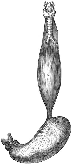
Fusiform Dilatation of Oesophagus (Luschka). A, Larynx; B, Thyroid gland; C, Trachea; D, Oesophagus; E, Stomach.
General dilatation, though sometimes congenital, is, as mentioned under Etiology, more frequently the mechanical result of distension of the oesophagus by food or drink prevented from ready entrance into the stomach by a constriction at the cardiac orifice. This form of dilatation is sometimes discovered as a post-mortem curiosity. The muscles have usually undergone great hypertrophy, and the mucous membrane some thickening and congestion, with erosions and sometimes ulcerations, indicative of chronic oesophagitis. In some instances all the coats of the oesophagus have undergone hypertrophy. The dilatation may vary from slight enlargement to the thickness of an ordinary man's arm or larger (Rokitansky31); in rare cases, even a capacity nearly equal to that of the stomach (Luschka32 and others). (See Fig. 19.)
31 Path. Anat.
32 Arch. für Anat., etc., March, 1868, p. 473.
The oesophagus is usually fusiform or spindle-shaped, being constricted at those portions at which it is normally slightly constricted. Sometimes the dilatation takes place between the lobes of the lungs (Raymond33).
33 Gaz. méd. de Paris, 1869, No. 7, p. 91.
Annular dilatation is usually due to circumferential distension just above a stricture. When not due to stricture its seat is usually just above the diaphragm, where the oesophagus is normally liable to constriction. The upper portion of the dilatation is larger than the lower portion, and the muscular walls are usually hypertrophied.
Pouched dilatation (diverticulum) is usually formed chiefly of mucous membrane and submucous tissue pushed through gaps in the fibres of the muscular coat, produced by distension. It sometimes involves the entire coat in cases in which the oesophageal wall has become adherent to enlarged lymphatic glands, which subsequently undergo subsidence in volume and drag the adherent portion of the wall after them (Rokitansky). The muscular walls are then usually hypertrophied, the mucous membrane sometimes hypertrophied, sometimes atrophied. The diverticulum is usually located in the upper portion of the oesophagus, just below the inferior constrictor muscle of the pharynx. It may thus be, in part, a pharyngocele also. It may be located behind the point of bifurcation of the trachea or where the oesophagus is crossed by the left bronchus. Its direction may be to the left side in the upper portion of the oesophagus, to the right side, or upon both sides; but when situated lower down it is usually directed backward, between the posterior wall of the tube and the spinal column. Hence its distension with food completely blocks up the calibre of the oesophagus. The orifice by which the oesophageal wall remains in communication with the pouch is round or elliptic in shape and variable in size, sometimes being about an inch in its long diameter, sometimes much smaller. The size of the diverticulum varies; a common size is that of a duck egg, but the size of a fist has been attained. Sometimes the diverticulum drags the oesophagus out of position and forms a sort of blind pouch in the direct line of its axis, so that it becomes filled with food which fails to reach the stomach. Sometimes there are several dilatations.
The dilatations become enlarged by retention of food, and are liable to undergo inflammation, ulceration, and perforation.
DIAGNOSIS.—The diagnosis will depend upon the symptoms of dysphagia, regurgitation, and so on, and upon the evidence furnished by auscultatory indications, palpation with the oesophageal sound, and, in some instances, the existence of a tumor in the neck, enlarging after meals, and from which food or mucus can be forced up into the pharynx by pressure externally.
Stethoscopic auscultation of the oesophagus during the deglutition of water indicates an alteration in the usual form of the gulp, which seems to trickle rapidly in a larger or smaller stream according to the degree of dilatation. If the dilatation be annular and located high up, auscultation is said to give the impression of a general sprinkling of fluid deflected from its course. The peculiar gurgle is often audible without the aid of stethoscopy. Palpation with the oesophageal bougie is competent to reveal the existence of a large sac by the facility with which the terminal extremity of the sound can be moved in the cavity. In the case of a diverticulum, however, the sound may glide past the mouth of the pouch without entering it, although arrested at the bottom of the sac in most instances.
In annular dilatation any constriction below it is usually perceptible to the touch through the sound; but, on the other hand, the ready passage of the bougie into the stomach, while excluding stricture, does not positively disprove the existence of a circumscribed dilatation. If high up, the dilatation may be detected externally by its enlargement when filled with food after a meal, and the subsidence of tumefaction when the sac is emptied by pressure from without, or by regurgitation. If the dilatation occupy a position which exercises compression of the trachea, dyspnoea will ensue when it is distended. The intermittence of the tumefaction serves to differentiate the swelling from abscess or morbid growth. From aneurism of the aorta, which it may simulate (Davy34), it is to be discriminated by absence of the usual stethoscopic and circulatory manifestations. The diagnosis of congenital dilatation is based upon a history of difficulty in deglutition dating from the earliest period of recollection.
34 Irish Hosp. Gaz., 1874, p. 129; Med. Press and Circular, May, 1874.
PROGNOSIS.—The prognosis is not favorable in any given case unless the cause can be removed, and not even then unless food can be prevented from accumulating in the distended portion of the tube. Nevertheless, cases sometimes go on into advanced age. On the other hand, they may terminate fatally within a year (Lindau35). The danger of perforation adds additional gravity to the prognosis, for life may be suddenly lost by this accident. Death usually takes place by inanition. A case of death by suffocation has been recorded, attributed to the pressure of the distended oesophagus upon the intrathoracic vessels (Hannay36).
35 Casper's Wochenschrift, 1840, No. 22; Arch. gén. de Méd., 1841, p. 498; Dict. de Méd et de Chir., xxiv. p. 410.
36 Edinb. Med. and Surg. Journ., July 1, 1833.
TREATMENT.—If the dilatation be due to stricture or to an impacted foreign body, the treatment should be directed to overcoming the one and removing the other.
General dilatation from chronic oesophagitis requires treatment for that disease.
Much depends upon preventing the accumulation of food in a sac or diverticle; the best means of accomplishing which is the systematic administration of all nutriment by means of the stomach-tube. When this is not advisable, care must be exercised in the selection of such food as is least likely to irritate the parts if detained in the pouch.
As far as general treatment is concerned, stimulants are usually indicated, as the patients become much reduced. If paralysis of the muscular coat of the oesophagus is believed to exist, the administration of preparations of phosphorus and of strychnine are indicated on general principles of therapeutics. Stimulation of muscular contractility by the oesophageal electrode has been recommended, but the prospects of success hardly justify the risks of serious injury in the domain of the pneumogastric nerve.
It has not yet been determined whether surgical procedures are competent to relieve dilatation. In cases of pouched dilatation high up it would not be difficult, as suggested by Michel,37 to expose the sac and excise it in such a manner that the sutures uniting the walls of the oesophagus shall occupy the site of the mouth of the diverticulum, and, thus obliterating it by cicatrization, restore the normal path of the food from the pharynx to the oesophagus. Gastrostomy, too, should hold out some hope of rescue, no matter what portion of the oesophagus be dilated.
37 Dict. Encyclop., xiv. p. 465.
FUNCTIONAL AND INFLAMMATORY DISEASES OF THE STOMACH.
BY SAMUEL G. ARMOR, M.D., LL.D.
Functional Dyspepsia (Atonic Dyspepsia, Indigestion).
To difficulty in the physiological process of digestion the familiar name of dyspepsia has been given, while to a merely disturbed condition of the function the term indigestion is more frequently applied. This distinction, difficult at all times to make, may appear more arbitrary than real; and inasmuch as it involves no important practical point, the author of the present article will use the terms interchangeably as indicating functional disturbance of the stomach—i.e. disturbance of the digestive process not associated with changes of an inflammatory character, so far as we know.
Since it is one of the most common of all complaints from its association with various other morbid conditions, the term is not unfrequently vaguely employed. It is difficult, of course, to define a disease whose etiology is so directly related to so many distinct morbid conditions. Indeed, there are few diseases, general or local, which are not at some time in their history associated with more or less derangement of the digestive process. For purposes of limitation, therefore, it will be understood that we now refer to chronic functional forms of indigestion which depend largely, at least, on a purely nervous element, and for this reason are not infrequently described as sympathetic dyspepsia. Doubt has been expressed as to whether such forms of disease ever exist, but that we encounter purely functional forms of dyspepsia, corresponding to the dyspepsia apyretica of Broussais, would appear to be a well-recognized clinical fact.
What the precise relation is between digestive disturbances and the nervous system we may not fully understand, no more than we understand how a healthy condition of nervous endowment is essential to all vital processes. Even lesions of nutrition are now known to depend upon primary disturbance of nervous influence. This is seen in certain skin diseases, such as herpes zoster, which closely follows the destruction of certain nerves. And it is well known that injury of nerve-trunks is not unfrequently followed by impaired nutrition and failure in reparative power in the parts to which such nerves are distributed. Indeed, so marked is the influence of the nervous system over the nutritive operations that the question has been considered as to whether there are trophic nerves distributed to tissue-elements themselves whose special function is to keep these elements in a healthy state of nutrition. The proof, at least, that the digestive process is, in some unexplained way, under the immediate influence of the nervous system, either cerebro-spinal or trophic, is both varied and abundant. The digestive secretions are known to be the products of living cells which are abundantly supplied with nerve-fibres, and we can readily believe that the potential energy of this cell-force is probably vital and trophic. At any rate, it is unknown in the domain of ordinary chemistry. The digestive ferments, as clearly pointed out by Roberts, are the direct products of living cells. Their mode of action, he claims, bears no resemblance to that of ordinary chemical affinity. It has a distinctly physiological character. Nor do they derive their vital endowments from material substances. "They give nothing material to, and take nothing from, the substances acted on. The albuminoid matter which constitutes their mass is evidently no more than the material substance of a special kind of energy—just as the steel of a magnet is the material substratum of the magnetic energy, but is not itself that energy" (Roberts). That this living cell-force is partly, at least, derived from the nervous system is clear from the well-known effects of mental emotion, such as acute grief, despair, etc., in putting an immediate stop to the digestive process. Experiments on the lower animals have also shown the direct influence of the nervous system over gastric secretion. Wilson Philip showed by various experiments on rabbits and other animals that if the eighth pair of nerves be divided in the neck, any food which the creatures may afterward eat remains in the stomach undigested, and after death, when the nerve has been divided, the coats of the stomach are not found digested, however long the animal may have been dead. Bernard also excited a copious secretion by galvanization of the pneumogastric, and by section of the same nerve stopped the process of digestion and produced "pallor and flaccidity of the stomach." Recently doubt has been thrown on these statements of Bernard and Frerichs. Goltz concludes, from observations made on frogs, that nerve-ganglia, connected by numerous intercommunicating bundles of nerve-fibres, exist in the walls of the stomach, the irritation of which gives rise to local contractions and peristaltic movements of the stomach, and that these ganglia influence the gastric secretion. However this may be, it still remains true that these gastric ganglia are in connection, through the vagi, with the medulla oblongata, and are thus influenced by the cerebro-spinal nerve-centres. And clinical observation confirms what theoretical considerations would suggest. Thus, strong mental impressions are known to produce sudden arrest of secretion, and that which arrests secretion may, if continued, lead to perversion of the same.
Impressions made upon the nerves of special sense are also known to affect the salivary and gastric secretions. The flow of saliva is stimulated by the sight, the smell, the taste, and even thought, of food. Bidder and Schmidt made interesting experiments on dogs bearing upon this point. They ascertained by placing meat before dogs that had been kept fasting that gastric juice was copiously effused into the stomach. Other secretions are known to be similarly affected. Carpenter by a series of well-observed cases has shown the direct influence of mental conditions on the mammary secretion. The nervous association of diabetes and chronic Bright's disease is interesting in this connection, and the direct nervous connection betwixt the brain and the liver has been shown by numerous experiments. It is maintained by modern physiologists that "the liver—indeed each of the viscera—has its representative area in the brain, just as much as the arm or leg is represented in a distant localized area" (Hughlings Jackson). And in harmony with this view Carpenter long since pointed out the fact that if the volitional direction of the consciousness to a part be automatically kept up for a length of time, both the functional action and the nutrition of the part may suffer. It has been described by him as expectant attention, and it has, as we shall see, important practical bearings on the management of gastric affections. Sympathetic disturbance of the stomach is also connected with direct disease of the brain. This is seen in cases of concussion. The almost immediate effects of a blow are nausea and vomiting, and the same thing is observed in local inflammation of the meninges of the brain.
Many forms of functional dyspepsia due to nervous disturbance of a reflex character will be pointed out when discussing the etiology of the disease.
ETIOLOGY.—Among the agencies affecting the digestive process in atonic forms of dyspepsia may be mentioned—
First, predisposing causes;
Second, exciting causes.
In general terms it may be said that all conditions of depressed vitality predispose to the varied forms of atonic dyspepsia. These conditions range through an endless combination of causes, both predisposing and exciting. There is not a disturbed condition of life, extrinsic or intrinsic, that may not contribute to this end. In some cases it may be the effects of hot and enervating climates; in others the alterations in the elementary constituents of the blood may be apparent; while in still others the cause may be exhausting discharges, hemorrhages, profuse suppuration, venereal excesses, sedentary occupations, and long-continued mental and moral emotions.
Heredity may also predispose to functional dyspepsia. Certain faulty states of the nervous system are specially liable to be transmitted from parent to offspring—not always in the exact form in which they appeared in the parent, but in forms determined by the individual life of the offspring. For obvious reasons, growing out of our modern American civilization, the inheritance of a faulty nervous organization is apt to spend itself upon the digestive apparatus. The inordinate mental activity, the active competitions of life, the struggle for existence, the haste to get rich, the disappointments of failure,—all contribute to this end. The general tendency of American life is also in the direction of a highly-developed and morbidly sensitive nervous system, and functional dyspepsia is a natural sequence of this. The symptoms of dyspepsia thus caused usually manifest themselves at an early period of life.
Age also predisposes to weak digestion. The stomach becomes weak as age advances, in common with all the functions of the body, and consequent upon this weakness there is diminished excitability of the gastric nerves, with diminished muscular action of the walls of the stomach and deficient secretion of the gastric juice. Chronic structural changes are also apt to occur in advanced life. The gastric glands become atrophied and the arteries become atheromatous, so that with symptoms of indigestion there are often associated loss of consciousness at times, vertigo, irregular action of the heart, etc. These general facts have an important bearing upon the hygienic management of dyspepsia in the aged. They require, as a rule, less food than the young and vigorous. In times when famine was more frequent than now it was found that the older a human being was, the better deficiency of food was borne. Hippocrates tells us, in his Aphorisms, that old men suffer least from abstinence. Their food should be such, both in quantity and quality, as the enfeebled stomach can digest. There is less demand for the materials of growth, and consequently for animal food. Moderate quantities of alcohol, judiciously used, are also specially adapted to the indigestion of the aged. It has the double effect of stimulating the digestive process and at the same time checking the activity of destructive assimilation, which in old age exhausts the vital force. And in order to more effectively arrest destructive metamorphosis great caution should be taken against excessive muscular fatigue, as well as against sudden extremes of temperature. Loss of appetite from deficient formation of gastric juice is a common symptom in old age. This is not often successfully treated by drugs, and yet medicines are not without value. The sesquicarbonate of ammonium acts as a stimulant to the mucous membrane and to the vaso-motor nerve, and in this way becomes a valuable addition to the simple vegetable bitters. Dilute hydrochloric acid with the vegetable bitters may also be tried. Condiments with the food directly stimulate the action of the enfeebled stomach. The old remedy of mustard-seed is not unfrequently useful, and pepper, cayenne, horseradish, and curries act in a similar manner in torpid digestion. And in cases of great exhaustion associated with anæmia benefit may be derived from small doses of iron added to tincture of columbo or gentian.
Nor should it be forgotten that in the opposite extreme of life the digestive capacity is extremely limited. The infant's digestion is readily disturbed by unsuitable alimentation. For obvious reasons it does not easily digest starchy substances. The diastasic ferment does not exist in the saliva of young sucking animals, at least to any extent. No food is so suitable for early infantile life as the mother's milk, provided the mother herself is healthy. It contains in an easily digestible form all the constituents necessary to the rapidly-growing young animal. Van Helmont's substitute of bread boiled in beer and honey for milk, or Baron Liebig's food for infants, cannot take the place of nature's type of food, which we find in milk. If a substitute has to be selected, there is nothing so good as cow's milk diluted with an equal quantity of soft water, or, what in many cases is better, barley-water, to which may be added a teaspoonful of powdered sugar of milk and a pinch of table-salt and phosphate of lime. Lime-water may be added with advantage. Dilution of alimentary substances is an important condition of absorption in the infant stomach.
Anæmia is a common predisposing cause of indigestion. Indeed, as a widely-prevailing pathological condition few causes stand out so prominent. It affects at once the great nutritive processes, and these in turn disturb the functional activity of all the organs of the body. Not only are the gastric and intestinal glands diminished in their functional activity by impoverished or altered blood, but the movements of the stomach are retarded by weakened muscular action. It is impossible to separate altered blood from perverted tissue-structure and altered secretion. Indigestion produced by anæmia is difficult of treatment, on account of the complexity of the pathological conditions usually present, the anæmia itself being generally a secondary condition. Careful inquiry should be made, therefore, into the probable cause of the anæmia, and this should, if possible, be removed as an important part of the treatment of the dyspepsia. Nothing will more promptly restore the digestive capacity in such cases than good, healthy, well-oxidized blood. Indeed, healthy blood is a condition precedent to the normal functional activity of the stomach.
To these general predisposing causes may be added indigestion occurring in febrile states of the system. The cause here is obvious. In all general febrile conditions the secretions are markedly disturbed; the tongue is dry and furred; the urine is scanty; the excretions lessened; the bowels constipated; and the appetite gone. The nervous system also participates in the general disturbance. In this condition the gastric juice is changed both quantitatively and qualitatively, and digestion, as a consequence, becomes weak and imperfect—a fact that should be taken into account in regulating the diet of febrile patients. From mere theoretical considerations there can be no doubt that fever patients are often overfed. To counteract the relatively increased tissue-metamorphosis known to exist, and the consequent excessive waste, forced nutrition is frequently resorted to. Then the traditional saying of the justly-celebrated Graves, that he fed fevers, has also rendered popular the practice. Within certain bounds alimentation is undoubtedly an important part of the treatment of all the essential forms of fever. But if more food is crowded upon the stomach than can be digested and assimilated, it merely imposes a burden instead of supplying a want. The excess of food beyond the digestive capacity decomposes, giving rise to fetid gases, and often to troublesome intestinal complications. The true mode of restoring strength in such cases is to administer only such quantities of food as the patient is capable of digesting and assimilating. To this end resort has been had to food in a partially predigested state, such as peptonized milk, milk gruel, soups, jellies, and beef-tea; and clinical experience has thus far shown encouraging results from such nutrition in the management of general fevers. In these febrile conditions, and in all cases of general debility, the weak digestion does not necessarily involve positive disease of the stomach, for by regulating the diet according to the digestive capacity healthy digestion may be obtained for an indefinite time.
Exhaustion of the nerves of organic life strongly predisposes to the atonic forms of dyspepsia. We have already seen how markedly the digestive process is influenced by certain mental states, and it is a well-recognized fact that the sympathetic system of nerves is intimately associated with all the vegetative functions of the body. Without a certain amount of nervous energy derived from this portion of the nervous system, there is failure of the two most important conditions of digestion—viz. muscular movements of the stomach and healthy secretion of gastric juice. This form of indigestion is peculiar to the ill-fed and badly-nourished. It follows in the wake of privation and want, and is often seen in the peculiarly careworn and sallow classes who throng our public dispensaries. In this dyspepsia of exhaustion the solvent power of the stomach is so diminished that if food is forced upon the patient it is apt to be followed by flatulence, headache, uneasy or painful sensations in the stomach, and sometimes by nausea and diarrhoea. It is best treated by improving in every possible way the general system of nutrition, and by adapting the food, both in quantity and quality, to the enfeebled condition of the digestive powers. Hygienic measures are also of great importance in the management of this form of dyspepsia, and especially such as restore the lost energy of the nervous system. If it occur in badly-nourished persons who take little outdoor exercise, the food should be adapted to the feeble digestive power. It should consist for a time largely of milk and eggs, oatmeal, peptonized milk gruels, stale bread; to which should be added digestible nitrogenous meat diet in proportion to increased muscular exercise. Systematic outdoor exercise should be insisted upon as a sine quâ non. Much benefit may be derived from the employment of electric currents, and hydrotherapy has also given excellent results. If the indigestion occur in the badly-fed outdoor day-laborer, his food should be more generous and mixed. It should consist largely, however, of digestible nitrogenous food, and meat, par excellence, should be increased in proportion to the exercise taken. Medicinally, such cases should be treated on general principles. Benefit may be derived from the mineral acids added to simple bitters, or in cases of extreme nervous prostration small doses of nux vomica are a valuable addition to dilute hydrochloric acid. The not unfrequent resort to phosphorus in such cases is of more than doubtful utility. Some interesting contributions have been recently made to this subject of gastric neuroses by Buchard, Sée, and Mathieu. Buchard claims that atonic dilatation of the stomach is a very frequent result of an adynamic state of the general system. He compares it to certain forms of cardiac dilatation—both expressions of myasthenia. It may result from profound anæmia or from psychical causes. Mathieu regards mental depression as only second in frequency. Much stress is laid upon poisons generated by fermenting food in the stomach in such cases. It may cause a true toxæmia, just as renal diseases give rise to uræmia. Of course treatment in such cases must be addressed principally to the general constitution.
But of all predisposing causes of dyspepsia, deficient gastric secretion, with resulting fermentation of food, is perhaps the most prevalent. It is true this deficient secretion may be, and often is, a secondary condition; many causes contribute to its production; but still, the practical fact remains that the immediate cause of the indigestion is disproportion between the quantity of gastric juice secreted and the amount of food taken into the stomach. In all such cases we have what is popularly known as torpidity of digestion, and the condition described is that of atony of the stomach. The two main constituents of gastric juice—namely, acid and pepsin—may be deficient in quantity or disturbed in their relative proportions. A certain amount of acid is absolutely essential to the digestive process, while a small amount of pepsin may be sufficient to digest a large amount of albuminoid food. Pure unmixed gastric juice was first analyzed by Bidder and Schmidt. The mean analyses of ten specimens free from saliva, procured from dogs, gave the following results:
Gastric Juice of a Dog.
Water 973.06
Solids 26.94
Containing— Peptone and pepsin 17.19
Free hydrochloric acid 3.05
Alkaline chlorides 4.26
Ammonium chloride 0.47
Chlorine 5.06
Phosphates— Lime 1.73
Magnesia 0.23
Iron 0.08
They proved by the most careful analyses that fresh gastric juice contains only one mineral acid—namely, hydrochloric; since which time Richet has been able to prove that "this acid does not exist in a free state, but in loose combination with an organic substance known as lucin," the chloride of lucin. And just here the curious and puzzling question arises as to the secretion of a mineral acid from alkaline blood. Ewald, the distinguished lecturer in the Royal University of Berlin, tells us that "a brilliant experiment of Maly's has thrown unexpected light upon this. There are fluids of alkaline reaction which may contain two acid and alkaline mutually inoffensive salts, but still have an alkaline reaction, because the acid reaction is to a certain extent eclipsed; for instance, a solution of neutral phosphate of soda (Na2HPO4) and acid phosphate of soda (NaH2PO4) is alkaline. Such a solution placed in a dialyzer after a short time gives up its acid salt to the surrounding distilled water, and one has in the dialyzer an alkaline fluid outside an acid fluid." He thus proved that the acid phosphate of sodium is present in the blood in spite of its alkaline reaction.
Lack of the normal amount of the gastric secretion must be met by restoring the physiological conditions upon which the secretion depends. In the mean time, hydrochloric and lactic acids may be tried for the purpose of strengthening the solvent powers of the gastric secretion.
EXCITING CAUSES.—The immediate causes of dyspepsia are such as act more directly on the stomach. They embrace all causes which produce conditions of gastric catarrh, such as excess in eating and drinking, imperfect mastication and insalivation, the use of indigestible or unwholesome food and of alcohol, the imperfect arrangement of meals, over-drugging, etc.
Of exciting causes, errors of diet are amongst the most constantly operative, and of these errors excess of food is doubtless the most common. The influence of this as an etiological factor in derangement of digestion can scarcely be exaggerated. In very many instances more food is taken into the stomach than is actually required to restore tissue-waste, and the effects of such excess upon the organism are as numerous as they are hurtful. Indeed, few elements of disease are more constantly operative in a great variety of ailments. In the first place, if food be introduced into the stomach beyond tissue-requirements, symptoms of indigestion at once manifest themselves. The natural balance betwixt supply and demand is disturbed; the general nutrition of the body is interfered with; local disturbances of nutrition follow; and mal-products of digestion find their way into the blood. Especially is this the case when the excessive amount of food contains a disproportionate amount of nitrogenous matter. All proteid principles require a considerable amount of chemical alteration before they are fitted for the metabolic changes of the organism; the processes of assimilative conversion are more complex than those undergone by fats and amyloids; and it follows that there is proportional danger of disturbance of these processes from overwork. Moreover, if nitrogenous food is in excess of tissue-requirement, it undergoes certain oxidation changes in the blood without becoming previously woven into tissue, with resulting compounds which become positive poisons in the economy. The kidneys and skin are largely concerned in the elimination of these compounds, and the frequency with which these organs become diseased is largely due, no doubt, to the excessive use of unassimilated nitrogenous food. Then, again, if food be introduced in excess of the digestive capacity, the undigested portion acts directly upon the stomach as a foreign body, and in undergoing decomposition and putrefying changes frets and irritates the mucous membrane. It can scarcely be a matter of doubt that large groups of diseases have for their principal causes excess of alimentation beyond the actual requirements of the system. All such patients suffer from symptoms of catarrhal indigestion, such as gastric uneasiness, headache, vertigo, a general feeling of lassitude, constipation, and high-colored urine with abundant urates, together with varied skin eruptions. Such cases are greatly relieved by reducing the amount of food taken, especially nitrogenous food, and by a systematic and somewhat prolonged course of purgative mineral waters. Europe is especially rich in these springs. The waters of Carlsbad, Ems, Seltzer, Friedrichshall, and Marienbad, and many of the alkaline purgative waters of our own country, not unfrequently prove valuable to those who can afford to try them, and their value shows how often deranged primary assimilation is at the foundation of many human ailments. The absurd height to which so-called restorative medicine has attained within the last twenty years or more has contributed largely to the production of inflammatory forms of indigestion, with all the evil consequences growing out of general deranged nutrition.
The use of indigestible and unwholesome food entails somewhat the same consequences. This may consist in the use of food essentially unhealthy or indigestible, or made so by imperfect preparation (cooking, etc.). Certain substances taken as food cannot be dissolved by the gastric or intestinal secretions: the seeds, the skins, and rinds of fruit, the husks of corn and bran, and gristle and elastic tissue, as well as hairs in animal food, are thrown off as they are swallowed, and if taken in excess they mechanically irritate the gastro-intestinal mucous membrane and excite symptoms of acute dyspepsia, and not unfrequently give rise to pain of a griping character accompanied by diarrhoea. Symptoms of acute dyspepsia also frequently follow the ingestion of special kinds of food, such as mushrooms, shellfish, or indeed fish of any kind; and food not adapted to the individual organism is apt to excite dyspeptic symptoms. Appetite and digestion are also very much influenced by the life and habits of the individual. The diet, for instance, of bodily labor should consist largely of digestible nitrogenous food, and meat, par excellence, should be increased in proportion as muscular exercise is increased. For all sorts of muscular laborers a mixed diet is best in which animal food enters as a prominent ingredient. Thus, it has been found, according to the researches of Chambers, that in forced military marches meat extract has greater sustaining properties than any other kind of food. But with those who do not take much outdoor exercise the error is apt to be, as already pointed out, in the direction of over-feeding. It cannot be doubted at the present time that over-eating (gluttony) is one of our popular vices. Hufeland says: "In general we find that men who live sparingly attain to the greatest age." While preventive medicine in the way of improved hygiene—better drainage, better ventilation, etc.—is contributing largely to the longevity of the race, we unfortunately encounter in more recent times an antagonizing influence in the elegant art of cookery. Every conceivable ingenuity is resorted to to tempt men to eat more than their stomachs can properly or easily digest or tissue-changes require. The injurious consequences of such over-feeding may finally correct itself by destroying the capacity of the stomach to digest the food.
But, on the other hand, in many nervous forms of dyspepsia the weak stomach is not unfrequently made weaker by severely restricted regimen, and especially is this the case with mental workers. Theoretical and fanciful considerations sometimes lead to physical starvation. This is apt to be the case with dyspeptics. Men who toil with their brain rather than their muscles, whether dyspeptic or not, require good, easily-digested mixed diet. It is a popular error to suppose that drugs can take the place of such food, especially drugs which are supposed to have a reconstructive influence over the nervous system, such as iron and phosphorus. The expression of Büchner, "No thinking without phosphorus," captivating to theoretical minds, has gained much notoriety, and has doubtless led to the excessive use of that drug in nervous forms of indigestion. There never was a period when phosphorus was so universally prescribed as the present. It enters into endless combinations with so-called nerve-tonics. Of the injurious influence of the drug in many cases of functional indigestion there can be no doubt; and the statement itself, so often quoted, that "the amount of phosphorus in the blood passing through the brain bears an exact proportion to the intensity of thought," is calculated to mislead. T. K. Chambers, author of the excellent Manual of Diet, makes the statement that "a captive lion, tiger, leopard, or hare assimilates and parts with a greater amount of phosphorus than a hard-thinking man; while the beaver, noted for its power of contrivance, excretes so little phosphorus that chemical analysis cannot find it in its excreta." In the wonderful adaptations and regulative mechanisms of nature we may trust largely to the natural law of supply and demand in maintaining a proper equilibrium. It may be doubted, indeed, whether we require at any time more phosphorus for brain- and nerve-tissue than can be found in such food as contains digestible phosphatic salts. The natural demand for food grows out of healthy tissue-change. An appetite to be healthy should commence in processes outside of the stomach.
Food may also be introduced into the stomach in an undigestible form from defects of cookery. The process of cooking food produces certain well-known chemical changes in alimentary substances which render them more digestible than in the uncooked state. By the use of fire in cooking his food new sources of strength have been opened up to man which have doubtless contributed immeasurably to his physical development, and has led to his classification as the cooking animal. With regard to most articles the practice of cooking his food beforehand is wellnigh universal; and especially is this the case with all farinaceous articles of food. The gluten of wheat is almost indigestible in the uncooked state. By the process of cooking the starchy matter of the grain is not only liberated from its protecting envelopes, but it is converted into a gelatinous condition which readily yields to the diastasic ferments. Roberts, in his lectures on the Digestive Ferments, points out the fact that when men under the stress of circumstances have been compelled to subsist on uncooked grains of the cereals, they soon fell into a state of inanition and disease.
Animal diet is also more easily digested in the cooked than in the raw state. The advantage consists chiefly in the effects of heat on the connective tissue and in the separation of the muscular fibre. In this respect cooking aids the digestive process. The gastric juice cannot get at the albumen-containing fibrillæ until the connective tissue is broken up, removed, or dissolved. Hot water softens and removes this connective tissue. Hence raw meat is less easily digestible. Carnivorous animals, that get their food at long intervals, digest it slowly. By cutting, bruising, and scraping meat we to a certain extent imitate the process of cooking. In many cases, indeed, ill-nourished children and dyspeptics digest raw beef thus comminuted better than cooked, and it is a matter of observation that steamed and underdone roast meats are more digestible than when submitted to greater heat.
Some interesting observations have been made by Roberts on the effects of the digestive ferments on cooked and uncooked albuminoids. He employed in his experiments a solution of egg albumen made by mixing white of egg with nine times its volume of water. "This solution," says Roberts, "when boiled in the water-bath does not coagulate nor sensibly change its appearance, but its behavior with the digestive ferments is completely altered. In the raw state this solution is attacked very slowly by pepsin and acid, and pancreatic extract has no effect on it; but after being cooked in the water-bath the albumen is rapidly and entirely digested by artificial gastric juice, and a moiety of it is rapidly digested by pancreatic extract."
It is a mistake, however, to suppose that cooking is equally necessary for all kinds of albuminoids. The oyster, at least, is quite exceptional, for it contains a digestive ferment—the hepatic diastase—which is wholly destroyed by cooking. Milk may be indifferently used either in the cooked or uncooked state, and fruits, which owe their value chiefly to sugar, are not altered by cooking.
The object in introducing here these remarks on cooking food is to show that it forms an important integral part of the work of digestion, and has a direct bearing on the management of all forms of dyspepsia.
Haste in eating, with imperfect mastication, is a common cause of indigestion in this country. Mastication is the first step in the digestive process. It is important, therefore, that we have good teeth and that we take time to thoroughly masticate our food, for by so doing we prepare it for being acted upon by the juices of the stomach. Time is also necessary in order that the salivary secretion may be incorporated with the alimentary substances. By the salivary diastase starch is converted into sugar and albuminoids are prepared for the action of the gastric juice. If these changes take place imperfectly, the stomach can scarcely regain in gastric digestion what was lost in imperfect mastication and insalivation. Haste in eating is one of the American vices. It grows out of the temperament of our people. We are jealous of lost time, and unfortunately this time is too often taken from the stomach. We bolt our food with unseemly haste, and pay the penalty in ruined stomachs. Many cases of indigestion are greatly relieved, if not permanently cured, by simply doubling or quadrupling the time occupied in eating.
Irregularity in the intervals between meals, such as taking one meal only in twenty-four hours or taking food before the preceding supply has been digested, is another fruitful source of indigestion. The digestive process, in the natural order of change, is confused; changes which should take place are delayed; and the results are such as arise from excessive eating. Moreover, the stomach lacks the rest so essential to digestion. The necessary interval, however, between meals varies with the nature of the food taken. "Between the extremes of the carnivoræ," says Ewald, "which feed once in twenty-four hours, and the herbivoræ, which never have done with the business of feeding, man holds a middle place, but not without permitting the recognition in the course of his life of a sort of transition from the herbivora to the carnivora. Infants should have the breast during the first three weeks as often as they wake; after that every two hours to the third month; then up to dentition every three hours; and later there should be five meals in twenty-four hours." But to this general statement there are, of course, many exceptions. Under certain pathological conditions food should be taken in small quantities at short intervals. This is especially the case in chronic gastric catarrh and in feeble digestion of nervous subjects. Such patients are not unfrequently improved by becoming again infants or herbivoræ. By the use of an exclusive milk diet or peptonized milk gruels, given in small quantities at comparatively short intervals of time, the stomach may be so accommodated that it will digest without discomfort a large amount of nourishment within a given time. To S. Weir Mitchell of Philadelphia we are indebted for some valuable observations bearing upon this point of forced alimentation.
To the causes of indigestion already alluded to may be added the habit of spirit-drinking, especially the habit of taking alcohol undiluted on an empty stomach, which rarely fails after a time to engender dyspeptic symptoms. It is a prominent factor in the production of chronic gastric catarrh—a condition more frequently present in painful indigestion than any that have been named. It is one of the most common diseases met with in practice. Indeed, all causes already alluded to involve, sooner or later, if they are constantly operative, irritative and catarrhal conditions of the mucous membrane of the stomach, so that we find it difficult at times—indeed impossible—to separate purely functional from subacute inflammatory forms of dyspepsia. Practically, we simply study the subject in the relative degrees of prominence of the one condition or the other.
But, in a still more comprehensive sense, indigestion is caused by disturbance of organs directly associated with the stomach in the digestive process. All organs closely associated with each other in their physiological functions are apt to become associated in morbid action. The clinical recognition of this is a matter of great importance in the management of gastric affections. And first in the order of importance in such association is the liver. So closely, indeed, are the liver and stomach functionally associated in the process of primary assimilation that they may be considered parts of the same great digestive apparatus. Hence disturbance of the liver—either in the formation of glycogen, the destruction of albuminoid matter, or the secretion of bile—is immediately communicated to the stomach. It may be difficult to say which of these separate and distinct functions of the liver is most at fault; that can only be a matter of physiological inference. In the one case, for instance, the dyspeptic may be fairly well nourished, yet his elimination may be bad. In the other there is no failure of the destructive and excreting functions, but those concerned in the assimilation of fat and peptones are disordered, so that the patient is not well nourished, so far as the fatty element is concerned. This is the more common form, and a form not unfrequently associated with pulmonary consumption. The liver finally becomes fatty—a condition usually found associated with the constitutional forms of phthisis.
The pancreas is also closely associated with the stomach, and its secretion is of essential value in the digestive process. It is to be regretted that our precise knowledge of its diseases is in such striking contrast with its importance in the animal economy, and yet it can scarcely be doubted that in dyspeptic symptoms associated with failure of digestion of starchy, albuminous, and fatty elements of food there is disorder of the secretion of the pancreas. Hence in the treatment of the early stages of pulmonary consumption and other disorders associated with deficient digestion and assimilation of fatty substances the importance of directing our attention to the condition of the liver and pancreas, as well as to the stomach.
That morbid states of the intestinal track occupy a prominent place in the etiology of dyspepsia is also a well-recognized clinical fact. Indeed, constipation of the bowels is an almost universal accompaniment of deranged digestion, and when persistent for years it is apt to lead to the most disastrous consequences. These are mainly in the direction of lessened elimination from the intestinal glandulæ. The general symptomatology of deficient excretion from these glandulæ is closely analogous to the same condition of the liver: there is impairment of the general health; the clear florid complexion disappears; the patient becomes of a greenish or sallow hue; the blood is altered in quality; fatigue is experienced after the slightest exertion; the nights are restless; and there is great tendency to mental despondency. Moreover, constipation often precedes the gastric symptoms. The diminished muscular activity of the intestinal track extends to the stomach; its movements are diminished; food is not properly mixed with the gastric juice, and by being too long retained in the stomach in a comparatively undigested state acetous fermentation in the saccharine and starchy articles of diet is set up, acid eructations and a sour taste in the mouth being commonly complained of. Dyspepsia associated with this condition of the intestinal track cannot be relieved until the constipation is relieved, and by overcoming the constipation the dyspeptic symptoms often disappear.
Mention has been made of the baneful influence of certain mental states in the production of dyspeptic symptoms. But there are forms of indigestion due to local nervous disturbance existing elsewhere than in the nerve-centres. This was ascribed by the older writers to what they termed consensus nervorum, or sympathy, by which "the operation of a stimulus is not limited to the nerves immediately irritated, but is extended to distant parts in known or unknown connection with the irritated nerves." An intimate acquaintance with this law of sympathy is of the utmost importance in the study of the functional forms of dyspepsia, for no other organ of the body is subjected to such a wide range of reflected nervous disturbance as the stomach. Morbid sympathetic impressions are transmitted mainly through branches of the vaso-motor nerve of the semilunar ganglia of the abdomen, and from the pneumogastric to the stomach. Thus, a pregnant uterus not unfrequently produces very troublesome vomiting; some females suffer from nausea and indigestion during each menstrual period; and the more chronic forms of pelvic irritation, such as a flexed uterus, and endometritis, cervicitis, or tender ovary, may be the continuous exciting cause of most troublesome forms of nervous dyspepsia. There is also close sympathy of the stomach with the lungs and heart through the distribution of the pneumogastric. So also may fixed points of irritation in any part of the nervous system be reflexly transmitted to the stomach, giving rise to most pronounced symptoms of indigestion. And it is evident that in all such cases but little can be accomplished in the way of relieving the dyspeptic symptoms until the cause upon which they depend is removed. The treatment must have reference mainly to the removal of such cause.
Lastly, all the causes mentioned finally concur in producing irregularities of the mechanism of digestion; and this may be done by disturbing either the muscular movements of the stomach or in suspending or perverting the gastric solvents, or in these two conditions combined.
SYMPTOMS.—1st. Referable to the Stomach.—The symptoms which attend and indicate the presence of functional dyspepsia are such as accompany in a greater or less degree almost all cases of chronic gastritis. Clinically, so far as the direct gastric symptoms are concerned, it is difficult to separate them. The more prominent of the local symptoms are—a sense of fulness and distension after meals, discomfort during the digestive process, derangement of appetite, acid eructations, flatulence, regurgitations of food, and sometimes nausea and vomiting. There is seldom severe pain; the sensation is rather that of uneasiness. Exceptionally, however, there may be pain, which radiates from the stomach to the shoulders, and may pass down the left arm so as to simulate angina pectoris. But it may be readily distinguished from that complaint by coming on after food, and not after exertion. In other cases a sense of constriction may be accompanied by dyspnoea, arising from impeded movements of the diaphragm from being pushed upward by the distended stomach, or there may be heartburn, with an ill-defined sense of burning felt in the epigastrium; but thirst, so frequently present in chronic gastritis, is, as a rule, absent in functional dyspepsia. These symptoms are manifested in varying degrees of prominence in individual cases, and some of them are rarely found present. Thus, nausea and vomiting are not characteristic features of the chronic forms of functional dyspepsia, and as a rule epigastric tenderness is entirely absent. In markedly hysterical subjects or in persons whose nervous system has been unduly excited by alcohol there may be shrinking from the slightest touch upon pressure; but in these cases the tenderness is not confined to the stomach, nor is it increased by deep pressure. In some cases there is an unnatural craving for food—a symptom rarely if ever observed in structural lesions of the stomach—and now and then it happens that the appetite becomes depraved, especially with hysterical patients. They crave indigestible and unnatural substances, such as earth, chalk, and substances wholly devoid of alimentary properties. Impairment of appetite, however, is the more common feature of this form of indigestion.
Flatulence and eructations are generally complained of, the flatulence being accompanied by a painful sense of fulness, affecting in equal degree the stomach and small intestines. It is derived principally from putrefactive or fermentative changes of the ingesta, which are imperfectly elaborated in the stomach. The gases consist of carbonic acid, sulphuretted hydrogen, hydrogen, nitrogen, and the hydrocarbons, the butyric and acetic fermentations furnishing the hydrogen and carbonic acid gas. In addition to these marsh gas is formed by a special fermentation, the basis of which exists in the cellulose taken with vegetable food. In excessive meteorism from paralysis of the intestines the gas is principally nitrogen; the marsh-gas fermentation results from the ingestion of certain easily-fermentable vegetables, such as cabbage, cauliflower, etc.