Part 2 — CHAPTER III.
Reading edition
Zoophytes, Cœlenterata, Medusæ, Corals, Hydrozoa.
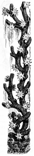
Fig. 344.—Gorgonia Nobilis.
A study of the earliest growth of the Cœlenterata has shown that their internal cavities are nothing more than regular radiate out-growths of the internal structures. The result of this development is a condition which does not occur again in the whole of the animal kingdom. There is a system of cavities all in open communication one with another, no closed blood vascular system, and no specialised respiratory apparatus. Again, all the animals that constitute this large group are radiate in structure, that is, when viewed from above they are typically star-shaped, and if cut across, every horizontal section shows a symmetrical arrangement of the several parts around a centre. There are other radiate animals, as the Echinoderms, but while in these five is the fundamental number of rays, in the Cœlenterata the rays are a multiple of four, six and upwards. The skeleton or framework of each differs, and when the Cœlenterata form calcareous structures, these are quite different from the tests of the sea-urchins; and in all cases the anterior portion of the body is crowned with one or more circles of tentacles, which remain perfectly flexible and flower-like. The most highly-developed of the free forms are the sea-anemones and the jelly-fish. These have no hard or calcareous skeleton whatever, but withal they are, in the opinion of naturalists and microscopists, the most beautiful objects among Zoophytes.
In spite of their variety of forms, the Cœlenterata seem to be as incapable of higher development as do the Echinoderms, and they have failed to make headway in fresh water, but it is not improbable that some of the simplest forms of the whole group may have given rise to higher animal forms, while the sea anemones, corals, &c., being those descendants of the primitive simple form, have retained the original type of organization almost unchanged.
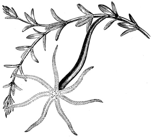
Fig. 345.—Hydra viridis, adhering to a stalk of Anacharis alsinastrum.
The type of the group is the Hydra, a fresh-water polyp, commonly found attached to the leaves and stems of many aquatic plants, or floating pieces of stick. Two species are well known to microscopists, the H. viridis, or green polyp, and the H. vulgaris, somewhat darker in colour, probably dependent upon the nature of its food. The third, less common species, the H. fasca, is distinguished from both by the length of its tentacles, which, when fully extended, greatly exceed those of either of the before-mentioned. The fresh-water group measures from one-eighth to the one-third of an inch in length, and form simple stocks of one, two or more branches. They almost exactly resemble in form the polyps of the Hydractinia, which are provided with a circle of tentacles. When placed in a vessel of water and left undisturbed they often attach themselves to the side, where they may be examined with a moderate power at leisure. They are then seen to spread out their tentacles like fine threads, and seize upon any small creature that may come in their way, and by the same means convey it to a mouth capable of great extension. All Hydra possess stinging-cells, by means of which they paralyse their prey. Many Hydra attain to a large size, and shoot out long poisonous filaments; they also possess smaller kinds of smooth cells, which appear to be employed for an entirely different purpose, but for what is not positively known. Hydra usually multiply by means of buds, an out-growth from the body, and these remain attached to the mother stalk for some time, often long enough to give rise to one or two smaller buds. Single eggs are also developed in the ectoderm beneath capsules, or wart-like prominences. The adult animal can be cut to pieces, and from each piece a new individual will be developed. This method of reproduction was first tried by the naturalist Trembley in 1739, whose experiments in this direction excited the greatest interest among the naturalists of the middle of the last century. Hydra fusca in various stages of development is given in outline in Fig. 346.
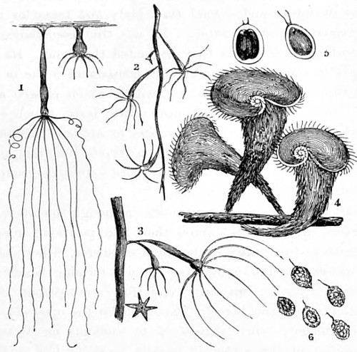
Fig. 346.
1, 2, 3. Hydra in various stages of development; 4. Group of Stentor polymorphus, many-shaped Stentor; 5. Englena; 6. Monads.
In the polyps belonging to this family the body-structure for the most part consists of a homogeneous aggregation of vesicular granules, held together by an intercellular sarcode, and capable of great extension and contraction, so that these animals can assume a variety of forms and extend their body and tentacles until the latter become almost invisible. It was the resemblance in this respect to the fabled Hydra that originated the name. Its organ of prehension is termed the hasta; this consists of a sac or opening at the terminal end of the tentacle, within which is seen a saucer-shaped vesicle, supporting a minute ovate body, which carries a sharp calcareous piece termed a sagitta or arrow. Although the fresh-water Hydra may be regarded as typical of this group of animals, marine fauna furnish a far more extensive group in the corals, jelly-fish, and sea-anemones.
A smaller group, the Ctenophora, although members of this sub-kingdom, have not yet found their true position; nevertheless they are interesting glassy, transparent creatures, either shaped like apples, melons, or Phrygian caps, or else forming bands of some considerable length; all are wonderfully transparent, with the single exception of the Beroë. These inhabit the open sea, and are only seen inshore when driven in by currents or strong winds. Their position in the water is usually more or less vertical, the mouth being turned downwards. The portion from which this group derives its name is the ribs, which are symmetrically arranged, and consist of rows of short transverse combs, each forming rows of cilia, which, as they wave to and fro, constitute a swimming or rowing plate, their activity in the water depending upon the will of the animal. They are also provided with an oral umbrella, and capturing filaments or tentacles with hair-like branches. These tentacles, attached to the sides of the animal, are capable of erection or withdrawal into pockets. Great variety is seen in these accessory organs of locomotion; for instance, the Cydippidæ (Plate XVII.) have only arms, but these are remarkable for their length, and serve for the purpose of capturing food as well as for steering. The most interesting, if not the most beautiful of the Ctenophora, are the Beroidæ; it is this family that bear a resemblance to the Phrygian cap (Plate XVII., e). The mouth is wide, but it appears to have no capturing tentacles, and yet their habits are carnivorous; they will even devour their own relations. Many of the genus are phosphorescent, and in place of stinging-cells have small spherical knobs beset with sticky globules, in which their food becomes entangled, and these are apparently in constant use.
PLATE XVII.
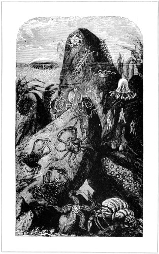
ZOOPHYTES, ASTEROIDS, NUDIBRANCHIS, ACALEPS, ECHINOIDS, CTENOPHORA, TUNICATA, AND CRUSTACEANS.
The Stinging Series, Cnidaria, comprise sea-anemones, corals, jelly-fish among marine animals, and Hydra among the fresh-water Cœlenterata; and derive their name from a remarkably curious feature, the so-called stinging capsules. These are not only offensive, but also defensive weapons with all the animals belonging to this group; the possession of which has converted the bell-like jelly-fish into a simple Cnidarian. The principal change is in the gelatinous layer between the outer wall and the inner digesting layer of the ectoderm. But without entering further into their structure and relations, the stinging-cells and batteries claim especial attention. These cells vary considerably in size without their characteristics being essentially changed. The protoplasm of the cell is modified into a tolerably firm substance, enclosing an oval or cylindrical vesicle. Closely associated with this is a pointed process, standing up far above the level of the outer covering, known as the cnidocil. Within the vesicle is found, either spirally rolled up or in an irregular tangle, a long filament or hollow tube, a prolongation of the vesicle, but turned outside in. This tube is more than twenty times as long as the cell, is pointed at the tip, and beset with two rows of fine spirally-arranged barbed hooks. When the cnidocil is touched or irritated, this filament is violently shot out, being then turned inside-out, like the fingers of a glove. So long as the thread remains rolled up within the vesicle the barbed hooks remain in their tube, but when shot out, they change to the outside. The rolled-up filament appears to be filled with some poisonous material, which is ejected when the tube is shot out, and where the point strikes a wound is inflicted, so that unless the prey is stronger than the attacker it cannot escape. The greater the struggle, the larger the number of capsules discharged in order to kill.
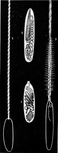
Fig. 347.—The Stinging Capsules of Cnidaria.
1 and 2. Retracted filaments; 3. Partly protruded; 4. Fully protruded. Magnified × 600. (Warne.)
Polypomedusæ.—Among the higher development of the stinging group is the jelly-fish. The Siphonophora, as represented by the Portuguese man-of-war, are in their turn the highest development of swimming-bells, and exhibit many modifications and combinations of individuals. The tentacles of the Physalia, the best known, are stiff with batteries of stinging-capsules, the sting of which is more like the shock of the electric current. The Challenger soundings brought to light some remarkably interesting forms, and these have furnished much work for the microscope, as all their larval forms are extremely curious. Among the Hydromedusæ there are many different life histories. Take the jelly-fish, the eggs of which have given up forming stocks, and are hatched out at once as Medusæ. There are others, the eggs of which form stocks; others, again, in which the sexual individuals do not swim away as jelly-fish. The last were at one time described under a new name, because of one or two curious forms being taken creeping on the ground. This creeping Medusa (Clavatella prolifera) has six arms, the tips of which are provided with true suckers, and on these it walks as on stilts, while from each arm a short stalk arises, the swollen end of which is beset with stinging capsules. It has an extensile mouth-tube, and feeds upon small crustaceans found on seaweeds.
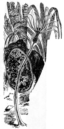
Fig. 348.—Plumularia primata. Doris tuberculata seen clinging to a fucus.
Among the forms that swim away as jelly-fish a very curious example is presented in Corymorpha mutans. These swim about for a time, and then firmly attach themselves by numerous thread-like appendages, forced into the sand, and where the young prepare for their next metamorphosis. As an example of the stocks of those representatives which do not swim away as jelly-fish, take the beautifully-feathered, plant-like creatures found erect along the seashore, the Sertularia (Fig. 358, No. 12) and Plumularia. Plumularia primata, Fig. 348. Other members of these groups will be found in Plate IV., Nos. 95-99.
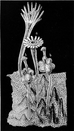
Fig. 349.—Group of female stock of Hydractinia echinata.
a, a. Nutritive individuals; b, b. Female individuals and groups of eggs. Highly magnified.—(Warne.)
In addition to the nutritive individuals, there are the egg-bearing; these do not become free-swimming individuals. One small family is neither branched nor feathered—the Hydractinia echinata, found in the North Sea and on the Norwegian coasts, where it attaches itself to the shells of gastropods, selecting those inhabited by hermit crabs. The part of the stock common to all the individuals is the skin-like portion which adheres to the surface of the shell. In some spiny processes are produced, and the nutritive canals running down the stems of the polyps are continued into the membrane belonging to the stock, as seen in Fig. 349.
The nutritive individuals are distinguished by long tentacles, mouths, and digestive canals. The females have no mouths, and are supplied with food through the system of canals running to them from the nutritive males. These reproductive members are furnished with stinging threads instead of tentacles for the protection of their ova. The ciliated larvæ, in a very short time, swim off to found new colonies.
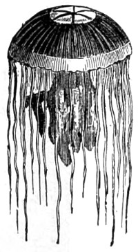
Fig. 350.—Medusæ, Jelly-fish.
The free-swimming jelly-fish (Fig. 350, and Plate XVII., c and d) belong to the order Scyphomedusæ. These are characterised by their delicate colouring, and from the arrangement of their nervous system, which can only be made out by staining. Some new and curious forms were dredged from a depth of more than 6,000 feet off the coast of New Zealand, varying in size from an inch to twenty inches; many having from four to eight or ten eyes arranged along the margin.
Anthozoa.—From the free-swimming we turn to a group of permanently fixed polyp forms, the sea-anemones and corals. The development of Monoxenia commences with the egg, repeatedly dividing into many parts (Fig. 351, C, D, and E), by a process common to the animal kingdom, termed egg-segmentation, in this particular instance proceeding from an apparently hollow sphere, A, enclosing a single layer of cells, G. Each cell sends out a long cilia, or whip-like process, F, by means of which the larva turns about and swims in the body fluid of the parent polyp. One half of the sphere now becomes enfolded into the other half, H, and forms what is termed a gastrula, I, K. The gastrula stage of Monoxenia is of the simplest kind, the larva forming a sac, with walls consisting of two layers, an outer, or ectoderm, and an inner, or endoderm. The transition from the flat dish shape, H, to the sac with a narrow mouth is at once clear, and the knowledge that all the Cœlenterates proceed from similar larvæ, and that all the complications of their various systems are developed from a simple gastrula, throws much light on their anatomy. During these transitions the endoderm, whose cells multiply, continues as an uninterrupted lining to the stomach and its appendages, while the ectoderm yields the cuticular elements.
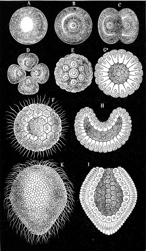
Fig. 351.—Stages in development of Monoxenia Darwinii, × 600.—(Warne.)
A third intermediate gelatinous layer, the mesoglæa, arises between the two layers in which muscles and connective interstitial tissue appear. In the mesoglæa of one species of coral calcification takes place; this internal calcification has but a small share in the work of the great rock-making corals, their most important calcification being external. In Monoxenia, although the transition from the gastrula larva to the adult animal has not been seen, there can be no doubt as to how this is carried out, the transformations having been watched throughout in other species. The larva attaches itself with the end opposite the mouth, the cilia disappear, and after the mouth-tube has been formed by the folding in of the anterior end along the longitudinal axis of the body, and has thus become marked off from the stomach, eight hollow tentacles rise round the mouth as outgrowths of the body cavity, or as direct continuations of the stomach.
Like all other corals, Monoxenia periodically multiply by means of eggs, which are formed either in the walls of the radiating partitions or septa, or along the free edges. These are ejected through the oral opening. As a rule, the polyps are either male or female; but in stock-forming species individuals of the two sexes are often mixed. Monoxenia may be taken as the simplest type of the regularly radiate polyps; in all the different organs being repeated in regular rings round a central axis; the mouth also is circular. From this interesting account, drawn by Haeckel, of a simple polyp, it will be at once seen what kind of radiate animal it is that builds up the coral reefs. “No garden on earth can match the gardens of the sea that circle the northern part of Australia. As the tide ebbs in azure sunset, coral-reefs peer out symmetrically arranged in beds and intersected by emerald pathways coursing through corals of all hues and tints fathoms deep in the channels.”
In a growing polyp-stock the individuals usually remain in organic connection; that is to say, each first provides for itself and then shares its superfluity with others, sometimes by means of a continuous reticulated system of canals perforating the calcareous substance which often separates the members of one stock from another. The whole colony may thus be physiologically one creature with many mouths. There are others that remain single, as the inverted pyramidal-looking bodies, Fungidæ, commonly called “Sea-mushrooms,” found in great variety. The colour of the polypidom is white, of a flattened round shape, made up of thin plates or scales, imbedded in a translucent jelly-like substance, and within is concealed a polyp; the footstalk, by means of which the animal is attached to the rock, is of a calcareous nature (Fig. 352, No. 1).
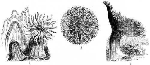
Fig. 352.—Sea-Anemones.
1. Actinia rubra, tentacles displayed and retracted; 2. Heticictis bellis; 3. H. bellis, seen from above.
Hexactinia (six-rayed polyps) are not limited to six rays, as the name given them may seem to imply; they are, in fact, very numerous in some of the largest and most gorgeous of the sea-anemones. All are distinguished by their solitary manner of life, their size, and their vivid and variedly beautiful colouring. The endoderm is firm, and when the animal withdraws its tentacles and shuts its body substance in, there is some difficulty in penetrating to the interior. It does not, however, secrete a calcareous skeleton inside or out, as do the true coral polyps. Among the Hexactinia the sea-anemone (Fig. 352) takes the first place.
These beautifully coloured creatures are, for the most part, found attached to the spot selected by the larvæ; a few species bore into the sand with the posterior part of the body, or build a sheath, which they inhabit. They are voracious feeders, and devour large pieces of flesh, and even mussel and oysters, sucking them in by means of their long grasping tentacles. Well-fed anemones change their skin frequently, during which process they remain closely retracted; the shed skin forms a loose girdle around the base. Actinia bellis not infrequently attach themselves to the shells of crabs and whelks, and are thus carried to pastures new.
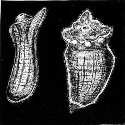
Fig. 353.—Larvæ of Sea-Anemones, Actinia effœta, highly magnified.
On account of the ease with which anemones are kept in captivity, their mode of reproduction can be closely observed. With but few exceptions they develop from eggs, and in the course of a few weeks are hatched into ciliated infusorial larvæ, presenting most curious and exquisite representations of jugs and jars, with cover lids (as seen in Fig. 353, Actinia effœta). These evince the handiwork of a master hand in the ceramic art. They are, however, of so translucent a nature as to permit of the internal structure being seen to consist of nerves and vessels, and which are rendered more apparent by staining. These settle down in a week or ten days, and then shed their cilia, the first tentacle appearing during the process of attachment.
In some species the young Actiniæ are seen to pass through their whole development within the body cavity of the parent. Most anemones are provided with several circles of more or less cylindrical tentacles, and there are a few specially beautiful species which, besides tentacles of the usual form, have, either within or without the ordinary circle of tentacles, lobed or leaf-like tactile and seizing organs. These belong to the family of the beautiful Crambactis of the Red Sea. Below these grasping tentacles comes a circle of thicker arms unlike the former, being spindle shaped. All the tentacles of the sea-anemones are hollow with a fine aperture at the tip, through which, on closing rapidly, it is seen to expel a jet of water.
True Corals.—It will have been noticed in the foregoing remarks that in the soft body-division of the Hexactinia there are both single individuals and colonies joined together to form stocks. The same diversity in this respect will be found among corals proper, with this difference, that the skeleton-forming polyps, by combining, build up substantial structures in the most secure and advantageous positions. Now it so happens that all the corals found about our coasts are generally small and solitary dwellers, one of the best known of which is the scarlet crisp coral, Flabellum, and is characterised by the slit-like form of the mouth. Viewed sideways it resembles a small fan fastened along the edges, and just inside a row of fully developed tentacles is seen protruding. An interesting form of budding occurs in these corals: the buds fall off, and in this budding condition the coral might pass, and indeed has been described as a different species of Flabellum. The colour of the coral is a beautifully transparent red. Remarkable as the solitary corals are, they are surpassed both in number and in form by those which form compound stocks, that is to say, in which the buds do not fall off, but go on building up coral islands and barrier reefs in the warmer seas. Some very few typical forms only are given in the group accompanying, shown in Fig. 358.
A different kind of stock is developed in a number of forms, some producing many buds, as in the Madrepores, in which selected polyps spring up above the rest, their sides also becoming covered with small buds, each one of which is a living, feeding, coral animal surrounded by a crown of tentacles. These Madrepores play a very important part in the building up of coral reefs.
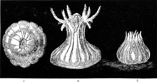
Fig. 354. Developmental stages of Larvæ, Astroides calycularis, × 40.
Another massive coral, the Astroides calycularis, has a different mode of growth, the tubes not being fused together. When seen standing out these yellowish-red polyps have been mistaken for small anemones. The larvæ of this coral leave the egg while still in the large chambered body cavity of the parent, where they swim about for a time, till they escape through the mouth. They are worm-like in form, and swim by means of cilia, which are thicker at the foremost end. The mouth first appears after leaving the parent, but as they soon become exhausted by the effort they assume a contracted form, and attach themselves, as do anemones, by pressing the thicker end of the body against a rock, the whole contracting into a thick round disc, while longitudinal furrows become visible at the upper part where the mouth sinks in. At the end of these furrows twelve tentacles appear. The accompanying illustration shows the various stages through which the larvæ pass in rapid succession (Fig. 354); at the same time it has already commenced to secrete its calcareous skeleton. This is not formed as a connected whole but from a number of separate centres of secretion formed between the polyp and the substance to which it has attached itself, and which become gradually fused into a perfect skeleton. A section of the polyp at this stage forms an interesting microscopical object.
The so-called eight-rayed corals consist of the one genus Tubipora, the members of which are few in number and not varied in form (Fig. 358, No. 10). In the structure, however, of skeletons they are unique among extant corals. Each individual secretes a smooth-walled tube without calcification of the vertical septa. These tubes, like the pipes of an organ, stand almost parallel, and are united to form a stock by means of transverse platforms. The formation of buds does not appear to take place in this family.
Another of the eight-rayed corals is Gorgoniidæ. These are permanently fixed to the spot on which they are found, and form a bush-like growth, giving no idea of the living coral, as it rises in graceful branching colonies, in deep water, and represents a portion of Gorgonia nobilis with polyps expanded (Figs. 344 and 358, No. 9).
Other corals present numerous other departures from the types we have been considering, but so far modified in form as that of the Sea-pen, Veretillum (Fig. 355), the stock part of which is surrounded by polyps continued down a portion of the cylindrical stalk. The best known of the species is Pennatula phosphorea of the Mediterranean.
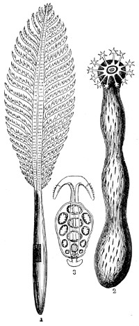
Fig. 355.
1. Pennatula phosphorea; 2. Synapta chirodata; 3. Anchor-shaped spiculum and plate from the ectoderm of same.
Pennatulidæ.—This family derives its name from penna, a quill. Their spicula also resemble a penholder in appearance, shown in Fig. 358, No. 3. The polyps are without colour, provided with eight rather long retractile tentacula, beautifully ciliated on the inner aspect with two series of short processes, and strengthened by these crystalline spicula, a row being carried up the stalk, together with a series of ciliated processes. The mouth, occupying the centre of the tentacula, is somewhat angular. The ova lie between the membranous part of the pinnæ; these are globular, of a yellowish colour, and by pressure can be made to pass through the mouth. Dr. Grant wrote:—“A more singular and beautiful spectacle could scarcely be conceived than that of a deep purple Pennatula phosphorea, with all its delicate transparent polyps expanded and emitting their usual brilliant phosphorescent light, sailing through the still and dark abyss, by the regular and synchronous pulsations of the minute fringed arms of the polyps.”
The spicula are seen to be a continuous series of cones fitting into each other.
Bryozoa, Moss-animals.
The exact position in which the Bryozoa, or moss-animals, should be placed in the animal kingdom has not been finally determined. They were at one time associated with corals; then with sponges; but, on further acquaintance, it became evident that they did not belong to either. Naturalists also claimed them as Rotifers and Ciliata, but this claim met with no better reception. Since they appear to have no settled classification, there can be no objection to linking them once more to corals, as they apparently resemble these animals by always living in colonies, the individual members of which are joined in a number of different ways to form stocks, the individuals themselves, however, being very much smaller than those of corals proper. The advantage is that the structure of the Bryozoans can be more readily studied, as many of them live in transparent chambers or cells, the walls of which, although somewhat firmly agglutinated together, are flexible enough to fold up, as the animals instantly withdraw their bodies and close up the top on the slightest alarm (Fig. 356).
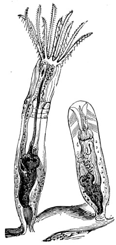
Fig. 356.—Paludicella, tentacles expanded and cell closed.
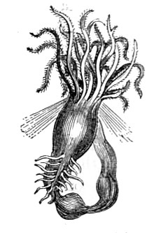
Fig. 357.—Sea-moss, Flustra, the body having been withdrawn from its cell.
The general structure of the Bryozoan individual, figured attached by its footstalk to a stem of wood, consists of a mouth at the anterior part of the body opening into a muscular pharynx in the alimentary canal, together occupying a considerable amount of space. The terminal portion turns upon itself towards the oral opening, its chief attachment being a short strand of tissue termed the funiculus (shown in Fig. 358, No. 11). In all adults two masses of cells are found attached to the wall of the chamber; the upper yields the eggs, within the lower the male elements are developed. Moss-animals are hermaphrodite, fertilisation being effected by the two elements mingling together in the body fluid. These are the essential points in the structure of the whole seventeen hundred species. Among the larger colonies a number of fresh-water genera are found attached to the roots and branches of aquatic plants, most of which, however, are inconspicuous. The beauty of these minute bodies can only be seen under the microscope. Many consist of delicate branching growths, the Sea-mats (Flustra), for instance; others again appear as attractive lace corals, between the open meshes of which multitudes of minute apertures crowned with tentacles are displayed. The several individuals of the genus Lepralia are arranged in rows, and further distinguished by the animals being developed only on one side of the stock. The marvellous variety of forms presented by these small animals is in a measure determined by the particular manner of their buddings. The greater number of fresh-water moss-animals belong to the order Phylactolæmata, so called because the mouth is provided with a tongue-shaped lid. The crown of tentacles is furnished with rows of cilia, and is horseshoe-shaped, the whole being surrounded at its base by an integument forming a kind of cup, which is either soft or horny. Those belonging to the wandering types (Cristatella, Plate IV., Nos. 95-98) form flattened elliptical colonies, some of which creep or move about on a kind of foot. A nervous system pervades the mass of polyps, while in each separate polyp a nerve ganglion is seen to be situated between the œsophagus and the posterior part of the alimentary canal. The colony nerve system regulates the movements of the stock.
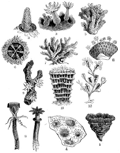
Fig. 358.—Typical forms of Corals.
1. Fungia agariciformis; 2. Alcyonium, Cydonium Mulleri; 3. Cydonium, polyps protruding and tentacles expanded, others closed; 4. A stock viewed from above; 5. Madrepore abrotanoide; 6. Madrepore, slightly magnified, showing oral opening; 7. Corallidæ; 8. Coral, polyps protruding from cells; 9. Gorgonia nobilis, with polyps expanded; 10. Tubipora musica; 11. Tubes of same, with polyps expanded, one cut longitudinally to show internal structure; 12. Sertularia, polyps protruded, and withdrawn into their polypidoms.
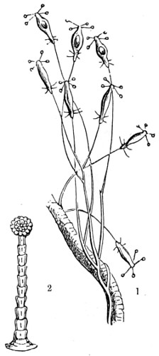
Fig. 359.
1. Coryne stauridia; 2. A tentacle detached and magnified 200 diameters.
There are many beautifully formed freshwater polyps deserving of more than a passing notice, as the slender Coryne (Coryne stauridia), found adhering to the footstalk of a Rhodymenia (Fig. 359), about which it creeps in the form of a white thread. On placing both under the microscope, the thread-like body of the little animal appears cylindrical and tubular, perfectly transparent, and permeated by a central core, apparently cellular in texture, hollow, and within which a rather slow circulation of globules is perceived. The parent Coryne sends off numerous branches, the terminal head of which is oblong, cylindrical, and at the extreme end there are arranged four tentacles, long and slender, each being furnished with a nodular head. A magnified view of one detached is shown erect (Fig. 359, No. 2). This polyp is much infested by parasites, vorticella growing on it in immense numbers, forming aggregated clusters here and there, individuals of the parasitic colony adhering to each other, and projecting outwards in every direction.
Alcyonella, another fresh-water polyp, is found in the autumn of the year in all the London Docks adhering to pieces of floating timber. A. stagnorum partakes of the character of a sponge rather than that of a polyp. It is usually found in gelatinous colonies, and when stood aside for a short time these put forth a number of ciliated tentacles (shown in Fig. 360, magnified 100 diameters).
The ova contained within the sac, and viewed by transmitted light, appear as opaque spheres surrounded by a thin transparent margin; these increase in thickness as the ova is developed, and such of the ova as lie in contact seem to unite and form a statoblast. A rapid current in the water around each animal, drawing with it loose particles and floating animalcules, is seen moving with some velocity as in other ciliated bodies; and a zone of very minute vibrating cilia surrounds the transparent margin of each tentacle.
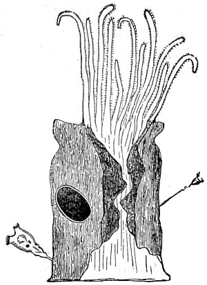
Fig. 360.—Alcyonella fluviatella.
Dr. Percival Wright discovered on the western coast of Ireland a new genus of Alcyonidæ, which he named after the well-known naturalist Harte, Hartea elegans (Plate IV., No. 86). This polyp is solitary, the body cylindrical, and fixed by its base to the rock; it has eight ciliated tentacles, which are knobbed at their base and most freely displayed. It is a very beautiful polyzoon of a clear white colour, and when fully expanded stands three-quarters of an inch high.
Lophopus crystallinus (Plate IV., No. 98) displays beautiful plumes of tentacles arranged in a double horseshoe-shaped series. When first observed these polyps resemble in many respects masses of the water snail ova, for which they are often mistaken. On placing these jelly-like masses into a glass trough with some of the clear water taken from the stream in which they are found, delicate tubes are seen to cautiously protrude, and the beautiful fringes of cilia are quickly brought into play. The organisation of L. crystallinus is simple, although it is provided with organs of digestion, circulation, respiration, and generation. The nervous70 and muscular systems are well developed. This polyp increases both by budding and by ova, both of which conditions are shown in Plate IV., No. 98. The ova are enclosed in the transparent case of the parent. In Lophopus and some other fresh-water genera, Cristatella, Plumatella, and Alcyonella, the neural margin of the Lophopore is extended into two triangular arms, giving it the appearance of a deep crescent.
Another family presents a contrast: there is no lid to the mouth, and the tentacles are arranged in a circle on a disc. An important rise in organisation is found in the Gymnolæmata, especially in the lip-mouthed forms; the individuals belonging to this order vary in structure and fulfil different physiological functions. There are structures known as zoæcia, stolons, avicularia, vibracula, and ovicells, some of which are merely modified individuals. The zoæcia are the normal individuals of the colony, fully developed for most of the functions of life; the stolons have a much humbler function, but are indispensable—they are the root-like outgrowths of the stock, and serve for attaching the colony to foreign objects. The most remarkable are those known as avicularia, so called because they resemble the head of a bird. This process acts as a pair of forceps, the large upper blade of which is very like the skull and upper jaw of a bird, and the smaller lower blade (like the lower jaw) constantly opens and shuts by means of a complicated arrangement of muscles (shown in Fig. 361). These avicularia are movably attached by short muscles to the neck, and are found near the entrance to a zoæcium. They turn from side to side, snapping in all directions, catching at every particle of food that may come near; at length the morsel is drawn into the mouth by the cilia on the tentacles. From this very peculiar structure the Chilostomata were originally named bird’s-head corallines, then specifically shepherd’s-purse corallines, Notamia bursaria. Equally interesting, again, are the vibracula, long thread-like structures, attached by short footstalks. These keep up a constant whip-like motion, the object of which is not quite clear. The ovicells, or egg receptacles, are found at the lower ends of the zoæcia in the form of shields, helmets, or vesicles. In Plate IV., Nos. 95 and 96, a front and edge view of the statoblast is shown highly magnified.
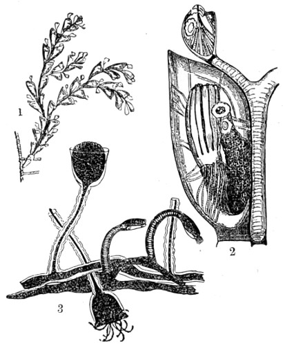
Fig. 361.
1. Notamia bursaria, shepherd’s-purse Bryozoa; 2. Polyp magnified and withdrawn into its cell; 3. Portion of a colony of Hydroid polyps.
Another sub-order consists of the Cyclostomata, or round-mouthed Bryozoans, of which the Tubulipora is the typical form. The stocks are cup-shaped incrustations, the individuals radiating outwards, as in Plate IV., No. 92. Tubularia dumortierii is a very interesting form, the germinal bodies, statoblasts, being formed as cell masses on the strand, or funiculus, which also maintains the stomach in its place. They are round or oval in shape, and brown or yellow in colour, and consist of two valves fitted one upon the other like watch glasses, as shown in No. 96. A number of other statoblasts are shown, Nos. 97, 98, and 99. The edge running round No. 95 is seen to have barbed tips; the ring itself contains small air chambers, and is termed the swimming belt. It is, in fact, a perfect hydrostatic apparatus, giving support to the winter buds or statoblasts on the surface of the water. The barbed hooks apparently act as anchors, and by their means they catch on at points suitable for their development during the coming spring. As soon as the time comes, the two halves split apart and the germinal mass emerges forth. Out of these winter buds and statoblasts asexually produced individuals arise, which reproduce themselves sexually, their descendants again yielding winter germs. In short, an alternation of generations is a continually recurring process.
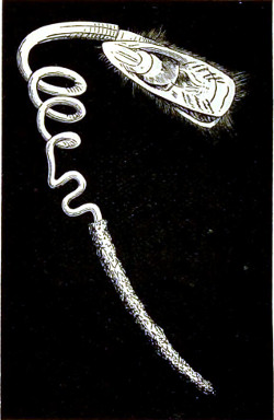
Fig. 362.—Lingula pyramidata.
Brachiopoda.—Here again we have to do with an enigmatical class of arm-footed animals, of which the Lamp-shells may be regarded as typical. These have remained unaltered from the earliest geological epochs. Brachiopods are divided into two orders: those having shells without hinges, and those with shells hinged together. On the whole they possess less interest for the microscopists than many other animals, except in their earliest developmental stages of existence.
One of the most interesting of the hinge-class group, living chiefly near the shores of the warmer seas, is the Lingulidæ. The valves are almost exactly similar, but are not hinged together, and have no processes for the support of the thick fleshy spiral arms of the animals. In L. pyramidata, found around the Philippine Islands (Fig. 362), the stalk is nine times longer than the body. The animal does not attach itself by this, but moves about like a worm, making tubes out of sand, into which it can withdraw itself and disappear. The cilia at the mantle edge form a fine sieve, thus preventing foreign particles from entering the gills. Its internal structure possesses points of interest, and the parasitic growths covering the cartilaginous structure, miscalled a shell, are curious, and excite the attention of the naturalist.
Another bivalve so unlike a crustacean, among which it has been placed, I may venture to describe among Lamp-shells. I refer to the barnacle (Lepas) generally met with covering the bottoms of ships. These, as in the former genus, are more interesting to the microscopist in the early stage of existence, and also for the curious parasites known to infest them. The barnacle protrudes through its two valves six pairs of slender, bristly, two-branched filamentous limbs, which keep up a constant sweeping motion, and whereby it secures its supply of food (Fig. 363). When first hatched the young are in the Nauplius stage, being furnished with a median eye and three pairs of flagellated appendages. After enjoying a free life the larva moults and passes into a second stage, in which with its two eyes and compressed carapace (shown in Fig. 364) it so nearly resembles a Daphnia. Before these thoracic appendages entirely disappear they first change places, and then each is seen to be provided with a sucker; by this means the larva fixes itself to its permanent resting-place, while a cement gland pours out a secretion that glues it firmly to the point of attachment chosen. These Cirripedes are not true parasites, inasmuch as they do not extract nourishment from the body to which they are attached.
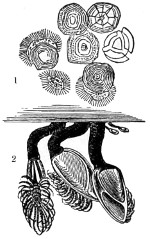
Fig. 363.
1. Spat of oyster, some ciliated; 2. Barnacles attached by footstalks.
One species, the Proteolepas, is in the adult stage a maggot-like, limbless, shell-less animal found living within the mantle chamber of other members of the same order, while the root-headed Cirripedes (Peltogaster curvatus, as Fig. 364, No. 1) live parasitically upon higher crustaceans.
Echinodermata.—This sub-kingdom includes the star-fishes, stone-lilies, sea-urchins, feather-stars, and sea-cucumbers, some of which have been already alluded to, and are so well known that they need no lengthy description, while of the fossil sea-urchins of our chalk formations, the Pentremites and Crinoids, whose silicious remains are so abundant and so familiar to naturalists and geologists, but little remains to be said. They are chiefly interesting to the microscopist from their calcareous and silicious appendages, known as spicula. In the sea-urchin, brittle-star, or feather-star, the outer body surface consists almost wholly of a deposit of calcium carbonate, combined in the form of little plates built up into a rigid “test,” whereas in the star-fish it usually forms a kind of scaffolding, between the layers of which there stretches a firm leathery skin. Among the sea-cucumbers, the living specimens of which present extraordinary variations both in form and character, the deposit consists chiefly of small spicules which grate when the skin is cut with a knife. If a thin section of the skin is examined under the microscope, the spicules are seen to be profusely distributed in the middle layer. The same deposit takes place in the stalked column of a crinoid and in sea-urchins (Echinodermata), which has tended to preserve them in the fossilised state. Fig. 365 is selected as exhibiting to perfection the Medusa-headed Pentacrinoid. This echinoderm differs in two characters: first, its microscopic structure is that of a meshwork deposited in the spaces of a network of soft tissue; secondly, that each element, whether a spicule or a plate, is, despite its trellised structure, deposited around regular lines of crystallisation (shown in Plate IV., Nos. 89 and 90). Owing to these characteristics the minutest portion of an echinoderm skeleton is readily recognised, even when fossilised, under the microscope. Even the species of the sea-cucumber can be determined by the shape of their spicules.
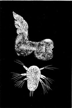
Fig. 364.—Parasitic Barnacles.
1. Peltogaster curvatus; 2. Nauplius larva of Parthenopea; × 200.
Another noticeable feature in the radiate structure is that in many cases it gives to the animal a star-shape, to which the names of star-fish and brittle-star are given (see Plate IV., No. 91, and Plate XVII., f and n). The ordinary five-rayed star-fish is found everywhere around the English coasts. This constant arrangement of organs holds good in the majority of the echinoderms; it can be detected in the Holothurians, where, beside the feathery tentacles of the head, rows of shorter sucker-like processes will be found, which in some instances extend the whole length of the body, the fixed number of rows being also five in their internal organs. Hence these animals were formerly grouped under Radiata. But if a sea-cucumber or sea-urchin be dissected, a marked distinction will be found between them, in one portion of the organism in particular: the intestine is shut off from the rest of the body-cavity, often coiling round inside. Examine a star-fish or sea-urchin on the under-surface of the rays, and, passing in five bands from top to bottom, a number of small cylindrical processes are seen gently waving about; these lie in two rows with a clear space between them, and are termed in consequence ambulacrum. They end in sucker-like discs, which enable the animal to attach itself, or pull itself against strong currents.
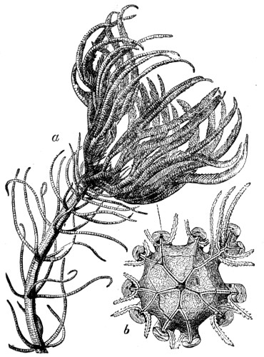
Fig. 365.—Medusa-headed Pentacrinoid.
a. Crown and part of stem; b. Upper surface of body, the arms broken away, showing the food grooves passing to the central mouth.—(Warne.)
Just one other special feature should be noticed: radial canals pass along under the ambulacra, and join a ring-canal around the mouth, well supplied by nerve cells.
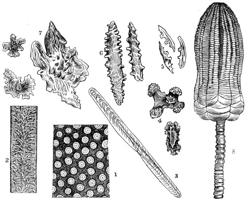
Fig. 366.
1. Transverse section of a branch of Myriapore; 2, and the others Section of the stem of Virgularia mirabilis; 3, Spiculum from the outer surface of Sea-pen; 4, Spicula from Isis hippuris; 5, from Gorgonia elongata; 6, from Alcyonium; 7, and from Gorgonia umbraculum; 8, Calcareous remains of a Crinoid.
Crinoids (stone-lilies), on the other hand, are formed of a series of flat rings, pierced through by a narrow canal. The ossicles, as they are termed, are joined by ligaments passing through their solid substance and endowed with muscular power; the central part serves for the passage of blood-vessels, and is surrounded by a sheath of nervous tissue that controls the movements of the stem, the latter being encrusted by a number of fine rootlets. The stems possess a limited power of bending. In the words of Professor Agassiz, “The stem itself passes slowly from a rigid vertical attitude to a curved or even a drooping position; the cirri move more rapidly than the arms, and the animal uses them as hooks to catch hold of objects, and on account of their sharp extremities they are well adapted to retain their hold of prey.” The rosy-feather star-fish is often found clinging to a tube of the Sabella worm; the food of crinoids consists of foraminifera, diatoms, and the larvæ of crustaceans. There are so many curious features in connection with the Echinodermata that my readers may with advantage consult “The Challenger Reports” and Warne’s “Natural History” on other points of interest.
Holothuroidea (sea-cucumbers) are elongated slug-like creatures, the skin being in structure similar to that of the slug, with a comparatively small amount of calcareous matter. Usually this occurs in small spicules, which assume very definite shapes, as the anchors of Synapta (Plate IV., No. 87, and in Fig. 355). There are also rings of calcareous plates around the gullet, five of which have the same relation to the radial water-vessels as the auricles round the jaws of a sea-urchin, and which likewise serve for the attachment of muscles. These plates are seen in Plate VIII., Nos. 171 and 172, as they appear coloured by selenite films under polarised light. Around the mouth in Cucumaria is a fringe of branched tentacles connected with the water-vascular ring; these appear to be used as a net to intercept floating organisms.
Correlated with the star-fishes is a small family based on the character of their pincer-like organs, called pedicellariæ, on the surface of the test (shown in Plate IV., Nos. 93 and 94, magnified × 25). Movable spines cover the surface of these echinoderms, varying in size from minute bristle-like structures to long rods. The pedicellariæ are, it is believed, derived from the smaller spines, and two of them are united at the base by muscles, slightly curved, and made to approach each other at their extremities. There is a gradual modification of this type through the whole series. Many uses have been assigned to them, as the holding of food, as they have been seen to hold to the fronds of seaweed and keep them steady until the spines and tube feet can be brought into action. The inner surface of the pedicellariæ are known to be the most sensitive, and the blades close on the minutest object touching the inner surface. Beside these peculiar bodies the surface of the skin has small tubular processes, and tubular feet with suckers at the end. At the extremity of each arm is a single tube-foot with an impaired tentacle, and above this again is a small eye coloured by red pigment.
Passing by many other points of interest in the Echinoidæ, the spines are seen to be attached to the test or shell by a ball and socket joint and well-arranged muscles, whereby the spines can be moved in any direction. The tubercles, however, do not cover the whole test, but are disposed chiefly in five broad zones extending from one pole to another. When a transverse section of a spine is examined by a medium power it is seen to be made up of a series of concentric and radiating layers (shown in Plate XVIII., Nos. 1 and 2), the centre being occupied by reticulated structure and structureless spots arranged at equal distances; these may be termed ribs or pillars. Passing towards the margin are other rows conveying the impression of a beautiful indented reticulated tissue. Many of the spines present no structure, while others exhibit a series of concentric rings of successive growth, which strongly remind one of the medullary rays of plants. When a vertical section of a spine is submitted to examination, it is seen to be composed of cones placed one above the other, the outer margin of each cone being formed by the series of pillars. In certain species of Echinus the number of cones is very considerable, while in others there are seldom more than one or two to be found; from these, transverse sections may when made show no concentric rings, only the external row of pillars.
The skeleton of echinoderms contains but a small amount of organic matter, as will be seen on dissolving out the calcareous portion in dilute nitric or hydrochloric acids. The residuum structure will appear to be meshes or areolæ, bounded by a substance having a fibrous appearance, intermingled with granulous matter; in fact, it bears a close resemblance to the areolar tissue of higher animals, and the test may be considered as formed, not by the consolidation of the cells of the ectoderm, as in the mollusc, but by the calcification of the fibro-areolar tissue of the endoderm. This calcification of a simple fibrous tissue by the deposit of a mineral substance, not in the meshes of areolæ but in intimate union with the organic basis, is a condition of much interest to the physiologist; it presents an example of a process which seems to have an important share in the formation and growth of bone, namely, in the progressive calcification of the fibrous tissue of the periosteum membrane covering of the bone.
The development of the sea-urchin from the fertilised egg first divides and then sub-divides, and in a short time the embryo issues forth with a small tuft of cilia, by means of which it swims off freely. The larvæ, in its full development, measures about one millimetre in diameter, and is a curious and remarkable creature.
The sub-kingdom Mollusca comprises some fifty thousand species, and fresh forms are being constantly discovered, the number of the aquatic genera being more than double that of the terrestrial species, for it matters not to what depth of ocean the dredge is let down, some new form is certain to be gathered. The Challenger expedition has enriched our knowledge of the deep-sea fauna to an enormous extent; so much so, that fifty volumes have already been published descriptive of animals brought to the surface. Nevertheless, we are told that the great coast lines of South America, Africa, Asia, and parts of Australia have been but imperfectly explored for smaller kinds of Mollusca.
Molluscs are soft-bodied, cold-blooded animals, without any internal skeleton, but this is compensated for by the external hardened shell, which at once serves the purpose of bones, and is a means of defence. These bodies are not divided into segments like those of worms and insects, but are enveloped in a muscular covering or skin, termed the mantle, the special function of which in most species is the formation and secretion of the shell. The foot, which serves the double purpose of locomotion and burrowing in the sand or rock, is an organ particularly characteristic of most molluscs. There are many departures from this rule, as, for instance, in the group Chitonidæ, where the shell takes the form of a series of eight adjacent plates; and in another, the Pholadidæ, there are one or more accessory pieces in addition to the two principal valves. Some are bivalved, others univalved, and concealed beneath the skin. All shells are mainly composed of carbonate of lime, with a small admixture of animal matter. Their microscopic examination reveals a great diversity of structure, as we shall presently see, and they are accordingly termed porcellaneous, nacreous, glassy, horny, and fibrous. Most molluscs have the power of repairing injuries to their shells; many exhibit an outer coat of animal matter, termed the peristracum, the special function of which is to preserve the shell from atmospheric and chemical action of the carbonic acid in the water in which they dwell.
The shells of gastropods are enlarged with the growth of the mollusc by the addition of fresh layers to the margin. In some species the periodic formation of spines occurs; a typical case will be found among Muricidæ. The varied colours of shells are due to glands situated on the margin of the mantle, and beneath the peristracum; occasionally the inner layer of porcellaneous shells is of a different colour to the outer, as, for example, in the helmet-shells (Cassis), much used by carvers of shell cameos. Light and warmth, as in the vegetable kingdom, are the great factors in the production of brilliant colours. In cold climates land snails bury themselves in winter time in the ground or beneath decaying vegetable matter, and in hot seasons they close up the aperture of the shells with a temporary lid, called an epiphragm. These exhibit great tenacity of life, as, for instance, in the Egyptian desert-snail, Helix desertorum. The reproductive system is in all cases effected by means of eggs. The ova are usually enclosed in capsules, and deposited in masses, and the number of eggs contained in the squid and the whelk have been stated to be thirty or forty thousand. The ova of molluscs may be gradually developed into the adult, or there may be a free-swimming ciliated larval stage, or a special larval form, as in the fresh-water mussel. Most are provided with a more or less distinct head; both cephalopods and gastropods are furnished with eyes. In land snails these are found placed on projecting stalks. In most cases the utility of molluscs far outweighs the injury occasioned by a few species, as, for instance, the Teredo, and the burrowing habits of the Pholas and Saxicava, compact marble having been found bored through by them.
Mr. J. Robertson wrote me in 1866:—“Having, while residing here (Brighton), opportunities of studying the Pholas dactylus, I have endeavoured during the last six months to discover how this mollusc makes its hole or crypt in the chalk—by a chemical solvent? by absorption? by ciliary currents? or by rotatory motions? My observations, dissections, and experiments set at rest controversy on this point. Between twenty and thirty of these creatures have been at work in lumps of chalk in sea water in a finger glass and a pan, at my window for the last three months. The Pholas dactylus makes its hole by grating the chalk with its rasp-like valves, licking it up when pulverised with its foot, forcing it up through its principal or branchial siphon, and squirting it out in oblong nodules. The crypt protects the Pholas from Conferveæ, often found growing parasitically not only outside the shell but even within the lips of the valves, thus preventing the action of the siphons. In the foot there is a spring, or style, which when removed is found to possess great elasticity, and this seems to be the mainspring of the motion of the Pholas.”
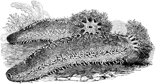
Fig. 367.—Hexabranchus.
I must pass by many groups and orders to more aberrant types, represented by the naked-gilled orders, Opisthobranchiata and Nudibranchiata. These gastropods constitute a large sub-order of extremely beautiful molluscs, remarkable in shape, and often brilliant in colour. The distinguishing character of these typical forms consists in the peculiar nature and situation of their breathing organs, which are exposed on the back of the animal or around the anterior part, and are not protected by the mantle. But the situation is varied, and the gills are sometimes placed on each side of the body, respiration being effected by the ciliated surface of the whole. For these and other reasons they have been placed in four groups. Nudibranchs are found in all parts of the world, and are most abundant in depths where the choicest seaweeds and corallines abound. Their fecundity is very great, as many as sixty thousand eggs being deposited by a single female at one time. They are eaten as a luxury where they most abound.
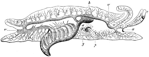
Fig. 368.—Longitudinal section of Pleurobranchus aurantiacus, showing circulation and gills or branchiæ.—(Warne.)
In the Opisthobranchs the branched veins as well as the auricle are placed behind the ventricle of the heart. They differ from Nudibranchs inasmuch as they are usually furnished with a pair of tentacles and labial palpi, or an expansion of the skin like the veil of the larval form. To clearly understand the character of the internal organisation of these curious animals, the longitudinal section given in Fig. 368 must be consulted: p is the foot; a the mouth, covered above with the veil-like expansion, over which are the tentacles, c; the branchial veins, v, carry the blood to the gills, from which it flows into the heart at h. This disposition is the opposite of that which characterises the Prosobranchus. Another anatomical peculiarity, which may here be referred to, is the direct communication of the system of blood vessels with the surrounding medium; a characteristic common to most other molluscs, and on which depends the changeable external appearance of the animal. In the illustration of Pleurobranchus here given, g indicates the opening of the duct which conveys water direct to the blood, and through which the blood vessels permeate the back and foot. Like the holes in the sponges, it can be filled or emptied at the will of the animal.
Although this, in the main, is the principle of the circulation in most of this order, one branch possesses no special breathing organs, respiration being carried on throughout the naked skin of the body.
With regard to the Nudibranchiata, the group having the most symmetrical form is the extensive family Dorididæ, characterised by differences in the branchiæ, the relative proportion of the mantle to the foot, and variations in the radula and jaws. The general aspect of the genus Doris, although drawn on a small scale, is represented in Plate XVII., Fig. b. The whole sub-order of Nudibranchs has become more generally known and admired since the publication of Alder and Hancock’s monograph with its many attractive coloured illustrations.
These gastropods can be kept alive for some time in a small aquarium if the precaution is observed of often changing the water and adding a little fresh seaweed. Numerous curious microscopic forms of life may be found adhering to them.
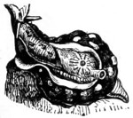
Fig. 369.—Aplysia dipilans.
Tunicata.—The most remarkable group of animals belonging to this sub-order are the Ascidians. They derive their name from the test or tunic, a membranous consistence, in which they dwell, and which often includes calcareous spicules. The test has two orifices, within which is the mantle. Few microscopic spectacles are more interesting than the circulation along this network of muslin-like fabric, and that of the ciliary movement by which the fluid is kept moving. In the transparent species, as Clavelina and Perophora, the ciliary movement is seen to greater advantage. The animals are found adhering to the broad fronds of fuci near low water-mark. They thrive in tanks, and multiply both by fission and budding. Two species are figured in Plate XVII., Figs. i and k, the zooids of which were found arranged in clusters, as represented.
Aplysiidæ (sea-hares), so called on account of a slight resemblance to a crouching hare. The body form is elongated with a partially developed neck and head, oral and dorsal tentacles, and furnished beneath the mantle with a shelly plate to protect the branchiæ. The mouth is provided with horny jaws, and the gizzard is armed with spines, to prepare the food for digestion. The side lobes are thin and large, and are either folded over the back or used in swimming. Fig. 369 is a reduced drawing of A. dipilans.
The Pectinibranchs are known as violet sea-snails, Ianthinidæ and Scalariidæ. The radula consists of numerous rows of pointed teeth arranged in cross series, forming an angle in the middle. There is no central or rachidian tooth, and they have thin trochiform shells adapted for a pelagic life. They are mostly of a violet colour, from which they derive their name, the colour being more vivid on the underside, which is turned up towards the light when the animal is swimming near the surface of the sea (Fig. 370).
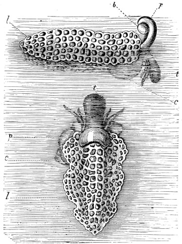
Fig. 370.—Ianthinia, Violet Sea-snail.—(Warne.)
The bubble b, drawn somewhat too large, is about to be joined to the anterior end of the float; c. Shell; l. Float; p. Foot; t. Head.
The most interesting feature in connection with these oceanic snails is the curious float which they construct to support their egg-capsules. It is a gelatinous raft, in fact, enclosing air-bubbles, which is attached to the foot, the egg capsules being suspended from its under-surface. They are unable to sink so long as they are in connection with their floats, and are therefore often cast on shore during storms, and furnish an endless series of microscopic specimens. The violet snails feed on various kinds of jelly-fish, and occur in shoals.
Pond Snails.—The three families, Limnœidæ, Physidæ, and Chilinidæ, form a special group of the pulminate, sessile-eyed fresh-water snails. The larger family of these belongs to the genus Limnœa, having a compressed and triangular head with two tentacles and eyes placed at their inner base. They are prolific and gregarious, and their ova are enclosed in transparent gelatinous capsules, deposited in continuous series, and firmly glued to submerged stems and leaves of aquatic plants. L. stagnalis is common in all ponds, marshes and slow-running rivers of Great Britain.
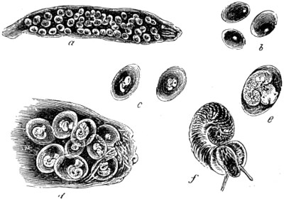
Fig. 371.—Ova and young of Limnæus stagnalis.
One of the species, L. trancatula, is the host of the liver-fluke so fatal to sheep. The fluke parasite passes one stage of its existence in the intestine of the pond snail.
Each ova-sac of Limnœa contains from fifty to sixty ova (represented in Fig. 371, at a). If examined with a low power soon after the eggs are deposited, they appear to consist simply of a pellucid protoplasmic substance. In about twenty-four hours a very minute yellowish spot, the nucleus, is discovered near the cell-wall. In another twenty-four hours the nucleus referred to is seen to have assumed a somewhat deeper colour and to contain within it a minute spot—a nucleolus.
On the fourth day the nucleus has changed its position, and is enlarged to double the size; a slightly magnified view is seen at b. On a closer examination a tranverse fissure is seen; this on the eighth day divides the small mass as at c, and the outer wall is thickened. The embryo becomes detached from the side of the cell, and moves with a rotatory motion around the interior; the direction of this motion is from the right to the left, and is always increased when sunlight falls upon it. The increase is gradual up to the eighteenth day, when the changes are more distinctly visible, and the ova crowd down to the mouth of the ova-sac, as at d. By employing a higher magnifying power a minute black spec, the future eye (e) and tentacles of the snail, is quite visible. Upon closely observing it, a fringe of cilia is noticed in motion near the edge of the shell. It is now apparent that the rotatory motion first observed must have been in a great measure due to this; and the current kept up in the fluid contents of the cell by the ciliary fringes. For days after the young animal has escaped from the egg, this ciliary motion is carried on, not alone by the fringe surrounding the mouth, but by cilia entirely surrounding the tentacles themselves, which whips up a supply of nourishment, and at the same time aeration of the blood is effected. From the twenty-sixth to the twenty-eighth day it appears actively engaged near the side of the egg, using force to break through the cell-wall, which at length it succeeds in accomplishing; leaving its shell in the ova-sac, and immediately attaching itself to the side of the glass its ciliary action recommences, and it appears to have advanced a stage, as at f. It is still some months before the embryo grows to the perfect form, Fig. 372; the animal is here shown with its sucker-like foot adhering closely to the glass of the aquarium. A single snail will deposit from two to three of these ova-sacs a week, producing, in the course of six weeks or two months, from 900 to 1,000 young.
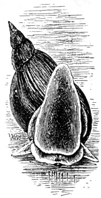
Fig. 372.—Limnæus stagnalis (natural size).
The shell itself is deposited in minute cells, which take up a circular position around the axis; on its under-surface a hyaline membrane is secreted. The integument expands, and at various points an internal colouring-matter or pigment is deposited. The increase of the animal goes on until the expanded foot is formed, the outer edge of which is rounded off and turned over by condensed tissue in the form of a twisted wire; this encloses a network of small vessels filled with a fluid in constant and rapid motion. The course of the blood or fluid, as it passes from the heart, may be traced through the larger branches to the respiratory organs, consisting of branchial-fringes placed near the mouth; the blood may also be seen returning through other vessels. The heart, a strong muscular apparatus, is pear-shaped, and enclosed within a pericardium or extremely thin and pellucid enveloping membrane. The heart is seen to be furnished with muscular bands of considerable strength, the action of which appears like the alternate to-and-fro motion occasioned by drawing out a band of indiarubber, and which, although so minute, are clearly analogous to the muscular fibres of the mammal heart; it beats or contracts at the rate of about sixty times a minute, and is placed rather far back in the body, towards the axis of the shell. The nervous system is made up of ganglia, or nervous centres, and distributed throughout the various portions of the body.
The singular arrangement of the eye cannot be omitted; it appears at an early stage of life to be within the tentacle, and consequently capable of being retracted into it. In the adult animal the eye is situated at the base of the tentacle; and although it can be protruded at pleasure for a short distance, it seems to depend much upon the tentacle for protection as a coverlid—it invariably draws down the tentacle over the eye when that organ needs protection. The eye itself is pyriform, somewhat resembling the round figure of the human eye-ball, with its optic-nerve attached. In colour it is very dark, having a central pupillary-opening for the admission of light. The tentacle, which is cylindrical in the young animal, becomes flat and triangular in shape in the adult. The tentacles serve in some respect to distinguish species. In Limnœa they are, as I have said, compressed and triangular, with the eyes at their inner base. In Physa they are cylindrical and slender and without lateral mantle lobes. The development of the lingual membrane is delayed; consequently, the young animal does not early take to a vegetable sustenance: in place of teeth it has two rows of cilia, as before stated, which drop off when the teeth are fully formed. The lingual band bearing the teeth, or the “tongue,” as it is termed, consists of several rows of cutting spines, pointed with silica.
It is a fact of some interest, physiologically, to know that if the young animal is kept in fresh water alone, without vegetable matter of any kind, it retains its cilia, and arrest of development follows, and it more slowly acquires gastric teeth, and attains to perfection in form or size. If, at the same time, it is confined within a narrow cell or space, it grows only to such a size as will enable it to move about freely; thus it is made to adapt itself to the necessities of a restricted state of existence. Some young animals in a narrow glass-cell, at the end of six months, were alive and well; the cilia were seen to be retained around the tentacles in constant activity, whilst other animals of the same brood and age, placed in a situation favourable to growth, attained their full size, and produced young, which grew in three weeks to the size of their elder relations.71
My experimental investigations were further extended to the development of the lingual membrane, or teeth, of Gastropoda, as well as the jaw and radula. In Limnœa, the teeth when fully developed resemble those of Helix; that is to say, in the fully grown animal are found several rows or bands of similar teeth, with simple obtuse cusps and a much suppressed central tooth. In the young snail a high power of the microscope is required to make them out. The dental band, however, in most Mollusca is disposed in longitudinal series, but varies a good deal in this respect, as will be seen on reference to my several papers, with illustrations of upwards of a hundred different species, published in “Linnæan Transactions” of 1866, and in the “Microscopical Society’s Transactions” of 1868. By way of example I may say, in the Pulmonata the lingual band usually consists of a single median row, the laterals on each side being broad and similar. But in many other groups the teeth are arranged in three, five, or seven dissimilar series. Taking Nerita as a type, the broad teeth on each side of the median are termed laterals; and the numerous small teeth on the outside of the band, known as the pleuræ, are termed uncini.
Since the investigations of Lovén into the lingual dentition of the Mollusca, various observers have studied the subject, with great advantage to our knowledge of the affinities of these animals. That these investigations have proved of value is shown by the light which has been shed on the true position of many species. When once we have ascertained the homology of a genus, whose relations were otherwise somewhat doubtful, it is surprising how other characteristics, even of the shell, probably misunderstood before, concur to bear out the affinities indicated by the lingual band. These tooth-bearing membranes, armed with sharp cutting points, admirably adapted for the division of the food on which they feed, are most of them beautiful objects for the microscope.
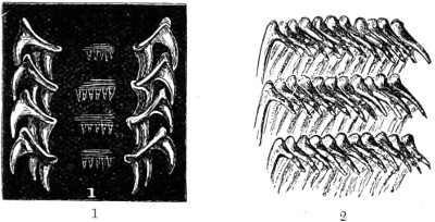
Fig. 373.
1. Palate of Buccinum undatum, common Whelk, seen under polarised light; 2. Palate of Doris tuberculata, Sea-slug.
The two ends of each longitudinal row of teeth are connected with muscles attached to the upper and lower surfaces of cartilaginous cushions; the alternate contractions and extensions of the muscles cause the bands of teeth to work backwards and forwards, after the fashion of a chain-saw, or rather of a rasp, upon any substance to which it is applied, and the resulting wear and tear of the anterior teeth are made good by a development of new teeth in the secreting sac in which the hinder end of the band is lodged. Besides the chain-saw-like motion of the band the lingual membrane has a kind of licking or scraping action as a whole. With the constant growth of the band new teeth are developed, when the teeth on the extreme portion of the band differ much in size and form from those in the median line.
As I have shown in the papers already referred to, that as each row is a repetition of the first, the arrangement of teeth admits of easy representation by a numerical formula, in which, when the uncini are very numerous, they are indicated by the sign ∞ (infinity), and the others by the proper figure. Thus, ∞ · 5 · 1 · 5 · ∞, which, in the genus Trochus, signifies that each row consists of one median, flanked on both sides by five lateral teeth, and these again by a large number of uncini. When only three areas are found, the outer ones must be considered the pleuræ, inasmuch as there is frequently a manifest division in the membrane between them and the lateral areas.
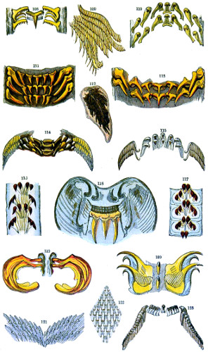
Most of the Cephalopod molluscs are provided with well-developed teeth, and they are, as we know, carnivorous. The teeth of the cuttle-fish, Sepia officinalis (Plate V., No. 111), resemble those of the Pteropoda, and have the same formula, 3 · 1 · 3. Sepia are also furnished with a retractile proboscis, and a prehensile spiny collar, apparently for the purpose of seizing and holding prey while the teeth are tearing it to pieces. In the squid Loligo (Plate V., No. 113) the median teeth are broad at the base, approach the tricuspid form with a prolonged acute central cusp, while the uncini are much prolonged and slightly curved. The lingual band increases in breadth towards the base, sometimes to twice that of the anterior portion. This band, mounted dry, forms an attractive object for black-ground illumination.
In another family, that of the rock-limpet, Patella radiata, the lingual band (Plate V., No. 116) well serves to distinguish it from the better-known common limpet. It is furnished with a remarkable long ribbon, studded by numerous rows of strong dark-brown tricuspid teeth. The lingual membrane when not in use lies folded up in the abdominal cavity. The teeth of Acmæa are somewhat differently arranged (Plate V., No. 117); their formula is 3 · 1 · 3.
Testacella maugei, belonging to Pulmonifera, is slug-like in appearance, and subterranean in its habits, chiefly feeding on earth-worms. During winter and in dry weather it forms a kind of cocoon, and thus completely encloses itself in an opaque white mantle; in this way it protects itself from frost and cold. Its lingual membrane is large, and covered with about fifty rows of divergent teeth, gradually diminishing in size towards the median row; each tooth is barbed and pointed, broader towards the base, and with an articulating nipple set in the basement membrane. A few rows are represented slightly magnified (Plate V., No. 121). Their formula is 0 0 · 1 · 0 0.
Tongues, etc., of Gasteropods.
Tuffen West, del. W. F. Maples, ad. nat. del. Edmund Evans.
Plate V.
The boat-shell, Cymba olla, belonging to the Velutinidæ, formula 0 · 1 · 0, or 1 · 1 · 1. The lingual band (Plate V., No. 118) is narrow and ribbon-like in its appearance, with numerous trident-shaped teeth set on a strong muscular membrane. The end of the band and its connection with the muscles at the extremity of the cartilaginous cushion is shown in the drawing. The blueish appearance is produced by a selenite film and polarised light. In Scapander ligniarius the band (Plate V., No. 119) is also narrow, but the teeth are bold and of extraordinary size; their formula is 1 · 0 · 1. This mollusc is said to be eyeless. Pleurobranchus plumula belongs to the same family; its teeth are simple, recurved, and convex, and arranged in numerous divergent rows, the medians of which are largest. The mandible (Plate V., No. 122) presents an exceedingly pretty tesselated appearance, and the numerous divergent rows of teeth are tricuspid.
The velvety-shell, Velutina lævigata, formula 3 · 1 · 3. The teeth (Plate V., No. 108) are small and fine; medians recurved, with a series of delicate denticulations on either side of the central cusp, which is much prolonged: 1st laterals, denticulate, with outer cusp prolonged; 2nd and 3rd laterals, simple curved or hooked-shaped. The mandible (No. 109), divided in the centre, forms two plates of divergent denticulations.
The ear-shell, Haliotis tuberculatus, is a well-known beautiful shell, much used for ornamental purposes. The lingual band (Plate V., No. 114), is well developed. The medians are flattened-out, recurved obtuse teeth; 1st laterals, trapezoidal or beam-like; uncini numerous, about sixty, denticulate, the few first pairs prolonged into strong pointed cusps.
The top-shell, Turbo marmoratus. After the outer layer of shell is removed, it presents a delicate pearly appearance. Its lingual band (No. 123) closely resembles Trochus; it is long and narrow, the median teeth are broadest, with five recurved laterals, and numerous rows of uncini, slender and hooked. A single row only is represented in the plate.
Cyclotus translucidus, a family of operculate land-shells, belongs to the Cyclostomatidæ. The teeth shown in No. 110, formula 3 · 1 · 3, are arranged in slightly divergent rows on a narrow band; they are more or less subquadrate, recurved, with their central cusps prolonged. Cistula catenata, one of the family Cyclophoridæ; its band (No. 115) formula, 2 · 1 · 2. Its teeth resemble those of Littorina. The lingual band of Cyclostomatidæ points out a near alliance to the Trochidæ; but this question can only be determined by an examination of several species, when it may, perhaps, be decided to give them rank as a sub-order. They are numerous enough; the West Indian islands alone furnish 200 species.
The length of the lingual band, and number of rows of teeth borne on it, vary greatly in different species. But it is among the Pulmonifera we meet with the most astonishing instances of large numbers of teeth. Limax maximus possesses 26,800, distributed through 180 rows of 160 each, the individual teeth measuring only one 10,000th of an inch. Helix pomatia has 21,000, and its comparatively dwarfed congener, H. absoluta, no less than 15,000.
Structure of the Shell of Mollusca.—In my opening sketch of the sub-order Mollusca an idea may have been gathered of the general character of the shell covering of these animals. The simplest form of shell occurs in the rudimentary oval plate of the common slug, Limax rufus. It is embedded in the shield situated at the back, near the head of the animal. In the Chitons, a small but singular group of molluscs allied to the univalve limpets, we have an ovoid shell, made up of eight segments, or movable plates, which give them a resemblance to enormous woodlice. These have been regarded as forming a transition series—a link between one division and the other. The shell in by far the greater portion of all the molluscs is developed from cells that in process of growth have become hardened by the deposition of calcareous matter in the interior. This earthy matter consists principally of calcium carbonate deposited in a crystalline state; and in certain shells, as in that of the oyster (Plate XVIII., Fig. 8), from the animal cell not having sufficiently controlled the mode of deposition of the earth particles, they have assumed the form of perfect rhomboidal crystals.72
PLATE XVIII.
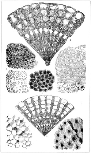
SECTIONS OF SHELL-STRUCTURE.
The shell of the wing-shells, Pinna ingens (Plate XVIII., No. 7), is composed of hexagonal cells, filled with partially translucent calcareous matter, the outer layer of which can be split up into prism-like columns. Figs. 3 and 6 are horizontal sections of the Haliotis splendens, with stellate pigment in a portion of the section, and wavy lines, as in the dentine of the human tooth, and of Terebratulata rubicuna, showing radiating perforations. Nos. 4 and 5, sections of the shell of a crab, show pigment granules beneath the articular layer and the general hexagonal structure of the next layer.
Some difference of opinion has been expressed with regard to the formation of pearls, but it is now generally understood to be a diseased condition. Pearls are matured on a nucleus, consisting of the same matter as that from which the new layers of shell proceed at the edge of the mussel or oyster. The finest kinds are formed in the body of the animal, or originate in the pearly-looking part of the shell. It is from the size, roundness, and brilliancy of pearls that their value is estimated.
The microscope discloses a difference in the structure of pearls: those having a prismatic cellular structure have a brown horny nucleus, surrounded by small imperfectly-formed prismatic cells; there is also a ring of horny matter, followed by other prisms, and so on, as represented in Fig. 374; and all transverse sections of pearls from oysters show the same successive rings of growth or deposit.
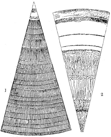
Fig. 374.
1. A transverse section of a Pearl from Oyster, showing its prismatic structure 2. A transverse section of another Pearl, showing its central cellular structure, with outside rings of true pearly matter. (Magnified 50 diameters.)
In a segment of a transverse section of a small purple pearl from a species of Mytilus (Fig. 375), all trace of prismatic structure has disappeared, and only a series of fine curved or radiating lines is seen. This pearl consists of a beautiful purple-coloured series of regular laminæ, many of which have a series of concentric zones, and are of a yellow tint. The most beautiful sections for microscopic examination are obtained from Scotch pearls.
Preparation of the Teeth and Shell of Mollusca for Microscopical Examination.—The method of preparing lingual membranes of Mollusca is as follows: Under a dissecting microscope, and with a large bull’s eye lens, cut open and expose to view the floor of the mouth; pin back the cut edges throughout its length, and work out the dental band with knife and forceps. The band being detached, place it in a watch-glass, and boil in caustic potash solution for a few minutes. Having by this process freed the tongue from its integuments, remove it, wash it well, and place it for a short time in a dilute acid solution, either acetic or hydrochloric. Wash it well and float it upon a slide; with a fine sable brush open it out flat, and remove whatever dirt or fibre may be adhering to it. Lastly, place it in weak spirit and water, and there let it remain for a few days before mounting in formalin. Canada balsam renders them rather too pellucid, and the finer teeth are thereby lost.
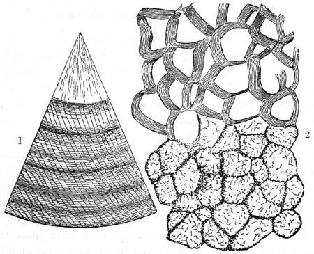
Fig. 375.
1. Transverse section of a small Pearl from a Mytilus; 2. Horizontal section magnified 240 diameters to show prismatic structure and transverse striæ.
The preparation of shell structure must be proceeded with with some amount of care and caution, or the delicate reticulated network membrane will be destroyed. If any acid solvent be used to remove the calcareous structure it should be much diluted, so that the action may proceed slowly rather than hastily. In the young hermit-crab, for example, where the calcareous and membranous portions of the shell are continuous, and the calcium carbonate in a relatively small proportion, a strong acid solution would entirely destroy the specimen. In the case of nacreous shells the process of cutting and grinding must also be proceeded with with some amount of caution. The operation should be examined as the process proceeds, and under polarised light. Sections of shell structure are usually mounted in Canada balsam. Under the heading Technique much useful information on this and kindred subjects will be found in the “Journal of the Royal Microscopical Society.”
Annulosa, Worms, and Entozoa.
The Annulosa of Huxley embraces the lowest grade of articulated animals, most of which are now grouped with Metazoa, while some writers place them in a sub-kingdom Vermes. It appears to me then only possible to describe this heterogeneous group of worm-like animals among those which resemble each other in certain negative features, but not possessing any of the distinctive characters of those previously described. There are numerous species among Entozoa, every one of which is of the highest interest to mankind in general, and to animal life as a whole. To these I shall devote some attention, from the wide-spread importance attached to them. They are characterised by having a soft absorbent body with little or no colour, in consequence of being excluded from light, living within the bodies of animals and absorbing their vital juices, thereby inflicting a large amount of injury and death upon the whole vertebrate kingdom. They bear in this respect a close analogy to parasitic Fungi in the nature of their destructive action upon plant life, which I have fully discussed in a previous chapter.
The relations which obtain between parasites and their hosts are in all respects conditioned by their natural history; and without a detailed knowledge of the organisation, the development, and the mode of life of the different species, it is impossible to determine the nature and extent of the pathological conditions to which they give rise, and at the same time find means of protection against guests in every way so unwelcome.
The nutritive system of the entozoa must be regarded as in the lowest state of development, yet there are some among them of a higher grade, as will be seen as we proceed. All are remarkable alike for their vast productiveness and for their peculiar metamorphoses. For example, the greater number of the Tænia begin their lives as sexless, encysted larvæ, and on entering their final abode, segments are successively added, until the worm has finally reached the adult stage. Again, the tapeworm of the cat has its origin in the encysted larvæ found in the livers of the mouse and rat. Another species of entozoa inhabit the stomach of the stickle-back, and only attain their perfect form in the stomachs of aquatic birds that feed exclusively on fish. Another infests the mantle of pond-snails, and through their agency, the embryos pass into the stomach of sheep.
An almost endless number of similar transformations take place in other genera. The simplest form among internal parasites is the Gregarinæ, formerly grouped among Protozoa. They consist of a simple limiting membrane, with a mass of granular matter enclosed and surrounding a nucleus (Plate III., No. 53). These parasites pass through a crystoid stage in the body of one of the lower animals, usually the earthworm, Lumbricus agricola. In the more mature organism an envelope, differentiated from the protoplasm within, can be made out (No. 54); this affords an indication of greater differentiation in the subjacent layer of protoplasm. An anterior portion is in many cases separated by a constriction from the cylindrical or band-like body (No. 56). Gregarinæ multiply when encysted, and divide into a multitude of minute pseudo-navicula, so named from their resemblance in shape to a well-known form of Diatomaceæ. When a young pseudo-navicule escapes it behaves somewhat like an amœba, and if perchance it is swallowed by an appropriate host, it develops at once into the higher stage. The various forms are represented in Plate III., Nos. 53—61. Miescher, in 1843, described suchlike bodies, taken from the muscles of a mouse. A good account of specimens obtained from the muscles of a pig was published by the late Mr. Rainey in the “Philosophical Transactions,” 1857. He regarded them as cestoid entozoa. They have been described under a variety of names, as worm-nodules, egg-sacs, eggs of the fluke, young measles, &c. M. Lieberkühn carefully traced the pseudo-naviculæ after leaving the perivisceral cavity of the earth-worm; he found large numbers of small corpuscles, exhibiting amœba-like movements, as well as pseudo-naviculæ, containing granules, formed in an encysted Gregarinæ. He imagines that these latter bodies burst, and that their contained granules develop into the amœbiform bodies which subsequently become Gregarinæ.
Professor Ray Lankester made a careful examination of more than a hundred worms for the purpose of studying these questions, but he succeeded in arriving at no other conclusion than that certain forms may be the by-products of encysted Gregarinæ. The G. lumbricus is one of those forms which are unilocular. The vesicle is not always very distinct, and is sometimes altogether absent; occasionally it contains no granules, sometimes several, one of which is generally nucleated. In other of these cysts a number of nucleated cells may be seen developing from the enclosed Gregarina, which gradually become fused together and broken up, until the entire mass is converted into nucleated bodies, often seen in different stages of development, assuming the form of a double cone, as that presented by certain species of Diatomaceæ. At length the cyst contains nothing but pseudo-naviculæ, sometimes enclosing granules; these gradually disappear, and finally the cyst bursts. Encystation seems to take place much more rarely among the bilocular forms of Gregarinæ than in the unilocular species found in the earthworm and other Annelids.73
Dr. J. Leidy published in the “Transactions of the Philadelphia Society,” 1853, the results of his examinations of several new species of Gregarinæ. He described a double membrane “within the parietal tunic of the posterior sac, this being transparent, colourless, and marked by a most beautiful set of exceedingly regular parallel longitudinal lines.”
Professor R. Leuckart is the latest writer on the parasites of animals, and to him we are indebted for a more systematic account of the whole group, and their life-history, than to any previous investigator. I can only attempt to give a mere outline of the developmental stages of a few typical forms of parasites, commencing with the cystic tapeworm, Tænia. These worms are ribbon-like in appearance, and are divided throughout the greater part of their length into segments, and their usual habitation is the intestinal cavity of vertebrate animals. The anterior extremity of a tænoid worm is usually called the head, and bears the organ by which the animal attaches itself to the mucous membrane of the creature which it infests. These organs are either suckers, or hooks, or both conjoined. In Tænia, four suckers are combined with a circlet of hooks, disposed around a median terminal prominence. The embryo passes through certain stages of development—viz., four forms or changes: but the embryo itself is very peculiar, consisting of an oval non-ciliated mass, provided with six hooks, three upon each side of the middle line. Tænia are found enclosed in various situations besides that of the alimentary canal: the eye, the brain, the muscular tissues, the liver, &c., of animals. The following cystic worms are usually included in this genera, Cysticercus Anthocephalus, Cœnurus, and T. Echinococcus. Plate IV., No. 100, shows an adult specimen of the latter with rostellum suckers, and three successive segments, the last of which is the ova sac. The water-vascular system is represented coloured by carmine. This parasite infests the human body as frequently as many other species. My accurately-drawn figure is copied from Cobbold’s “Introduction to the Study of Entozoa.”
Cysticercus fasciolaris is developed within the liver of white mice; Cysticercus cellulosæ in the muscles of the pig; hence we have the diseased state of pork familiarly known as “measly pork.” Should a lamb become infested with Tænia the final transformation will be different; within a fortnight symptoms of a disease known as “staggers” manifest themselves, and in the course of a few weeks the Cœnurus cerebralis will be developed within the brain. Von Siebold pointed out the bearing of this fact upon the important practical problem of the prevention of “staggers.” Others belonging to the same class of parasites are quite as remarkable in their preference for the alimentary canal of fishes. The Echinorhynchus is developed in the intestinal canal of the flounder, Triænophorus nodulus in the liver of the salmon. Thus, by careful and repeated observation with the microscope, a close connection is found to exist between the cystic and cestoid entozoa.
The Echinococcus (Plate IV., No. 101) infests the human liver. These parasites are always found in cysts, and in closed cavities in the interior of the body. They are united in fours by a very short stalk or pedicle, common to the whole. By an increase of magnification the contents of a cyst present the several structures represented in Fig. 376.
Echinorhynchus, or spiny-headed threadworms, constitute a group of entozoa which undergo a metamorphosis hardly, perhaps, less remarkable than that known to take place in other Nematode worms. Leuckart instituted, in 1861, a series of experiments with the ova of Echinorhynchus proteus found parasitic upon the Gammarus pulex. The ova of E. proteus resemble in form and structure those of allied species. They are of a fusiform shape, surrounded with two membranes, an external of a more albuminous nature, and an internal chitinous one. When the eggs reach the intestine the outer of these membranes is absent, being in fact digested, while the inner remains intact until ruptured by the embryo.
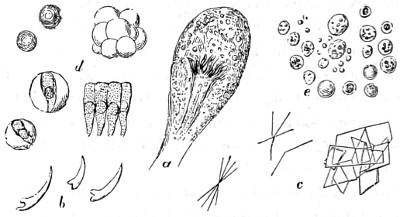
Fig. 376.—Cystic Disease of Liver (Human).
a. Cyst with Echinococcus enclosed; b. detached hooklets from the head of Echinococcus, magnified 250 diameters; c. crystals found in cyst, chiefly cholesterine; d. cylindrical epithelium, some enclosed in structureless vesicles; e. Puro-muculent granules, fat and blood corpuscles.
The typical Threadworm belonging to the order Nematoidea infest the intestines of children, and are a source of much suffering. The egg is elliptical, and contains a mass of granular protoplasm, the external wall of which soon becomes marked out into a layer of cells. The mouth of the worm appears as a depression at the end of the blunt head. When the muscular system and alimentary canal are developed the embryo hatches out, some few of which are free living forms; most of them lead a parasitic life. Their reproduction is enormous, representing thousands of eggs and embryos.
Of the non-parasitic species of thread-worm, the common vinegar eel, Anguillula,74 affords an example. This is found in polluted water, bog-moss, and moist earth, as well as in vinegar; also in the alimentary canal of the pond-snail, the frog, fish, &c. Another species is met with in the ears of wheat affected with a blight termed the “cockle”; another, the A. glutinis, in sour paste. If grains of the affected wheat are soaked in water for an hour or two before they are cut open, the so called “eels” will be found. The paste-eel makes its appearance spontaneously just as the pasty mass is turning sour; the means of securing a supply for microscopical examination consists in allowing a portion of the paste in which they show themselves to dry up, and laying it by for stock; if at any time a portion of this is introduced into a little fresh-made paste, and the whole kept warm and moist for a few hours, it will be found to swarm with these wriggling little worms. A small portion of paste spread over the face of a Coddington lens is a ready way of viewing them.
Trichina spiralis.—One of the smallest and most dangerous of all human internal parasites is T. spiralis, since it finds its way into the muscles throughout the human body. The young animal presents the form of a spirally-coiled worm in the interior of a minute oval-shaped cyst (Plate IV., No. 104), a mere speck scarcely visible to the naked eye. In the muscular structure it resembles a small millet seed, somewhat calcareous in composition. The history of the development of Trichina in the human muscle is briefly that in a few hours after the ingestion of infected pork, Trichina, disengaged from the muscle, will be found in the stomach: hence they pass into the small intestine, where they are further developed. Continuing their migrations, they penetrate far into the interior of the primitive muscular fasciculi, where they will be found, in about three days after ingestion, in considerable numbers, and so far developed that the young entozoa have almost attained a size equal to that of the full-grown Trichina (Plate IV., No. 105). They quickly advance into the interior of the muscular fasciculi, where they live and multiply in continuous series, while the surrounding structures as well as the muscular tissue undergo a process of histolysis. The destructive nature of the parasite is very great.
The number of progeny produced by one female may amount to several thousands, and as soon as they leave the egg they either penetrate through the blood-vessels, or are carried on by the circulation, and ultimately become lodged in the muscles situated in the most distant parts of the body. Here, as already explained, they become encysted.
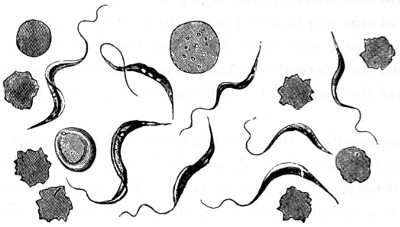
Fig. 377.—Monads in Rat’s Blood, stained with methyl violet, showing membrane under different aspects; blood-corpuscles, some crenated and others with stained discs (× 1,200).—(Crookshank.)
Professor Virchow draws the following conclusions:—“1. The ingestion of pig’s flesh, fresh or badly dressed, containing Trichinæ, is attended with the greatest danger, and may prove the proximate cause of death. 2. The Trichinæ maintain their living properties in decomposed flesh; they resist immersion in water for weeks together, and when encysted may, without injury to their vitality, be plunged in a sufficiently dilute solution of chromic acid for at least ten days. 3. On the contrary, they perish and are deprived of all noxious influence in ham which has been well smoked, kept a sufficient length of time, and then well boiled before it is consumed.”
A more minute Filarian worm has been detected in the human blood-vessels, known as Filaria sanguinis hominis. This worm carries on its work of destruction throughout the night; during the day it remains perfectly passive. It increases rapidly, and produces swellings of the glandular structures of the body, somewhat after the nature of those characteristic of the Bombay plague, with a slight difference, that after death the swellings are seen to be due to the vast accumulations of the Filaria sanguinis blocking the blood-vessels. The accompanying Fig. 377 shows a similar infiltration of monads in the blood of rats dying of plague in Bombay.