Part 1 — CHAPTER III. (continued 3)
Reading edition
In 1814, Fräunhofer,35 a German optician, discovered these lines quite independently, and mapped out 576 of them, calling the more prominent of them A, B, C, D, E, F, G, H, which lines he used as marks of comparison. He also found that the distances of these lines from each other may vary according to the nature of the substance composing the prism; thus, their relative distances are not the same in prisms of flint-glass, crown-glass, and bisulphide of carbon, but they always occupy the same position relatively to the colours of the spectrum. Kirschoff and Angström had mapped out in 1880 no less a number than 2,000 Fräunhofer lines, a portion of which are correctly shown in the accompanying chart (Fig. 191).
In 1830, Simms, a London optician, made an improvement in the construction of the spectroscope by placing a lens in front of the prism, so arranged that the slit was in the focus of the lens. This lens turns the light, after it has passed through the slit, into a cylindrical beam before entering the prism. Another lens, also introduced by him, receives the circular beam emerging from the prism, and compels it to throw an image of the slit, which may be magnified at pleasure for each ray. The lens between the prism and the slit is termed the collimating lens. Thus the following are the essential parts of a chemical spectroscope:—(1) a slit, the edges of which are two knife-edges of steel very truly ground, and exactly parallel to each other, and in a direction parallel to the refracting edge of the prism, to admit a pencil of rays. (2) A collimating lens; a convex lens with the slit at its principal focus, which renders the rays parallel before entering the prism. (3) A prism of dense glass, in which the rays are refracted and dispersed. (4) An observing telescope constructed like an astronomical refractor of small size, and placed so that the rays shall traverse it after emerging from the prism. Such are the essentials of a one-prism chemical spectroscope.
The form of instrument in use with the microscope is the “direct vision” spectroscope, consisting of two prisms of flint-glass, placed between three of crown-glass cemented together by Canada balsam; the spectrum being viewed directly by the eye. The earliest constructed form of micro-spectroscope is shown in Fig. 192, the Browning-Huggins.
It was, however, Mr. Sorby who suggested that the prism should be made of dense flint-glass and of such a form that it could be used in two different positions, and that in one it should give twice the dispersion that it would in the other, but that the angle made by the incident and emergent rays should be the same in both positions.
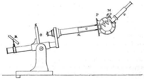
Fig. 192.—The Browning-Huggins Micro-spectroscope.
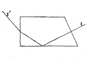
Fig. 193.
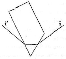
Fig. 193a.
Figs. 193 and 193a represent prisms of the kind arranged to use in two different positions, i and i′ being the same angle as I and I′.
For most absorption-bands, particularly if faint, the prism should be used in the first position, in which it gives the least dispersion; when greater dispersion is required, so as to separate some particular lines more widely, or to show the spectra of the metals, or Fräunhofer’s lines in the solar spectrum, then the prism must be used as in Fig. 193a. This answers well for liquids or transparent objects, but it is, of course, not applicable to opaque objects.
To combine both purposes, some form of direct vision-prisms that maybe applied to the body of the microscope is required. Fig. 194 represents an arrangement of direct vision-prisms, invented by Herschel. The line R R′ shows the path of a ray of light through the prisms, where it would be seen that the emergent ray R′ is parallel and coincident with the incident ray R.
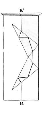
Fig. 194.
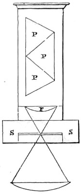
Fig. 194a.
Another very compact combination is shown in Fig. 194a. Any number of these prisms (P P P) may be used, according to the amount of dispersion required. They are mounted in a similar way to a Nicol’s prism, and are applied directly over the eye-piece of the microscope. The slit S S is placed in the focus of the first glass (F) if a negative, or below the second glass if a positive eye-piece be employed. One edge of the slit is movable, and, in using the instrument, the slit is first opened wide, so that a clear view of the object is obtained. The part of the object of which the spectrum is to be examined is then made to coincide with the fixed edge of the slit, and the movable edge is screwed up, until a brilliant coloured spectrum is produced. The absorption-bands will then be readily found by slightly altering the focus. This contrivance answers perfectly for opaque objects, without any preparation; and, when desirable, the same prism can be placed below the stage, and a micrometer used in the eye-piece of the microscope, thus avoiding a multiplicity of apparatus.
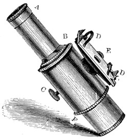
Fig. 195.—The Sorby-Browning Micro-spectroscopic Eye-piece.
A later and better form of instrument is the Sorby-Browning eye-piece (Fig. 195), shown in section (Fig. 196) ready for inserting into the body-tube of the microscope, the prism of which is contained in a small tube, removable at pleasure. Below the prism is an achromatic eye-piece, having an adjustable slit between the two lenses, the upper lens being furnished with a screw motion to focus the slit. A side slit, capable of adjustment, admits, when required, a second beam of light from any object whose spectrum it is desired to compare with that of the object placed on the stage of the microscope. This second beam of light strikes against a very small prism, suitably placed inside the apparatus, and is reflected up through the compound prism, forming a spectrum in the same field with that obtained from the object on the stage.
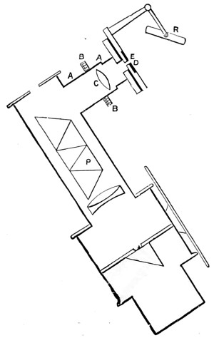
Fig. 196.—Sectional view of bright-line Spectroscope; the letters also apply to the standard spectrum scale (Fig. 198).
A is a brass tube, carrying the compound direct vision prism; B, a milled head, with screw motion to adjust the focus of the achromatic eye lens C, seen in the sectional view as a triple combination of prisms. Another screw at right angles to C, which from its position cannot be well shown in the figure, regulates the slit horizontally. This screw has a larger head, and when once recognised cannot be mistaken for the other. D D is a clip and ledge for holding a small tube, so that the spectrum given by its contents may be compared with one from an object on the stage. E is a round hole for a square-headed screw, opening and shutting a slit, admitting the quantity of light required to form the second spectrum. A light entering the round hole near E strikes against the right-angled prism, which is placed inside the apparatus, and is reflected up through the slit belonging to the compound prism. If any incandescent object be placed in a suitable position with reference to the round hole, its spectrum will be obtained. F shows the position of the field lens of the eye-piece. The tube is made to fit the microscope to which the instrument is applied. To use this instrument insert F as an eye-piece in the microscope tube, taking care that the slit at the top of the eye-piece is in the same direction as the slit below the prism. Screw on to the microscope the object-glass required, and place the object whose spectrum is to be viewed on the stage. Illuminate with the stage mirror if it be transparent; with mirror, Lieberkühn, and dark well, by side reflector, or bull’s-eye condenser if opaque. Remove A, and open the slit by means of the milled-head, not shown in figure, but which is at right angles to D D. When the slit is sufficiently open the rest of the apparatus acts as an ordinary eye-piece, and any object can be focussed in the usual way. Having focussed the object, replace A, and gradually close the slit till a good spectrum is obtained. The spectrum will be much improved by throwing the object a little out of focus.
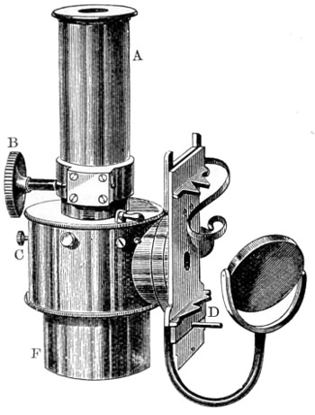
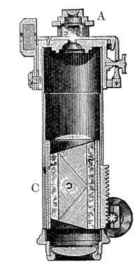
Sectional View.
Figs. 197 and 197a.—The Beck-Sorby Micro-spectroscope Eye-piece, drawn on a scale of one half size.
Every part of the spectrum differs a little from adjacent parts in refrangibility, and delicate bands or lines can only be brought out by accurately focussing that particular part of the spectrum. This can be done by the milled-head B. Disappointment will occur in any attempt at delicate investigation if the directions given be not carefully followed out.
Opposite E a small mirror is attached. It is like the mirror below the stage of a microscope, and is mounted in a similar manner. By means of this mirror light may be reflected into the eye-piece, and in this way two spectra may be procured from one lamp.
Method of using the Micro-Spectroscope.
A beginner with the micro-spectroscope should first make himself fully acquainted with the spectroscope by holding it up to the sky and noting the effects of opening and regulating the slit, by rotating the screw C, Figs. 195 and 197. The lines will be well seen on closing down the opening. This screw diminishes the length of the slit, when the spectrum is seen as a narrow ribbon of prismatic colours. The screw E regulates the admission of light through the aperture above D. The better objects with which to commence the study of the absorption bands are, aniline dye, much diluted, madder, permanganate of potash, and blood. As each colour varies in refrangibility, the focus must be adjusted by the screw E. When it is desired to view the spectrum of a very minute object, the prisms should be removed by withdrawing the tube containing them, the slit set open, and the object brought into the centre of the field; the vertical and horizontal slits must then be partially closed up, and the prisms replaced, when a suitable objective is employed to examine the spectrum. For ordinary observations a magnifying power of an inch and a half or two inches will be suitable, but for small quantities of material a higher power must be employed, when a single blood corpuscle can be made to show its characteristic absorption band. After having obtained the best image of any object on stage, throw it slightly out of focus, and substitute the micro-spectroscopic eye-piece for the Huyghenian. Opaque objects should be examined by reflected light, by means of the bull’s-eye condenser, or side reflector. Mr. Sorby uses a binocular microscope, which enables him to regulate the focussing and throwing out of focus of the object.
In examining crystals or other small objects, a small cardboard diaphragm should be placed beneath them; and when examining the spectra of liquids in cells, slip a small cap with a perforation of 1⁄10-inch in diameter over the tube containing the ½-inch or 2-inch objective. Substances which give absorption bands or lines in the red are best seen by artificial light, while those which show bands in the violet are better seen by daylight. By following rules of the kind we are less likely to mix the bands of the absorption spectrum with the Fräunhofer lines. For example, if the edge of a band happens to coincide with a Fräunhofer line, the observer is apt to imagine that the band is better defined and more abruptly shaded on one side than it really is.
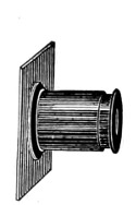
Standard Spectrum Scale.
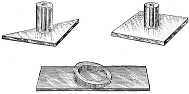
Cells for use with Spectroscope.
Fig. 198.
Cells and Tubes.—These are either supplied ready-made by the optician, or can be formed out of small pieces of barometer tubing, with the edges ground down and cemented on ordinary glass slips. In Fig. 198 is seen the several kinds of cells and tubes usually employed, while the little flat tubes commonly in use as bouquet holders will be found of use, with the side stage reflecting spectrum as comparison tubes; being of different diameters they allow of two or more depths of colour in the fluid intended for examination.
In the case of many other fluids the sloping form of cell (Fig. 198) will be useful, as different shades of fluids can be examined without removal from the stage of the microscope. The deeper cells are cut from a piece of barometer tubing of about half to an inch long, one end being cemented to a piece of flatted glass, and the other covered over temporarily or permanently with a thin piece of glass on the top, held in its place by capillary attraction, thus admitting of the tube being turned upside down.
Re-agents required.—A diluted solution of ammonia, citric acid, double tartrate of potash and soda (the last being used to prevent the precipitation of oxide of iron), and the double sulphate of the protoxide of iron and ammonia (employed to deoxidise blood, etc.). In some special cases, dilute hydrochloric acid, purified boric acid, and sulphate of soda are required.
The character of stains of blood varies with age and with the nature of the substance with which it happens to be combined. This is important to remember in connection with Jurisprudence, when the micro-spectroscope is brought into use for the detection of blood stains. The spectrum used in important cases of the kind should have a compound prism, with enough, but not too great dispersive power, otherwise the bands become, as it were, diluted, and less distinct.
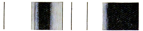
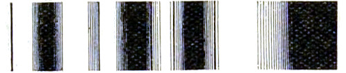
If the blood stain is quite recent, the colouring matter will be hæmoglobin only. This easily dissolves out in water, and when sufficiently diluted gives the spectrum of oxy-hæmoglobin, which on the addition of ammonia, together with a small quantity of the double tartrate, a small piece of ferrous salt, and stirring carefully without the admission of air, changes the spectrum of reduced hæmoglobin. When stirred again, so as to expose the solution as much as possible to air, the two bands reappear; on gradually adding citric acid in small quantities the colour begins to change, and the bands are seen to gradually fade away; if there should have been much blood present, a band appears in the red; the further addition of ammonia makes all clear again, but does not restore the original bands, because the hæmoglobin has been permanently changed into hæmatin. This reaction alone distinguishes blood from most other colouring matters, since other substances after being changed by acids are restored by alkalies to their original state. There are many other curious facts connected with the spectroscopic analysis of blood, which are fully explained and illustrated by Dr. Maemunn in his book on “The Use of the Spectroscope in Medicine,” and also in Dr. Thudicum’s36 reports and charts, which are the most complete. Sir George Stokes, F.R.S., was one of the first to show the essential value of the spectral phenomena of hematine, and who proved, after Hoppe had first drawn attention to the fact, that this colouring matter is capable of existing in two states of oxidation, and that a very different spectrum is produced according as the substance, which he termed cruorine, is in a more or less oxidised condition. The chart appended to his paper37 affords an imperfect representation of the changes seen in the spectrum.
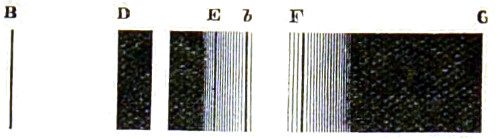
No. 1.—Arterial Blood, Scarlet Cruorine.
No. 2.—Venous Blood, Purple Cruorine.
No. 3.—Blood treated with Acetic Acid.
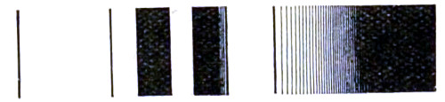
No. 4.—Solution of Hæmatin.
Fig. 199.—Sir George Stokes’ Chart of the Absorption Bands of Blood.
Proto-sulphate of iron, or proto-chloride of tin, causes the reduction of the colouring-matter, but, on exposure to air, oxygen is absorbed, and the solution again exhibits the spectrum characteristic of the more oxidised state. The different substances obtained from blood colouring-matter produce different bands. Thus, hæmatin gives rise to a band in the red spectrum D; hæmato-globulin produces two bands, the second twice the breadth of the first in the yellow portion of the spectrum between the lines D and E, No. 1. The absorption-bands differ according to the strength of the solution employed, and the medium in which the blood-salt is dissolved; but an exceedingly minute proportion dissolved in water is sufficient to bring out very distinct bands. B represents the red end of the spectrum and G the green as it approaches the violet end.
Mapping the Spectra.—In the sectional view given of the micro-spectroscope (Fig. 196), the internal construction of the instrument is shown, and the arrangement made for throwing a bright point on to the surface of the upper prism is clearly seen. The mapping out is accomplished by means of a photographic scale fixed as a standard spectrum (Fig. 198), in the position of A A, illuminated by the small mirror at R, and focussed by a small lens at C, so that on looking into the instrument one can see the spectrum accurately divided into one hundred equal parts, and scale readings can be made at once; the only precaution needed is to be sure the D (or the sodium line, if D cannot be got) always stands at the same number on the scale. To map absorption spectra on this scale we have to lay down a line, as many millimetres long as there are divisions in the scale, and mark the position of the bands on this line. Mr. Browning supplies scales printed off ready for use. But the mapping out of spectra, as Mr. Sorby pointed out, requires some consideration; since the number of divisions depends on the thickness of the interference-plate, it becomes necessary to decide what number should be adopted. Ten it was thought would be most suitable; but, on trial, it appeared to be too few for practical work. Twenty is too many, since it then becomes extremely difficult to count them. Twelve is as many as can well be counted; it is a number easily remembered, is sufficiently accurate, and has other practical advantages. With twelve divisions the sodium-line 0 comes very accurately at 3½; thus, by adjusting the plate so that a bright sodium-light is brought into the centre of the band, when the Nicol’s prisms are also crossed accurately at 3½, parallelism is secured, together with a wider field of observation. The general character of the scale will be best understood from the following figure, in which the bands are numbered, and given below the principal Fräunhofer lines. The centre of the bands is black, and they are shaded off gradually at each side, so that the shaded part is about equal to the intermediate bright spaces. Taking, then, the centres of the black bands as 1, 2, 3, &c., the centres of the spaces are 1½, 2½, 3½, &c., the lower edges of each ¾, 1¾, &c., and the upper 1¼, 2¼, &c., we can easily divide these quarters into eighths by the eye: and this is as near as is required in the subject before us, and corresponds as nearly as possible to 1⁄100th part of the whole spectrum, visible under ordinary circumstances by gaslight and daylight. Absorption-bands at the red end are best seen by lamp-light, and those at the blue end by daylight.
(Red end.) (Bue end.)
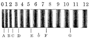
Fig. 200.—
On this scale the position of some of the principal lines of the solar spectrum is about as follows:—
A ¾ B 1½ C 23⁄8 D 3½
E 511⁄16 b 63⁄16 F 7½ G 105⁄8
At first plates of selenite, which are easily prepared, were used, because they can be split to nearly the requisite thickness with parallel faces; but their depolarising power varied much with temperature. Even the ordinary atmospheric changes alter the position of the bands. However, quartz cut parallel to the principal axis of the crystal is but slightly affected, and is not open to the same objection; but this is prepared with some difficulty. The sides should be perfectly parallel, the thickness about ·043-inch, and gradually polished down with rouge until the sodium-line is seen in its proper place. This must be done with care, since a difference of 1⁄10000-inch in thickness would make it almost worthless.
The two Nicol’s prisms and the intervening plate are mounted in a tube, and attached to a piece of brass in such a manner that the centre of the aperture exactly corresponds to the centre of any of the cells used in the experiments, and must be made to correspond with equal care, so that any of them, or this apparatus in particular, may be placed on the stage and in proper position without further adjustment, whereby both time and trouble are saved.
Absorption Spectrum of Chromule.
In 1869 I published in the Journal of the Royal Microscopical Society38 a paper on results obtained by the spectrum analysis of the colouring-matter of plants and flowers, some of which were of considerable interest in many respects. My examinations extended to several hundred different specimens, from which I was led to conclude that the chromule of flowers is, for the most part, due to the chemical action of the actinic rays of light over the protoplasm of the plant, more so than to that of soil. But as certain roots of plants, as those of the alkanet, yield their colouring-matter to oil, and in a much smaller degree to spirit or water, it follows then that conclusions of any kind can only be drawn after a long and careful study of the question. Some of the results obtained were, however, of some interest at the time, that, for example, seen in three different solutions of the chlorophyll of Cinchona succirubra, one of three solutions in alcohol, scarcely coloured, having in fact only a faint tinge of green colour, and the spectrum of which much astonished me at the time. It gave four well-marked absorption-bands; one deep sharp line in the red; another, rather narrower, in the orange, coincident with D, or the sodium-line; one in the green, about b, coincident with the Thallium green band; and a fourth on the blue line F, nearly as broad as that in the red. The ethereal solution gave different results. It showed only three bands of absorption, nearly the same as in the last case (though all of them fainter); but the fourth in the blue was not apparent, the whole of that end of the spectrum being absorbed a little beyond the green line b. This solution was deep emerald-green, and even dilution did not alter the phenomena. The acid alcoholic solution was as deeply green as the last, but gave only the sharp broad absorption-band in the red, and two very faint ghostly bands in the position described above of the D and b lines respectively.
Further additional researches on the chlorophyll of plants furnished curious results, the chlorophyll being dissolved out by alcohol, digested for some hours, and without heat; some plants being fresh, and others dried. Five classes of phenomena exhibited themselves, but all agreed in having the red absorption-band broad, sharp, and well defined, some having this one band only, the Lilac being of this type.
There are two classes in which two absorption-bands occur. One has the red and the orange bands, of which the Fuchsia, Guelder-rose, and Tansy are examples; another, in which the red and the green bands are alone co-existent. Ivy is the type of the class, and it is immaterial whether we take last year’s leaves or those of the early spring; the results are the same.
The fourth class consists of the two former spectra superposed. Three lines occur, the red, the orange, and the green bands, at C, D, and b, as before. This is by far the largest class, and I have thirty or forty examples of it. Œnothera biennis, Laurestinus, &c., are types with the ethereal solution of the leaves of Red Bark.
The fifth class consists of those having properties similar to the alcoholic solution of Red Bark described. But I only found eight of these, and not all equal in colour power, namely: Berberry, Sloe, Tea, Hyoscyamus, Digitalis, Senna, and Red Bark. The results obtained appeared at the time to be well worth following up to a more practical conclusion than that arrived at. It should be noted that in the preparation of vegetable colouring matters for the micro-spectroscope, care must be taken to employ only a small quantity of spirits of wine to filter the solution, and evaporate it at once to dryness at a very gentle heat, otherwise if we attempt to keep the colouring matters in a fluid state they quickly decompose. It is necessary also to employ various re-agents in developing characteristic spectra. The most valuable re-agent is sulphite of soda. This admits of the division of colours into groups.
It is better to use a dilute alcoholic solution for the extraction of colour from plants, and to observe the spectrum in a column of about three-quarters of an inch in height. By this means it is quite possible to ascertain that the spectrum of chlorophyll presents seven distinct absorption bands.
For further information on this interesting subject I must refer the reader to Mr. Sorby’s paper “On a Definite Method of Qualitative Analysis of Vegetable and Animal Colouring Matter by means of the Spectrum Microscope,” “Proc. Roy. Soc.,” No. 92, 1867.