Part 1 — CHAPTER IV. (continued 2)
Reading edition
Rock Sections.—Small pieces of rock may be ground down by the aid of the lathe, or on a zinc plate, with emery powder and water, until one side is rendered smooth and flat. Then fasten the polished side of the section to a square of glass on the metal holder of the lathe, with dried Canada balsam, as directed for bone, and allow it time to become consolidated. When moderately thin take a piece of plate-glass and some fine emery or putty-powder and rub the section down as thin as possible. When found to be thin enough wash it well in water, and put it aside to dry, or warm it over a spirit-lamp, and with a needle push the section off the glass into a watch-glass of benzole or turpentine, and allow it to soak until all the balsam is dissolved out. Wash again in turpentine, and mount in Canada balsam, with or without a cover-glass. Sections of echinus spines, shells, stones of fruits, &c., are prepared in the same way as those of bones and teeth; but when the grinding is finished, the sections must be passed through alcohol into oil of cloves, after which they should be mounted in Canada balsam. If tolerably thin, sections of these substances can be cut in the lathe; in the first instance, there will be no actual occasion to attach them to glass at all, except for the purpose of obtaining a hold upon the specimen for polishing, but the surface thus attached must afterwards be completely removed in order to bring into view a stratum which the Canada balsam may not have penetrated.
With regard to smaller bodies, these can scarcely be treated in any other way than by attaching a number of them to slips of glass at once, and in such a way as to make them mutually support each other. Thus in making horizontal and vertical sections of foraminifera, it would be impossible to slice them through unless they were laid close together in a bed of hardened Canada balsam, and first grinding away one side and then turning and rubbing down the other. My friend, Dr. Wallich, many years ago communicated to me the ingenious plan adopted by himself when mounting and turning a number of these minute objects together. The specimens being cemented with Canada balsam, in the first instance, to a thin film of mica, and then attached to a glass slide by the same means, when ground down to the thinness desired, the slide must be gradually heated just sufficiently to allow of the detachment of the mica-film and the specimen it carries; a clean slide with a thin layer of hardened balsam having been prepared, the mica-film is transferred to it with the ground surface downward. Its adhesion by drying having been complete, the grinding and polishing should be proceeded with; and as the mica-film will yield to the stone without any difficulty, the specimen now reversed in position may be further reduced to the requisite thickness for mounting as a permanent object.
Staining and Mounting Vegetable Tissues.—Bacteria I propose to treat of in a separate section. Vegetable tissues generally will first receive attention, and their differentiation is based on the employment of delicate gradations of colour stains. The more striking results are obtained by Multiple Staining, while the cell contents are rendered more palpable. On this account colouring media have been divided into nuclear, plasmic, and specific. The first are chiefly valued in proportion as they prove to have a selective affinity for the nuclei of cells, and leaving the protoplasm comparatively unstained. Such stains are needful for fresh and young tissues. On the other hand, plasmic stains colour tissue uniformly, and are used to give a ground colour by way of contrast; and specific stains are chiefly employed to distinguish certain elementary structures from the mass of cellulose which forms the basis of vegetable tissue, and which is also met with to a slight extent in animal membranes.
Cellulose, as it occurs in plant life, presents a variety of physical properties: sometimes it is soft, as in young plants, and again quite dense in older structures. This fact accounts for the varying results obtained when cellulose is subjected to the action of staining fluids, and whether the cellulose occurs in a nearly pure form, as in cotton fibre, or in the modified form of lignine or woody-fibre. Stains which readily attack young tissue have little or no effect upon it in its maturer form. It is of much importance, then, in the staining of fibres, as well as sections for the microscope, that the cellulose should take the stain uniformly.
The staining of tissues may be effected in four ways. First, when the stain has sufficient affinity for the tissue to be retained by it without the intervention of any outside agent. Second, when the stain and mordant are mixed and applied to the tissue in one solution. These two are the simplest and easiest methods of staining. Third, when the tissue is first immersed in the staining liquid and then transferred to some other liquid which shall fix the colour upon the tissue. Fourth, when the tissue is first impregnated with the mordant, or fixing agent, and then immersed in the stain. The last method is the one usually followed in commercial works, and it is to be recommended in the staining of microscopical preparations which do not readily take the stain.
Nuclear Stains.—As in both vegetable and animal sections it is generally the nuclei which form the landmarks of the structure, so the most important class of reagents which are used in any of the branches of microscopical work are the “nuclear stains.” There are several of these stains, the most important of which is the hæmatoxylin, and when proper solutions are used the results are very satisfactory. Many formulæ have been given, but there are three only reliable, Delafield’s, Kleinenberg’s, and Ehrlich’s, in all of which alum is present as an ingredient; the idea being that the alumina forms with the colouring matter an insoluble lake, and so acts as a mordant.
In Delafield’s solutions a large proportion of alum to hæmatoxylin is used, and methylic alcohol (wood-spirit in the place of rectified spirit).
For Kleinenberg’s solution many different formulæ exist. Squire’s improved formulæ for both stains is given in the Appendix, “Formulæ and Methods.”
Hæmatoxylin solutions stain the nuclei violet, and in order to change this into blue, the sections should be transferred to water taken from the house supply, not distilled water; but as the alkalinity of the water varies in different localities, a better and more uniform result is obtained by using a weak solution of bicarbonate of sodium (half a grain to the ounce).
Carmine is also much in vogue as a nuclear stain, and the two solutions more generally employed are Greenacher’s alcoholic borax carmine, and Orth’s lithium carmine. Under ordinary circumstances they act as general stains, affecting the ground tissue as well as the nuclei. By subsequent treatment with acidulated alcohol or acidulated glycerine the colour is discharged from the ground tissue without seriously affecting the nuclei. Used in this way, carmine becomes a good nuclear stain. It should be remembered that the sections must not be washed in pure water, as the colour will to a great extent be discharged; nor in acidulated water, as the carmine will be precipitated.
Alum carmine and alum cochineal are useful nuclear stains, not requiring after-treatment.
Picro-carmines are also largely used. The following formulæ will be found the most useful:—
Ammonia Picro-carmine.—Carmine, one gramme; strong solution of ammonia, three cc.; distilled water, five cc. Dissolve the carmine in the ammonia and water with a gentle heat, then add saturated aqueous solution of picric acid, 200 cc.; heat to boiling, and filter.
Picro-Lithium Carmine.—The following is generally preferred for use—Lithium carmine solution, 100 cc.; saturated solution of picric acid, 270 cc.
There are several aniline dyes which are used for nuclear staining: methylene blue, methyl green, safranine, gentian violet, vesuvine, fuchsine, and Hoffmann’s blue.
The usual process is to stain in ¼ or ½ per cent. aqueous solutions and wash in methylated spirit. Methylene blue and methyl green have the reputation of being so readily washed out in the methylated spirit as to be worthless. This is obviated by washing the sections (when removed from the stain) in distilled water, previous to the differentiation in methylated spirit. Treated in this manner, the nuclear staining is very beautiful. This also applies to Hoffmann’s blue and partly to vesuvine; with the latter, however, it is not a necessity. Safranine and gentian violet worked better by transferring the sections directly from the stain into 90 per cent. alcohol.
Contrast Stains.—Very frequently other stains are used to dye the ground a colour which is in contrast to that employed for the nuclei. Brown, orange, or pink are used after nuclear blue or green. Carmine is generally counterstained yellow or indigo-blue; and fuchsine red, as in tubercle bacilli, is counterstained with nuclear blue. It is important that the ground stain should be made weaker than the principal stain, so that the whole tissue may be shown without detracting from the nuclei. The following colours are used as counterstains for animal sections, but they prove less useful for vegetable sections: benzo-purpurine, eosin, erythrosine, orange, acid rubin, and picric acid.
Examples of specific stains are fuchsine, methylene blue, and gentian violet for bacteria; osmic acid for fatty elements; victoria blue and rose bengale, for demonstrating elastic tissue; methyl violet, iodine, and safranine, for amyloid degeneration. Methylene blue is one of the most useful of aniline dyes, and one of the most variable in composition.
Iodine green, or methyl green, has long been in use as a reagent for amyloid, starchy matters, in ignorance of the fact that the reaction is due to the methyl violet, contained as an impurity in the iodine green. It is exceedingly difficult to obtain a green quite free from violet. As nuclear stains they are identical, and the amyloid reaction, being dependent wholly upon the contained violet, varies, not with the formula of the green, but with the extent to which it has been purified.
Cellulose reactions.—After the nuclear stains, the most important reagents to the botanist are those which affect cellulose and its several modifications. Pure cellulose is coloured yellow by iodine, the colour being changed to a blue on the addition of slightly dilute sulphuric acid, or a strong solution of zinc. Solutions containing iodine, iodide of potassium, and chloride of zinc, give a violet reaction with unaltered cellulose, and yellow with lignine.
Schulze’s zinc re-agent must be used with a certain amount of caution, as the chloride of zinc and potassium undergo decomposition. The formula now in use is as follows: Take of zinc chloride solution (sp. gr. 1·85) 70 cc., potassium iodide 10 grammes, iodine 0·1 gramme; but this solution can only be employed as a re-agent and not as a dye, and structures stained with it cannot be mounted in any of the ordinary media, and the only fluid for ringing them down is caoutchouc cement.
Cellulose can be stained permanently by carmine, hæmatoxylin, nigrosine, methylene blue, safranine, and fuchsine. The aniline dyes are used in dilute aqueous solutions containing one-eighth or one-fourth per cent. of dye. When the cellulose undergoes the change known as lignification its reactions are altered. It is coloured yellow by chloro-zinc iodine, red by phloroglucin, yellow by aniline chloride. The two latter are much assisted by hydrochloric acid. The results of these reactions also cannot be preserved in the usual mounting media.
Sections containing mixed tissue, partly unaltered cellulose and partly lignified, give striking results with aniline dyes, and with this additional advantage can be preserved for years.
Double Staining.—When a section is passed through methyl green solution and afterwards one of carmine, the lignified portion is coloured green and the unlignified red. Acid green may be used in the place of methyl green, with a similar result. When picric acid is used with carmine, ingrosine, or Hoffmann’s blue, the picric acid dyes the ligneous portion and the others colour the unlignified structure, red, black, and blue respectively.
Eosin stain is the most useful for sieve-tubes and plates. Make a strong solution of eosin in equal parts of water and alcohol, and stain the section for five or ten minutes. Wash well in methylated spirit, dehydrate, clean in oil of cloves, and mount in Canada balsam.
Bleaching Process.—The bleaching and clearing of vegetable structures before staining is a very necessary process, especially so if starch be present in any quantity. Clearing agents are of two kinds—those which act by virtue of their property of strongly refracting light, and those which disintegrate and dissolve the amyloid cell contents. To the first class belong the essential oils, as oil of cloves, Canada balsam, glycerine, and other similar bodies; to the second class, solutions of potash, phenol, and chloral hydrate. The actual value of some of these agents is questionable. The process usually preferred is as follows: Place the sections in a fresh clear solution of chlorinated lime, allowing them to remain until quite bleached, say from two to four or five minutes; then gently warm in a test-tube for a few seconds, and quickly replace the solution with distilled water and boil for two or three minutes; repeat the treatment with boiling water three times; wash with a one per cent. solution of acetic acid, and finally with distilled water. The sections are then quite ready for staining operations.
When the stem is hard and brown, a solution of chloride of lime should be used—a quarter of an ounce of chloride dissolved in a pint of water, well shaken and stood by to settle down, then pour off the clear fluid for use. For hard tissues this solution answers well, but it is not suitable for leaves, as they require not only bleaching, but the cell contents should be dissolved out to render them transparent. A solution of chlorinated soda answers well for both stems and leaves. It is prepared as follows:—
To one pint of water add two ounces of fresh chloride of lime, shake or stir it well two or three times, then allow it to stand till the lime has settled. Prepare meanwhile a saturated solution of carbonate of soda—common washing soda. Now pour off the clear supernatant fluid from the chloride of lime, and add to it, by degrees, the soda solution, when a precipitate of carbonate of lime will be thrown down; continue to add the soda solution till no further precipitate is formed. Filter the solution, and keep it in a well-stoppered bottle in the dark, otherwise it speedily spoils.
Sections bleached in chlorinated soda must, when white enough, be washed in distilled water, and allowed to remain in it for twenty-four hours, changing the water four or five times, and adding a few drops of nitric acid, or at the rate of eight or ten drops to the half-pint, to the water employed before the final washing takes place. From water transfer them to alcohol, in which they must remain for an hour or more.
Although alkaline glycerine has been recommended for several purposes in micro-technique, it is not so well known as it should be how serviceable it is as an extempore mounting solution in vegetable histology. The best mixture for general use is composed of glycerine 2 ozs., distilled water 1½ oz., solution of potash, B.P., ½ oz. This combines the refringent property of the glycerine with the clearing action of the caustic potash, while the swelling action of the potash is considerably diminished.
Cutting Sections of Hard Woods.—The lathe and circular saw will be found as useful for cutting sections of the harder kinds of woods, as for bone structure. It may be necessary to subject the older and consequently harder pieces of wood to the action of steam for a few hours to soften them, and afterwards transfer them to methylated spirit, before making an attempt to cut sections. But the more open woods, of one, two, or three years’ growth, will show all that may be required, and these can be cut by hand, or with the microtome, as already described.
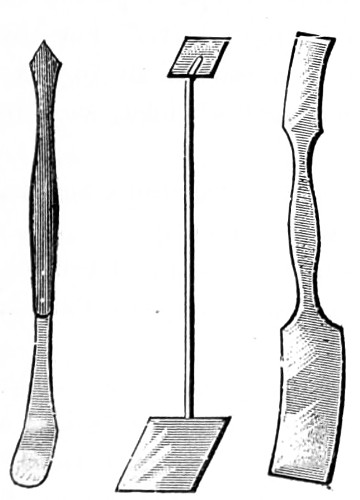
With a little practice the finest and thinnest possible slices may be cut by hand. It is usual to take off the first slice to give a smooth and even surface to the specimen. Then turn the screw to raise it a little, sprinkle the surface with spirit and water, and cut with a light hand. Remove the cut sections with a fine camel’s-hair brush or a section lifter (Fig. 250) to a small vessel containing water, when the thinnest will float on the surface, and remove to methylated spirit and water, where they should remain until they can be mounted. Sections of hard woods, and those containing gum-resins, or other insoluble material, must first be kept in methylated spirit or alcohol, and finally transferred to oil of cloves, to render them sufficiently transparent for mounting in Canada balsam.
If the structure of an exogenous wood is required to be examined, the sections should be made in at least three different ways: the transverse, the longitudinal, and the oblique, or, as they are sometimes called, the horizontal, vertical, and tangential, each of which will exhibit different appearances, as seen in Fig. 245: b is a vertical section through the pith of a coniferous plant, and exhibits the medullary rays known to the cabinet-maker as the silver grain; e is a magnified view of a part of the same; the woody fibres are seen with their dots l, and the horizontal lines k indicating the medullary rays cut lengthwise; c is a tangential section, and f a portion of the same; the medullary rays m m, and the woody fibres with vertical slices of the dots, are shown. Instructive preparations will be secured by cutting oblique sections of the stem. The sections seen are made from the pine. All exogenous stems, however, exhibit three different appearances, according to the direction in which the section is made.
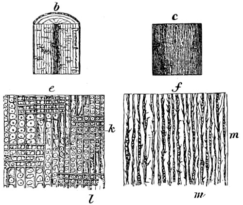
Fig. 245.—Sections of Wood.
Bacteria Cultivation, Sterilising, and Preparing for Microscopical Examination.
That branch of mycology which is now looked upon as a separate department of science, termed bacteriology, took shape in the years 1875-9, when its founder, the veteran botanist Cohn, who recognised that the protoplasm of plants corresponded to the animal sarcode, published his exact mode of studying bacteria. But it was a pupil of his, Dr. Koch, who a year later discovered that a specific cattle disease, anthrax, was due to a bacillus, and it was he also who gave us the useful modification of gelatine as a medium in which to grow bacteria; he hit upon the method of pouring melted gelatine containing distributed germs on to plates, and thus isolating the colonies and ensuring the further isolation of the spores, and so facilitate the preparation of pure cultures on a large scale, and with great saving of time.
The difficulty of isolating a bacterium and tracing its life history under the microscope must at first sight appear great. A further objection that such work is slow and difficult has no more weight here than in any other department of science, as will be seen on proceeding to follow out the directions I am about to furnish for the use of the student.
Apparatus, Material, and Reagents employed in Bacteriological Investigations.
A good microscope with a wide-angled sub-stage condenser, and objectives of an inch, ¼-inch, or 1⁄6-inch, and a 1⁄12-inch homogeneous oil-immersion.
A large bell-glass for covering the same when fuming acids are in use in the laboratory.
About a square foot of blackened plate-glass.
A white porcelain slab, or a shallow photographic dish of some size.
Glass bottles with ground stoppers for alcoholic solutions and aniline dyes.
Glass bottles with funnels for filtering solutions of stains, with pipettes.
A specialised form of pipette for the micro-chemical filtration of solutions (Fig. 246).
A small stoppered bottle of cedar oil (Fig. 247).
Set of small glass dishes or watch-glasses for section staining.
Stock of glass slides sterilised, together with round thin glass-covers, in boxes (Fig. 248).
Needle holders and platinum needles, with a packet of ordinary sewing needles (Fig. 249).
Platinum, or plated copper section-lifters (Fig. 250).
Glass rods, drawn out to a fine point, for manipulating sections when acids are employed.
Fig. 246.—Pipette for Micro-chemical Filtration. Fig. 247.—Bottle and Dipper for Cedar Oil. Fig. 249.—Needle Holders, fine Lifter and Hook for Manipulating Structure.
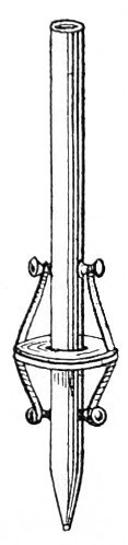
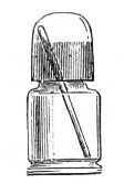
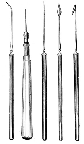
Fig. 248.—Box for keeping Glass-covers.
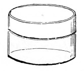
Fig. 250.—Section Lifters. Fig. 251.—Spring Flat Forceps. Fig. 251a.—Forceps with fine Points.
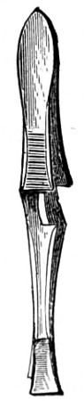
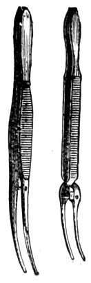
A pair of small spring steel platinum-pointed forceps for holding glass-covers (Fig. 251).
One or two pairs of fine-pointed forceps (Fig. 251a).
Collapsible tubes for containing Canada balsam and dammar.
Soft rags or old pocket handkerchiefs for removing cedar oil from lenses and cover-glasses. Chamois leather for wiping lenses and removing dust.
Reagents, alcohol, bergamot oil, celloidin, dissolved in equal parts of ether and alcohol.
Ebner’s solution. (See Appendix.)
Formalin, glycerine, gelatine, Klebs’ and Kleinenberg’s solutions. (See Appendix.) The latter consisting of a watery solution of picric acid 100 parts; strong sulphuric acid two parts; filter, and add distilled water 300 parts.
Muller fluid. (See Appendix.)
Osmic acid, a five per cent. solution.
Paraffin, spermaceti and xylol, acetic acid, hydrochloric acid, a one per cent. solution with alcohol.
Ammonia liquid, ether, picro-lithium carmine, potash solution.
Safranine, concentrated alcoholic solution of, and a watery solution.
Turpentine, vesuvin, water distilled and sterilised.
Aqueous solutions of the several dyes may be kept in bottles ready for use.
To both aqueous and alcoholic solutions a few drops of phenol, or a crystal of thymol, should be added as a preservative. For the rapid staining of cover-glass preparations, it is convenient to have the most frequently used stains—fuchsine, methyl-violet, &c.—in bottles provided with pipette stoppers.
Clearing Agents.—Oils of cedar wood, cloves, origanum, aniline, terebene, toluol and xylol, benzol and spirits of turpentine.
Mounting Media.—Acetate of potash solution concentrated, benzole, balsam, glycerine jelly, Fanant’s medium, dammar and mastic, Canada balsam in xylol, Hollis’s glue, zinc white.
Cement for fixing small specimens temporarily to a glass slide. Remove all traces of moisture, place upon it a drop or two of a medium prepared as follows:—Dissolve over a water bath 15 grammes of white lac in 100 grammes of absolute alcohol, decant off the clear liquid, and stand it by for a while.
As the alcohol evaporates from the warmed surface of the glass slide a hard transparent coating is left. This may be slightly softened at any time by means of a drop of oil of lavender. After arranging the objects the heat of a spirit-lamp will cause the oil to evaporate, leaving them firmly attached. Objects may be mounted on cover-glasses in a similar way. A resinous mounting medium may then be employed in the usual manner. If glycerine or glycerine jelly is the mounting medium employed, collodion diluted with two or three times its volume of oil of lavender may be found preferable as the fixing agent. The section should be placed in position before the preparation dries and the oil is evaporated.
Methylated spirit is often so largely adulterated with rock-oil as to render it unsuitable for technical purposes. Even to varnishes it imparts a fluorescent appearance as it dries off.
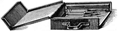
Fig. 252.—Iron Box for holding Sterilised Instruments and Glass Plates.
The needles and instruments used must not be passed through a Bunsen burner flame, which is most destructive, but enclosed in a sheet-iron box made for the purpose (Fig. 252), and placed in the hot-air steriliser for an hour at 150°C. The box can be opened at the side, and each instrument withdrawn with a pair of sterilised forceps when required for use.
Glass plates are sterilised in the same iron box, and the platinum needles for inoculating nutrient media, examining cultivations, &c., are served in the same manner before being used. The needles consist of two or three inches of platinum wire fixed to the end of a glass rod. Several of these needles should be made by fixing pieces of wire into a glass rod about six inches long. The glass rod must be heated at the extreme end in the flame of a Bunsen burner, or blow-pipe, and the platinum wire held near one extremity with forceps, and fused into the end of the glass rod. Some of these rods should be straight, and some bent, and others provided with a loop, and kept especially ready for inoculating test-tubes of nutrient jelly.
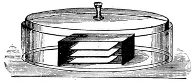
Fig. 253.—Damp Chamber for Plate-cultivations.
Glass Dishes.—Several shallow glass dishes are required for preparing damp chamber cultivations, the upper covers fitting over the under (as in Fig. 253), in the centre of which culture-plates are stacked one above the other, and when necessary placed in the incubator.
Apparatus for Incubation and Cultivations in Liquid Media.
Lister’s Flasks.—Lister devised a globe-shaped flask with two necks, a vertical and a lateral one, the lateral being a bent spout, tapering towards the extremity. When the vessel is restored to the erect position after pouring out some of its contents, a drop of liquid remains behind in the end of the nozzle, and thus prevents the regurgitation of air through the spout. A cap of cotton-wool is tied over the orifice, and the residue left in the flask for future use. The vertical neck of the flask is plugged with sterilised cotton-wool in the ordinary way.
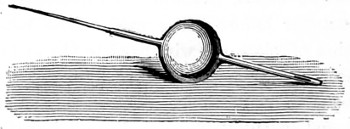
Fig. 254.—Pasteur’s Bulb Pipette.
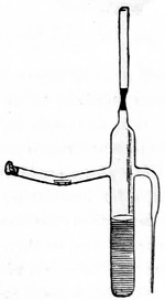
Fig. 255.—Storing Cultivation Tube.
Sternberg advocates the use of a glass bulb, provided with a slender neck drawn out to a fine point and hermetically sealed. Special forms of tubes, bulbs, and pipettes were devised by Pasteur, and are still in use at the Bacteriological Institute, Paris, and known as the Pasteur’s bulb pipette (Fig. 254).
Others are provided with lateral or with curved arms, one of which is drawn out to a fine point, and the slender neck plugged with cotton-wool, as in Fig. 255.
THE WARM CHAMBER, STERILISER, AND INCUBATOR.
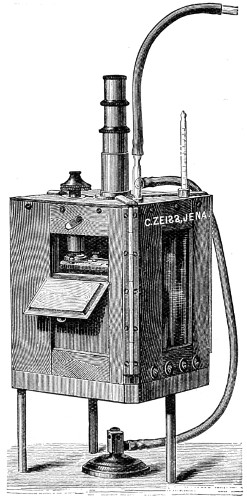
Fig. 256.—Pfeiffer’s Warm Chamber.
The Warm Chamber.—This is an accessory of importance in bacteriological work. For the continuous heating of specimens during cultivation it is an absolute necessity. Pfeiffer’s warm chamber (Fig. 256) is suitable for microscopical work generally. It consists of a hard-wood box, made air-tight, with doors and glass windows to allow of the specimen being moved from time to time, and kept under constant observation. The box is mounted on a metal plate tripod stand, and is heated from below by a small gas burner, with a thermo-regulator. A paraffin lamp will do as well, so long as it maintains a temperature of from 25° to 45°C., and without danger of injury to the stand and lenses of the microscope. A thermometer is placed in the air space to mark the temperature.
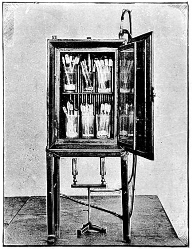
Fig. 257.—Crookshank’s Incubator.
Hot-air Incubators and Sterilisers are usually made of sheet-iron, in the form of a cubical chest, with double walls, supported on four legs, as that of Dr. Crookshank’s (Fig. 257). They are heated by gas or a lamp from below, while the temperature is indicated by a thermometer inserted through a hole in the top, as in that of the Hearson’s incubator. Test-tubes, flasks, funnels, cotton-wool, &c., must be sterilised by exposure to a temperature of 150°C. for an hour or more.
Wire Cages or crates are used for containing test-tubes, especially when they are to be sterilised in the hot-air steriliser, or for lowering tubes of nutrient jelly into the steam steriliser. All instruments, needles, scalpels, &c., before using must be carefully sterilised.
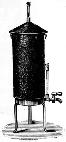
Fig. 258.—Dr. Koch’s Steam Steriliser.
Steam Sterilisers are made either of iron or tin, jacketed with thick felt, and provided with a conical cap or lid perforated at the apex to receive a thermometer (Fig. 258). Inside the vessel is an iron grating or diaphragm about two-thirds of the way down, which divides the interior into two chambers, the upper or steam chamber, and the lower or water chamber. A gauge outside marks the level of the water in the lower chamber; this should be kept about two-thirds full. The apparatus stands upon three legs, and is heated from below with a Bunsen burner or a lamp. It is employed for sterilising nutrient media in tubes or flasks, for cooking potatoes or hastening the filtration of agar-agar. When the thermometer indicates 100° C. the lid is removed, and test-tubes are lowered in a wire-basket by means of a hook and string, and the lid quickly replaced. Potatoes or small flasks are lowered into the cylinder in a tin receiver with a perforated bottom, which rests upon the grating, and admits of the contents being exposed to the steam generated.
One of the most efficient forms of incubators introduced into the bacteriological laboratory is that known as Hearson’s (Fig. 259). This consists of a chamber surrounded by a water-jacket, with water space below, to afford room for the pipe, L, which conveys the heated products from the flame of the lamp, T, through the water and back again to the lantern. A is the water-jacket surrounding the chamber containing the cultures; O, the pipe through which the water supply is admitted; N, the tap for employing the same; M, the overflow pipe; S, the capsule in a case attached by a tube to the lower plate outside; D, a lever pivoted on the left, carrying at its free end a damper, F, which, when resting on the chimney, V, effectually closes it; P, a screw for adjusting the damper when starting the apparatus; H, a lead weight for bringing more pressure on the capsule; K, a thermometer, the bulb of which is inside and the scale outside the chamber.
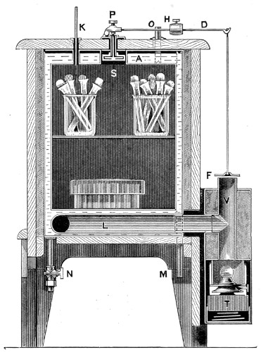
Fig. 259.—The Baird-Hearson Biological Incubator.
The treated products of combustion move in the direction indicated until the water and chamber are sufficiently heated to distend the capsule. When this point is reached the wire between S and P is pushed up by the capsule, and the lever causes the damper to rise more or less off the chimney, V, and on examining the thermometer the inside of the chamber is at length found to remain steadily at the required temperature.
When the thermometer registers the desired temperature, the lead weight must be damped to the lever by means of the milled-head screw which goes through it. After having been once adjusted the heat in the interior will remain constant, notwithstanding the utmost changes of temperature occurring in these latitudes, nor will very great alterations in the size of the lamp-flame seriously interfere with the results. The milled-head screw, P, must be turned, after the first adjustment, during the whole time that the incubator is in use. Observe the temperature before opening the door; observations taken afterwards are worthless.
Preparation of Nutrient Media—Separation, and Cultivation of Bacteria.
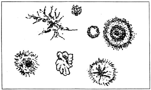
Fig. 260.—Plate Cultivation Showing Colonies.
To cultivate micro-organisms artificially they must be supplied with the proper nutrient material, perfectly free from pre-existing organisms. The secret of Koch’s methods greatly depends upon the possibility, in the case of starting with a mixture of micro-organisms, of being able to isolate them completely one from another, and to obtain an absolutely pure growth of each cultivable species. When sterile nutrient gelatine has been liquefied in a test-tube and inoculated with a mixture of bacteria in such a way that the individual micro-organisms are distributed throughout it, and the liquid is poured out on a glass plate and allowed to solidify, the individual bacteria, instead of moving about freely as in a liquid medium, are fixed to one spot, where they develop their own species. In this way colonies are formed, each possessing its own biological characteristics and morphological appearances (Fig. 260).
To maintain individuals isolated from each other during growth, and free from contamination, it is only necessary to thin out the cultivation to protect the plates from the air, and to have facilities for examining them from time to time, and observing the characteristic microscopical appearances. The colonies on nutrient gelatine examined with a low power (Fig. 260), if micro-organisms such as Bacillus anthraces and Proteus mirabilis, the naked eye appearances in test-tubes of the growth of the bacilli of anthrax and tubercle, and the brilliant growth of micro-coccus prodigiosus, may be given as examples in which the appearances are often very striking and sometimes quite characteristic. I must, however, first direct attention to a well-recognised fact, that bacteriology only touches animal pathology at a few points, and that so far from bacteria being synonymous with disease germs, the majority of these remarkable organisms appear to be beneficent rather than inimical to man. This is of immense importance to science, as I shall attempt to show further on; although even a brief description of all the useful ferments due to bacteria and brought into use would occupy a volume to themselves, and call for a school of bacteriology quite apart from that involved in the medical aspect of the question, for the purpose of fully investigating problems raised by the agriculturist, the forester, the gardener, the dairyman, brewer, dyer, tanner, and other industries, which open up vistas of practical application, and to some extent are already being taken advantage of in commerce.
The Preparation of Nutrient Gelatine and Agar-agar.—Take half a kilogramme (one pound) of beef as free as possible from fat, chop finely, transfer to a flask or cylindrical vessel, and shake up well with a litre of distilled water. Place the vessel in an ice-pail, or ice-cupboard, or in winter in a cold cellar, and leave for the night. Next morning commence with the preparation of all requisite apparatus. Thoroughly wash and rinse with alcohol about 100 test-tubes, and allow them to dry. Plug the mouths of the test-tubes with cotton-wool, place them in their wire cages in the hot-air steriliser, to be heated for an hour at a temperature of 150°C. In the same manner cleanse and sterilise several flasks, and a small glass funnel. In the meantime, the meat infusion must be well shaken, and the liquid portion separated by filtering and squeezing through a linen cloth or a meat press. The red juice thus obtained must be brought up to a litre by transferring it to a large measuring glass and adding distilled water. It is then poured into a sufficiently large and strong beaker, and set aside after the addition of ten grammes of peptone, five grammes of common salt, and 100 grammes of best gelatine.
In about half an hour the gelatine is sufficiently softened, and subsequent heating in a water bath causes it to be completely dissolved.
The next process requires the greatest care and attention. Some micro-organisms grow best in a slightly acid, others in a slightly alkaline, medium. For example, for the growth and characteristic appearances of the comma bacillus of Asiatic cholera a faintly alkaline soil is absolutely essential. This slightly alkaline medium will be found to answer best for most micro-organisms, and may be obtained as follows:—With a clean glass rod dipped in the mixture, the reaction upon litmus-paper may be obtained, and a concentrated solution of carbonate of soda must be added drop by drop until red litmus-paper becomes faintly blue. If it is too alkaline, it can be neutralised by the addition of lactic acid.
Finally, the mixture is heated for an hour in the water-bath. Ten minutes before the boiling is completed the white of an egg beaten up with the shell is added, and the liquid is then filtered while hot.
During filtration the funnel should be covered over with a plate of glass, and the process of filtering must be repeated, if necessary, until a pale straw-coloured, perfectly transparent filtrate results. The sterilised test-tubes are filled to about a third of their depth by pouring in the gelatine carefully and steadily. The object of this care is to prevent the mixture touching the part of the tube with which the plug comes into contact; otherwise, when the gelatine sets, the cotton-wool adheres to the tubes and becomes a source of embarrassment to subsequent procedures. As the tubes are filled they are placed in a basket, and then sterilised. They are either lowered into the steam steriliser, when the thermometer indicates 100 cc., for twelve minutes, for four or five successive days, or they may be transferred to the test-tube water-bath, and heated for an hour or two for three successive days.
If the gelatine shows any turbidity after, it must be poured back into a flask, boiled for ten minutes, and filtered again, and the process of sterilisation repeated.
Nutrient Agar-agar is a substance prepared from seaweed which grows on the coasts of Japan and India, and is supplied in long crinkled strips. It boils at 90° C., and remains solid up to a temperature of about 45° C. It is therefore substituted for gelatine in the preparation of a jelly for the cultivation of those bacteria which will grow best in the incubator at the temperature of the blood, and also at ordinary temperature for bacteria which lignify gelatine. The preparation is conducted on much the same principles as those already described. Instead, however, of 100 grammes of gelatine, only about twenty grammes of agar-agar (1·5 to 2 per cent.), and to facilitate the solution it must be allowed to soak in salt water overnight. Flannel is substituted for filter paper. The hot-water apparatus is invariably employed. The final results, when solid, should be colourless and clear; but if slightly milky, it may still be employed.
Wort-gelatine is used in studying the bacteria of fermentation. It is made by adding from five to ten per cent. of gelatine to beer-wort.
Glycerine Agar-agar.—This is made by adding five per cent. of glycerine to nutrient agar-agar, after the boiling and before the filtration.
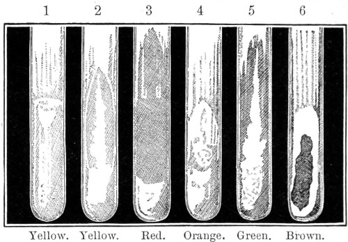
Fig. 261.—Pure Cultivation in Tubes (Crookshank).
Test-tube Cultivations.—To inoculate test-tubes containing nutrient jelly, the cotton-wool plug is removed. A sterilised needle, charged, for example, with blood or pus containing bacteria, is thrust once in the middle line into the nutrient jelly, and steadily withdrawn. The tube should be held horizontally or with its mouth downwards, and the plug replaced as quickly as possible, and an india-rubber cap fitted over the mouth of the tube.
The appearance produced by the growths in the test-tubes can be in most cases sufficiently examined with the naked eye (Fig. 261). In some cases the jelly is partially liquefied, while in others it remains solid. The growths may be abundant or scanty, coloured or colourless. When liquefaction slowly takes place in the needle tracts, the appearances which result are often very delicate and in some very characteristic. The appearance of a simple white thread with branching lateral filaments, of a cloudiness, or of a string of beads in the track of the needle, may be given as examples. In some cases much may be learnt by means of a magnifying-glass.
Beneke recommends that gelatine culture tubes should be inoculated by making a puncture quite at the side of the medium, close to the glass. The advantage of this method over the plan of inoculating the mass in the middle is that the growing culture can be microscopically examined from the outside, and various details made out, such as the nature of the growth, the comparative appearance of colonies near the surface and those situated more deeply, and the presence of one or more distinct organisms. If the tubes used have the opposite sides flat and parallel, such examinations will be still further facilitated.
Plate Cultivations.—By this method a mixture of bacteria, whether in fluids, excreta, or in cultivations on solid media, can be so treated that the different species are isolated one from the other, and perfectly pure cultivations of each of the cultivable bacteria in the original mixture established in various nutrient media. We are enabled also to examine under a low power of the microscope the individual colonies of bacteria. The same process, with slight modification, is also employed in the examination of air, soil, and water.
In order to spread out the liquid jelly evenly on the surface of a glass plate, and to hasten its solidification, it is necessary to place the plate upon a level and cool surface. The glass plates are sterilised in an iron box placed in the hot-air steriliser, at 150° C., from one to two hours.
The damp chambers for the reception of the inoculated plates are prepared by cleansing and washing out with one in twenty carbolic acid the shallow glass dish and bell-cover (Fig. 253). A piece of filter-paper should cover the bottom of dish, moistened with the same solution.
“In a glass-beaker with pad of cotton-wool at bottom place tube containing cultivation, the three tubes to be inoculated, three glass rods which have to be sterilised, and a thermometer. Liquefy the gelatine in the three tubes by placing them in a beaker containing water 30° C. Keep the tubes, both before and after the inoculation, in the warm water to maintain the gelatine in a state of liquefaction. Remove the plug from the culture and also the plug of test-tube with liquefied jelly. With the needle take up a droplet of the cultivation and stir it round in the liquefied jelly. Replace both plugs, and set aside the cultivation. Hold the freshly-inoculated tube almost horizontally, then raise it to the vertical, so that the liquid gelatine gently flows back. By repeating this motion, and rolling the tube, the micro-organisms which have been introduced are distributed throughout the gelatine. Any violent shaking, and consequent formation of bubbles, must be carefully avoided. Inoculate the second tube, and also third, in the same way, but with three droplets from a sterilised needle. The next process consists in pouring out the gelatine on glass plates and allowing it to solidify.
“Remove cover of box containing sterilised plates, withdraw a plate with sterilised forceps, and rapidly transfer it to the filter-paper under the bell-glass and quickly replace cover of box. Remove plug from the test-tube which was first inoculated, and the contents are poured out on the plate. With a glass rod the gelatine must be then rapidly spread out in an even layer within about half an inch of the margin of the plate, the bell-glass is replaced, and the gelatine is allowed to set. Meanwhile a glass bench is placed in damp chamber, upon which the plate is placed when the gelatine is quite solid; precisely the same process is repeated with the other tubes.
“The colonies will be found to develop in the course of a day or two, the time varying with the temperature of the room. The lower plate will contain a countless number of colonies, which, if the micro-organisms liquefy gelatine, speedily commingle, and produce in a very short time a complete liquefaction of the whole gelatine. On the middle plate the colonies will also be very numerous, but retain their isolated positions for a longer time; while on the uppermost plate the colonies are completely isolated from one another, with an appreciable surface of gelatine intervening.
“The microscopical appearances of the colonies are best studied by placing the plate on a slab of blackened glass, or on a porcelain slab if the colonies are coloured. A small diaphragm is used, and the appearances studied principally with a low power. A much simpler method of plate-cultivation is to pour the liquefied jelly into shallow flat dishes; they take up much less room, and in many ways are more convenient.
“Nutrient agar-agar can also be employed for the preparation of plate-cultivations, but it is much more difficult to obtain satisfactory results.”
Microscopical Examination of Bacteria.
Bacteria in Liquids, Cultures, and Fresh Tissues.—In conducting bacteriological researches, the importance of absolute cleanliness cannot be too strongly insisted upon. All instruments, glass vessels, slides, and cover-glasses should be thoroughly cleansed before use. The same applies to the preparation and employment of culture media; any laxity in the processes of sterilisation, or insufficient attention to minute technical details, will be followed with disappointing results by contamination of the cultures, resulting in the loss of much time.
For the preparation of microscopical specimens it will be found convenient to use a platinum inoculating needle, sterilised, as before directed, in the sheet-iron box; in a few moments it will be cool enough not to destroy the bacteria with which it is brought into contact.
Unstained Bacteria.—The bacteria in liquids, such as blood and culture-fluids, can be investigated in the unstained condition by transferring a drop with a looped platinum needle, or a capillary pipette, to a slide, covering it with a clean cover-glass, and examining without further treatment. If it is desirable to keep the specimen under prolonged observation, a drop of sterilised water or salt solution must be run in at the margin of the cover-glass to counteract the tendency to dry.
Cultures on the solid media can be examined by transferring a small portion with a sterilised needle to a drop of sterilised water on a slide, thinning it out, and covering with cover-glass as already described. Tissues in the fresh state may be teased out with needles (Fig. 249) in sterilised salt solution, and pressed out into a sufficiently thin layer between the slide and cover-glass. Glycerine may in many cases be substituted for salt solution, especially for such as actinomyces and mould fungi.
Very small bacilli and micro-cocci are distinguished from granular matter or fat-crystals, or vice versâ, by the fact that the latter are altered or dispersed by the addition of acetic acid, and changed by solution of potash; ether dissolves out fatty particles, while micro-organisms remain unaffected. Baumgarten demonstrated tubercle bacilli in sections by treating them with potash, which clarified the tissues and brought the bacilli clearly into view. In examining unstained bacteria the iris-diaphragm should be used, and the sub-stage condenser carefully centred and focussed.
His’s Method of Staining.—A slide is prepared as for bacteria in the fresh state; the reagents are then applied by placing them with a pipette drop by drop at a margin of the cover-glass, and causing them to flow through the preparation by means of a strip of filter-paper placed at the opposite margin.
Babès’ Method is as follows: A little of the growth spread out on a cover-glass into as thin a film as possible; when almost dry, apply a drop or two of a weak aqueous solution of methyl-violet from a pipette to the film; any excess of the stain must be removed by gentle pressure with a strip of filter-paper.
Cover-glass Preparations.—A cover-glass is smeared with the substance to be examined spread out into a sufficiently thin layer; in the case of cultures on solid media, diffuse the bacteria in a little sterilised water. By means of another cover-glass the juice or fluid is squeezed out from between them into a thin layer, and on sliding them apart each cover-glass bears on it a thin film of the material. The cover-glass is then placed with its film side upwards and allowed to dry. After a few minutes it is passed from above downwards through the flame of a Bunsen burner three times. Apply two or three drops of an aqueous solution of fuchsine or methyl-violet to cover the film, wash away any surplus stain after a few minutes with distilled water. The cover-glass is then allowed to dry, when the preparation may be mounted in Canada balsam, or while still wet, turned over on a slide, and the excess of water removed with filter-paper.
If necessary to apply stain for a much larger period, pour staining solution into a watch glass and allow cover-glass to swim on surface with prepared side downwards.
Crookshank, instead of watery solutions of aniline dyes, prefers to use stronger solutions, and to reduce the staining by a momentary immersion in alcohol. The method is as follows: cover-glass preparations are stained with carbolised fuchsine (Neelsen’s solution) for about two minutes, rinsed in alcohol for a few seconds, and quickly washed in water. This method is specially valuable for sarcinæ and streptococci.
Gram’s Method.—The whole film is first stained violet with gentian-violet, fixed by a solution of iodine, in iodide of potassium in the bacilli, but not in any débris, pus cells, or tissue elements present. Transfer cover-glass to alcohol, the bacilli alone remain stained, the violet colour being changed to blue. By employing a contrast colour, such as eosin, a double staining is obtained.
For staining preparations with gentian-violet Crookshank employs the following useful method:—Place four or five drops of pure aniline in a test-tube, add distilled water to three-quarters full, close mouth with thumb, shake thoroughly. Filter the emulsion twice, pour filtrate into watch-glass. To the perfectly clear aniline water thus obtained, add, drop by drop, a concentrated alcoholic solution of gentian-violet till precipitation commences. Cover-glasses must be left in this solution ten minutes, transferred to iodine-potassic-iodide until the film becomes uniformly brown, then rinsed in alcohol. The decolourisation may be hastened by dipping the cover-glass in clove oil and returning to alcohol. Again immerse cover-glass in clove oil, dry by gently pressing between two layers of filter-paper, and mount in Canada balsam.
Double-staining of cover-glass preparations.—They can be treated by Ehrlich’s method for staining tubercular sputum, or by Neelsen’s modification, or by staining with eosin after treatment by the method of Gram.
Ehrlich’s Method is as follows: Five parts of aniline oil are shaken up with one hundred parts of distilled water, and the emulsion filtered through moistened filter-paper. A saturated alcoholic solution of fuchsine, methyl-violet, or gentian-violet, is added to filtrate in watch-glass, drop by drop, until precipitation commences. Cover-glass preparations are floated in this mixture for fifteen minutes to half an hour, then washed for a few seconds in dilute nitric acid (one part of nitric acid to two of water), then rinsed in distilled water.
Neelsen’s Solution and Methylene Blue.—Ziehl suggested the use of carbolic acid as a substitute for aniline blue. Neelsen recommended a solution of carbolic acid, absolute alcohol and fuchsine. (See Appendix.)
Gram’s Solution and Eosin.—After using Gram’s method as above and decolourising in alcohol, the cover-glass is placed in a weak solution of eosin for two or three minutes, washed in alcohol, immersed in clove oil, dried, and mounted in balsam.
Staining of Spores.—The cover-glass preparation must be heated to 210° C. for half an hour, or passed about twelve times through the flame of a Bunsen burner, or exposed to the action of strong sulphuric acid for several seconds, then a few drops of a watery solution of aniline dye applied in the usual way. To double-stain spore-bearing bacilli the cover-glass preparation must be floated from twenty minutes to an hour on Ehrlich’s fuchsine-aniline-water, or on the Ziehl-Neelsen solution. The stain must be heated until steam arises.
Staining of Flagella.
Koch first stained flagella by floating the cover-glass on a watery solution of hæmatoxylin, transferring them to a five per cent. solution of chromic acid, or to Müller’s fluid, by which they obtained a brownish-black coloration.
Löffler’s Method.—Add together aqueous solutions of ferrous-sulphate and tannin (twenty per cent.) until the mixture turns a violet-black colour, then add three or four cc. of a one-in-eight aqueous solution of logwood; a few drops of carbolic acid may be added before transferring to a stoppered bottle; that is the mordant. The dye consists of 1 cc. of a one per cent. solution of caustic soda, added to 100 cc. of aniline water, in which four or five grammes of either methyl-violet, methylene blue, or fuchsine, are dissolved. A cover-glass preparation is made in the usual way, then the film is covered with mordant, and cover-glass held over flame until steam rises, the mordant is then washed off with distilled water. The stain is filtered and a few drops allowed to fall on film, after a few minutes the cover-glass is again warmed until steam rises. The stain is then washed off with distilled water, and the preparation is ready to be mounted for examination.
As Löffler’s process is somewhat complicated, a modification has been said to afford more satisfactory results. A specimen is taken from a recent gelatine culture and diluted with water. A little of the fluid is then transferred to a warm cover-glass by means of a pipette and allowed to dry, after which a drop of the following mordant is applied:—Aqueous solution of tannin (twenty per cent.), ten cc.; cold saturated solution of ferrous sulphate, five cc.; saturated solution of fuchsine in absolute alcohol, one cc. The cover is next heated gently for a short time until vapours are given off, then washed carefully. This process is repeated two or three times, and the specimen washed after each application. Subsequently, staining is effected by means of Ziehl’s fuchsine solution, the cover is afterwards warmed once or twice for about fifteen seconds, then washed, and the specimen examined in water to ascertain if the colour is sufficiently intense. If satisfactory, the preparation may then be dried and finally mounted in Canada balsam or dammar.
Preservation of Preparations.—After examining a cover-glass preparation with an oil-immersion objective the cedar oil must be carefully wiped off, and the slide set aside for the Canada balsam to set. At a convenient time these preparations should be sealed with a ring of Hollis’s glue.
Bacteria in Sections of Tissues.
Method of Hardening and Decalcifying Tissues.—To harden small organs, such as the viscera of a mouse, they should be placed on a piece of filter-paper at the bottom of a small wide-mouthed glass jar, and covered with about twenty times their volume of absolute alcohol. Larger organs are treated in the same way, but must be cut up into small pieces. Müller’s fluid, methylated spirit, or formalin may be used.
Teeth, or osseous structures, must first be placed in a decalcifying solution, as Kleinenberg’s. When sufficiently softened, soak in water, to wash out picric acid, and transfer to weak spirit. Ebner’s solution gives good results.
Methods of embedding, fixing, and cutting.—Crookshank finds that after hardening, the pieces of tissue are embedded in a mixture of ether and alcohol for an hour or more, then transferred to a solution of celloidin in equal parts of ether and alcohol, and left there for several hours.
The piece of tissue is then placed in a glass capsule, and some of the celloidin solution poured over it. The capsule can be placed bodily in 60 to 80 per cent. alcohol, and left there until the following morning. The celloidin should be of the consistency of wax. The piece of tissue is next cut out, and after trimming is put into water until it sinks, then transferred to gum, and cut with the freezing microtome.
Sections of fresh tissues are to be floated in ·8 per cent. salt solution, and then carefully transferred by a platinum lifter to a watch-glass containing absolute alcohol.
Staining Bacteria in Tissue Sections.—Weigert’s method is as follows:—Place sections for from six to eighteen hours in a one per cent. watery solution of any of the basic aniline dyes. To hasten, place the capsule containing solution in the incubator, or heat it to 45° C., or a stronger solution may be used. In the latter case the sections must be treated with a half-saturated solution of carbonate of potash, as they are easily over-stained. In either case the sections are next washed with distilled water, passed through sixty per cent. alcohol into absolute alcohol. When almost decolourised, spread out on a platinum lifter and transfer to clove oil, or stain with picro-carmine solution (Weigert’s) for half an hour, wash in water, alcohol, and treat with clove oil, and transfer to clean glass slide.
Gram’s Method.—Sections are stained for ten minutes in a capsule containing aniline-gentian-violet solution, then placed in the iodine and iodide solution until uniformly brown, then placed in absolute alcohol, and washed by carefully moving sections in the liquid with a glass rod. When completely decolourised, they are transferred to clove oil and then to a slide.
Double-staining is obtained by transferring the sections after decolourisation to eosin, Bismarck brown, or vesuvin (Crookshank).
Formalin is an excellent preservative fluid; one part to 20,000 is sufficient to prevent fermentation. For the preservation of vegetable sections, a one per cent. solution is required; even the fresh appearance of vegetable structures is preserved for some time when immersed in it. In the nutrient gelatine for biological specimens, if used early, will arrest the liquefaction of the gelatine by bacteria. For hardening it saves time, and is even better than alcohol, chromic acid, pot. bich., and many others. It does not cause shrinkage of the cells. Tissue ½ to ¾ inch thick hardens in twenty-four hours in pure formalin; five to ten per cent. is best for loose tissue. In another method, by which time can be saved, instead of placing the specimen in the formalin and afterwards in mucilage, prior to cutting sections, make the mucilage with two per cent. (or stronger) formalin water, and it will then answer both purposes at the same time.
Preparing, Mounting, Cementing and Collecting Objects.
Various materials are required for preparing and mounting microscopic objects, as slips of glass, patent flatted plate measuring 3 × 1 inch, thin glass covers, glass cells, preservative media, varnishes, cements, a glazier’s diamond, and a Shadbolt’s turn-table.
The glass slides and covers, although sent out packed ready for use, should be immersed in an alkaline solution to ensure perfect freedom from any greasiness derived from touching by the fingers. Dr. Seller recommends a particular solution for this purpose. (See Formulæ, Appendix.)
Varnishes and cements must be selected with care, as these are not only expected to adhere firmly to the glass slide, but also to resist the action of the preservative fluid in which the specimen may be mounted. Among the numerous preparations employed, I may enumerate Canada balsam, gum dammar, Venice turpentine, Japanners’ gold size, used for closing up cells, asphalte varnish, Brunswick black, shellac, glue and honey, Hollis’ liquid glue, and marine glue. To give a finish to the mounted specimen, coloured varnishes are sometimes resorted to. A red varnish of sealing-wax is made by digesting powdered sealing-wax in strong alcohol. Filter, and place the solution in a dish, and evaporate by means of a sand bath to reduce it to a proper consistency. This is said to resist the action of cedar oil. For white, zinc, cement is the best. This is made of benzole, gum dammar, oxide of zinc, and turpentine. Cole gives another formula, but either of these may be obtained of Squire, who supplies every kind of staining and mounting material.
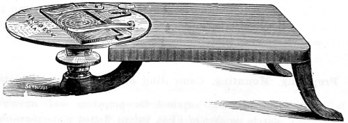
Fig. 262.—Walmsley’s Cell-making Turn-table.
Cells for Mounting.—The minuter forms of life should be mounted in thin cells, which may be readily made with Japanners’ gold size, dammar or asphalte, and a Shadbolt or Walmsley’s turntable. The glass slide being placed under the metal springs in such a manner that its two ends shall be equi-distant from the centre (a guide to the position is afforded by the circles traced out on the brass), take a camel’s hair pencil and dip it into the Japanner’s gold-size, holding it firmly between the finger and thumb, and set the wheel in motion, when a perfect circle will be formed; put it aside to dry, or place it in the warm chamber to harden. To cut cover-glasses place a sheet of thin glass under the brass springs, and substitute for the pencil a cutting diamond. A cutting diamond is not only useful to the microscopist for the above purpose, but also for writing the names of mounted objects on one end of the slide.
It will be found convenient to make a number of such cells, and keep a stock ready for use. There are many objects whose structure is very transparent. These should be mounted dry. Scales from the wings of butterflies and moths, of the podura and lepisma, and some of the diatomaceæ are of this class. All that is necessary in preparing objects for dry mounting is to take care that they are free from extraneous matter, and fix them permanently in the position in which their structure will show to the best advantage.
For mounting specimens of greater thickness it is desirable to use deeper cells. It will then be found convenient to make a second or a third application of the gold-size, allowing sufficient time between applications for the varnish to dry. Cells of a still deeper kind are made up by cementing rings of glass or metal to the glass-slides with marine glue or Brunswick black. The latter will be rendered more durable by mixing in a small quantity of indiarubber varnish (made by dissolving small strips of caoutchouc in gas-tar). The process of mounting in glass-cells is similar to that employed in making varnish-cells, except that a somewhat larger quantity of cementing medium is required. Objects mounted in this way should be kept for a time in the horizontal position, and a little fresh varnish must be applied if the cement shows a tendency to crack. In mounting objects in balsam, care must be taken to have the specimen quite dry before transferring it to turpentine. Objects mounted in cells should become perfectly saturated with the mounting fluid before being finally cemented down.
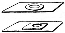
Fig. 263.—Glass-cells for Mounting.
It is preferable to mount and preserve specimens of animal tissues in shallow cells, to avoid undue pressure on the preparation. Cells intended to contain preparations immersed in fluid must be made of a substance impervious to the fluid used, such as here represented (Fig. 263). The surface of the fixed glass-circle should be slightly roughened before applying the cement.
Different modes of mounting may be employed with advantage; for instance, entomological specimens, as legs, wings, spiracles, tracheæ, ovipositors, stings, tongues, palates, corneæ, should be mounted in balsam; the trachea of the house-cricket, however, should be mounted dry. Sections of bone may either be mounted dry or in a fluid. Other objects, as sections of wood and stones of fruit, exhibit their structure best in Canada balsam.
In mounting entomological specimens, the first thing, of course, is the dissection of the insect. This is best accomplished by the aid of a dissecting microscope, a pair of small brass forceps, and finely-pointed scissors; the parts to be prepared and mounted should first be carefully detached from the insect with the scissors, then immersed in a solution of caustic alkali (liquor potassæ) for a few days, to soften and dissolve out the fat and soft parts. The length of time necessary for their immersion can only be determined by experience, but, as a general rule, the objects assume a certain amount of transparency when they have been long enough in the alkali; when this is ascertained, the object must be placed in a proper receptacle and put by to soak for two or three hours in soft or distilled water. It should then be placed between two slips of glass, and gently pressed till the softer parts are removed. Should any adhere to the edge of the object, it will be necessary to wash the specimen carefully in water, a process that will be much assisted by the delicate touches of a camel’s-hair brush. Place the object now and then under the microscope to see that all extraneous matter is removed, and when this is accomplished take the specimen up carefully with the camel’s-hair brush, or a lifter, and place it on a piece of very smooth paper (thick ivory note is the best for the purpose), arrange it carefully with the brush and a finely pointed needle, place a second piece of paper over it, and press it flat between two slips of glass, and compress it by a small spring clip (Fig. 264). A dozen clips may be had for a few pence. When thoroughly dry (which it will probably be in about twenty-four hours, if in a warm room), separate the glasses, and gently unfold the paper; then, with a little careful manipulation, the object may be readily detached, and placed in a little spirit of turpentine, where it should be allowed to remain until rendered transparent and fit for mounting. The time during which it should remain in this liquid will depend on the structure; some objects, such as wings of flies, will be quickly permeated, while horny and dense objects require an immersion of a fortnight or even longer. A pomatum pot with a concave bottom and well-fitting lid will answer admirably for conducting the soaking process in; and it is well, in preparing several specimens at a time, to have two pots, one for large and medium, the other for very small objects, otherwise the smaller will adhere to the larger.
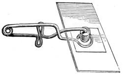
Fig. 264.—Spring Clip for Mounting.
In mounting objects in fluid, the glass cover should come nearly, but not quite, to the edge of the cell, a slight margin being left for the cement, which should project slightly over the edge of the cover, in order to secure it to the cell.
Media for Preserving Algæ.—The most useful preservative media for algæ are chrome-alum, formalin, and camphor water. The solution should consist of one per cent. of chrome-alum and one per cent. of formalin; this will render the gelatinous sheath and matrix form clear, while it will retain the colour of the algæ in most cases. The Chlorophyceæ do well in any of these media; but other species, as Ulva Lactuca, are rendered somewhat brittle. For such use formalin alone. The Phæophyceæ should be placed while fresh in the formalin; the larger forms are better fixed by placing them for an hour or two in chrome-alum solution. The Florideæ do well in any of the three solutions, but the more delicate species, Griffithsia, require a two per cent. formalin solution in sea-water; the plant preserves its natural appearance in this medium.
To preserve and mount diatomaceæ in as nearly as possible a natural condition, they should be first well washed in distilled water and mounted in a medium composed of one part of spirits of wine to seven parts of distilled water. The siliceous coverings of the diatoms, however, which show various beautiful forms under the higher powers of the microscope, require more care in preparation. The guano, or infusorial earth containing them, should first be washed several times in water till the water is colourless, allowing sufficient time for precipitation between each washing. The deposit must then be put into a test tube and nitro-hydrochloric acid (equal parts of nitric and hydrochloric acids) added to it, when a violent effervescence will take place. When this has subsided, the whole should be subjected to heat, brought nearly to the boiling point for six or eight hours. The acid must now be carefully poured off, and the precipitate washed in a large quantity of water, allowing some three or four hours between each washing, for the subsidence of some of the lighter forms. The sediment must be examined under the microscope with an inch object-glass, and the siliceous valves of the diatoms picked out with a coarse hair or bristle.
Dr. Rezner’s Mechanical Finger (Fig. 265) for selecting and arranging diatoms, adaptable to any microscope, is made to slip on to the objective far enough to have a firm bearing, and so that the bristle point can be brought into focus when depressed to its limit. It is clamped in its place by a small thumb-screw. The bristle holder slides into its place, and is carefully adjusted to the centre of the field. When using the finger, the bristle is first raised by means of the micrometer screw till so far within focus as to be nearly or quite invisible, then the objective is focussed on to the slide, and the desired object sought for and brought into the centre of the field; the bristle point is then lowered by the screw until it reaches the object, which usually adheres to it at once, and can then be examined by rotating the bristle wire by means of the milled head.
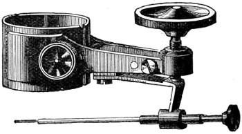
Fig. 265.—Rezner’s Mechanical Finger.
The medium used for mounting diatomaceæ is of considerable importance, inasmuch as their visibility is either diminished or much increased thereby. Professor Abbe, experimenting with the more minute test objects, diatoms, &c., found monobromide of naphthaline gave increased definition to most of them. This liquid is colourless, somewhat of an oleaginous nature, and is soluble in alcohol. Its density is 1·555, and refractive index 1·6. Its index of visibility is about twice that of Canada balsam.
Taking the refractive index of air as 1·0, and diatomaceous silex as 1·43, the visibility may be expressed by the difference ·43.
The following table may be constructed :—
Refractive indices (taken approximately). Visibility of silex (Refr. index = 1·43).
Water .. = 1·33 .. 10
Canada balsam .. = 1·54 .. 11
Bisulphide of carbon .. = 1·68 .. 25
Sol. of sulphur in bisulph. .. = 1·75 .. 32
"phosphorus" .. = 2·11 .. 67
These data relating to visibility must be taken in connection with the numerical aperture of the objectives and of the illuminating pencil. The effect produced on diatoms is very remarkable, the markings on their siliceous frustules being visible under much lower powers.
So that the visibility of the diatom mounted in phosphorus as compared with balsam is as sixty-seven to eleven; in other words, the image is six times more visible. Mr. Stephenson’s phosphorus medium is composed of a solution of solid or stick phosphorous dissolved in bisulphide of carbon. Great care is required in preparing the solution owing to the very inflammable nature of the materials. So small a quantity of the bisulphide of carbon is required to dissolve the phosphorus that the diatom may be said to be mounted in nearly pure phosphorus. Remarkable enough, this medium has the reverse effect upon such test-objects as podura and lepisma scales. These lose their characteristic markings.
For mounting minute objects, carbolic acid solution will be found a useful medium—the purest crystals of carbolic acid dissolved in just sufficient water to render them fluid. No more should be dissolved than may be wanted for the time being, as if left standing exposed to the light it changes colour. Small crustacean foraminifera, the palates of moluscs, after boiling a short time in liquid potash and well washing to remove all traces of alkali, may be preserved in carbolic acid solution. Should the specimens appear cloudy gently warm the slide over a spirit lamp.
Preserving and Killing Rotatoria with cilia in situ.—Mr. C. Rousselet’s method of preserving and mounting the Rotatoria47 has been attended with so much success that the old difficulty attendant upon the preservation of these various beautiful forms of infusorial life has been practically overcome. The process resorted to consists of four stages, namely, narcotising, killing, fixing, and preserving. In dealing with rotifers hitherto, the difficulty has been that of successfully killing them with their rotating organs fully extended. It has been found needful to have recourse in the first instance to a narcotising agent, and one that acts slowly. The most suitable is a weak solution of the hydrochlorate of cocaine, a one per cent. solution, or even weaker. This was first proposed by Mr. Weber for keeping these active little bodies quiet while under observation. Mr. Rousselet carries this agent further; he applied it to narcotise them prior to killing, and this it does most effectually. The rotifers are seen to sink to the bottom of the live-cell, and the cilia gradually to slacken in motion, and the time for killing has arrived. This is effected by Flemming’s chromo-aceto-osmic acid. A rather weak solution must be employed—consisting of 1 per cent. solution of chromic acid, 15 parts; 2 per cent. osmic acid, 4 parts; glacial acetic acid, 1 part—which is at the same time a killing and fixing medium. The word “fixing” must not be taken to imply simply fixing, as it includes rapidly killing and hardening and preventing further change in the tissues of the rotifers by subsequent treatment, as mounting. The animal, therefore, must remain quietly for a few minutes, and then taken out and washed in five or six changes of distilled water, and hence transferred to the preservative fluid. All this must be effected with great care. The best preservative fluid is simply distilled water, rendered antiseptic by a trace of the fixing solution (about eight drops to an ounce of water) giving the slightest tinge of yellow to the solution. This slight tinge of colour is imparted to the rotifers, otherwise they remain transparent and unchanged, while the nervous tissue throughout the body is brought out to perfection.
Some slight difference in treatment is required by certain species, as that of Asplanchna priodonta; after the application of the cocaine solution, which should be added slowly, that is, by letting a few drops trickle down the side of the live-trough; this, being heavier than water, sinks to the bottom, thus narcotising the rotifers, and assisting to kill them with the cilia fully expanded. They should be left quietly for fifteen minutes, then thoroughly washed with distilled water. On further experimenting, Mr. Rousselet found that a weaker solution of osmic acid alone, ¼ per cent., answers quite as well as, if not better than, Flemming’s fluid; even this must be allowed to act for only a very short time—a minute at most; the rotifers then remain white and transparent, excepting the ova, in which a fat-like substance, lecithene, is secreted. If they become too much stained, they may be decolourised by passing them through peroxide of hydrogen. For narcotising the following solution has been found most useful:—Take a 2 per cent. solution of cocaine hydrochlorate, 3 parts; methylated spirit of wood naphtha, 1 part; and distilled water, 6 parts. This must be added as before directed, drop by drop, watching the effect upon the rotifers under the microscope.
All the rotatoria may be killed and preserved in the same way. For mounting, Mr. Rousselet prefers a slightly hollowed-out glass cell, the advantage of which is that the rotifers are kept to the centre, and cannot move to the edge. A little difficulty at first presents itself to exclude air-bubbles, but this, with a little care, can be overcome by placing a drop of a two or three per cent. solution of formalin, just sufficient to fill the cell. Then transfer the rotifers with a dipping pipette to the cell, and lower the cover-glass down very gently, removing any excess of fluid by blotting-paper. The best cement for the cover-glass is gold-size.
Method of Cementing.—After many years’ experience, I have arrived at the conclusion that for cementing down the cover-glass there is nothing better than either gold size or gum dammar varnish. The latter, for some preparations, will be improved by the addition of a small proportion of indiarubber dissolved in naphtha. (See Appendix.)
Should glycerine be preferred, carefully wash away any surplus quantity by gently syringing; then apply a ring of waterproof cement round the cover-glass. An inexpensive one can be made by dissolving ten grains of gum-ammoniac in an ounce of acetic acid, and adding to this solution two drachms of Cox’s gelatine. This liquid flows easily from the brush and is waterproof, rendered more so if subsequently brushed over with a solution of ten grains of bichromate of potash in an ounce of water. An especial recommendation to this cement is its adhesiveness to glass, even should there be a little glycerine left behind on the cover. After the gelatine ring is thoroughly dry any kind of cement may be employed.
A useful cement for fixing minute objects, diatoms, &c., temporarily to thin glass covers, before permanently mounting them in Canada balsam, is made as follows:—Dissolve, without heat, two or three grains of gum arabic in one ounce of distilled water, then add glacial acetic acid, three minims, and the least trace of sugar. Filter carefully through filter paper, and repeat this in the course of three or four weeks. This cement will be unaffected by the balsam.
Mounting Chara.—It is often found difficult to preserve and mount the fruit of chara, but this can be successfully accomplished in glycerine jelly, by taking the following precautions. After cleaning the specimen place it in 92 per cent. of alcohol for several hours, then transfer it to a mixture of equal parts of spirit and glycerine for several hours longer, pour off nearly all the mixture, and add pure glycerine at intervals till the glycerine becomes concentrated. The specimen is then mounted in glycerine jelly in a cell just deep enough to take it without pressure.
There are some objects much more difficult to prepare than others, and which tax the patience of the beginner in a manner which can hardly be imagined by any one who has never made the attempt. The structure of many creatures is so delicate as to require the very greatest care to prevent mutilation, and consequent spoliation, of the specimen. The beginner, therefore, must not be discouraged by a few failures in commencing, but should persevere in his attempts, and constant practice will soon teach him the best way of managing intricate and difficult objects. The room in which he operates should be free from dust, smoke, and intrusion, and everything used should be kept scrupulously clean, since a very small speck of dirt, which may be almost invisible to the naked eye, will assume unpleasant proportions under the microscope, and not only mar, but possibly spoil a fine and delicate preparation.
Few students on commencing to work with the microscope will fully realise the fact that under medium or high powers the natural appearance of almost all objects is changed by the refractive nature of the fluid medium in which they are immersed and which enters more or less into their composition. The remarkable changes effected by the law of diffusion, when alkaloid substances enter into their composition, show the necessity of taking every precaution in the employment of preservative fluids. Glycerine affords an example of the chemical change induced, should the preparation have been passed through an alkaline solution.
Air Bubbles are a constant source of annoyance both in preparing and mounting. These may be removed from the specimen by gently warming the under part of the slide over a spirit lamp, or placing the slide in the warm chamber, when the bubbles will move towards the edge of the cover-glass and ultimately disappear. The air-pump is preferred by many microscopists.
Collection of Objects.
Infusorial Life, with all its fascinations, was fully unveiled to naturalists by the celebrated Ehrenberg. It was he who termed it infusorial, because he first met with the more interesting forms of minute life in infusions of hay and other vegetable substances. Since his day it is a well-known experience of those who take up the microscope that the most interesting objects to commence with are infusorial living creatures of sufficient dimensions to be easily understood and seen with moderate magnifying powers. Moreover, infusoria are more readily found in almost any pool or running stream of water, either near the surface or clinging to the under surfaces of aquatic plants. At one time all the small shallow pools in the neighbourhood of London—Hampstead Heath, Clapham, Wandsworth, and other commons—abounded in the most interesting forms of life, were famous hunting grounds for the marvellous volvox, the charming dismid and diatom, the wonderful budding and self-dividing hydra. A few hours’ ramble furnished the microscopist with a bountiful supply of these and many other forms of life. Now all is changed; our commons have been devoted to other purposes, and with the general levelling up all the little pools have disappeared, and the microscopist has been warned off and driven further afield, or seeks the good offices of a country friend for an occasional peep into pond life.48
A teaspoonful, however, judiciously taken from a well-chosen locality will often be found to contain a variety of living forms, every one of which will deserve a careful and patient study.
Of the microscopic organisms, the collection of which requires no other methods than those ordinarily pursued by the naturalist, most of them must be sought for in pools or running waters, basking in the sunshine, clinging to leaves and rootlets of all aquatic plants; some freely moving about, others clinging to stones or pieces of wood at the bottom. Dismids congregate in shallow waters or rise to the surface in a quiet nook, while the diatomaceæ are seen covering the bottom of clear water, to which they give a yellowish-brown tinge of colour.
Infusorial animal life, as vorticellæ, stentors, rotifers, and various polyzoa, cling, as also do hydra, in colonies to vallisneria, duck-weed, frogbit, or small branches dipping down under water; and if some of the water-weed is brought home the little creatures will live and thrive for several weeks. No waters, however, are so full of minute animal life as the sphagnum bog. A number of species of diatoms, as well as protozoids and the smaller molluscs, will be found in all peat bogs. It is remarkable, too, that the same species, everywhere, are associated with this kind of moss. Lord Sidney Godolphin Osborn supplied his friends with moss growing in a damp part of the garden walk of his rectory; this always furnished the same species of rotifers. These proved to be most interesting objects to my friends, and in an early communication I described them as indestructible, since they will bear any amount of desiccation; nevertheless, they were revived when a drop of water was introduced into the glass-cell.
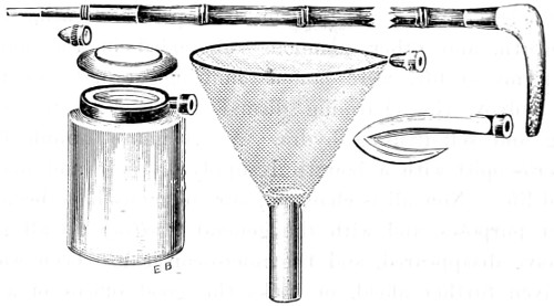
Fig. 266.—Collecting Stick, Bottle, Hook, and Net.
The Thames mud always furnishes a number of beautiful forms of triceratum. Lower down the river, as brackish water is reached, greater varieties of diatoms appear. But to secure them the collector must be provided with a collecting stick. A convenient form is furnished by Messrs. Baker (Fig. 266). This consists of an ordinary walking-stick, together with a lengthening rod, a cutting hook to clear away weeds, ringed bottles with screw tops, and a net with a glass tube attached. Their uses are too obvious to need further description.
The siliceous skeletons of diatoms are met with in the fossil state. Among the first discovered of the infusorial strata were the polishing slates of Bilin and Tripoli, the berg-mehl or mountain meal, the entire mass of which is composed of the siliceous skeletons of different species of diatoms. Richmond, Virginia, is rich in the same organisms, while the great mass of our chalk cliffs are composed of foraminiferous shells, xanthidiæ, &c. One remarkable fact in connection with fossil infusoria is that most of the forms are still found in the recent state. The beautiful engine-turned discs, Coscinodisci, so abundant in the Richmond earth, may be met with in our own seas, and in great profusion in the deposits of guano on the African and American coasts, and in the stomachs of the oyster, scallop, and other salt-water molluscous animals common to our shores.
A great number of infusorial earths may be mounted as dry objects, while others require careful washing and digesting in appropriate media. The finer portions of the sediments will be found to contain the better and more perfect siliceous shells.
Preparing and Mounting Apparatus.
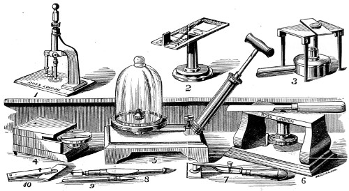
Fig. 267.—Mounting Apparatus.
1.—Ross’s instrument for cutting thin covering-glass for objects. This apparatus consists of a bent arm supporting the cutting portion of this apparatus, which consists of a vertical rod with a soft cork at one end. A brass arm at right angles carries the diamond parallel with and close to the main rod.
2.—Covering-glass measurer. To measure the thickness of covering-glass, place it between the brass plate and the steel bearing; the long end of the lever will then indicate the thickness on the scale, to 1⁄50-th, 1⁄100-th, or 1⁄1000-th inch.
3.—Brass table on folding legs, with lamp for mounting objects.
4.—Whirling table with eccentric adjustment for making cells and finishing off slides.
5.—Air-pump with glass receiver, 3½-inch brass plate for mounting objects and withdrawing air-bubbles.
6.—Improved table with knife for cutting soft sections. This consists of an absolutely flat brass table, with a square hole to receive the wood, or other matter, on a movable screw, which adjusts the thickness of the section.
7.—Smith’s holder with spring and screw for adjusting pressure when mounting objects.
8.—Cutting diamonds for cell-making and cutting slips of glass.
9.—Writing diamonds for cutting thin covering-glass and naming objects.
10.—Page’s wooden forceps, for holding glass slips or objects when heated, during mounting.