Part 2 — CHAPTER I. (continued 2)
Reading edition
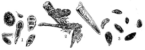
Fig. 281.—Ærobic Spores × 200.
1. Ærobic fungi caught over a sewer; 2. Fragments of Penicillium spores; 3. Ærobic fungi taken in the time of the cholera visitation, 1854.
With reference to the ærobic bacteria I have only to add that in addition to the simple mode of taking them on glass slides smeared over with glycerine, special forms of æroscopes are now in use for the purpose, consisting of a small cylinder in which a current of air is produced by an aspirator and diffused through a glass vessel containing a sterilised fluid. These are in constant use in all bacteriological laboratories. The results obtained are transferred to sterilised flasks or tubes as those shown in a former chapter.
Miquel, who has given considerable attention to the subject of ærobic and anærobic bacteria, reckons that the number of spores that find their way into the human system by respiration, even should health be perfectly sound, may be estimated at 300,000 a day.
One of the most commonly met with forms of micro-organisms is Leptothrix buccalis. It chiefly finds its nutritive material in the interstices of the teeth, and is composed of short rods and tufted stems of vigorous growth, to which the name of Bacillus subtilis has been given (Fig. 282). Among numerous other fungoid bodies discovered in the mouth, Sarcinæ have been found. See Plate IX., No. 7.
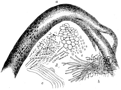
Fig. 282.—Section of the Mucous Membrane of the Mouth, × 350.
Showing: a. The denser connective tissue; b. Teased out tissue; c. Muscular fibre; d. Leptothrix buccalis, together with minute forms of bacteria and micrococci; e. Ascomycetes and starch granules.
The Beggiatoa, a sewage fungus, found by me in the river Lea water of 1884 growing in great profusion, consists chiefly of mycelial threads and a number of globular, highly refractive bodies, and may be regarded as evidence of the presence in the water of an abnormal amount of sulphates which set free a gas, sulphuretted hydrogen, of a dangerous and offensive character. Another curious body closely allied to Beggiatoa alba is Cladothrix; this assumes a whitish pellicle on the surface of putrefying liquids.
These saprophytes obtain nourishment from organic matter; nevertheless they are not true parasites in the first stage of their existence, during which they live freely in the water or in damp soil; they, however, become pathogenic parasites when they penetrate into the tissues of animals, and necessarily live at the expense of their host.
Fungi, Algæ, Lichens, etc.
Tuffen West, del. Edmund Evans.
Plate I.
Bacteria, as I have said, were for a long time classed with fungi under the name of Schizomycetes. But the more recent researches into their organisation, and more especially into their mode of reproduction, show that they rather more resemble a group of algæ devoid of chlorophyll. Zopf asserts that the same species of algals may at one time be presented in the form of a plant living freely in water, or in damp ground, in association with chlorophyllaceous protoplasm, and at another in the form of a bacterium devoid of green colouring matter, and receiving nourishment from organic substances previously elaborated by plants or animals, thus accommodating itself, according to circumstances, to two very different modes of existence.
That widely-distributed single-cell plant, the Palmoglœa macrococca of Kützing, that spreads itself as a green slime over damp stones, walls, and other bodies, affords an example. If a small portion be scraped off and placed on a slip of glass, and examined with a half or a quarter-inch power, it will be seen to consist of a number of ovoid cells, having a transparent structureless envelope, nearly filled by granular matter of a greenish colour. At certain periods this mass divides into two parts, and ultimately the cell becomes two. Sometimes the cells are united end to end, just as we see them united in the actively-growing yeast plant; but in this case the growth is accelerated, apparently, by cold and damp. Another plant belonging to the same species, the Protococcus pluvialis, is found in every pool of water, the spores of which must be always floating in the air, since they appear after every shower of rain.
Protococcus pluvialis is furnished with motile organs—two or more vibratile flagella passing through perforations in the cell-wall—whereby, at certain stages, they move rapidly about. The flagella are distinctly seen on the application of the smallest drop of iodine. The more remarkable of the several forms presented by the plant is that of naked spores, termed by Flotow Hæmatococcus porphyrocephalus. These minute bodies are usually seen to consist of green, red, and colourless granules in equal proportions, and occupying different portions of the cell. They seem to have some share in the after subdivision of the cell (Fig. 283). There are also still-cells, which sub-divide into two, while the motile cells divide into four or eight. It is not quite clear what becomes of the motile zoospores, B, but as they have been seen to become encysted, they doubtless have a special function, or become still-cells under certain circumstances.
It appears that both longitudinal and transverse division of the primordial cell takes place; and that the vibratile flagella of the parent cell retain to the last their function and their motion after the primordial cell has become detached and transformed into an independent secondary cell (Fig. 283, G).
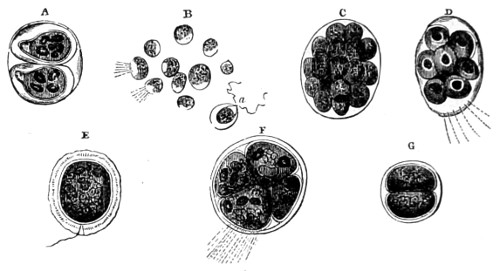
Fig. 283.—Cell Development. (Protococcus pluvialis.)
Protococcus pluvialis, Kützing. Hæmatococcus pluvialis, Flotow. Chlamidococcus versatilis, A. Braun. Chlamidococcus pluvialis, Flotow and Braun.
A. Division of a simple cell into two, each primordial vesicle having developed a cellulose envelope; B. Zoospores, having escaped from a cell; C. Division of an encysted cell into segments; D. Division of another cell, with vibratile flagella projecting through cell-wall; E. An encysted flagellate cell; F. Division of an encysted nucleated cell into four parts, with vibratile filaments projecting; G. Fission of a young cell.
The most striking of the vital phenomena presented by Protococcus is that of periodicity. Certain forms—for instance, encysted zoospores, of a certain colour, appear in a given infusion, at first exclusively, then they gradually diminish, become more and more rare, and finally disappear altogether. After some time their number again increases, and this may be repeated. Thus, a cell which at one time presented only still forms at another contained only motile ones. The same may be said with respect to segmentation. If a number of motile cells be transferred from a larger vessel into a smaller one, in the course of a few hours most of them will have subsided to the bottom, and in the course of the day observed to be on the point of sub-division. On the following morning division will have become completed; on the next day the bottom of the vessel will be found covered with a new generation of self-dividing cells, which, again, will produce another generation. This regularity, however, is not always observed. The influence of every change in the external conditions of life upon the plant is very remarkable. It is only necessary to pour water from a smaller into a larger or shallower vessel to at once induce segmentation of cells. The same phenomenon occurs in other algals; thus Vaucheria almost always develops zoospores at whatever time of year they may be brought from their natural habitat into a warm room. Light is conducive to the manifestation of vital action in the motile spores; they usually collect in great numbers on the surface of the water, and at that part exposed to the strongest light.
But in the act of propagation, on the contrary, and when about to pass into the still condition, the motile Protococcus cell seems to shun light, and falls to the bottom of the vessel. Too strong sunlight, as when concentrated by a lens, quickly kills the young zoospores. A temperature of undue elevation is injurious to the development of their vital activity and the formation of the zoospores. Frost destroys motile, but not still zoospores.55
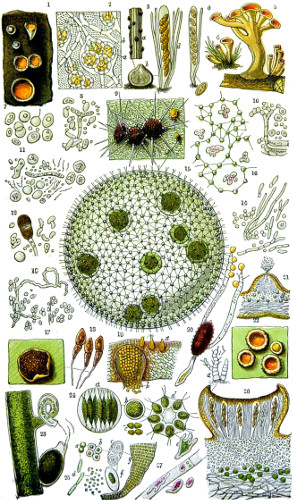
Stephanosphæra pluvialis is a conspicuous variety of the fresh-water algals, described by Cohn. It consists of a cell containing eight primordial cells filled with chlorophyll, uniformly arranged (see Plate I., No. 24 d). The globular mother-cell rotates, somewhat in the same way as the volvox, by vibratile flagella, two of which are seen projecting from each cell and piercing the transparent outer cell wall. Every cell divides first into two, then four, and lastly eight cells, each one of which again divides into a number of micro-gonidia, which have a motion within the mother-cell, and ultimately escape from it. Under certain circumstances each of the eight young cells is observed to change places in the interior of the cell; eventually they escape, lose their flagella, form a thicker membrane as at b, and for a time remain motionless, and sink to the bottom of the vessel in which they are contained. If the vessel is permitted to become thoroughly dry, and then again has water poured into it, motile cells reappear; from which circumstance it is probable that these represent the resting spores of the plant. When in the condition of greatest activity its division into eight is perfected during the night, and early in the morning light the young cells escape and pass through similar changes. It is calculated that in eight days, under favourable circumstances, 16,777,216 families may be formed from one resting-cell of Stephanosphæra. In certain of the cells, and at particular periods, remarkable amœboid bodies (Plate I., No. 24 c) make their appearance. There is a marked difference between Stephanosphæra and Chlamydococcus, for while in the latter the individual portions of a primordial cell separate entirely from one another, each developing its own enveloping membrane, and ultimately escaping as a unicellular individual; in the former, on the other hand, the eight portions remain for a time living in companionship.
Volvocineæ.—A fresh-water unicellular plant of singular beauty and interest to the microscopist is the Volvox globator (Plate I., No. 15). No. 16 represents a portion of another cell, with brownish amœboid bodies enclosed in the protoplasmic web. It is common to our fresh-water pools, and attains a diameter of about 1⁄20th or 1⁄30th of an inch. Its movement is peculiar, a continued roll onwards, or a rotation like that of a top; at other times it glides along smoothly. When examined under a sufficiently high power, it is seen to be a hollow sphere, studded with green spots, and traversed by green threads connecting each of the spots or spores with the maternal cell. From each of the spores proceed two long flagella, lashing filaments, which keep the globular body on the move. After a time the sphere bursts, and the contained sporules issue forth and speedily pass through a similar stage of development. These interesting cells were long taken to be animal bodies. Ehrenberg described them as Monads, possessing a mouth, stomach, and an eye.
The setting free of the young volvox is essentially a process of cell division, occurring during the warmer periods of the year, and, as Professor Cohn shows, is a considerable advance upon the simpler conjugation of two smaller cells in desmids; it more closely resembles that which prevails among the higher algæ and a large number of cryptogams. As autumn advances the volvox spherules usually cease to multiply by the formation of zoosporanges, and certain of their ordinary cells begin to undergo changes by which they are converted, some into male or sperm-cells, others into germ-cells, but the greater number appear to remain sterile. Both kinds of cells at first so nearly resemble each other that it is only when the sperm cells begin to undergo sub-division that they are seen to be about three times the size of the sterile cells. Then the primary cell resolves itself into a cluster of peculiar secondary cells, each consisting of an elongated body containing an orange-coloured endochrome and a pair of long flagella, as seen in the antherozoids of the higher cryptogams. As the sperm-cells approach maturity the clusters may be seen to move within them; the bundles then separate and show an independent active movement while still within the cavity of the primary cell, and finally escape through a rupture in the cell-wall, rapidly diffusing themselves as they pass through the cavity. The germ-cells continue to increase in size without undergoing sub-division, at first showing large vacuoles in their protoplasm, but subsequently becoming filled with a darker coloured endochrome. The form of the cell also changes from its flask-like shape to the globular, and at the same time seems to acquire a firmer envelope. Over this the swarming antherozoids diffuse themselves and penetrate the substance to the interior, and are then lost to view. The product of this fusion, Cohn tells us, is a reproductive cell, or “oospore,” which speedily becomes enveloped in another membrane with a thicker external coat, beset with conical-pointed processes; and now the chlorophyll of the young cell gives place, as in Palmoglæ, to starch and reddish or orange-coloured, and a more highly refractive, fluid. As many as forty of such oospores have been counted in a single sphere of volvox, which then acquires the peculiar appearance observed by Ehrenberg, and described by him under the name of Volvox stellatus. The further history of this wonderful spheroid unicellular plant has been traced out by Kirchner, who found that their germination commences in the early months of the year—in February—with the liberation of the spherical endospore from its envelope and its division into four cells. A remarkable phenomenon has been observed by Dr. Braxton Hicks—the conversion of an ordinary volvox cell into a moving mass of protoplasm that bears a striking resemblance to the well-known amœba. “Towards the end of the autumn the endochrome mass of the volvox increases to nearly double its ordinary size, but instead of undergoing the usual sub-division so as to produce a macrogonidium, it loses its colour and regularity of form, and becomes an irregular mass of colourless protoplasm, containing a number of brownish granules.” The final change and the ultimate destination of these curious amœboid bodies have not been satisfactorily made out, but from other observations on the protoplasmic contents of the cells of the roots of mosses, which in the course of two hours become changed into ciliated bodies, it is believed that this is the mode in which these fragile structures are enabled to retain life and to resist all the external conditions, such as damp, dryness, and the alternations of heat and cold.
It would be quite impossible to deny the great similarity there is between the structure of volvox and that of the motile cell of Protococcus pluvialis. The influence of reagents will sometimes cause the connecting processes of the young cells, as in Protococcus, to be drawn back into the central mass, and the connecting threads are sometimes seen as double lines, or tubular prolongations of the membrane. At other times they appear to be connected by star-like prolongations to the parent cell (Plate I., No. 15), presenting an almost identical appearance with Pediastrum pertusum. Another body designated by Ehrenberg Sphærosira volvox is an ordinary volvox in a different stage of development; its only features of dissimilarity being that a large proportion of the green cells, instead of being single, are double or quadruple, and that the groups of flagellate cells form by their aggregation discoid bodies, each furnished with a single flagellum. These clusters separate themselves from the parent cell, and swim off freely under the forms which have been designated Uvella and Syncrypta by Ehrenberg. Mr. Henry Carter, F.R.S., who made a careful investigation of unicellular plants, described Sphærosira as the male, or spermatic form of volvox.
Among other organisms closely allied to volvox and included in Volvocineæ, affording the microscopist many interesting transitional forms in their various modes of fructification, are the Eudorina, still-water organisms that pass through a similar process of reproduction as the volvox. In the Pandorina morum, its reproduction is curiously intermediate between the lower and the higher types; as within each cell is a mulberry-like mass, composed of cells possessing a definite number of swarm spores, sixteen usually, which rupture the mother cell, and swim off furnished with a pair of flagella. A similar process takes place in some of the Confervaceæ and other fresh-water algæ. The Palmella, again, consist of (Plate I., No. 21) minute organisms of very simple structure, which grow either on damp surfaces or in fresh water. The stonework of some of our churches is often seen to be covered with a species of Palmella, that take the form of an indefinite slimy film. The “red snow” of Arctic or Alpine regions, considered to be a species of Protococcus, is frequently placed among Palmella. A more characteristic form of the P. cruenta is the Hæmatococcus sanguinis, the whole mass of which is sub-divided by partitions enclosing a larger or smaller number of cells, which diffuse their granular contents through the gelatinous mass in which their several changes take place. The albuminoid envelope of these masses is seen to contain parasitic growths, which have given rise to some discussion, especially when their filaments are observed to radiate in various directions.
The Oscillariaceæ constitute a genus of Confervaceæ which have always had very great interest for the microscopist in consequence of their very remarkable animal-like movements, and from which they derive their generic name. For more than a century these Bacillaria have excited the curiosity of all observers without any one having derived more than an approximate idea of their remarkable rhythmical movements. The frustule consists of a number of very fine short threads attached together by a gelatinous sheath, in one species all of equal length. Their backward and forward movement is of a most singular character; the only other conferva in which I have seen a motion of a similar kind is the Schizonema. In this species the frustules are packed together in regular series, the front and side views being always in the same direction. These several bodies move along within the filamentous sheath without leaving their respective places. On carefully following the movement, it is seen at first much extended, and then more compressed, while the frustules become more linear in their arrangement, and present a closer resemblance to Bacillaria paradoxa, augmented by the circumstance that the frustules are seen to move in both directions. A frustule of Schizonema can move independently of the sheath, and so will a detached frustule of bacillaria. This peculiar and exceptionally anomalous phenomenon as that of the movements of bacillaria can hardly be confined to a solitary species. The movements of the frustules are much accelerated by warmth and light. The longer filaments of other minute species only slightly exhibit any motion of the kind, but have peculiar undulating motions.
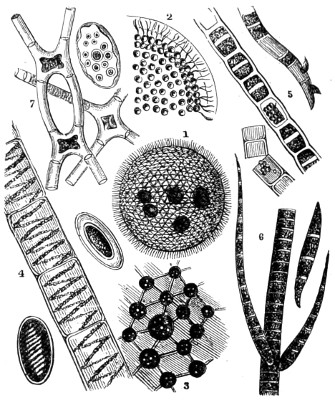
Fig. 284.—Confervaceæ.
1. Volvox globator; 2. A section of volvox, showing the flagellate margin of the cell; 3. A portion more highly magnified, to show the young volvocina, with their nuclei and thread-like attachments; 4. Spirogyra, near which are spores in different stages of development; 5. Conferva floccosa; 6. Stigeoclonium protensum, jointed filaments and single zoospores; 7. Staurocarpus gracilis, conjugating filaments and spores.
Confervaceæ are a genus of algals. The species consist of unbranched filaments composed of cylindrical or moniliform cells, with starch granules. Many are vesicular, and all multiply by zoospores generated in the interior of the plant at the expense of the granular matter. They are, for the most part, found in fresh water attached or floating, some in salt water, and a few in both, in colour usually green, but occasionally olive, violet, and red. The Confervaceæ proper are often divided into four families: 1. Hydrodictidæ; 2. Zygnemidæ; 3. Confervidæ; 4. Chætophoridæ. To the microscopist all the plants of this genera are extremely interesting as subjects for the study of cell multiplication. The process usually takes place in the terminal cell, the first step in which is the division of the endochrome, and then follows a sort of hour-glass contraction across the cavity of the parent cell, whereby it is divided into two equal parts. This is better seen in some of the desmids than in Fig. 284, Nos. 4, 5, and 6. Some species are characterised by a different mode of reproduction; these possess a number of nuclei, and multiply by zoospores of two kinds, the largest of which have either two or four cilia, which germinate directly the smaller are biciliated; conjugation has been seen to take place in a few instances.
Allied to the Confervaceæ is an interesting plant, Sphæroplea annulina, which has received careful attention from Cohn. The oospores of this plant are the product of a process partaking of a sexual nature, and when mature are filled with reddish fat vesicles which divide by segmentation.
The Ædogoniaceæ also closely resemble Confervaceæ in habits of life, but differ in some particulars, especially so in the mode of reproduction (only a single large zoospore being set free from each cell) and by the almost complete fission of the cell-wall or one of the rings which serve as a hinge. The zoospores are the largest known among algals, and each is described as having a red eye-spot. The Chætophoraceæ form an interesting group of confervoid plants, and are usually found in running streams, as they prefer pure water. One of the characteristics of the group is that the extremities of the branches are prolonged into an acute-shaped termination, as represented in Fig. 284, No. 6. A very pretty object under the microscope is Draparnaldia glomerata, belonging to this species. It consists of an axis composed of a row of cells, and at regular intervals whorls of slender prolongations, containing chlorophyll or endochrome of a deeper green; these attain to an extraordinary length.
The Batrachospermæ bear a strong resemblance to frog-spawn, from which they derive their name, and are chiefly a marine group of algals allied to the Rhodespermeæ or red seaweeds. The late Dr. A. Hassall first described them; they have since received more careful attention from M. Sirodot. They are reddish-green, extremely flexible, and nothing can surpass the grace of their movements in water; but when removed from their element they lose all form, and resemble a jelly-like substance without a trace of organisation; but if allowed to remain quiet they regain their original shape.
The presence of the cell-membrane will be best demonstrated by breaking up the filaments, either by moving the thin glass cover, or by cutting through a mass of them in all directions with a fine dissecting knife. On now examining the slide, in most instances many detached empty pieces of the cell-membrane, with its striæ, will be seen, as well as filaments partly deprived of protoplasm. On the application of iodine all these appearances become more distinguishable in consequence of the filament turning red or brown, while the empty cells remain either unaffected, or present a slight yellowish tint, as is frequently the case with cellulose when old.
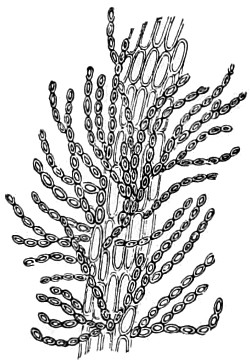
Fig. 285.—Mesoglia vermicularis.
With regard to the contents of the cell, the endochrome is coloured in the Oscillatoriæ, and is distinguishable by circular bands or rings around the axis of the cylindrical filament. Iodine stains them brown or red, and syrup and dilute sulphuric acid produce a beautiful rose colour. As to their mode of propagation, nothing positive is known. If kept for some time they gradually lose their green colour; a portion of the mass, becoming brown, sinks to the bottom of the vessel, and presents a granular layer.
Mesoglia vermicularis (Fig. 285) consists of strings of cells cohering and held together by their membranous covering. In the lowly organised plant Vaucheria (Plate I., No. 23, V. sessilis)—so named after its discoverer Vaucher, a German botanist—a genus of Siphonaceæ, we have an example of true processes of sexual generation. The branching filaments are often seen to bear at their sides peculiar globular bodies or oval protuberances, nipple-shaped buddings-out of the cell-wall, filled with a dark-coloured endochrome and distributed in pairs, one of which curves round to meet the other, when conjugation is seen to take place. Near these bodies others are found with pointed projections, which have been described as “horns,” but these, Pringshelm says, are “antherids which produce antherozoids in their interior,” while the capsule-like bodies constituting the oospores become, when fertilised, a new generation, which swarm out through a cavity or aperture in the parent cell-wall.
The fruit of fresh-water and most olive-green algals is enclosed in spherical cavities under the epidermis of the frond, termed conceptacles, and may be either male or female. The zoids are bottle-shaped and have flagella; the transparent vesicle in which they are contained is itself enclosed in a second of similar form. In monœcious and diœcious algals the female conceptacles are distinguished from the male by their olive colour. The spores, when developed, are borne on a pedicle emanating from the inner wall of the conceptacle. They rupture the outer wall at its apex; at first the spore appears simple, but soon after a series of changes takes place, consisting in a splitting up of the endochrome into six or eight masses of spheroidal bodies. A budding-out occurs in a few hours’ time, and ultimately elongates into a cylindrical thread. The Vaucheria present a double mode of reproduction, and their fronds consist of branching tubes resembling in their general character that of the Bryophyta, from which indeed they differ only in respect of the arrangement of their green contents. In that most remarkable plant Saprolegnia ferox, which is structurally so closely allied to Vaucheria, though separated from them by the absence of green colouring matter, a corresponding analogy in the processes of development takes place. In the formation of its zoospores, an intermediate step is presented between that of the algæ and a class of plants formally placed among fungi.
The Ulvaceæ.—The typical form of seaweeds is the Ulva lactuca, well known from its fronds of dark-green “laver” on every coast throughout the world. Ulvæ are seen to differ but little from the preceding group of fresh-water algals. The specific difference is that the cells, when multiplied by binary subdivision, not only remain in firm connection with each other but possess a more regular arrangement. The frond plane of the algal is either more simple or lobed, and is formed of a double layer of cells closely packed together and producing zoospores. The whole group is chiefly distinguished from Porphyra by their green colour, the latter being roseate or purple. Ulvæ are mostly marine, with one or two exceptions. One species (U. thermalis) grows in the hot springs of Gastein, in a temperature of about 117° Fahr. The development of Ulvæ is seen in Fig. 286. The isolated cells, A, resemble in some points those of the Protococcus; these give rise to successive subdivisions determining the clusters seen at B and C, and by their aggregation to the confervoid filament shown at D. These filaments increase in length and breadth by successive additions, and finally take the form of fronds, or rows of cells.
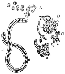
Fig. 286.—Successive Stages of Development of Ulvæ.
A. Isolated spores; B and C. Clusters of cells; D. Cells in the filamentous stage.
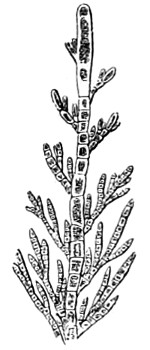
Fig. 287.—Sphacelaria cirrhosa, with spores borne at the sides of the branchlets.
The marine greenish-olive algæ present a general appearance which might at first sight be mistaken for plants of a higher order of cryptogams. Their fronds have no longer the form of a filament, but assume that of a membranous expansion of cells. The cells in which zoospores are found have an increased quantity of coloured protoplasm accumulated towards one point of the cell-wall; while the zoospores are observed to converge with their apices towards the same point. In some algæ, which seem to be closely related in form and structure to the Bryophyta, we notice this important difference, that the zoospores are developed in an organ specially destined for the purpose, presenting peculiarities of form and distinguishing it from other parts of the branching tubular frond. In the genus Derbesia distinct spore cases develop, a young branch of which, when destined to become a spore case, instead of elongating indefinitely, begins, after having arrived at a certain length, to swell out into an ovoid vesicle, in the cavity of which a considerable accumulation of protoplasm takes place. This is separated from the rest of the plant, and becomes an opaque mass, surrounded by a distinct membrane. After a time a division of the mass takes place, and a number of pyriform zoospores, each of which is furnished with flagella, are set free.
Desmidiaceæ, Diatomaceæ, Algæ.
Tuffen West, del. Edmund Evans.
Plate II.
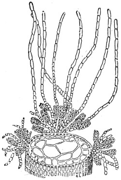
Fig. 288.—Cutleria dichotoma. Section of lacinia of a frond, showing the stalked eight-chambered oosporanges growing on tufts with intercalated filaments. Magnified 50 diameters.
In Cutleria (Fig. 288) we have a special feature of interest with two kinds of organs, seemingly opposed to each other with regard to their reproductive functions. The sporangia not only differ from those of other species, but the frond consists of olive-coloured irregularly-divided flagella, on each side of which tufts (sori) consisting of the reproductive organs, intermixed with hair-like bodies, are scattered. The zoospores are divided by transverse partitions into four cavities, each of which is again bisected by a longitudinal median septum. When first thrown off they are in appearance so much like the spores of Puccinia that they may be mistaken for them, although so very much larger than those of other olive-coloured algæ.
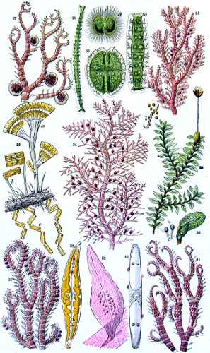
Florideæ, the red algæ (Plate II.), present many varieties of structure, although less appears to be known of their reproductive processes than of lower forms of cryptogamic plants. These are, however, of three kinds. The first, to which the term polyspore has been applied, is that of a gelatinous or membranous pericarp or conceptacle, in which an indefinite number of zoospores are contained. This organ may be either at the summit or base of a branch, or it may be concealed in or below the cortical layer of the stem. In some cases a number of spore-bearing filaments emanate from a kind of membrane at the base of a spheroidal cellular perisporangium, by the rupture of which the zoospores formed from the endochrome of the filaments make their escape. Other changes have been observed; however, they all agree in one particular, namely, that the zoospore is developed in the interior of a cell, the wall of which forms its perispore, and the internal protoplasmic membrane endochrome, the zoospore itself, for the escape of which the perispore opens out at its apex.
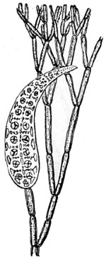
Fig. 289.—Dasya kutzingiana, with seed vessel and two rows of tetraspores. Magnified 50 diameters.
The second form is more simple, and consists of a globular or ovoid cell, containing a central granular mass; this ultimately divides into four quadrate-shaped spores; these, on attaining maturity, escape by rupture of the cell-wall. Another organ, called a tetraspore, takes its origin in the cortical layer. The tetraspores are arranged either in an isolated manner along the branches, or in numbers together; in some instances the branches that contain them are so modified in form they look like special organs, and have been called stichidia; as, for example, in Dasya (Fig. 289). Of the third kind of reproductive organ a difference of opinion exists as to the signification of their antheridia; although always produced in precisely the same situations as the tetraspores and polyspores, they are agglomerations of little colourless cells, either united in a bunch, as in Griffithsia, or enclosed in a transparent cylinder, as in Polysiphonia, or covering a kind of expanded disc of peculiar form, as in Laurencia. According to competent observers, the cells contain spermatozoids. Nägeli describes the spermatozoid as a spiral fibre, which, as it escapes, lengthens itself in the form of a screw. Thuret, on the contrary, says the contents are granular, and offer no trace of a spiral filament, but are expelled from the cells by a slow motion. The antheridia appear in their most simple form in Callithamnion (Plate II., Nos. 32 and 34), being reduced to a small mass of cells composed by numerous little bunches which are sessile on the bifurcations of the terminal branches. The spores are simpler structures than the tetraspores, and mostly occupy a more important position. They are not scattered through the frond, but grouped in definite masses, and generally enclosed in a special capsule or conceptacle, which may be mistaken for a tetraspore case. The simplest form of the spore fruit consists of spherical masses of spores attached to the wall of the frond, or imbedded in its substance, without a proper conceptacle; such a fruit is called a favellidium, and occurs in Halymenia; the same name is applied to the fruits of similar structures not perfectly immersed, as those of Gigartina, Gelidium, &c., where they form tubercular swellings on the lobes. In some, the tubercles present a pore at the summit, through which the spores emerge forth. In other cases, as in Ceramium (Plate II., Nos. 27 and 37), the spores occupy a more conspicuous place; a characteristic species is Delessaria (Plate II., No. 39), the coccidium either occurring on lateral branches, or is sessile on the face of the frond, when it consists of a case filled with angular-shaped spores attached to the wall of the case. The general external appearance of the red algæ is very varied, but it seems to attain to its deepest colouring in the Red Sea, which, it is said, is entirely due to the peculiarly vivid red seaweed. They are all exquisite objects for the microscope, as may be surmised from the few varieties presented in Plate II. The Florideæ of the warmer seas exhibit most elegantly formed fronds, as will be seen on reference to the “Phycologia Australica” of the late Dr. William Harvey, F.R.S.
The Characeæ may be placed among the highest of the algals, if only for the complexity of their reproductive organs, which certainly offer a contrast in their simplicity of structure. Chara vulgaris, stonewort, is a simple fresh-water plant, preferring still freshwater ponds or slow-moving rivers running over a chalky soil. It thus derives the calcareous matter found in the axis of the plant, together with a small portion of silica. Its filaments (or branches, as some botanists prefer to call them) are given off in whorls. The Characeæ are a small family of acrogens, consisting of only two or three at most. They are monœcious and diœcious, the two kinds of fruit being often placed close together. They may easily be grown in a tall glass jar for observation. All that is necessary is to put the jar occasionally under the house tap and let the water run slowly over the top for a short time, thus renewing the contents without disturbing the plant. The hard water supplied to London suits chara better than softer water. Both chara and nitella are objects of great interest to microscopists, since in the former the important fact of vegetable circulation was first observed. A portion of the plant of the natural size is shown in Fig. 290, No. 1.
Characeæ.
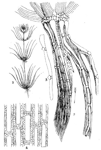
Fig. 290.—Diagrammatic sketch of Chara.
1. A stem of Chara vulgaris, natural size; 2. Magnified view (arrows indicating the course taken by the chlorophyll); 3. A limb, with buds protruding; 4. Portion of a leaf of Vallisneria spiralis, showing cyclosis of chlorophyll granules.
Each plant is composed of an assemblage of long tubiform cells placed end to end, with fixed intervals, around which the branchlets are disposed with great regularity. In nitella the stem and branches are composed of simple cells, which sometimes attain to several inches in length. Each node, or zone, from which the branches spring, consists of a single plate, or layer, of small cells, which are a continuation of the cortical layer of the internode (Fig. 290, No. 3) as an outgrowth. Each cell is partially filled with chlorophyll granules, and it is these that are seen under the microscope taking the course shown by the arrows (Fig. 290, No. 2). The rate of movement of the granules is accelerated by moderate warmth and retarded by cold. It is in viewing the circulation in water plants that the warm stage of the microscope is brought into use. Borne along with the protoplasmic stream are a number of solid particles consisting of starch granules and other matters. The method of viewing the circulation is by cutting sections off a portion of the plant with a very sharp knife, and arranging them in a growing cell with a few drops of water, and covering over with a thin cover-glass.
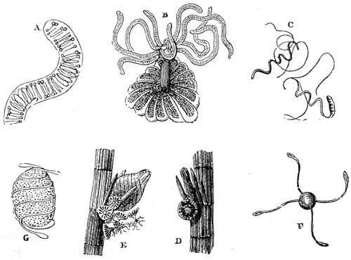
Fig. 291.—The Fructification of Chara fragilis.
A. Portion of filament containing “antheroids”; B. A group of antheridial filaments, composed of a series of cells, within each of which antherozoids are formed; C. The escape of mature antherozoids, with whip-like prolongations, about to swim off; D. Antherid supported on flask-shaped pedicle; E. Nucule enlarging, and seen to contain oospores; F. Spores and elaters of Equisetum; G. Spores surrounded by elaters of Equisetum.
The reproductive process of Chara is effected by two sets of bodies, both of which are placed at the base of the branches (Fig. 291, E and D) either on the same or different plants, one set known as globules or antherids, and the other as nucules, containing the oospores or archegones. These are often of a bright red colour, and have covering plates, or shields (B and E), curiously marked, and the central portion is composed of a number of filaments rolled up (as in E) or free (as seen at B), projecting out from the centre of the sphere. The antherid is supported on a short flask-shaped pedicle, which projects into the interior. At the apex of each of the eight manubria is a roundish hyaline cell, termed a capitulum, and at its apex again six smaller or secondary capitula. The long whip-shaped filaments are divided by transverse septa into a hundred or more compartments, every one of which is filled with an antherozoid (as at A), consisting of a spiral thread of protoplasm packed into two or three coils; these escape and become free (as seen at C), each having two long fine flagella. The young antherozoid swims off with a lashing action, and the whole field appears for a time filled with life. They swim about freely, but their motion gradually ceases, and soon they arrive at a state of inaction.
Nitella appears to have a somewhat different mode of fructification to that of its congener. It puts forth a long filamentous branch from one of its joints, which, on reaching the surface of the water, terminates in a whitish fruit-like cluster. It is even a more delicate and less robust algal than chara, and every care should be taken to imitate the still water in which it grows. It delights in shady woods and in calcareous open pools.
Similar care is requisite with regard to Vallisneria; and a more equal temperature is better suited to the growth of this aquatic plant. It should be planted in the middle of the jar or aquarium, about two inches deep in mould, closely pressed down, then gently fill the jar with water. When the water requires changing, a small portion only should be run off at a time. It appears to thrive in proportion to the frequency of changing the water, and taking care that the water added rather increases the temperature than lowers it.
The natural habitat of the Frog-bit, another water-plant of much interest, is found on the surface of ponds and ditches; in the autumn its seeds fall, and become buried in the mud at the bottom during the winter; in the spring these plants rise to the surface, produce flowers, and grow throughout the summer. Chara may be found in many places around London, and in the upper reaches of the Thames.
Anacharis alsinastrum.—This remarkable plant is so unlike any other water-plant that it may be at once recognised by its leaves growing in threes round a slender stem. It is also known as “Waterthyme,” from a resemblance it bears to that plant.
The colour of the plant is deep green; the leaves are nearly half an inch long, by an eighth wide, egg-shaped at the point, with serrated edges. Its powers of increase are prodigious, as every fragment is capable of becoming an independent plant, producing roots and stems, and extending itself indefinitely in every direction. The specific gravity of it is so nearly that of water, that it is more disposed to sink than float. A small branch of the plant is represented, with a hydra attached to it, in a subsequent chapter.
The special cells in which the circulation is most readily seen are the elongated cells around the margin of the leaf and those of the midrib. On examining the leaf with polarised light, the cells are observed to contain a large proportion of silica, and present a very interesting appearance. A bright band of light encircles the leaf, and traverses its centre. In fact, the leaf is set, as it were, in a framework of silica. By boiling the leaf for a short time in equal parts of nitric acid and water, a portion of the vegetable tissue is destroyed, and the silica rendered more distinct, without changing the form of the leaf.
It is necessary to make a thin section or strip from the leaf of Vallisneria for the purpose of exhibiting the circulation in the cells, as shown in Fig. 290, No. 4. Among the cell granules, a few of a more transparent character than the rest, are seen to have a nucleolus within.
The phenomenon of cell cyclosis occurs in other plants beside those growing in water. The leaf of the common plantain or dock, Plantago, furnishes a good example, the movement being seen both in the cells of the plant and hairs of the cuticle torn from the midrib.
Cell-division.—In order to study the process of cell-division the hairs on the stamens of Tradescantia should be taken. Remove one from a bud on a warm day and let a drop of a one per cent. sugar solution fall upon it, and cover it with a thin glass cover. Place it for a short time in a moist-chamber (Fig. 256), and then examine it with a magnifying power of 500 diameters. The nucleus of the cell will be seen, near its terminal position, to gradually elongate in the direction of the longer axis of the cell and become more granular, while the protoplasm moves towards the extreme end; the nucleus at the same time will present a striated appearance, with the fibrilla arranged parallel to the longer axis of the nucleus, and at length approach each other at the poles. A nuclear spindle will now be produced, and the fibres ruptured in the equatorial plane, so that two nuclei will be found in place of the one. The best preparations of nuclei are obtained by making thin longitudinal sections of actively-growing plants (young rootlets of Pinus, for example), and staining them with hæmatoxylin in the manner described in a former chapter.
Desmidiaceæ and Diatomaceæ.
The two groups of Desmidiaceæ and Diatomaceæ differ so little in their general characters that they may be spoken of as members or representative families of microscopic and unicellular algæ alike in their remarkable beauty and bilateral symmetry, and of such peculiar interest as to call for special notice. Desmids differ from diatoms chiefly in colour, in lacking a non-silicious skeleton, and in their generative process, which for the most part consists in the conjugation of two similar cells. Diatoms, on the other hand, have dense silicious skeletons and a general absence of green colouring matter. Ralfs, in his systematic monograph, enumerates twenty genera of desmids. The limiting membrane is alike firm and flexible, since it exhibits some elasticity and resistance to pressure, and is not readily decomposable. Traces of silica are found in only a few of the desmids, while the frustule of the diatom is chiefly composed of this substance; both have an external membranous covering, so transparent and homogeneous in structure as to be in danger of being entirely overlooked, unless some staining material is used, together with a high-power objective possessing considerable penetration. In some species, however, the mucous covering is more clearly defined, as in Staurastrum and Didymoprium Grevelli. Openings occur in the outer membrane of other species, as the Closterium.
PLATE X.
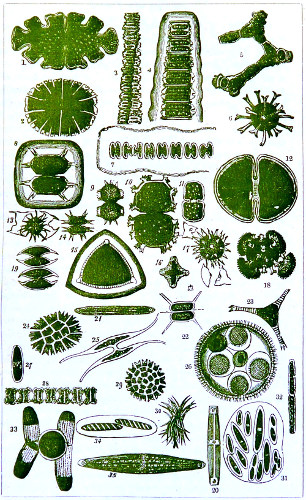
DESMIDIACEÆ.
Many species of desmids have a power of motion, the cause of which must be due either to cilia or a flagellate organ. This, however, is denied by some observers, who regard their movements as due to an exudation of the mucilaginous contents of the cell, to exosmose, or diffusion, neither of which hypotheses will at all help us to understand the gliding movements of the Oscillariæ or the sharp jerky movement of the Schizonema. The movements of desmids are especially exerted when in the act of dividing, and by sunlight, towards which they are always observed to move. The force with which some diatoms move about is very great, and this can only be satisfactorily explained by admitting a specialised organ.
The appearance of the Desmidiaceæ (Plate X.) is much modified by their eminences, depressions, and processes, as well as that of the surface, the margin of the fronds, and the depth and width of the central constriction. The surfaces may be dotted over irregularly, the dots themselves being elevated or depressed points in their structural character. The margins of some have a dentate appearance, as in Cosmarium. In the elongated forms, such as Penium, the puncta are disposed in lines parallel to the length. In several these lines are either elevations or furrows, it is not always easy to say which; they are peculiar, however, to the elongated forms of Closterium. When the lines are fine they produce a striation of the surface, but in order to discover this the fronds should be viewed when empty and by a fairly good power. The modification of surface in several genera seems to be due, not to mere simple appendages, but to expansion of the limiting membrance into thickened processes, and which may terminate in spines, as in Xanthidium and Staurastrum (Plate X., Nos. 8-19 and 22). A general distribution over the surface is characteristic of the former, but in Euastrum the surfaces are very irregular, and therefore described as “swellings or inflations.” Micrasterias has its margin deeply incised into lobes, which in some have a radiating arrangement; when the lobes on the margin are small they constitute crenations or dentations. The fronds of Euastrum binatum are bicrenate on the sides, as are those of Desmidium and Hyalotheca and other species. Another variety of margin exists, known as undulating or wavy, while the general concavity or convexity of the margins furnish other specific characteristics.
Pediastreæ (Plate X., Nos. 24-29).—The members of this family formerly included the Micrasterias and Arthrodesmius of Ehrenberg. From their arrangement of cells in determinate numbers and definite forms, it has been thought by some observers that they should be removed from the desmids to a special or sub-family. The points of difference consist in the firmness of the outer covering, in the frequent interruptions on the margin of the cells, and in the protrusion of “horns,” or rather a notch more or less deep. It is true that the cells are not made up of two symmetrical halves, and that they are in aggregation, which is not (except in the Scenedesmus, a genus that distinctly connects this group with desmids) in linear series, but in the form of discoidal fronds. They, however, divide into 8, 16, or 32 gonidia, and these move about for some time before the formation of a new frond. It was Nägeli who first instituted a sub-genus of Pediastrum, under the designation of Anomopedium, the chief characteristic of which is the absence of bilobed peripheral cells. In Cœlastrum the cells are hexangular, the central ones very regularly so; in Sorastrum they are wedge-shaped, or triangular, with rounded-off angles. Viewed laterally the cells appear oblong. The cells of Pediastrum are considerably compressed, so that when aggregated they form a flattened tubular structure; in figure they are polygonal, frequently hexagonal, a shape owing, in all probability, to mutual lateral pressure during growth. There is a pervading uniformity in the contents of the cells of the different genera, which consist of protoplasmic endochrome. At first the colour is pale green, but it becomes deeper with full maturity, while the decaying cells are seen to change to a deep reddish-yellow or brown. The protoplasm is also clear and homogeneous, but in time granules appear, enlarge, and multiply in number; moreover, each cell presents a single bright green vesicle, around which are collected clear circular spaces or globules, recalling those of Closterium, and varying in number from two to six or more, their position not being regulated by the partition wall as in Palmellæ, but by the centre of the entire frond. Oil globules are also contained in the cells; their presence is indicated by the addition of a drop of tincture of iodine. On one occasion Nägeli saw in Pediastrum boryanum the endochrome disposed in a radiating manner, an arrangement which often obtains in algals and in other vegetable cells with a central nucleus. The cells of Pediastreæ are always united together in compound fronds, as represented in Plate X., Nos. 24 and 29.56
The differences pointed out in no way constitute a claim to remove Pediastreæ from among Desmidiaceæ, certainly not to rank as a distinct species.
Reproduction of Desmidiaceæ.—A true reproductive act is presented by the conjugation or coupling of two fronds, and by the resulting development of a sporangium and subsequent interchange of the contents of the two cells. At another time self-division is frequently seen to take place in all respects as in the cells of other algæ. The proceeding is varied in some essential particulars by the form of the fronds and by other circumstances; as in fission of Euastrum, for instance (seen in Plate X., Nos. 1, 2, and 12), when the narrow connecting bands between the two segments of the fronds are rapidly pushed aside by growth and finally divide. Two modes of conjugation of fronds are represented in Plate X., Nos. 25 and 33, in Closterium and Penium. The act of conjugation admits of variations in character, as shown in Staurastrum and Microsterias; the contents of both fronds are discharged into a delicate intermediate sac; this gradually thickens and produces spines (Plate X., Nos. 8 and 19). In Didymoprium the separate joints unite by a narrow process pushed out from each other, often of considerable length, through which the endochrome of one cell is transferred to the other, and thus a sporangium is produced within one of two cells, just as in the conjugatæ (No. 5). In Penium Jennereri the conjugation takes a varied form; the fronds do not open and gape at the suture, but couple by small but distinct cylindrical tubes (No. 27).
Among those enumerated, the compressed and deeply constricted cells of Euastrum offer the more favourable opportunities for studying the manner of their division; for although the frond is really a single cell, in all its stages it appears like two, the segments being always distinct, from the earliest stage. The segments, however, are separated by a connecting link, which is subsequently converted into two somewhat round hyaline bodies. These bodies gradually increase and acquire colour, and as they grow the original segments are further divided, and at length become disconnected, each taking a new segment to supply the place of that from which it is separated. It is curious to trace the progressive development of the newer portions, which at first are devoid of all colour; but as they become larger a faint green tint is observed, which gradually darkens, and then assumes a granular appearance. Soon the new segments attain their normal size, while the covering in some species shows the presence of puncta. In Xanthidium, Plate X., Nos. 9, 10, and Staurastrum, Nos. 15-18, the spines and processes make their appearance last, beginning as mere tubercles, and then lengthening until they attain their perfect form and size, armed with setæ; but complete separation frequently occurs before growth is fully completed. This singular process is repeated again and again, so that the older segments are united successively, as it were, with many generations. When the cells approach maturity, molecular movements may be at times noticed in their contents, precisely similar to what Agardh and others aptly term “swarming.” Meyen describes this granular matter as starch.57 Closterium, early in the spring, when freshly secured and exposed to light, presents a wonderful appearance, these bodies being kept continually in motion at both ends of the frustule by the ciliary action within the cell, and the whole frond is seen brilliantly glittering with active cilia. When a gleam of stronger light is allowed for a moment to fall on the frond, the rapid undulations of the cilia produce a series of most delicate prismatic Newton’s rings. The action and distribution of the cilia, together with the cyclosis of the granular bodies in the frond, are better seen by the aid of Wenham’s parabola or a good condenser with a central stop. One of the wide angular objectives shows the circulation around the marginal portions of the whole frond. The stream is seen to be running up the more external portion, internal to which is another stream following a contrary direction; this action, confined to the space between the mass of endochrome and the outer portion of the cell-wall, is seen to pass above or around the space in which cyclosis of the spores is taking place.
During the summer of 1854, the late Rev. Lord Sidney Godolphin Osborne and myself became much interested in the remarkable family of Closteria. Fig. 292 is a highly magnified view of Closterium lunula which I drew by the aid of the camera-lucida at the time. There could be no doubt about the ciliary action within the frond: it was in every way similar to that of the branchiæ of the muscle, the same wavy motion, which gradually became slower as the death of the desmid drew near. This was brought about earlier when the cell was not kept supplied with fresh water.
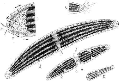
Fig. 292.—Closterium lunula.
In diagram A, line b points to a cluster of ovoid bodies; these are seen at intervals throughout the endochrome within the investing membrane. These bodies are attached to the membrane by small pedicles, and are occasionally seen in motion about the spot, from which they eventually break away, and are carried off, by the circulating fluid, to the chambers at the extremities of the frond; there they join a crowd of similar bodies, in constant motion within the chambers, when the specimen is quite fresh. That the action of these free granules or spores is “Brownian,” as surmised by some writers, is in my opinion entirely fallacious. It is doubtless in a measure due to the current brought about by the ciliary motion of the more fluid contents of the cell.
The circulation, when made out over the centre of the frond, for instance at a, is in appearance of a wholly different nature from that seen at the edges. In the latter the matter circulated is that of granules, passing each other in distinct lines, but in opposite directions; in the circulation as seen at a, the streams are broad, tortuous, of far greater body, and passing with much less rapidity. To see the centre circulation, use a Gillett’s illuminator and a 1⁄8th or a 1⁄10th immersion; work the fine adjustment so as to bring the centre of the frond into focus, then almost lose it by raising the objective; after this, with great care, work the milled head until the darker body of the endochrome is clearly brought out.
At B is an enlarged sketch of one extremity of the frond. The arrows within the chamber pointing to b denote the direction of a strong current of fluid, which can be occasionally followed throughout. It is acted upon by cilia at the edges of the chamber, the greater impetus appearing to come from the centre of the endochrome. The fluid is here acting in positive jets, that is, with an almost arterial action; and according to the strength with which it is propelled at the time, the loose floating bodies are sent to a greater or less distance from the end of the frustule; the fluid is thus impelled from a centre, and kept in activity by the lateral cilia, that create a rapid current and give a turning motion to the free bodies. The line—a, in this diagram, denotes the outline of the membrane which encloses the endochrome; on both sides cilia can be seen. The circulation exterior to it passes and repasses in opposite directions, in three or four distinct courses; these, when they arrive at—c, seem to encounter a stream making its way towards an aperture at the apex of the chamber; then they appear to be driven back again by a stronger force. Some, however, do occasionally enter the chamber, but very rarely will one of the bodies escape into the outer current, and should it do so, is carried about until it becomes adherent to the side wall of the frond.
With regard to the propagation of the C. lunula, I have never seen anything like conjugation; but I have repeatedly seen self-division (shown at D a a). This act is chiefly the work of one half of the frond. Having watched for some time, one half is seen to remain passive, while the other has a lateral motion from side to side, as if moving on an axis at the point of juncture; the motion increases, is more active, until at last with a jerk one segment separates itself from the other, as seen at E. It will be noticed that each end of the segment is perfectly closed before separation finally takes place; there is, however, only one perfect chamber, that belonging to the extremity of the original entire frond. The circulation continues for some time previous to and after subdivision, in both fronds, and by almost imperceptible degrees increases in volume. From the end of the endochrome symptoms of elongation of the frond take place, the semi-lunar form gradually changes, elongates, and is more defined, until it takes the form and outline of the fully-formed frustule at the extremity. The obtuse end—b of the other portion of frond is at the same time elongating and contracting, and in a few hours from the division of the one segment from the other the appearance of each half is that of a nearly perfect frustule, the chamber at the new end is complete, the globular circulation exterior to it becomes affected by the circulation from within the said chamber, and, shortly afterwards, some of the free bodies descend, and become exposed to the current already going on in the chamber. E is a diagram of one end of a C. didymotocum, in which the same process was well marked, and completed while it was under observation.
It will appear to most observers that if the continuation of the widely-spread family of Desmidiaceæ was wholly dependent upon conjugation and subdivision of their frustules, a process requiring several hours for its completion, the whole species must have long ago disappeared. It may be presumed then that some other mode of reproduction must prevail. In the fresh-water algæ the two more general methods of multiplication are clearly governed by the conditions of the seasons; the resting-spores securing continuity of life during the winter, the swarm-spores spreading the plant profusely during the warmer portion of the year, when rapid growth is possible. I therefore regard the actively swarming bodies seen in continuous motion at the two extreme portions of the frustule of Closterium lunula as being either oospores or zoospores, by means of which reproduction takes place.
Diatomaceæ, commonly called brittleworts, Plate XI., are chiefly composed of two symmetrical valves, narrow and wand-like, navicular, miniature boat-shaped, hence their name Navicula (little ship). Hitherto they have excited the deepest interest among microscopists because of their wonderfully minute structure, and the difficulty involved in determining their exact nature and formation. Each individual diatom has a silicious skeleton, spoken of as a frustule, frond, or cell, having a rectangular or prismatic form, which mostly obtains in the whole family, the angles of the junction of the two united valves being, as a rule, acute, and enclosing a yellowish-brown endochrome. Deeply-notched frustules, like those of the Desmidiaceæ, do not occur, and the production of spines and tubercles so common in that family is rare in the Diatomaceæ. Great variety of outline prevails, so much so that no rule in this respect can be formulated.
The frustules, however, are usually composed of two equal and similar halves, but exceptions to this are found in the Actinomtheæ, Cocconcidæ, and one or two other families. The extremities of some species, e.g., Nitzshia and Pleurosigma, are extremely elongated, forming long, filiform, tubular processes; in Biddulphia and Rizoselenia, short tubular processes from their margins. Great variety of outline may prevail in a genus, so considerable indeed that no accurate definition can be given, the characteristics shading off through several species until the similarity to an assumed typical form is much diminished, which may again be modified by accidental circumstances that surround the development of the silicious frustule. It must not be forgotten that the figure is greatly modified or entirely changed by the position of the valves, whether seen in one position or another, as already explained in connection with “Errors of Interpretation.” Again, in the genera Navicula, Pinnularia (Plate II., Nos. 33, 38, and 40), and others, the frustules are in one aspect boat-shaped, but in the other either oblong with truncated ends, or prismatic. In the genus Triceratium (Plate XI., No. 10), the difference of figure is very remarkable as the front or side view is examined.
The sudden change in appearance presented to the eye as the frustule is seen to roll over is rather peculiar. As a rule, therefore, we must examine all specimens in every aspect, to accomplish which very shallow cells should be selected, say of 1⁄100th of an inch deep, and covered with glass 1⁄250th of an inch thick. A good penetrating objective should be used, and careful illumination obtained. The Diatomaceæ are perhaps more widely distributed than any other class of infusorial life; they are found in fresh, salt, and brackish water; many grow attached to other bodies by a stalk (Plate II., No. 33, Licmophora and Achnanthidium); while others, as Pleurosigma, No. 40, swim about freely.
PLATE XI.
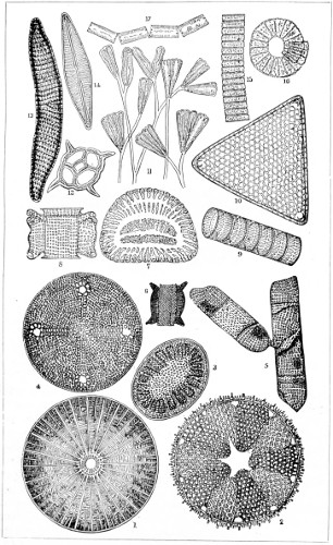
DIATOMACEÆ, RECENT AND FOSSIL.
There are a considerable number of Diatomaceæ which, when in the young state, are enclosed in a muco-gelatinous sheath; while others are attached by stipes or stalk to algæ. It would be vain, in a limited space, to attempt a description of this numerous and extensive family. Nägeli and other observers describe a “mucilaginous pellicle on the inner layer of the valves,” while, as Menghine observes, “an organic membrane ought to exist both inside and outside, for the silica could not become solid except by crystallizing or depositing itself on some pre-existing substance.” The surface of the frustules is generally very beautifully sculptured, and the markings assume the appearance of dots (puncta), stripes (striæ), ribs (costæ), pinnules (pinnæ), of furrows and fine lines; longitudinal, transverse, and radiating bands; canals or canaliculi; and of cells or areolæ; whilst all present striking varieties and modifications in their form, character, and degree of development. Again, the fine lines or striæ of many frustules are resolvable into rows of minute dots or perforations, as occur in Pleurosigma angulatum, delineated in the accompanying microphotograph (Fig. 294), taken for the author purposely to show the markings on this especially selected test diatom.
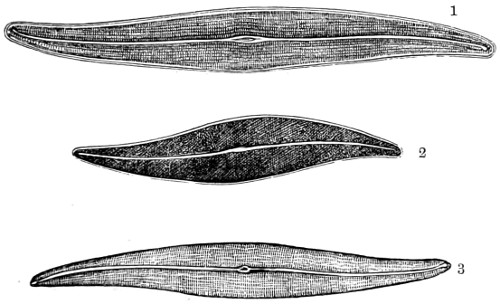
Fig. 293.
1. Pleurosigma attenuatum; 2. Pleurosigma angulatum; 3. Pleurosigma Spencerii. Magnified 450 diameters.
The nature of the markings on the diatom valves is one of considerable interest, and attempts have been made to produce them artificially, but without success.
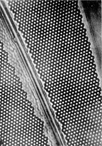
Fig. 294.—Pleurosigma angulatum, magnified 4500 diameters.
(From a microphotograph taken by Zeiss with the 2 mm. aprochromatic objective, 1·30 numerical aperture, and projection eye-piece, No. 4.)
Professor Max Schultze devoted a great amount of time to the investigation of the subject, and has recorded in a voluminous paper58 the results of his observations. He says, “Most of the species of the Diatomaceæ are characterised by the presence on their outer surface of certain differences of relief, referable either to elevations or to depressions disposed in rows. The opinions of microscopists with respect to the nature of these markings are still somewhat divided. Whilst in the larger forms, and those distinguished by their coarser dots, the appearance is manifestly due to the existence of thinner spots in the valve, we cannot so easily explain the cause of the striation or punctation in Pleurosigma angulatum and similar finely-marked forms.”
Dr. R. Zeiss some time ago furnished me with a microphotograph of a frustule magnified 4500 diameters that seemed to confirm Mr. T. F. Smith’s view of the structure of these valves. Dr. Van Heurck has also made a study of this diatom, and concludes that the valves consist of two membranes of thin films, and of an intermediate layer, the outer being pierced with openings. The outer membrane is, he believes, “so delicate that it is easily destroyed by acid or by friction, and the several processes employed in cleaning and preparing it for microscopical examination. When the openings or apertures of the internal portion are arranged in alternate rows they assume the hexagonal form; when in straight rows, the openings are seen to be square or oblong.” A description hardly in accord with Fig. 294.
Movements of Diatoms.
The late Professor Smith, in his “Synopsis of Diatoms,” refers to their movements in the following terms: “I am constrained to believe that the movements observed in the Diatomaceæ are due to forces operating within the frustule, and are probably connected with the endosmotic and exosmotic action of the cells. The fluids which are concerned in these actions must enter, and be emitted through the minute foramina at the extremities of the silicious valves.” Schultze’s researches, which were made at a later date, carried this debatable question somewhat further. He is of opinion “that a sarcode (protoplasmic) substance envelops the external surface of the diatoms, and its movements are due to this agent exclusively.” His investigations were mainly confined to P. angulatum, and to the larger P. attenuatum (Fig. 293, 1 and 2), as the transverse markings on the frustule do not impede to so great an extent the observation of what is going on within. The living specimen of P. angulatum under the microscope usually has its broad side turned to view, with one long curved “raphe” uppermost, and the other in contact with the glass cover (Fig. 293). Within the frustule the yellow colouring matter, “endochrome,” fills the cavity more or less completely. In the broader part of the frustule these bands of endochrome describe one or two complicated windings. It is only possible in those specimens in which the bands are narrow to properly trace their foldings, and determine their number. The next objects which strike the eye on examining a freshly-gathered Pleurosigma are numerous highly refractive oil-globules. These are not, however, all in the same place, and one globule appears nearer the observer than the other; their relative position is best seen when a view of the narrow side of the frustule can be obtained, so that one raphe is to the left and the other to the right. The blue-black colour which is assumed by these globules after treating with acid demonstrates their oleaginous nature. The middle of the cavity of the frustule is occupied, in the larger navicula, by two large oil-globules (seen in the diagrammatic Fig. 295), and by a colourless finely granular mass, whose position in the body is not so clearly seen in the flat view as in the side view. Besides the central mass, the conical cavities at either end of the frustule are seen to enclose granular substance, and two linear extensions from each of three masses are developed, closely underlying the raphæ. In the side view, therefore, they appear attached to the right and left edges of the interior of the frustule. This colourless granular substance carries in its centre, near the middle part of the diatom, an imperfectly developed nucleus which is not very easy to see, but may be demonstrated by the application of an acid. The colourless substance is protoplasm, and encloses numerous small refractive particles; this, on adding a drop of a one per cent. solution of osmic acid, is coloured blue-black, and proves to be fat. It is, however, exceedingly difficult to determine the exact limitations of the protoplasm, on account of the highly refractive character of the silicious skeleton, and the obstruction to the light presented by the endochrome.
At a short distance the protoplasm reappears, contracted into a considerable mass, within the terminal ends of the frustule. Schultze observed in this part of the protoplasm a rapid molecular movement, “cyclosis,” such as occurs in Closterium, and also a current of the granules of the protoplasm along the raphe. “Pleurosigma angulatum ‘crawls,’ as do all diatoms possessing a raphe, along this line of suture. To crawl along, it must have a fixed support.” “There is obviously,” adds Schultze, “but one explanation; it is clear that there must be a band of protoplasm lying along the raphe, which causes the particles of colouring matter to adhere, and gives rise to a gliding movement. For there is but one phenomenon which can be compared with the gliding motion of foreign bodies on the Diatomaceæ, and that is, the clinging to and casting off of particles by the pseudopodia of the rhizopod, as observed, for instance, on placing a living Gromia or Miliolina in still water with finely-powdered carmine. The nature of the adhesion and of the motion is in both cases the same. And since, with diatoms as unicellular organisms, protoplasm forms a large part of the cell (in many cases two distinctly moving protoplasms), this implies that the external movements are referable to the movements of the protoplasm.” It is quite evident to those who have studied the movements of diatoms that they are surrounded by a sarcode structure of a more pellucid character than that of Amœba. Six years before Schultze’s observations were published, I wrote in a third edition of my book, page 307, “The act of progression favours the notion of contractile tentacular filaments—pseudopodia—as the organs of locomotion and prehension.”
Since my former observations on the movements of diatoms, I have given much attention to two forms, P. angulatum and Pinnularia. The powers used were Hartnack’s No. 8, and Gunlack’s 1⁄16-inch immersion; Gillett’s condenser illumination, with lamp flame edge turned to mirror and bull’s-eye lens; a perforated slide with a square of thin glass ·006 cemented to it, and a cover-glass of ·005. So far as I could satisfy myself, no terminal space, as in the Closteria, could be seen, otherwise the course of the gemmules is as freely traced as in that form. They are more minute than the Closterium lunula granules, more steadily or slowly seen to pass up and down one half the frustule towards the extremity, one half of the current seeming to turn round upon its axis and descending towards the other. The granules were thickly scattered at the apex, but gradually became fewer, and the ascending and descending current tapered away towards the central nodule, which became more filled up or closed in.
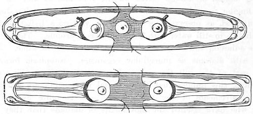
Fig. 295.—Outline sketches of Pinnulariæ, showing vesicles.
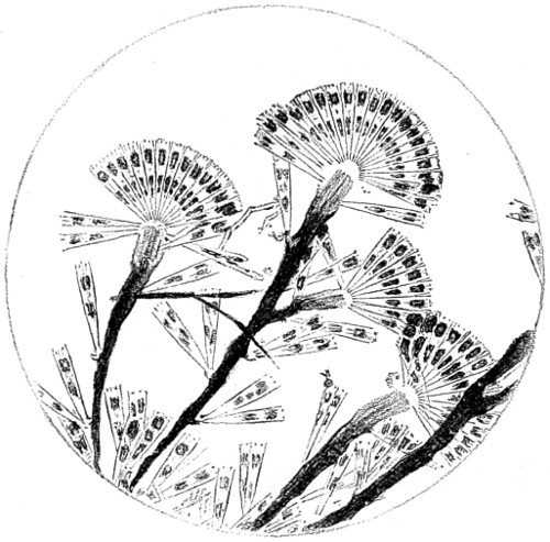
Fig. 296.—Gomphonema constrictum. (From a microphotograph.)
This beautiful sight was not confined to one frustule, but was exhibited in all that were in a healthy condition. I examined several, and watched them for a long time. The phenomenon described depends much upon the healthy condition of the frustule at the time; as the movements of the diatoms became sluggish, the circulation gradually slackens and then ceases altogether. I also saw a somewhat similar action in the more active specimens of P. hippocampus and Navicula cuspidata, but the coarser markings and thickness of the wall of these diatoms seemed to place greater difficulties in the way of observation than the finer valves of the P. angulatum. One thing I believe is certain, that the circulation described is precisely similar to that seen in the Closteria, or, on a much larger scale, in Chara and the leaf of the Anacharis, bearing in mind also that in the Closterium the cell is divided by a transverse suture, and in P. angulatum by a longitudinal one (Plate II., Nos. 38-40). About the same time some very lively specimens of the Pinnulariæ were sent to me, and the movements of these frustules were more closely observed. One or two of the more active would attack a body relatively larger than itself, it would also force its way into a mass of granular matter, and then recede from it with a jerky motion. In more than one instance a cell of Palmoglæa was seized and carried away by the Pinnularia, the former at the time being actively engaged in the process of cell division. Other diatoms present among my specimens were also in an active condition, and the circulation of granular matter in all was distinctly visible. In the Pinnulariæ two large colourless vesicles were seen on either side of the median nodule, each having a central nucleus, as represented in the accompanying sketch, made while under observation in two positions. The central portion of each frustule was closely packed with a rich yellowish-brown coloured endochrome, interspersed with a few fat globules. The phenomenon of cyclosis was not seen in any of these diatoms, but I have satisfied myself, by staining, of the presence of a delicately fine external protoplasmic covering in many diatoms. That their movements resemble the gliding movements exhibited by the Amœba can scarcely be doubted. Numerous forms of Diatomaceæ are found growing on or attached to water-plants or pieces of detached stalks, which, although generally simple, are sometimes compound, dividing and subdividing in a beautiful ramous manner. Pinnulariæ, Nitzschia, &c., are seen adherent by one extremity, about which they turn or bend themselves as on a hinge. By the process of cell-division, groups of Synedræ become attached by a point, in a fan-like form. The fan-like collection of frustules is said to be flabellate, or radiate. In Licmophora, Achnanthes and other species (Plate II., Nos. 29-33) the double condition of union of frustules and of attachment by a pedicle are illustrated. When a stipe branches it does so normally in a dichotomous manner, each new individual being produced by a secondary pedicle. This regular dichotomy is seen in several genera: Cocconema and Gomphonema, the latter more perfectly in Fig. 296, from a microphotograph, in which a branching, or rather longitudinal, rupture of the pedicle takes place at intervals, and the entire organism presents a more or less complete flabella, or fan-like cluster, on the summit of the branches, and imperfect or single frustules irregularly scattered throughout the whole length of the pedicle.
Isthmia enervis (Fig. 297).—The unicellular frustule of this species is extremely difficult to define, owing to the large areolations of the valves; it has a remarkable internal structure. The olive-brown cell contents are found collected, for the most part, into a central mass, from which radiating, branched, granular threads extend to and unite with the periphery. When viewed by a magnifying power of 600 or 700 diameters, these prolongations are seen to be composed of aggregations of ovate or spindle-shaped corpuscles, held together by protoplasmic matter. These bodies are sometimes quiescent, but more often travel slowly to and fro from the central mass. The general aspect under these conditions so nearly corresponds to the characteristic circulation in the frustules of unicellular plants and of certain rhizopoda, that it is difficult to realise that the object when under examination is an elegant marine diatom.
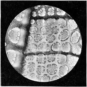
Fig. 297.—Isthmia enervis. Microphotograph.
There is a large section of diatoms in which the frustules are diffused throughout a muco-gelatinous envelope in a definite manner. Histologically this is homologous with the pedicles and connecting nodules thrown out during the act of self-division, and in some species (Cocconeis, Fragillaria, &c.) it often persists after that act is complete.
Diatomaceæ, Recent and Fossil.
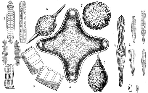
Fig. 298.—Fossil Diatoms from Springfield (Barbadoes).
1, Achnanthidium; 2, Diatoma vulgare, side view and front view; 3, Biddulphia; 4, 5, 6, 7, Amphitetias antediluviana, front view, with globular and oval forms; Gomphonema elongatum and capitatum.
Fossilised Diatomaceæ.—Dr. Gregory was of opinion that a large number of diatoms separated into species are only transition forms, and more extended observations have proved that form and outline are not always to be trusted in this matter. Species-making is a modern invention, and can hardly apply to the indestructible fossilised forms of the frustules of Diatomaceæ, with their beautiful sculpturings and geometrical constructions, which have not been materially changed since they were first deposited. Startling and almost incredible as the assertion may appear to some, it is none the less a fact established beyond all question, that some of the most gigantic mountain-ranges, as the mighty Andes, towering into space 25,250 feet above the level of the sea, their base occupying vast areas of land; as also massive limestone rocks; the sand that covers boundless deserts; and the soil of many wide-extended plains, are each and all principally composed of Diatomaceæ. And, as Dr. Buckland once observed: “The remains of such minute animals have added much more to the mass of materials which compose the exterior crust of the globe than the bones of elephants, hippopotami, and whales.”
In 1841 the late Mr. Sollitt, of Hull, discovered the beautiful longitudinal and transverse striæ (markings) on the Pleurosigma hippocampus. A curved graceful line runs down the shell, in the centre of which is an expanded oval opening. Near to the central opening the dots elongate crossways, presenting the appearance of small short bands.
In the vicinity of this town many interesting varieties of Diatomaceæ have been found, the beauty of the varied forms of which are constantly under investigation; at the same time some of them are highly useful, as forming that class of test objects which are better calculated than many others for determining the excellence and powers of certain objectives. Mr. Sollitt carefully measured the markings on some of the frustules and found they ranged between the 1⁄30000th and 1⁄130000th of an inch; the Pleurosigma strigilis having the strongest markings, and the Pleurosigma acus the finest.
Mr. J. D. Sollitt not only first proposed their use, but he also furnished the measurements of the lines of the several members of this family, as follows:—
Amphipleura pellucida, or Acus, 130,000 in the inch, cross lines. " sigmoidea, 70,000 in the inch. Navicula rhomboides, 111,000 in the inch, cross lines. Pleurosigma fasciola, fine shell, 86,000 in the inch, cross lines. "" strong shell, 64,000 in the inch, cross lines. " strigosum, 72,000 in the inch, diagonal lines. " angulatum, 51,000 in the inch, diagonal lines. " quadratum, 50,000 in the inch, diagonal lines. " Spencerii, 50,000 in the inch, cross lines. " attenuatum, 42,000 in the inch, cross lines. " Balticum, 40,000 in the inch, cross lines. " formosum, 32,000 in the inch, diagonal lines. " strigilis, 30,000 in the inch, cross lines.
PLATE XII.
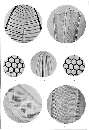
MICRO-PHOTOGRAPH OF TEST DIATOMS.
Lichenaceæ.
The lichens are a family of autonomous plants, an intermediary group of algals or cellular cryptogams, drawing their nourishment from the air through their whole surface medium, and propagating by spores usually enclosed in asci, and always having green gonidia in their thallus. Their gonidia, bright coloured globular cells, form layers under the cortical covering of the thallus, and generally develop in the form of incrustations, which cover stones, wood, and the bark of trees, or penetrate into the lamellæ of the epidermis of woody plants. The gonidia of lichens partake of both the character of vegetative and reproductive cells.
The thallus in the fructicose group attaches itself by a narrow base, growing in the form of a miniature shrub. Another group is met with in a slimy condition—the gelatinous lichens. These species, for the most part, furnished dyes before the discovery of the coal-tar dyes. In many of the Palmella cruenta, commonly found growing on the walls and roofs of houses, a colourless acid liquid is found, which, on being treated with alkali, produces a bright yellow colour; and another, Avernia vulpina, furnishes a brown dye; the Rocella fuciformis and R. tinctoria yield the purple dye substance known as orchil, or archil, from which the useful blue paper of the chemist for testing acidity is manufactured. Usnic acid, combined with green and yellow resins, seems to be more or less a constituent of many lichens.
A vertical section of Palmella stellata is given in Plate I., No. 26, in which the emission of the ripe spores of the lichens is seen to be not unlike that which takes place in some of the fungi, Pezizæ, Sphæriæ, &c. If a portion of the thallus be moistened and placed in a common phial, with the apotheca turned toward one side, in a few hours the opposite surface of the glass will be found covered with patches of spores, easily perceptible by their colour; or if placed on a moistened surface, and one of the usual glass slips laid over it, the latter will be covered in a short time. As to the powers of dissemination of these lowly organised plants, an observation led to the conclusion that the gonidia of lichens have greater powers in this direction than was formerly supposed. It is found that by placing a clean sheet of glass in the open air during a fall of snow, and receiving the melting water in a tube or bottle, quantities of what has been looked upon as a “unicellular plant” can be taken, the cells of which may be kept in a dormant condition for a long time during cold weather, but upon the return of spring warmth and moisture they begin to increase, by a process of subdivision, into two, four or eight portions; these soon assume a rounded form, and burst the parent cell-wall open; these secondary cells then begin to divide and subdivide again, and the process may go on without much variation for a long time. The phenomena described may be watched by taking a portion of the bark of a tree on which Chlorococcus has been deposited, and placing it under a glass to keep it in a moderately moist atmosphere; the only difference being a change in colour, caused by the growth of the fibres, as may be seen on microscopical examination. “And this,” says Dr. Hicks, who first observed this phenomenon in plant life, “is an instructive point, because it will be found that the colour varies notably according to the lichen prevalent in its neighbourhood.”59 He believes there can be no doubt that what has been called Chlorococcus is nothing more than the gonidia of a lichen; and that under suitable conditions, chiefly drought and warmth, the gonidium often throws out from its external envelope a small fibre, which, adhering and branching, forms a “soridium.” “The soridia remain dormant for a very long time, and do not develop into thalli unless in a favourable situation, in some cases it may be for years. It will be perceived that the soridium contains all the elements of a thallus in miniature; in fact, a thallus does frequently arise from one alone, and the fibres of neighbouring soridia interlace; thus a thallus is matured very rapidly. This is one of the causes of the variation of appearance so common in many species of lichens, more readily seen towards the centre of the parent thallus. When the gonidia remain attached to the parent thallus, the circumstances are, of course, more favourable, and they develop into secondary thalli, attached more or less to the older one, which, in many instances, decays beneath them. This process being continued year after year gives an apparent thickness and spongy appearance to the lichen, and is the principal cause of the various modifications in the external aspect of the lichens which caused them formerly to be misunderstood and wrongly classified.”60
The erratic lichens are found among the genus Palmella, some of which grow among boulders of the primary and metamorphic formations, curled up into a ball, and only fixed to their matrix by a slender thread. The globular Lecanora esculenta will at times suddenly cover large tracts of country in Persia and Tartary, where it is eaten by the cattle. During a scarcity of food a shower of these lichens, Mr. Berkeley tells us, fell at Erzeroum, and saved the cattle from starvation.61
Another group of the Palmella, or Peltigeri, so named from the target-like discs on their surface, spread their foliaceous fronds over the ground, and as the fruit is marginal, it gives the thallus a digitate appearance. These are often spotted over by a little red fungus. The Lecidinei contains numerous species of the most varied habits, and always crustaceous, and so closely adherent to the hard rocks and stones on which they grow, that at length they disintegrate them. From this low species a higher form arises, with erect branching stems, and clothed with foliaceous, brightly-coloured scales.
The Coccocarpei is mainly distinguished by having orbicular discs entirely deprived of the cortical envelope called an excipulum. The discs spring at once from the medullary stratum, and contain asci and sporidia similar to those of the minute fungi Sphæriæ. Some of the lichens are themselves parasitic, and begin existence under the thick skin of the leaves of tropical plants, and spread encrusting thallus over their surface, the excipulum and perithecia being black; but in most cases these are beautifully sculptured, and are interesting objects for the microscope. Indeed, the sphere-bearing lichens, with upright stems bearing globular fruit at the extremity of their branches, are at first indicated by a swelling, but in time the outer layer bursts and exposes sporidia, which are beautiful objects under the microscope on account of their spherical form and more or less deep blue tint. Humble and lowly as lichens may appear to be, they have been divided into fifty-eight or more genera and 2,500 species. The brothers Tulasne, De Bary, the Rev. Mr. Berkeley, and others, devoted great attention to the peculiarities of their structure and natural history.
Hepaticæ.—An intermediary group of much interest to the microscopist are the Hepaticæ (liverworts). These are found growing on damp rocks in the neighbourhood of springs and dripping banks. The scale-moss, the Marchantia polymorphia (Fig. 299), may be taken as typical of this little group, with its gemmiparous conceptacles and lobed receptacles, bearing archegones on transparent glass-like fruit stalks, carrying on their summits either round shield-like discs or radiating bodies with a striking resemblance to a wheel without its tyre.
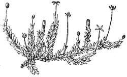
Fig. 299.—Marchantia polymorphia.
The liverworts are closely allied to the mosses, and as much difficulty was experienced in dividing the two, Hooker placed the whole under one genus, the Jungermannia. More recently, however, they have been divided into those with a stem and leaves confluent in a frond, Marchantia; those with stem and leaves distinct, Jungermannia; and those with a solitary capsule, filiform, bivalved, stalked, with a free central placentation, Anthocotaceæ. Some botanists have further divided them, but they are all extensively propagated by gemmæ.
The fronds carry the male organs, or antherids, and the disc, in the first instance, bears the female organs, or archegones, and after a time gives place to the sporanges, or spore cases. It is these bodies which are of so much interest to microscopists; if the plant is brought into a warm room, they suddenly burst open with some violence the moment a drop of water is applied to them, and the sporanges are dispersed in a small cloud of brownish dust. If this dust is examined under a medium power, it is seen to consist of a number of chain-like bodies, somewhat like the spring of a small watch; and if the process of bursting be closely watched, these minute springs will be found twisting and curling about in every direction. The structure of the frond itself will be seen to be interesting when cut in the vertical direction and placed under the microscope.
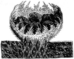
Fig. 300.—Gemmiparous conceptacle of Marchantia polymorphia, expanding and rising from the surface of a frond. In the interior are seen gopidial gemmæ already detached by the splitting of the epiderm.
The gemmæ of Marchantia polymorphia are produced in elegant membranous cups, with a toothed margin growing on the upper surface of the frond, especially in very damp courtyards between the stones, or near running water, where its lobed fronds are found covering extensive tracts of moist soil. At the period of fructification the fronds send up stalks, which carry at their summit round shield-like radiating discs, which bear upon their surface a number of little open basket-shaped “conceptacles.” These again expand into singularly graceful cups (as in Fig. 300), and are found in all stages of development. When mature, the basket contains a number of little green round or oblong discs, each composed of two or more layers of cells; the wall itself being surmounted by a glistening fringe of teeth, whose edges are themselves regularly fringed with minute outgrowths. The cup seems to be formed by a development of the superior epidermis, which is raised up, and finally bursts and spreads out, laying bare the seeds.
The archegones of Marchantia are very curious bodies, while the elater and spores are even still more so. These are elongated cells, each containing a double spiral fibre coiled up in the interior. It is the elasticity of this which tears apart the cell-membrane, and sends forth the spores with a jerk, and thus assists in their dispersion. Marchantia is the type of the malloid Hepaticæ.
Musci, Bryophyta.
Mosses are a beautiful class of non-vascular cryptogams. Linnæus called them servi, servants or workmen, as they seem to labour to produce vegetation in places where soil is not already formed. The Bryophyta form three natural divisions: the Bryinæ, or true mosses; the Sphagnaceæ, or peat-mosses; and the Hepaticæ, or liverworts. The two first are commonly united. In these the sexual organs consist of antheridia and archegonia, but they are of simpler structure than will be found in ferns; and the first generation from the spore is asexual.
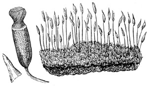
Fig. 301.—Screw-moss.
The common or wall screw-moss (Fig. 301) grows almost everywhere, and if examined closely, is seen to have springing from its base numerous very slender stems, each terminating in a dark brown case, which encloses antheroids. If a patch of the moss is gathered when in this state, and the green part of the base is put into water, the threads of the fringe will uncoil and disentangle themselves in a most curious and beautiful manner; from this circumstance the plant takes its popular name of screw-moss. The leaf usually consists of either a single or a double layer of cells, having flattened sides, by which they adhere one to another. The leaf-cells (Fig. 302) of the Sphagnum or bog-moss exhibit a curious departure from the ordinary type; they are large, polygonal, and elongated, and contain spiral fibres loosely coiled in their interior. The young leaf does not differ from the older; both are evolved by a gradual process of differentiation.
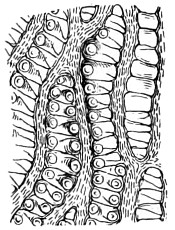
Fig. 302.—Section of leaf of Sphagnum moss, showing large cells of spiral fibres and connecting apertures.
Mosses, like liverworts, possess both antheridia and pistillida, which are engaged in the process of fructification. The fertilized cell becomes gradually developed into a conical body elevated upon a footstalk, the walls of the flask-shaped body carrying the higher part upwards as a calyptra or hood upon its summit, while the lower part remains to form a kind of collar round the base. These spore-capsules are closed on their summit by opercula or lids, and their mouths when laid open are surrounded by a beautiful toothed fringe, termed the peristome. This fringe is shown in Fig. 303, in the centre of a capsule of Funaria, with its peristome in situ. The fringes of teeth are variously constructed, and are of great service in discriminating the genera. In Neckera antipyretica the peristome is double, the inner being composed of teeth united by cross bars, forming a very pretty trellis. The seed spores are contained in the upper part of the capsule, where they are clustered round the central pillar, termed the columella; and at the time of maturity, the interior of the capsule is almost entirely occupied by spores.
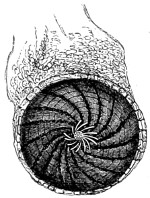
Fig. 303.—Mouth of Capsule of Funaria, showing Peristome.
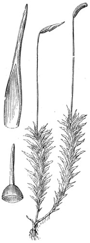
Fig. 304.—Hair-moss in Fruit.
The undulating hair-moss, Polytrichum undulatum (Fig. 304), is found on moist, shady banks of pools and rivulets. The seed-vessel has a curious shaggy cap; but in its construction it is very similar to that of the screw-moss, except that the fringe around its opening is not twisted. The reproductive organs of mosses are of two kinds; the capsule containing minute spores, archegonia, and the antheridia, or male efflorescence. The capsule, theca, or sporangium, is lateral or terminal, sessile, or on a fruit stalk (seta) of various shapes, indehiscent, or bursting by four valves at the sides, or more commonly by a deciduous cup, operculum. When this falls the mouth of the capsule becomes exposed. The rim is crowned with tooth-like or cilia-like appendages in sets of four or multiples of that number—peristome. These are often brightly coloured and hydroscopic. By simply breathing upon them they suddenly fly open, and are endowed with motion, that is, if they contain spores. The spores on germination produce a green confervoid-like mass of threads, from which the young plant arises.
The Sphagnaceæ, or “bog mosses,” have been separated from true mosses from the marked differences they present. The stem is more widely differentiated, and throughout its structure a rapid passage of fluid takes place. It has the power of absorbing moisture from the atmosphere, so that if a plant be placed dry in a glass of water with its rosette of leaves hanging over the edge, it acts like a syphon, and the water will drop from it until the glass is emptied. As may be supposed, the leaf is composed of large open cells, and it absorbs more water than the root. The antherids or male organs of Sphagnaceæ resemble those of liverworts, rather than those of mosses, both in form and arrangement; they are grouped in “catkins” at the tips of the lateral branches, each of the imbricated perigonal leaves enclosing a single globose antherid on a slender foot-stalk, and surrounded by long branched paraphyses of cobweb-like tenuity. The female organs, or archegones, do not differ materially in structure from those of mosses; they are grouped together in a sheath of deep green leaves at the end of the shorter lateral branchlets at the side of the rosette or terminal crown of leaves. The sporange is very uniform in all the species, and the spores are in groups of fours, as in mosses, around a hemispherical columella. These plants grow so rapidly that they soon cover a pool with thin matted bundles of branches, and as they decay they fall to the bottom, and become the foundation of the future bog or peat moss.