Part 2 — CHAPTER III. (continued 2)
Reading edition
Trematode Worms.—In the order Trematoda, to which the fluke belongs, the body is unsegmented, and to the naked eye smooth throughout, with a blood circulatory system, and two suctorial discs at the hinder end. There is a distinct digestive canal, usually forked, furnished with only one aperture, the mouth. The excretory organs open out as in tape worms, and the male and female organs co-exist in the same individual.
The Fluke (shown in Plate IV., No. 103) is cone-shaped, and is the Amphistome conicum of Rudolphi. This parasite is common in oxen, sheep, and deer, and it has also been found in the Dorcas antelope. It invariably takes up its abode in the first stomach, or rumen, attaching itself to the papillated folds of the mucous membrane. In the full-grown, adult stage, it rarely exceeds half an inch in length. It is certainly one of the most remarkable in form and organisation of any of the internal parasites.
The larger fluke (Fasciola hepatica) often attains to an inch or more in size. It is not only of frequent occurrence in all varieties of grazing cattle, but has likewise been found in the horse, the ass, and also in the hare and rabbit and other animals. Its occurrence in man has been recorded by more than one observer. The oral sucker forming the mouth leads to a short œsophagus, which very soon divides into two primary stomachal or intestinal trunks, the latter in their turn sending off branches; the whole together forming that attractive dendritic system of vessels so often compared to plant-venation. This remarkably-formed digestive apparatus is represented in Plate IV., Nos. 106 and 107, Fasciola gigantea of Cobbold, and should be contrasted with the somewhat similarly racemose character of the water-vascular system. Let it be expressly noted, however, that in the digestive system the majority of the tubes branch out in a direction obliquely downwards, whereas those of the vascular system slope obliquely upwards. A further comparison of the disposition of these two systems of structure, with the same systems figured and described as characteristic of the Amphistoma, will at once serve to demonstrate the important differences which subsist between the several members of the two genera, if we turn to the consideration of the habits of Fasciola hepatica, which, in so far as they relate to excitation of the liver disease in sheep, acquire the highest practical importance. Intelligent cattle-breeders, agriculturists, and veterinarians have all along observed that the rot, as this disease is commonly called, is particularly prevalent after long-continued wet weather, and more especially so if there have been a succession of wet seasons; and from this circumstance they have very naturally inferred that the humidity of the atmosphere, coupled with a moist condition of the soil, forms the sole cause of the malady. Co-ordinating with these facts, it has likewise been noticed that the flocks grazing in low pastures and marshy districts are much more liable to the invasion of this endemic disease than are those pasturing on higher and drier grounds; a noteworthy exception occurring in the case of those flocks feeding in the salt-water marshes on our eastern shores. Plate IV., No. 106, Fasciola gigantea: the anterior surface is exposed to display oral and ventral suckers, and the dendriform digestive apparatus injected with ultra-marine; No. 107 shows the dorsal aspect of the specimen and the multiramose character of the water-vascular system, the vessels being injected with vermilion.
In their larval condition the Amphistoma live in or upon the body of the pond-snail. This we infer from the circumstance that the larvæ, or cercariæ, of a closely-allied species, the Amphistoma subclavatum, are known to infest the alimentary canal of frogs and newts, and have also been found on the body of the Planorbis by myself. The cercariæ larvæ are taken, it is believed, by the sheep and the cattle while drinking. The earliest embryotic stage in which I have found the embryo fluke is represented at Fig. 378, No. 1. In the year 1854, whilst observing the habits of Limnœa and other water-snails, I brought home specimens from the ornamental water in the Botanic Gardens; upon these were discovered thousands of minute thread-like worms, subsequently met with on other embryos, and at first taken to be simple infusorial animals, but upon placing them in a glass vessel these minute bodies were observed to detach themselves and commence a free-swimming existence. A fringe of cilia was seen to surround the flask-shaped body (No. 1).
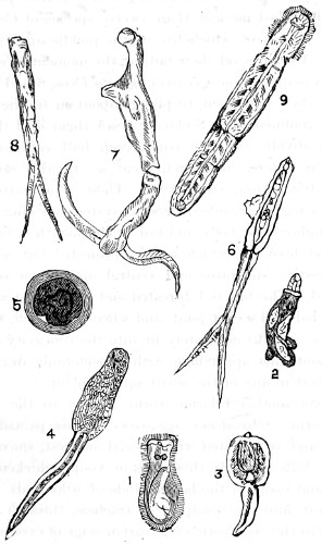
Fig. 378.—Forms of Cercaria; stages in the development of the Fluke.
1. An infusorial embryo; 2. a Trematode embryo having quite recently escaped from the egg; 3. embryo cercaria; 4. fully-formed cercaria, showing alimentary canal and sucker-like head; 5. encysted form of same; 6. Cercaria furcata, with the nervous system and forked tail displayed; 7. in the act of breaking up; 8. tail portion half an hour after division; 9. parasitic worm of another species of Trematoda. (Magnified from 10 to 25 diameters.)
The study of these embryos throws a flood of light upon the obscure history of Cercariæ. After a short period of wandering, their embryos fasten upon the water-snail, and compel it to act as a wet-nurse, and prepare it for a further and higher stage of life. The earliest condition in which I have discovered them concealed about the body of the water-snail is shown at No. 2; in appearance, a simple elongated sac filled with ova or germs, and which in a short time develop into the caudate worms already spoken of; their tails gradually attaining to the length of the mature embryos, Nos. 3 and 4, the latter being a full-grown Cercaria ephemera.
Diesing described no less than twelve species of Cercariæ, some of the most curious of which live on the puddle-snail, in colonies of thousands. All throw off their tails at the moment of changing into a fluke. On placing some Cercaria furcata (Nos. 6 and 7) under the microscope, they were seen to plunge about in frantic attempts to escape from confinement. Suddenly I saw them shed their tails and their bodies divide into two parts, each half swimming about as vigorously as before, quite indifferent as to the severance, and apparently dying from exhaustion. Those represented in Nos. 6 and 7 have a highly-organised nervous system, forming a continuous circuit throughout the body and tail. The mouth is furnished with a sucker and hooklets, which can be projected out some distance, while a digestive apparatus and ventral opening or sucker can be differentiated. The tail is bifurcated and articulated with the body by a sort of ball-and-socket joint, and when broken off, the convexity of one part is seen to accurately fit into the concavity of the other; it lashes about this appendage with considerable dexterity, rarely attaching itself to any of the small aquatic plants.75
There is yet another Filarian worm, a pest to the poultry-yard, the Gape-worm, Sclerostoma syngamus. This parasite is widely distributed, and is invested with special interest, since it produces disease, and kills annually thousands of young chickens, pheasants, partridges, and many of the larger kinds of wild birds. The worms find their way into the windpipe or tracheæ, through the drinking water, while in the embryotic or cercarian stage of existence, and their increase is so rapid, the birds quickly die of suffocation. The female gape-worm often attains to a considerable size, and when full grown resembles the well-known mud-worm of the Thames (Gordius aquaticus). She measures full six-eighths of an inch in length, while the male only measures one-eighth. So insignificantly small is he that the female carries him about tucked into a side pocket. The ova sac occupies a considerable portion of the internal body space, and is always found loaded with eggs in all stages of development, numbering some five hundred or a thousand. In shape these are ovoid. On cutting open the windpipe of chicken and partridges, I have found their tracheæ literally swarming with the gape-worm.76
A remarkable form of the Trematode worm is Bilharzia hæmatobra of Cobbold, Distomia hæmatobium of other authors (Plate IV., No. 102). This genus of fluke, discovered by Dr. Bilharz in the human portal system of blood vessels, gives rise to a very serious state of disease among the Egyptians. So common is the occurrence of this worm, that this physician expressed his belief that half the grown-up population of Egypt suffer from it. Griesinger conjectures that the young of the parasite exist in the waters of the Nile, and in the fish which abound. Dr. Cobbold thinks “it more probable that the larvæ, in the form of cercariæ, rediæ, and sporocysts, will be found in certain gasteropod mollusca proper to the locality.” The anatomy of this fluke is fully described by Küchenmeister in his book on parasites, by Leuckart,77 and by Cobbold. The eggs and embryos of Bilharzia are peculiar in possessing the power of altering their forms in both stages of life; and it is more than probable that the embryo form has been mistaken for some extraordinary form of ciliated infusorial animal, its movements being quick and lively. We cannot fail to notice the curious form of the male animal, and, unlike the Filarian previously described, it is he who carries the female about and feeds her. The whip-like appendage seen in the figure is a portion of the body of the female. The disease produced by this parasite is said to be more virulent in the summer months, probably owing to the greater abundance of cercarian larvæ at this period of the year.
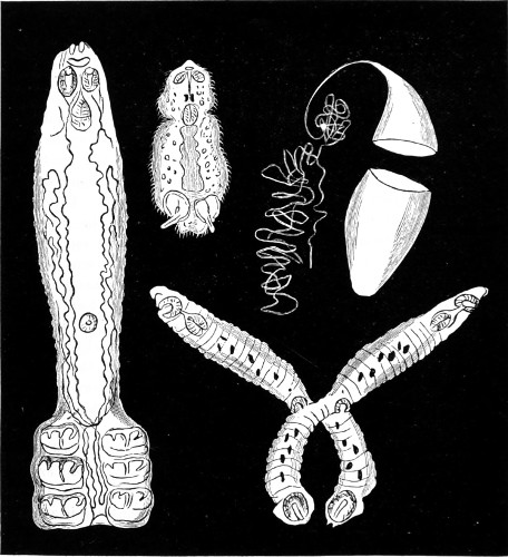
Fig. 379.—The double parasitic worm (Diplozoum paradoxum).
There are also double parasitic worms, which may be described as a sub-order of Trematoda, differing very much from those previously described. These live on the gills of several species of fresh-water fish, the gudgeon and minnow, for instance. Among them is a most remarkable creature well deserving the name of Diplozoum paradoxum which has been bestowed upon it. It consists of two complete mature similar halves, each possessing every attribute of a perfect animal (a). Each of the pointed front ends has a mouth aperture, and close to it two small sucking discs; while each individual has a separate intestine, consisting of a medium tube and innumerable side-branches. At the hinder end of the body are two suckers sunk in a depression, and protected by four hard buckle-shaped organs. The eggs are elongated, and provided at one end with a fine thread-like appendage (b). In this egg the young (c)—which at the time of hatching is only about one-hundredth of an inch—takes about a fortnight to develop. It is covered with cilia, has two eyes and two suckers; after quitting the egg, the larvæ are very lively and restless in their movements, gliding about and then swimming off with rapidity. If unable to find the fish into which they are destined to live, they grow feeble and perish, but if successful they grow into the Diporpa (d), which is flattened and lancet-shaped, and bears a small sucking disc on the under surface and a conical excrescence on the back. After living in this state for some weeks, and gaining nourishment by sucking the blood from the fish’s gills, the worms begin to join together in pairs, one specimen seizing the conical excrescence of another by its ventrical sucker; then, by a truly acrobatic feat, the second twists itself to the dorsal excrescence of the first, and in this state an inseparable fusion takes place between the suckers and the excrescences involved in the adhesion.78
In the group Vermes, the more highly-organised Annelida must be included. These, for the most part, live either in fresh or salt water. The Annelids are various, while the Planaria, a genus of Turbellaria, are very common in pools, and resemble minute leeches; their motion is continuous and gliding, and they are always found crawling over the surfaces of aquatic plants and animals, both in fresh and salt water. The body has the flattened sole-like shape of the Trematode entozoa (Fig. 378, No. 9), the mouth is surrounded by a circular sucker; this is applied to the surface of the plant from which the animal draws its nourishment; it is also furnished with a rather long proboscis, which is probably employed for a similar purpose.
Planariæ multiply by eggs, and by spontaneous fissuration in a transverse direction, each segment becoming a perfect animal. Professor Agassiz believes that the infusorial animals, Paramæcium and Kolpoda, are simply planarian larvæ.
Hirudinidæ, the leech tribe, are usually believed to form a link between the Annelida on the one hand, and the Trematoda on the other; their affinities place them closer with the latter than the former. Although deprived of the characteristic setæ of the Annelida, and exhibiting no sectional divisions, they are provided with a sucker-like mouth possessed by Trematoda, but they present no resemblance to them in their reproductive organs. On the other hand, in the arrangement of the nervous system and in their vascular system, the Hirudinidæ resemble Annelida. The head in most of the Annelida is distinctly marked, and furnished with eyes, tentacles, mouth, and teeth, and in some instances with auditory vesicles, containing otolithes. The nervous system consists of a series of ganglia running along the ventral portion of the animal, and communicating with a central mass of brain.
Hirudina medicinalis puts forth a claim for special attention on the ground of services rendered to mankind. The whole of the family live by sucking the blood of other animals; and for this purpose the mouth of the leech is furnished with a number of strong horny teeth, by which they cut through the skin. In the common leech three rows of teeth exist, arranged in a triangular, or rather triradiate form, a structure that accounts for the peculiar appearance of leech bites. The most interesting part of the anatomy of the leech to microscopists is certainly the structure of the mouth (Fig. 380). This is a muscular dilatable orifice, within which three beautiful little semi-circular saws are situated, arranged so that their edges meet in the centre. It is by means of these saws that the leech makes the incisions whence blood is to be procured, an operation which is performed in the following manner. No sooner is the sucker firmly fixed to the skin, than the mouth becomes slightly everted, and the edges of the saws are thus made to press upon the tense skin, a sawing movement being at the same time given to each, whereby it is made gradually to pierce the surface, and cut its way to the capillary blood-vessels beneath.
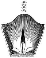
Fig. 380.—Mouth of Leech.
In Clepsinidæ the body is of a leech-like form, but very much narrowed in front, and the mouth is furnished with a prehensile proboscis. These animals live in fresh water, where they may often be seen creeping over aquatic plants. Their prey is the pond-snail.
Tubicola.—The worms belonging to this series of branchiferous Annelida are all marine, and distinguished by their invariable habit of forming a tube or case, within which the soft parts of the animal can be entirely retracted. This tube is usually attached to stones or other submarine bodies. Externally it is composed of various foreign materials, sand, crystalline bodies, and the débris of shells; internally it is lined with a smooth coating of sarcode, sometimes of a harder consistency. The Tubicola generally live in societies, winding their tubes into a mass which often attains a considerable size; only a few are solitary in their habits. They retain their position in their cases by means of tufts of bristles and spines; the latter, in the tubicular Annelids, are usually hooked, so that by applying them to the walls of the case, the animal is enabled to oppose a considerable resistance to any effort made to withdraw it. In the best known family of the order (Sabellia), the branchiæ are placed in the head, and form a circle of plumes, or a tuft of branched organs. The Serpulidæ form irregularly twisted calcareous tubes, and often grow together in large masses, when they secure themselves to shells and similar objects; other species, Terebellidæ, which build their cases of sand and stones, appear to prefer a life of solitude. The best known form is Terebella littoralis.79 The curious little spiral shells seen upon the fronds of seaweeds are formed by an animal belonging to the Spirorbis.
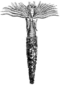
Fig. 381.—Serpula with extended tentacles and body protruding from calcareous case.
If the animals be placed in a vessel of sea-water a very pleasing spectacle will soon be witnessed. The top part of the tube is seen to open, and the creature cautiously protrudes a fringe of tentacles; these gradually spread out two beautiful fan-like rows of tentacles, surrounded by cilia of a rich purple or red colour. These serve the double purpose of breathing and feeding organs. When withdrawn from its calcareous case, the soft body is seen to be constructed of a series of rings, with a terminal prehensile foot by which it attaches itself.
Many Annelids are without tubes or cells of any kind, simply burying their bodies in the sand near tidal mark. The Arenicola, lob-worm, is a well-known specimen of the class; its body is so transparent that the circulating fluids can be distinctly seen under a moderate magnifying power. Two kinds of fluids flow through the vessels, one nearly colourless, the other red; the vessels through which the latter circulate are described as blood-vessels.
Not very much interest attaches to the developmental stage of the Annelida. They issue forth from ova, and the embryo so closely resemble ciliated polypes, that competent observers have mistaken them for animals belonging to a lower class; a few hours’ careful watching is sufficient to dispel a belief of the kind, when the embryonic, globular, or shapeless mass is seen to assume a form of segmentation, and soon the various internal organs become more and more developed, eye spots appear, and the young animal arrives at the adult stage of its existence.
Crustacea.
The crustaceans comprise a large assemblage of Arthropods, presenting great diversity of structure. Some of the parasitic species have become so simplified in organisation that they appear to present no relationship with the higher members of the class, yet it is certain that all the species, whether terrestrial or aquatic, belong to the same stock, and may have had origin in the same fundamental plan of structure. Essentially, the body consists of a large number of segments, to each of which is attached a pair of two-branched appendages; the external branch is termed the exopodite and the internal the endopodite. Five segments at the front end of the body unite to form a head, the appendages of the first two being situated in front of the mouth, and performing the office of feelers or antennæ, while those of the remaining three segments are transformed into jaws, the first pair of jaws being the mandibles and the following two pairs the maxillæ. The rest of the appendages are variously modified and to some are attached respiratory organs in the form of gills. Crustaceans are broadly divided from Centipedes, Millipedes, Insects, &c., by the presence of two pairs instead of one pair of antennæ, and by the possession of branchial and not tubular (tracheal) respiratory organs. Arachnida and some other species are again widely separated. The majority of the young on leaving the egg are quite unlike the parent, and only acquires their definite form after undergoing a series of changes. The earliest stage, which has been called the Nauplius, already referred to in connection with the barnacle, is a minute body showing no trace of segmentation, and provided with a single eye, and three pairs of swimming appendages, which become the two pairs of antennæ and the mandibles of the adult. This stage is by no means of invariable occurrence, but is chiefly characteristic of the lowest members, the Entomostraca, and is rare in the higher, Malacostraca. The typical crustaceans are shrimps, crayfish, &c., so familiarly described by Huxley. The zoæa stage of the crab, a minute transparent creature, which undergoes several changes, swims about flapping its long jointed abdomen, like some of the Entomostraca, and the shrimp in particular. The larva of crayfish, the so-called glass-crab, is very peculiar and interesting. The sessile-eyed series, in which the compound eyes are never mounted on a movable stalk, and to which the Isopoda belong, exhibits great diversity of structure as well as of habits and habitat. Some live in fresh water, most are marine, while others live on land and take to a parasitic life.
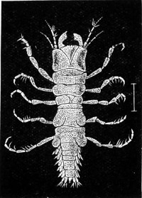
Fig. 382.—Male Gnathia, enlarged.
This genus contains Gnathia, in which the male and female are so dissimilar, that they are frequently referred to as members of two families. In the adult male the mandibles are powerful and prominent, and the head is large, squared, and as wide as the thorax. In the female, on the contrary, the head is curiously small and triangular, without visible mandibles, and the thorax is much dilated. The creatures are about one-sixth of an inch long, and of a greyish colour, and the destruction they bring about is due to their habit of boring into timber below water mark. Fig. 382 represents an enlarged view of the male Gnathia. These crustaceans are vegetarians, and feed on wood. Other members of the group, known as fish lice, are much larger in size, and chiefly infest the cetacea, and bear in addition two large eyes. By means of their powerful fore feet the Cymothordæ attach themselves to both marine and fresh-water fish, showing a preference for the inside of the mouth of their host.
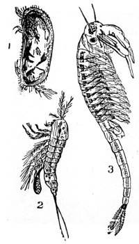
Fig. 383.—1. Cypris; 2. Cyclops; 3. Branchipus grubei.
The bar-footed group Copepoda are free living, and the thorax bears four or five swimming feet; the abdomen is without appendages. The best known fresh-water form is Cyclops, the structure of which serves as a type of the order. The body is, as is well known to microscopists, broad in front and tapering behind, being thus, when viewed swimming, pear-shaped in outline. The dorsal elements of the head are fused to form a carapace, which bears a single eye, from which circumstance it derives its name. The eggs are carried by the female in a couple of ova-sacs attached to the last segment of the thorax, and so prolific are these creatures that a female will produce over four thousand million young. The young when hatched is an oval Nauplius, which after two or three moults acquires the adult state. In the family of the Apodidæ we have an equally well-known crustacean, the Branchipus. In the Branchipodidæ the body is also elongated, but there are no appendages to the abdomen, which consists of nine segments, while there are eleven pairs of thoracic appendages. The head shield is not developed backwards, and the large separated eyes are supported on distinct stalks. In the male the second antennæ are converted into claspers. These crustaceans swim upside down (Fig. 383).
Cladocera (Daphniadæ of Dr. Baird).—The water-flea (Daphnia pulex) may be taken as the best known example of the order. The body of this little active animal is narrowed in front, and at the posterior end, where the carapace is deeply notched, is the tip of the abdomen bearing the pair of rigid barbed setæ from which the genus takes its name. At the front of the head is a large compound eye and two pairs of branched plumed appendages, antennæ. The first pair of these are small and simple. The jaws consist of the mandibles and the first pair of maxillæ, the second pair of maxillæ being obsolete in the adult. The thorax comprises five segments, each bearing a pair of leaf-like swimming limbs. The abdomen consists of three segments, and is destitute of limbs. The males are usually smaller than the females, and much rarer, being rarely met with before the end of summer.
Eggs are laid both in summer and winter, and are passed into a brood-pouch, separating the upper surface of the thorax from the backward extension of the carapace. Here the summer eggs hatch, but the winter set are enclosed in a kind of capsule developed from the carapace. This capsule, termed the ephippium, is cast off with the next moult of the mother’s integument (a process necessary for the gradual growth of the crustacean), and falling to the bottom of the water, gives exit to the embryos, which hatch in its interior, and the young born from these “ephippial” eggs produce young, which in their turn become mothers. It appears, then, the winter eggs are enclosed in capsules of more than usual hardness to enable them to withstand any degree of cold that might otherwise prove fatal to the parent. Dr. Baird found, on examining ponds that had been again filled up by rain after remaining two months dry, numerous specimens of Daphnia and Cyclops quadricornis in all stages of growth.80
We learn also from his investigations that the Daphnia have many enemies. “The larva of the Corethra plumicornis, known to microscopical observers as the skeleton larva, is exceedingly rapacious of Daphnia. Pritchard says they are the choice food of a species of Nais; and Dr. Parnell states that the Lochleven trout owes its superior sweetness and richness of flavour to its food, which consists of small shell-fish and Entomostraca.” These crustaceans abound in fresh and salt water. Artemiæ are formed exclusively in salt water, in salt marshes, and in water highly charged with salt. Myriads of these Entomostraca are found in the salterns at Lymington, in the open tanks or reservoirs where the brine is deposited previous to boiling. A pint of the fluid contains about a quarter of a pound of salt, and this concentrated solution destroys most other marine animals. During the fine days in summer Artemiæ may be observed in immense numbers near the surface of the water, and, as they are frequently of a lively red colour, the water appears tinged with the same hue. The movements of this little animal are peculiar. It swims about on its back, and by means of its tail, its feet being at the same time in constant motion. They are both oviparous and ovoviviparous, according to the season of the year. At certain periods they only lay eggs, while during the hot summer months they produce their young alive. In about fifteen days the eggs are expelled in numbers varying from 50 to 150. As is the case with many of the Entomostraca, the young present a very different appearance from the adult animals; and they are so exactly like the young of Chirocephalus, that with difficulty are they distinguishable one from the other. The ova of other species are furnished with thick capsules, and imbedded in a dark opaque substance, presenting a minutely cellular appearance, and occupying the interspace between the body of the animal and the back of the shell; this is called the ephippium. The shell is often beautifully transparent, sometimes spotted with pigment; it consists of a substance known as chitine, impregnated with a variable amount of calcium carbonate, which produces a copious effervescence on the addition of a small quantity of a strong acid to the water in which the shell is immersed. When boiled, Artemiæ turn red as their congeners, lobsters. Their shells may be said to consist of two valves united at the back, resembling the bivalve shell of a mussel, or simply folded at the back to appear like a bivalve, but are really not so; or they may consist of a number of rings or segments. The body of Cypris presents a reticulated appearance, somewhat resembling cell structure. Entomostraca should be narcotised and prepared for examination under the microscope as directed by Mr. Rousselet at pages 345, 346.
Insects’ Eggs, etc.
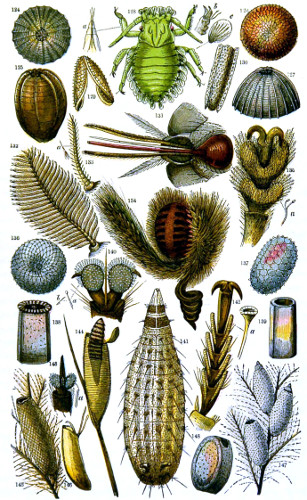
Tuffen West, del. Edmund Evans.
Plate VI.