Part 2 — CHAPTER I.
Reading edition

Microscopic Forms of Life—Thallophytes—Pteridophyta, Phanerogamæ—Structure and Properties of the Cell.
The time has long since passed by since the value of the microscope as an instrument of scientific research might have been called in question. By its aid the foundation of mycology has been securely laid, and cryptogamic botany in particular has, during the last quarter of a century, made surprising progress in the hands of those devoted to pursuits which confer benefits upon mankind.
Little more than thirty years ago practically nothing was known of the life history of a fungus, nothing of parasitism, of infectious diseases, or even of fermentation. Our knowledge of the physiology of nutrition was in its infancy; even the significance of starches and sugars in the green plant was as yet not understood, while a number of the most important facts relating to plants and the physiology of animals were unknown and undiscovered. When we reflect on these matters, and remember that bacteria were regarded merely as curious animalculæ, that rusts and smuts were supposed to be emanations of diseased states, and that spontaneous generation still-survived among us, some idea may be formed of the condition of cryptogamic botany and the lower forms of animal life some eight or ten years after my book on the microscope made its first appearance (1854).
Indeed, long prior to this time, dating from that of even the earliest workers with the microscope, it was known that the water of pools and ditches, and especially infusions of plants and animals of all kinds, teem with living organisms, but it was not recognised definitely that vast numbers of these microscopic living beings (and even actively moving ones) are plants, growing on and in the various solid and liquid matters examined, and as truly as visible and accepted plants grow on soil and in the air and water. Perhaps the most important discovery in the history of cryptogamic botany was initiated here. The change, then, that has come over our knowledge of microscopic plant life during this last busy quarter of a century has been almost entirely due to the initiation and improvement, first in methods of growing them, and in the methods of “Microscopic Gardening”; and secondly, to the greater knowledge gained in the use of the microscope.
“If we look at the great groups of plants from a broad point of view, it is remarkable that the fungi and the phanerogams occupy attention on quite other grounds than do the algæ, mosses, and ferns. Algæ are especially a physiologist’s group, employed in questions on nutrition, reproduction, and cell division and growth; the Bryophyta and Pteridophyta are, on the other hand, the domain of the morphologist. Fungi and Phanerogams, while equally or even more employed by specialists in morphology and physiology, appeal widely to general interest on the ground of utility.
“It is very significant that a group like the fungi should have attracted so much scientific attention, and aroused so general an interest at the same time. But the fact that fungi affect our lives directly has been driven home; and whether as poisons or foods, destructive moulds or fermentation agents, parasitic mildews or disease germs, they occupy more interest than all other cryptogams put together, the flowering plants alone rivalling them in this respect. A marked feature of the period in which we live will be the great advances made in our knowledge of the uses of plants, for, of course, this development of economic botany has gone hand in hand with the progress of geological botany, the extension of our planting, and the useful applications of botany to the processes of home industries.”49
The intimate organic structure of the vegetable world is seen to consist of a variety of different materials indeterminable by unassisted vision, and for the most part requiring high magnification for their discrimination. Chemical analysis had, however, shown that vegetables are composed of a few simple substances, water, carbonic acid gas, oxygen, nitric acid, and a small portion of inorganic salts. Out of these simple elements the whole of the immense variety of substances produced by the vegetable kingdom are constructed. No part of the plant contains fewer than three of these universally distributed elements, hence the greater uniformity in their chemical constituents. It will be seen, then, that the methods of plant chemistry are of supreme interest both to the chemist and the physiologist, or biologist. Plants, while they borrow materials from the inorganic, and powers from the physical world, whereby they are enabled to pass through the several stages of germination, growth, and reproduction, could not accomplish these transformations without the all-important aid of light and heat, the combined functions of which are indispensable to the perfect development of the vegetable world.
Light, then, enables plants to decompose, change into living matter, and consolidate, the inorganic elements of carbonic acid gas, water, and ammonia, which are absorbed by the leaves and roots from the atmosphere and earth; the quantity of carbon consolidated being exactly in proportion to the intensity of the light. Nevertheless, light in its chemical character is a deoxidising agent, by which the numerous neutral compounds common to vegetables are formed. It is the principal agent in preparing the food of plants, and it is during the chemical changes spoken of that the specific heat of plants is slowly evolved, which, though generally feeble, is sometimes very sensibly evolved, especially so when flowers and fruits are forming, on account of the increase of chemical energy at this period.
The action of heat is measurable throughout the whole course of vegetable life, although its manifestations take on various forms—those suited to the period and circumstances of growth. Upon the heat generated depends the formation of protein and nitrogenous substances, which abound more directly in the seed buds, the points of the roots, and in all those organs of plants which are in the greatest state of activity. The whole chemistry of plant life, in fact, is manifest in this production of energy for drawing material from its surroundings; therefore the organising power of plants bears a direct ratio to the amount of light and heat acting upon them.
The living medium, then, which possesses the marvellous property of being primarily aroused into life and energy, and which either forms the whole or the greater portion of every plant, is in its earliest and simplest form nothing more than a microscopic cell, consisting of one or two colourless particles of matter, in closest contact, and wholly immersed in a transparent substance somewhat resembling albumen (white of egg), termed protoplasm, but differing essentially in its character and properties. This nearly colourless organisable matter is the life-blood of the cell. It is sufficiently viscid to maintain its globular form, and under high powers is seen to have a slightly consolidated film enclosing semi-transparent particles, together with vacuoles which are of a highly refractive nature. These small bodies are termed nuclei, and they appear to be furnished with an extremely delicate enveloping film. In a short time the nuclei increase in number and split up the parent body. The protoplasmic mass, however, is undoubtedly the true formative material, and is rightly regarded as “the physical basis of life” of both the vegetable and animal kingdoms.
There are, however, certain members of the vegetable kingdom which somewhat resemble animals in their dependence upon receiving organic compounds already formed for them, being themselves unable to effect the fixation of the carbon needed to effect the first stage in their after chemical transformations. Such is the case with a large class of flowering plants, among Phanerogams, and the leafless parasites which draw their support chiefly from the tissues of their hosts. It is likewise the case with regard to the whole group of fungi; the lower cryptogams, which derive the greater portion of their nutritive materials from organic matter undergoing some form of histolysis; while others belonging to this group have the power of originating decomposition by a fermentative (zymotic) action peculiarly their own. There are many other protophytes which live by absorption, and which appear to take in no solid matter, but draw nourishment from the atmosphere or the water in which they exist.
With regard to motion, this was at one time considered the distinctive attribute of animal life, but many protophytes possess a spontaneity of power and motion, while others are furnished with curious motile organs termed cilia, or whip-like appendages, flagella, by which their bodies are propelled with considerable force through the water in which they live.
Henceforth this protoplasmic substance was destined to take an important position in the physiological world. It is, then, desirable to enter somewhat more fully into the life history of so remarkable a body. It has a limiting membrane, composed of a substance somewhat allied to starch, termed cellulose, one of the group of compounds known as carbo-hydrates. The mode of formation and growth of this cell wall is not yet definitely determined; nevertheless, it is the universal framework or skeleton of the vegetable world, although it appears to play no special part in their vital functions. It merely serves the purpose of a protecting membrane to the globular body called the “primordial cell,” which permanently constitutes the living principle upon which the whole fundamental phenomena of growth and reproduction depend.
Sometimes this protoplasmic material is seen to constitute the whole plant; and so with regard to the simplest known forms of animal life—the amœba, for example. That so simple and minute an organism should be capable of independent motion is indeed surprising. Dujardin, a French physiologist, termed this animated matter sarcode. On a closer study of the numerous forms of animal life it was found that all were alike composed of this sarcode substance, some apparently not having a cell wall. The same seemed to hold good of certain higher forms of cells, the colourless blood corpuscles for instance, which under high powers of the microscope are seen to change their shape, moving about by the streaming out of this sarcode. At length the truth dawned on histologists that the cell contents, rather than the closing wall, must be the essential structure. On further investigation it became apparent that a far closer similarity existed between vegetables and animals than was before supposed. Ultimately it was made clear that the vegetable protoplasm and the animal sarcode were one and the same structure. Max Schultz found this to be the case, and to all intents and purposes they are identical.
We have now to retrace our steps and look somewhat more closely into the discovery of that important body, the cell-nucleus. It was an English botanist, Dr. Robert Brown, who, in 1833, during his microscopical studies of the epidermis of orchids, discovered in their cells “an opaque spot,” to which soon afterwards he gave the name of nucleus. Schleiden and Schwann’s later researches led them to the conclusion that the nucleus is the most characteristic formative element in all vegetable and animal tissues in the incipient phase of existence. It then began to be taught that there is one universal principle of development for the elementary parts of all organisms, however different, and that is the formation of cells. Thus was enunciated a doctrine which was for all practical purposes absolutely new, and which opened out a wide field of further investigation for the physiologist, and led up to a fuller knowledge of the cell contents. In fact, it became a question as to whether the cell contents rather than the enclosing wall should not be considered the basis of life, since the cell at this time had by no means lost its importance, although it no longer signified the minute cavity it did when originally discovered by Schwann. It now implied, as Schultz defined it, “a small mass of viscid matter, protoplasm, endowed with the attributes of life.” The nucleus was once more restored to its original importance, and with even greater significance. In place of being a structure generated de novo from non-cellular substance, and disappearing as soon as its function of cell formation is accomplished, the nucleus is now known as the central permanent feature of every cell, and indestructible while the cell lives, and the parent, by division of its substance, of other generations of nuclei and cells. The word cell has at the same time received its final definition as “a small mass of protoplasm supplied with a nucleus.” In short, all the activities of plant and animal life are really the product of energy liberated solely through histolysis, or destructive processes, amounting to the combustion that takes place in the ultimate cells of the organisms.
But there are other points of especial interest involved in the question of cell formation beside those already mentioned.
The cell and its contents collectively are termed the endoplasm, or when coloured, as in algæ, endochrome. With regard to the outer layer of the cell and its growth nothing satisfactory has been clearly determined and finally accepted.
The cell as a whole is a protoplasmic mass, and not an emulsion, as some observers would have us suppose. It is, in fact, a reticulated tissue of the most delicate structure, made up of canaliculate spiral fibrils with hyaline walls capable of expansion and contraction. These fibrils are probably composed of still finer spirals. The visible granulated portion of the protoplasm, the only part that takes a stain under ordinary circumstances, is simply the contents of these canals. It is the chromatin of Flemming, and is capable of motion within the canals. The nucleus, then, is probably nothing more than a granule of the extra-cellular net, and is formed by the junction of the several bands of wall-threads which traverse it in different directions. The cell wall of plants possesses the same structure as protoplasm, and is probably protoplasm impregnated by cellulose.
It is this portion of the protoplasmic mass that is now recognised under the term octoplasm, or primordial utricle, and is of so fine and delicate a nature that it is only brought into view when separated from the cell wall either by further developmental changes, or by reagents and certain stains or dyes. It was, in fact, discovered to be a slightly condensed portion of the protoplasmic layer corresponding to the octosare of the lower forms of animal life. The octoplasm and cell wall can only be distinguished from each other by chemical tests. Both nucleus and nucleoli are only rendered visible in the same way, that is, by staining for several hours in a carmine solution, and washing in a weak acetic acid solution.
With the enlargement of the cell by the imbibition of water, clear spaces, termed vacuoles, are seen to occupy a small portion of the cell, while the nucleus and nucleoli lie close to the parietal layer.
The interesting phenomenon of cyclosis, to which I shall have occasion to refer further on, is now believed to be due to the contractility of certain wall-threads stretching from the nucleus to the outermost layers of the cell. An intimate relationship is thereby established between the nucleus, the nucleolus, and the parietal layer. This much has been made clear by the more scientific methods of investigation pursued in the use of the microscope. Nevertheless a large and important class of cells, forming a kind of borderland between the vegetable and animal kingdoms, still remains to be dealt with, in which the cell contents are only imperfectly differentiated, while numerous other unicellular organisms, owing to their extreme minuteness, tenuity, and want of all colour, are apparently devoid of any nucleus, and when present can only be differentiated by a resort to a specially conducted method of preparation and staining. There is, however, a remarkable feature in connection with many micro-organisms—that certain of these protophytes possess motive organs, cilia or flagella, bodies at one time supposed to be characteristic of, and belonging to, the protozoa.
This being the case, the methods of plant chemistry are of supreme interest, the more so because physiologists are in a position to isolate a single bacterial cell, grow it in certain media, and thus devote special attention to it, and keep it for some time under observation. In this way it has become possible to further grasp facts in connection with cell nutrition and the nature of its waste products. We have, then, arrived at a stage when the history of the chemical changes brought about by bacteria can be more definitely determined, as we have here to do with the vegetable cell in its simplest form. The chemical work performed by these micro-organisms has as yet occupied only a few years; nevertheless, the results have been of the most remarkable and encouraging character.
At an earlier period an interesting discovery in connection with the pathogenic action of these bodies was, by the labours of Schöenlein, Robin, and others, brought to the notice of the medical profession, viz., that certain diseases affecting the human body were due to vegetable parasites. In 1856 an opportunity offered itself for a thorough investigation, and the microscopical part of the work fell into my hands, with the result that I was able to add considerably to Schöenlein’s list of parasitic skin diseases. My observations were in the first instance communicated to the medical journals. But the generalisation arrived at was that “If there be any exceptions to the law that parasites select for their sustenance the subjects of debility and decay, such exceptions are rarely to be found among the vegetations belonging to fungi, which invariably derive nutrition from matter in a state of lowered vitality, passing into degeneration, or wherein decomposition has already taken place to a certain extent.... It scarcely admits of a doubt that all diseases observed of late years among plants have been due to parasites of the same class favoured by want of vigour of growth and atmospheric conditions, and that the cause of the various murrains of which so much has been heard is also due to similar causes.”50
Herein, then, is to be found the solution of a difficulty that so long surrounded the question, but which subsequently culminated in the specialisation and scientific development of bacteriology, due to the unceasing labours of Pasteur, whose solid genius enabled him to overcome the prejudices of those who were at work on other lines, and who had no conception of the functions that parasitic organisms fulfil in nature.
Going back to my earlier experimental researches to determine the part taken by saccharomycetes and saprophytes in fermentation, I find, from correspondence in my possession, that in 1859 I demonstrated to the satisfaction of Dr. Bell, F.R.S., the then head of the chemical laboratory of Somerset House, that a very small portion of putrefactive matter taken from an animal body, a parasitic fungus (Achorion Schöenleinii), a mould (Aspergillus or Penicillium), and a yeast (Torula cerevisiæ) would in a short time, and indifferently, set up a ferment in sweet-wort and transform its saccharine elements into alcohol, differing only in degree (quantitative), and not in kind or quality. This, then, was the first step in the direction towards proving symbiotic action between these several parasitic organisms. The only apparent difference observed during the fermentative processes was that putrefactive (saprophytic) action commenced at a somewhat earlier stage, and that the percentage of alcohol was also somewhat less.51
In 1856, also, the ærobic bacteria attracted my attention, and, together with the late Rev. Lord Sidney Godolphin Osborne, I exposed plates of glass (microscopical slides), covered with glycerine and grape sugar, in every conceivable place where we thought it possible to arrest micro-organisms. The result is known, viz., that fungoid bodies (moulds and bacterial) were taken in great numbers, and varying with the seasons. The air of the hospital and sick-room likewise engaged attention, each of which proved especially rich in parasitic bodies. During the cholera visitation of 1858 the air was rich in ærobic and anærobic bacteria, while a blue mist which prevailed throughout the epidemic yielded a far greater number than at any former period (represented in Plate I., No. 13). This blue mist attracted the especial attention of meteorologists. At a somewhat later period a more remarkable fungoid disease, the fungus foot of India, mycetoma, came under my observation, a detailed description of which I contributed to the medical journals, and also, with further details, to the “Monthly Microscopical Journal” of 1871. Interlacing mycelia, ending in hyphæ, in this destructive form of parasitic disease were seen to pervade the whole of the tissues of the foot, the bony structures being involved, and it was only possible to stay the action of the parasite by amputation.
So far, then, the study of parasitic organisms had at an early period shared largely in my microscopical work, extending over several years, and with the result that these micro-organisms were found to exhibit on occasions great diversity of character, and that different members of the bacteria in particular flourish under great diversity of action, and often under entirely opposite conditions; that they feed upon wholly different materials, and perform an immense variety of chemical work in the media in which they live.
The study of the chemistry (chemotaxis) of bacteria has, however, greatly enlarged our conception of the chemical value and power of the vegetable cell, while it is obvious that no more appropriate or remunerative field of study could engage the attention of the microscopist, as well as the chemist, than that of bacterial life, and which is so well calculated to enlarge our views of created organisms, whether belonging to the vegetable or animal kingdom.
Pathogenic Fungi and Moulds.
It is scarcely necessary to go back to the history of the parasitic fungi to which diseases of various kinds were early attributable. The rude microscopes of two and a half centuries ago revealed the simple fact that all decomposable substances swarmed with countless multitudes of organisms, invisible to ordinary vision. Leuwenhoek, the father of microscopy, and whose researches were generally known and accepted in 1675, tells of his discovery of extremely minute organisms in rain-water, in vegetable infusions, in saliva, and in scrapings from the teeth; further, he differentiated these living organisms by their size and form, and illustrated them by means of woodcuts; and there can be no doubt that his figures are intended to represent leptothrix filaments, vibrios, and spirilla. In other of his writings attempts are made to give an idea of the size of these “animalcules”; he described them as a thousand times smaller than a grain of sand. From his investigations a belief sprung up that malaria was produced by “animalcules,” and that the plague which visited Toulon and Marseilles in 1721 arose from a similar cause. Somewhat later on the natural history of micro-organisms was more diligently studied, and with increasing interest. Müller, in 1786, pointed out that they had been too much given to occupy themselves in finding new organisms, he therefore devoted himself to the study of their forms and biological characters, and it was on such data he based a classification. Thus the scientific knowledge gained of these minute bodies was considerably advanced, and the subject now entered upon a new phase: the origin of micro-organisms. It further resolved itself into two rival theories—spontaneous generation, and development from pre-existing germs—the discussion over which lasted more than a century. Indeed, it only ended in 1871, when the originator of the Abiogenesis theory withdrew from the contest, and the more scientific investigations of Pasteur (1861) found general acceptance. This indefatigable worker had been investigating fermentation, and studying the so-called diseases of wines and a contagious disease which was committing ravages among silkworms. Pasteur in time was able to confirm the belief that the “muscadine disease” of silkworms was due to the presence of micro-organisms, discernible only by the microscope. The oval, shining bodies in the moth, worm, and eggs had been previously observed and described by Nägeli and others, but it was reserved for Pasteur to show that when a silkworm whose body contained these organisms was pounded up in a mortar with water, and painted over the leaves of the tree upon which healthy worms were fed, all took the disease and died.
PLATE IX.
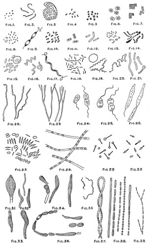
AFTER DR CROOKSHANK J. T. Balcomb. del.
TYPICAL FORMS OF BACTERIA, SCHIZOMYCETES, OR FISSION-FUNGI.
As the contagious particles were transmitted to the eggs, the method adopted for preventing the spread of the disease was as follows:—Each female moth was kept separate from the others, and allowed to deposit her eggs, and after death her body was crushed up in a mortar as before, and a drop of the fluid examined under the microscope. When any trace of muscadine was found present, the whole of the eggs and body were burnt. In this way the disease was combated, and ultimately stamped out.
Pasteur also pointed out that one form or cause of disease must not be confounded with another. For example, muscadine, a true fungus (Botrytis bassiana), should not be confounded with another disease known to attack silkworms, termed pebrin, this being caused by a bacterium, and, according to the more recent researches of Balbiani, by a Psorospermia. Botrytis is a true mould, belonging to the Oomycetes, and allied to the potato fungus, Peronospora. It is propagated by spores, which, falling on a silkworm, germinate and penetrate its body. A mycelium is then developed, which spreads throughout the body. Hyphæ appear through the skin, and bear white chalky-looking spores; these become detached, and float in the air as an impalpable dust-like smoke. Damp further develops the fungus.
Insects suffer much from the ravages of fungi. The house-fly sticking to the window-pane is seen to be surrounded by the mycelia of Penicillium racemosum (Sporendonema muscæ, or Saprolegnia feræ). In other cases Cordiceps attacks certain caterpillars belonging to the genera Cossus and Hepialus when they are buried in the sand and before their metamorphosis into chrysalides; they are killed by the rapid development of hyphæ and mycelium in their tissues.
Sphæria miletaris, a parasite of Bombyx pilyocarpa, the caterpillar of which is found on pine-trees, is one of the few fungi which may be regarded as beneficial to man, since it aids in the destruction of multitudes of these caterpillars, which otherwise would devour the young shoots and pine needles. Giard specialises other parasites of insects, which he terms Entomophoreæ. Others, E. rimosa, attack grasshoppers and the diptera, enveloping them in a dense coating of mycelium and spores, which speedily kills the victim.
The study, then, of the life-history of germs, microbes, micro-organisms, or bacteria (as they are indifferently termed), opened up a new science, that of Bacteriology. By the more recent advances in this science we are enabled to understand the very important part these minute organisms fill in the great scheme of Nature, for almost exclusively by their agency the soil is supplied with the requisite nutritive material for plant life. And, as already pointed out, wherever organic matter is present—that is, the dead and useless substances which are the refuse of life—such material is promptly seized upon by micro-organisms, by means of which histolysis is rapidly accomplished.
Bacteria require a power of from 600 to 1,000 diameters or more for the determination of the species to which they belong. The number of species has been so much increased of late that a bulky volume is found to be insufficient for their enumeration. I am, however, by the courtesy of Professor Crookshank, enabled to present my readers with the typical forms of thirty-nine species of Bacteria, Schizomycetes, or fission-fungi, a selection, it will be seen, chiefly taken from among pathogenic organisms—those believed to originate disease. But many of the supposed Saprophytic forms often described as originating disease are merely accidental associates, that is, living in companionship for a time.
Size.—In ordinary terms of measurement, bacteria are on an average from 1⁄25000th to about 1⁄5000th of an inch long. These measurements do not convey a definite impression to the mind. It is calculated that a thousand million of them could be contained in a space of 1⁄25th of an inch. The best impression of the size of the bacteria is, perhaps, obtained when it is stated that a 1⁄25-inch immersion objective gives a magnification of nearly 2,200 diameters, and that under this power the bacteria appear to be about the size of very small print. The standard of measurement accepted by bacteriologists is the micro-millimeter. One millimeter is equal to about 1⁄25000th an English inch. The number of micrococci in a milligramme of a culture of Staphylococcus pyogenes aurens has been estimated by Bujwid by counting at eight thousand millions. Not only do various species differ in dimensions, but considerable differences may be noted in a pure culture of the same species. On the other hand, there are numerous species which so closely resemble each other in size and shape that they cannot be differentiated by microscopic examination alone, and we have to look to other characteristics, as colour, growth in various culture media, pathogenic power, chemical products, &c., in order to decide the question of identity.
Reproduction.—The reproduction of bacteria takes place for the most part by fission and by spore formation. Fission is a process of splitting up or division, whereby an organism divides into two or more parts, each of which lives and divides in its turn. If certain organisms are watched under the microscope, a coccus or bacillus will be seen to elongate and at the same time become narrower, until its two halves become free, the two individual organisms again dividing and subdividing in their turn. This kind of reproduction is more readily seen in a higher class of unicellular organisms, the desmids. If, however, the new organisms do not break away from each other, but remain connected in groups or clusters, they are termed Staphylococci; if they remain connected in the form of a chain, or like a string of beads, they are termed Streptococci. If the division takes place in one plane, Diplococci are formed; if in two directions Tetracocci, or Tablet-cocci, are formed. On account of this multiplication by fission, the generic name of Schizomycetes, or fission-fungi, has been given to bacteria.
Spores.—A second method by which bacteria propagate is by spores. These bodies are distinguished by their remarkable power of resistance to the influence of temperature and the action of chemical reagents. Some of them will resist their immersion in strong acid solutions for many hours; also freezing and very high temperatures. Spore formation may take place in two ways: firstly, by “endogenous spores” (internal spores); secondly, by “arthrospores.”
Endogenous Spores.—When the formation of the spores takes place in the mother-cell, the protoplasm is seen to contract, giving rise to one or more highly refractive bodies, which are the spores. The enclosing membrane of the organism then breaks away, leaving the spores free.
Arthrospores.—When the spore is not formed in the parent bacillus, but when entire cells (owing to lack of favourable conditions of growth) become converted into spores, the formation is known as “arthrogenous,” the single individual being called an arthrospore. When the conditions are again favourable, spores germinate, giving rise to new bacilli. The germinating spore becomes elongated, and loses its bright appearance, the outer membrane becomes ruptured, and the young bacillus is set free. Certain conditions, such as the presence of oxygen in the case of the anthrax bacillus, give rise to the formation of spores; while various kinds of bacteria secure continuous existence by developing spores when there is lack of proper food material.
With reference to the incredible rapidity with which the bacteria multiply under conditions favourable to the growth and development, Cohn writes as follows:—“Let us assume that a microbe divides into two within an hour, then again into eight in the third hour, and so on. The number of microbes thus produced in twenty-four hours would exceed sixteen and a half millions; in two days they would increase to forty-seven trillions; and in a week the number expressing them would be made up of fifty-one figures. At the end of twenty-four hours the microbes descended from a single individual would occupy 1⁄40th of a hollow cube, with edges 1⁄25th of an inch long, but at the end of the following day would fill a space of twenty-seven cubic inches, and in less than five days their volume would equal that of the entire ocean.”
Again, Cohn estimated that a single bacillus weighs about 0·000,000,000,024,243,672 of a grain; forty thousand millions, 1 grain; 289 billions, 1 pound. After twenty-four hours the descendants from a single bacillus would weigh 1⁄2666th of a grain; after two days, over a pound; after three days, sixteen and a half million pounds, or 7,366 tons. It is quite unneccessary to state that these figures are purely theoretical, and could only be realised if there were no impediment to such rapid increase.
Fortunately, however, various checks, such as lack of food and unfavourable physical conditions, intervene to prevent unmanageable multiplication of these bodies.
These figures show, however, what a tremendous vital activity micro-organisms do possess, and it will be seen later at what great speed they increase in water, milk, broth, and other suitable media.
The following bacilli, among others, have numerous flagella distributed over the whole of the organism: the bacillus of blue milk (Bacillus cyanogenus)52; the bacillus of malignant œdema; the hay bacillus (Bacillus subtilis); Proteus vulgaris, &c.
The following have only one or two flagella at the poles: the Bacillus pyocyaneus, the Spirillum finkleri, the Spirillum choleræ Asiaticæ, &c.
The Spirillum undala, Spirillum rubrum, Spirillum concentricum, and Sarcinæ, pocket-cocci, have several flagella.
Micrococcus agilis have also several flagella; these possibly arise from one point. As I have already pointed out, the classification of the bacteria is one of great difficulty, since new kinds are being constantly discovered, and at present any attempt made in this direction can only be considered as quite of a provisional nature.
The difficulties which stand in the way may be surmised from the fact that Sarcinæ, pocket-cocci, were originally believed to be a single species, described by me, under the name of Sarcina ventriculi, in the fourth edition of my book, “as remarkable bodies invading the human and animal stomach, and seriously interfering with its functions.”
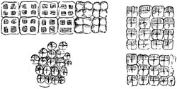
Fig. 268.—Sarcinæ.
The original woodcut of these curious parasites is reproduced in Fig. 268, also in Plate IX., No. 7, and which evidently belong to a different species, numbering thirty-nine altogether. Quite recently Mr. G. H. Broadbent, M.R.C.S., Manchester, sent me a supply of these interesting bodies lately discovered by him in an infusion of cow manure. On examining a drop with a power of 1500 diameters they were discovered moving over the field of the microscope with a gyrating motion by the aid of flagella projecting from each corner of the pocket. After some days, having attained their full growth of four, eight or sixteen in a pocket, they break up, and recommence the formative process. Sarcinæ are certainly pathogenic in their nature. Cocci in groups, or asso-cocci, are similarly associated. These several forms of spiro-bacteria are enclosed in a transparent cell-wall, and are sometimes described as zooglæa.
Of bacteria the most characteristic groups are bacillus, bacterium, and a species of clostridium, a bottle-shaped bacillus. It is, however, difficult to draw a sharp line between so-called species.
Spiro-bacteria, or spirilla, possess short or long filaments, rigid or flexible, and their movements are accordingly rotatory, or in the long axis of the filaments. These bodies are again divided into comma bacilli, or vibrios—a name invented by the older microscopists who first described them—some species of which have a flagellate appendage, to which their movements are due.
Anthrax, Splenic Fever, has been long known to be prevalent among cattle at certain seasons of the year, and is believed to originate from peculiar conditions of climate and soil. This view of splenic fever on microscopical examination proved an entire fallacy. Bollinger in 1872 discovered that the blood of the affected animal was still virulent after death, owing to the presence of the spores of the bacillus, and that the soil also became infected and impregnated by the disease germs wherever the fever first broke out. In 1877 Dr. Koch made a more careful investigation into the source of the disease, and was able to give a complete demonstration of the life-history of the splenic fever bacillus, and to offer definite proofs of its pathogenic properties. He pointed out that the rods grew in the blood and tissues by lengthening and by cross division. Further, that they not only grew into long leptothrix filaments but they produced enormous numbers of seeds or spores. He watched the fusion of the rods to the formation of spores and the sprouting of fresh rods. He furthermore inoculated a mouse, watched the effect through several generations, and fully demonstrated that in the blood and swollen spleen of the animal the same rods were always present. Pasteur and Paul Bret pursued the same course of investigations, which were always followed with precisely similar results. It was, however, principally due to the researches of Koch that the doctrine of contagium vivum was placed on a scientific basis.
Subsequently Koch formulated methods of cultivation, and dictated the microscopical apparatus needful. Furthermore, he furnished postulates for proving beyond doubt the existence of specific pathogenic micro-organisms.
“The chain of evidence regarded by Dr. Koch as essential for proving the existence of a pathogenic organism is as follows:—1. The micro-organism must be found in the blood, lymph, or diseased tissue of man or animal suffering from, or dead of the disease. 2. The micro-organism must be isolated from the blood or tissue, and cultivated in suitable media—i.e., outside the animal body. These pure cultivations must be carried on through successive generations of the organism. 3. Pure cultivation thus obtained must, when introduced into the body of a healthy animal, produce the disease in question. 4. In the inoculated animal the same micro-organism must again be found. The chain of evidence will be still more complete if, from artificial culture, a chemical substance is obtained capable of producing the disease quite independently of the living organism. It is not enough to merely detect, or even artificially cultivate, a bacterium associated with disease. An endeavour must be made to establish the exact relationship of the bacteria to disease processes. In many instances disease bacteria regarded as the actual contagia have been found, on a further searching inquiry, to be entirely misleading. It is almost needless to remind the enthusiast that the actual contagion of the disease must be fully demonstrated.”
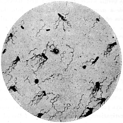
Fig. 269.—Micro-Photograph of Typhoid Fever Bacteria. Magnified 1000 ×. Taken by Leitz’s oil immersion 1⁄12-inch ocular No. 4, and sunlight exposure of one minute.
Typhoid Bacillus (Fig. 269).—Rods 1 to 3µ in length, and ·5 to ·8µ in breadth, and threads. Spore-formation has not been observed, but the protoplasm may be broken up, producing appearances which may be mistaken for spores. Actively motile, provided, some with a single and others with very numerous flagella, which are from three to five times as long as the bacillus itself. They stain readily in aqueous solutions of aniline dyes; and grow rapidly at a temperature of about 60° Fahr. In plate cultivations minute colonies are visible in thirty-six to forty-eight hours; they are circular or oval, with an irregular margin. On agar they form a whitish transparent layer, and they flourish in milk.
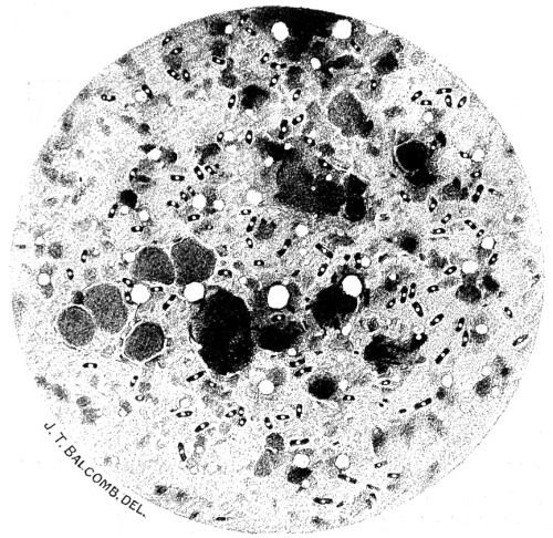
Fig. 270.—Plague Bacillus, Bombay, 1897. Magnified 1200 ×.
The Plague (Pestis Bacillus).—The Bombay plague of 1897-98 will ever be remembered as one of the most appalling visitations ever known. The number of deaths will never be accurately determined, as the native population, among whom the disease chiefly prevailed and became so fatal, concealed their dead or carried them away by night. The outbreak from the first proved to be most infectious, its incubation lasting from a few hours to a week only. It prevailed in all the over-crowded native quarters of the city. The rats and mice that infested the dwellings of the poor were found to be equally susceptible with human beings, and these vermin also died by hundreds. Those that survived left their holes and made off, in this way helping to spread the infective virus. On examining the bodies of dead rats, they were found to have swollen legs, the blood being filled by bacilli and curious monads, with whip-like appendages. The bacillus of plague was discovered by Kitasato in 1894; it is characterised by short rods with rounded ends, and a clear space in the middle. The bacilli stain readily with aniline dyes, and when cultivated on agar, white transparent colonies are formed which present an iridescent appearance when examined by reflected light. In addition to the bubonic swellings, the neighbouring lymphatic glands were also swollen and blocked by bacilli.
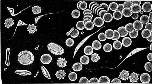
Fig. 271.—Monads in Rat’s Blood, 1,200 ×. (Crookshank.)
a. Monad threading its way among the blood-corpuscles; b. Another with pendulum movement attached to a corpuscle; c. Angular forms; d. Encysted forms; e and f. The same seen edgeways.
My illustration (Fig. 270) is from a micro-photograph taken in 1897, when the death rate stood very high. The general distribution of the bacilli, together with phagocytes and the contents of swollen lymphatic glands, magnified 1,200 ×, is from a preparation made in hospital. The monads from the rat’s blood, 1200 ×, seen threading their way among the blood corpuscles of a rat, and represented in Fig. 271, are somewhat larger than those found in the Bombay rats, but the flagella in the latter were quite as marked, while the encysted forms were wholly absent and the blood corpuscles less crenated. The white bodies (Fig. 270) were in some preparations, together with the lymphatic bodies, more numerous and more swollen.
With regard to the conditions of life of the bacteria, they may be divided broadly into two classes. When the organisms draw their nourishment from some living body or “host,” they are known as “parasites.” These are further termed “obligate” parasites if they exclusively live on their “host.” If the bacteria draw their nourishment from dead organic matter, they are called “saprophytes.” These are also divided into “obligate” and “facultative” saprophytes. Thus it will be apparent that a parasite under certain circumstances may readily become a saprophyte.
Some of the more important saprophytes are those organisms which play an important and useful part in our every-day life, such as, for instance, in the phenomena associated with fermentation, and putrefaction agents which transform dead and decomposing organic matter into their simpler elements, thus completing the great life cycle, and rendering the dead and effete matter again ready for the vital processes.
Among other life manifestations of certain bacteria may be mentioned those which have the property of generating colouring matter, though not chlorophyll. The bacteria themselves are colourless and transparent, and the pigment is merely formed as a product of their metabolism, especially under the influence of light. Many of the bacteria give rise to various gases and odours, particularly the anærobic organisms, which originate those foul putrefactive gases (ammonia, sulphuretted hydrogen, &c.). The blood-rain, Micrococcus prodigiosus, gives off an odour resembling trimethylamin. Micro-organisms have the property of producing various changes in the medium on which they are grown. In many cases albuminous bodies are peptonized and gelatine is liquefied. Many bacteria have the faculty of resolving organic bodies into their simplest elements; others, again, have the property of converting ammonia into nitric and nitrous acid. Certain microbes have the property of becoming phosphorescent in the dark. These phosphorescent bacteria are often seen on decaying plants and wood; sometimes in tropical climates the sea becomes luminous owing to the presence of countless numbers of these organisms. Again, they are frequently seen on the surface of dead fish, particularly mackerel, which often become so bright as to strongly illuminate the cupboard in which they lie.
The particular class of fungi that produce disease in man and the higher animals are generally known as “pathogenic.” These pathogenic organisms may exert their pernicious power in several ways. They may be injurious on account of their abstracting nourishment from the blood or tissues, or for the purely mechanical reason of their stopping up the minute capillaries and blood-vessels by their excessive multiplication. But the poisonous action of most of the pathogenic bacteria is due to the chemical products secreted by the organisms, and it is to the circulation and absorption within the body of these poisons that the disturbances of the animal system, which characterise disease, decay, and dissolution of every organism, must be traced.
Parasitic Diseases of Plants.
The subject of fungoid diseases and fungus epidemics are of worldwide interest, if only because of the annual losses to agriculturists from parasitic diseases of plants, amounting to millions of pounds sterling. The history of wheat-rust, and that of oats and rye, each equally susceptible to the ravages of the same Rufus, can be traced back to Genesis. A description of it was given in 1805 by Sir Joseph Banks. He suggested that the germs entered the stomata, and he warned farmers against the use of rusted litter, and made important experiments on the sowing of rusted wheat-grains. A great discussion on the barberry question followed, Fries particularly insisting on the difference between Æcidium berberidis and Puccinia graminis. Tulasne confirmed the statement made by Henslow that the uredo and puccinia stages belong to the same fungus, and are not mixed species. De Bary’s investigations in 1860-64 proved that the sporidia of some Uredinieæ (e.g., Coleosporium) will not infect the plant which bears the spores, and that the æcidia of certain other forms are stages in the life-history of species of Uromyces and Puccinia. Furthermore, De Bary in 1864 attacked the question of wheat rust, and by means of numerous sowings of the telentospores on barberry proved that they bring about the infection.
This led to the discovery of the phenomenon of Heterœcism (colonisation), introducing a new idea, and clearing up many difficulties. In 1890 the rust question entered on a new phase: it was taken up by men of science all over the world, and active inquiries were set on foot. The result has been the confirmation of De Bary’s results, but with the further discovery that our four common cereals are attacked by no less than ten different forms of rust belonging to five separate species or “form species,” and with several physiological varieties, capable of turning the table upon the barberry by infecting it. Some of these are found to be strictly confined to one or other of the four common cereals, infecting two or more of them, while others can infect various kinds of our common wild grasses.
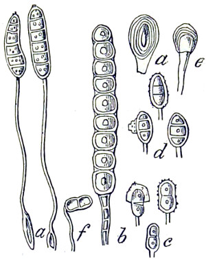
Fig. 272.—Puccinia, displaying uredospores and telentospores.
a. Aregma speciosum; b. Xenodochus paradoxus; c. P. Amorphæ; d. Triphœmium dubens; e. Younger spores; f. P. lateripes; magnified 450 diameters.
The fact is, that what has usually gone by the name of Puccinia graminis is an aggregate of several species, and that varietal forms of this exist so especially adapted to the host, that, although no morphical differences can be detected between them, they cannot be transferred from one cereal to another, pointing to physiological variations of a kind met with among bacteria and yeasts, but hitherto unsuspected in these higher parasitic fungi. It now appears we must be prepared for similar specialisation of varietal forms among Ustilagineæ as well as among Uredineæ.
Moreover, it has been found that different sorts of wheat, oats, barley, and rye are susceptible to their particular rusts in different degrees, at the bottom of which, it is suggested, there must be some complex physiological causes. De Bary gave proof, in 1886, that Peziza (Plate I., Nos. 1, 4, 5, 6) succeeds in becoming parasitic only after saprophytic culture to a strong mycelium, and that its form is altered thereby—probably by the excretion of a poison. Professor Marshall Ward showed that similar results took place in the case of the lily disease. Reinhardt, in 1892, showed that the apical growth of a peziza is disturbed and interrupted if the culture solution is employed concentrated; and Büsgen, in 1893, showed that Botrytis cinerea excretes poison at the tips of the hyphæ, thus confirming Professor Ward’s results with the lily disease in 1888, and of later years, that a similar excretion occurs in rust-fungus. He further found that the water contents of the infected plant exercises an influence, as in the case of Botrytis attacking chrysanthemums and other plants in the autumn, and that cold increases the germinating capacity of the spores.
Pfeiffer, in his work on “Chemotaxis,” shows that bacteria will congregate in the neighbourhood of an algal cell evolving oxygen. He also found that many motile antherozoids, zoospores, bacteria, &c., when free to move in a liquid, are attracted towards a point whence a given chemical substance is diffusing. He was concerning himself less with the evolution of oxygen or movements of bacteria than with a fundamental question of stimulation to movement in general. He found the attractive power of different chemical substances vary with the organism, and that various other bodies beside oxygen attract bacteria—peptone, dextrose, potassium salts, &c.; that swarm spores of the fungus Saprolegnia are powerfully attracted towards the muscles of a fly’s leg placed in the water in which they are swimming about; also, that in many cases where the hyphæ of fungi suddenly and sharply bend out of their original course to enter the body of a plant or animal, the cause of the bending lies in a powerful chemotropic action, due to the attraction of some substance escaping from the body. Professor Ward has seen zoospores of a Pythium suddenly dart out on to the cut surface of a bean-stem, and there fix themselves.
This will be better understood by referring to the course pursued by these bodies generally. When the spore of a parasitic fungus settles on a plant, it frequently behaves as follows:—The spore germinates and forms a slender tube of delicate consistency, blunt at the end, and containing colourless protoplasm, as shown, highly magnified in Fig. 272, and in Figs. 273 and 274 much less magnified. De Bary long ago showed that such a tube—the germinal-hypha—only grows for a short time along the surface of the organ, and its tip soon bends down and enters the plant, either through one of the stomata or by boring its way directly through the cell-walls. Professor Ward says these phenomena suggested to himself that the end of the tube is attracted in some way, and by some force which brings its tip out of the previous direction, and De Bary has suggested that this attraction is due to some chemical substance excreted by the host plant. It is remarkable with what ease the tube penetrates the cell-walls, and which Ward believes to be due to the solvent action of an enzyme, capable of dissolving cellulose.
“Miyoshi carried these observations a step further when, in 1894, he showed that if a leaf is injected with a substance such as ammonium-chloride, dextrine, or cane-sugar (all substances capable of exerting chemotropic attraction on fungus-hyphæ), and spores of a fungus which is not parasitic are then sown on it, the hyphæ of the fungus penetrate the stomata and behave exactly as if the fungus were a true parasite.
“So surprising a result lets in a flood of light on many known cases of fungi, which are, as a rule, non-parasitic, becoming so, in fact, only when the host plant is in an abnormal condition, e.g., the entry of species of Botrytis into living tissues when the weather is cold and damp and the light dull; the entry of Mucor into various fruits, tomatoes, apples, pears, &c., when the hyphæ meet with a slight crack or wound, through which the juices are exposed. It is exceedingly probable that the rapid infection of potato leaves in damp weather in July is traceable not merely to the favouring effect of the moisture on the fungus, but that the state of super-saturation of the cell-walls of the potato leaf—the tissues of which are now unduly filled with water and dissolved sugars, &c., owing to the dull light and diminished transpiration—is the primary factor which determines the easy victory of the parasite, and, as Professor Ward suggested some time ago, that the suppressed life of Ustilagineæ in the stems of grasses is due to the want of particular carbo-hydrates in the vegetative tissues, but which are present in the grain. A year later Miyoshi carried proof to demonstration, and showed that a fungus-hypha is actually so attracted by substances on the other side of a membrane, and that its tip pierces the latter; for the hyphæ were made to grow through films of artificial cellulose, of collodion, of cellulose impregnated with paraffin, of parchment paper, and even the chitinous coat of an insect, simply by placing the intact films on gelatine impregnated with the attracting substance, and laying the spores on the opposite side of the membrane.
“Now this is obviously a point of the highest importance in the theory of parasitism and parasitic diseases, because it suggests at once that in the varying conditions of the cells, the contents of which are separated only by membranous walls from the fungus-hyphæ, whose entrance means ruin and destruction, there may be found circumstances which sometimes favour and sometimes disfavour the entrance of the hyphæ; and it is, at least, a remarkable fact that some of the substances which experiments prove to be highly attractive to such hyphæ—e.g., sugars, the sap of plums, phosphates, nitrates, &c.—are just the substances found in plants; and the discovery that the action depends upon the nature of the substance as well as on the kind of fungus, and is affected by its concentration, the temperature, and other circumstances, only confirms us in this idea.”
Moreover, there is one other fact which it is important to notice, viz., that there are substances which repel instead of attract the hyphæ. Is it not, then, asks Professor Ward, natural to conclude that the differences in behaviour of different parasites towards different host-plants, and towards the same host-plant under different conditions, probably depend on the chemotropic irritability of the hyphæ towards the substance formed in the cells on the other side of the membranous cell-walls? And when, as often happens, the effusion of substances, such as the cells contain, to the exterior is facilitated by over-distension and super-saturation, or by actual wounds, we cannot be surprised at the consequences when a fungus, hitherto unable to enter the plant, suddenly does so. To this proposition my answer is emphatically in the affirmative, since in my investigations into the “fungus-foot disease” (“Mycetoma”), 1871, of India, the entry of the fungus was in almost every case shown to be through an abrasion of the skin or a direct open wound; the majority of the cases reported were among the agricultural classes. When, then, as often happens, the effusion of substances, such as the cells contain, to the exterior is facilitated by over-distension and super-saturation, or by actual wounds, we cannot be surprised at the consequences when a fungus, hitherto unable to enter the plant, suddenly does so. Nevertheless, it must be admitted that the knowledge gained of parasites does not satisfactorily account for epidemic visitations over large areas.
Habitat of Fungi and Moulds.
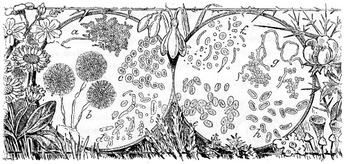
Fig. 273.—Fungi and Moulds.
Description of Figures.—a. Fungi Spores, taken in a sick chamber; b. Aspergillus glaucus; c. Yeast, recent state; d. Exhausted yeast, budding; e. Penicillium spores more highly magnified; g. Aerobic spores and mould mycelium; h. Aspergillus spore, grown on melon.
Habitat, Specialised Forms of Parasites.
Habitat.—The habitat of vegetable parasitic fungi is extremely variable. Fungi are found everywhere, living and flourishing on all the families of the vegetable and animal kingdoms. They attack our houses, foods, clothes, utensils of every kind, wall papers and books, the paste of which, to my astonishment, affords a sufficient supply of nourishment. Members of the parasitic tribe of bacteria, by a combined effort of countless myriads, have given rise to a sense of supernatural agency. Bacillus prodigiosus, described also as Palmella mirifica and Zoogalactina imetropia, from its attacking milk and other alimentary substances, the spores of which are often of a deep red colour, have been found to cover whole tracts of country in a single night with what is called a “gory dew,” changing in daylight to a deep green colour. This was at one time regarded with superstitious awe as a miracle, as it has been known to attack bread and even the sacred wafer, and which in mediæval ages was described as the “bleeding-host.” This parasitic plant belongs to anærobic bacteria, and is only developed in the dark. The nitrogen required for nutrition must be derived from the air. An algal form gives rise to the red scum seen in ponds and reservoirs in the autumn. The discharge from wounds is coloured blue by Bacterium pyocyanine. There are many other forms, some of which have an orange colour, and the genus is recognised as “chromogenic microbes.”
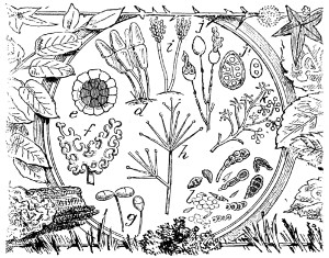
Fig. 274.—Fungi and Moulds.
Description of Figures.—d. Puccinia graminis on wheat; c. Polycystis spore of rye-smut; f. Alder fungus spores, Microspheria penicellula; g. Dactylium roseum, rose-coloured mould; h. Verticillium distans, whorled mould found on herbaceous plants; i. Botrytis, vine and lily fungus; j, j′. Peronospora infestans, potato fungus; k. P. gangliformis, mould of herbaceous plants; l. Various Penicillium and other spores taken in a bean-field.
A cryptogam belonging to anærobic bacteria, described as Protococcus invalis, on being set aside in a bottle, and a little rain water added, was seen to set up spontaneous fermentation, and in a very short time exhibited remarkable activity. The colour of the infusion changed, it assumed a delicate pink hue in direct light, which deepened to a red in reflected light. The fluid contents were now observed to be dichoric, and the spectroscopic appearance subsequently presented was one of much interest. The spectrum was a well-marked one, and might be taken to determine the presence of a nitrogenous element or of glucose.
Among all the various plants known to suffer from the attacks of parasites, the vine has been the greatest sufferer. The oïdium, or Erysiphe Tuckeri, so called from the name of the discoverer by whom it was first described, has been longest known to the vine grower. This really belongs to the group Ascomycetes, and appears to have been brought from America in 1845, whence it was passed on to France, where it soon threatened to entirely destroy the vineyards. This was followed by another parasite, belonging in this instance to the animal kingdom, Phylloxera vastatrix. This oïdium appears on the grape in the form of greyish filaments, terminating in an enlarged head, which contains an agglomeration of spores, not free or in a chaplet, as in Aspergillus (Fig. 273). These spores when ripe burst from the capsule as fine dust, and are diffused by the air in all directions, thus spreading the disease far and away. Another of the parasitic moulds, Peronospora viticola, is a kind of mildew, differing from oïdium. The hyphæ penetrate more deeply than that of oïdium. On the upper surface of the leaf brown patches appear; these branch out and ramify as seen in the potato-fungus, P. infestans (Fig. 274). The parasite destroys the tissue of the leaf, and it withers and dies. There are other well-known parasites, the black-rot, Phomauvicola, belonging to the Ascomycetes. This appears in early shoots in the form of round black spots, and gradually spreads over leaves and young fruit. This same rot, one year, devastated the American vineyards.
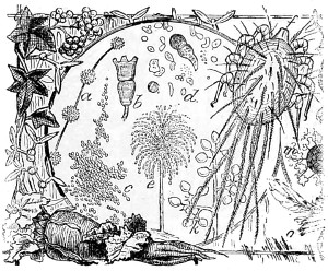
Fig. 275.—Fungi, Moulds.
a. Clustered Spores, Gonatobotrys simplex; b. Spore of Puccinia coronata, the mildew of grapes; c. Barley smut; d. Puccinia althæa; e. Penicillium glaucum; m. Ixodes farinæ, found in damaged flour together with smut.
Cereals, wheats and grasses, suffer from other well-known forms of microscopic fungi termed rusts and smuts, which cover the blades or infect the full ear of the fruit. The name given indicates their colour, and these belong, for the most part, to the genus Uredo and the family of the Basidiomycetes. They have no endogenous spores but as many as four forms of exogenous. This is also the case with wheat and barley, whereby they are distinguished as Uredo or Puccinia graminis (see Figs. 273 and 274, and Plate I., Nos. 19 and 22, Æcidium berberidis). For a long time it was believed that Uredo linearis and Puccinia graminis were so many distinct species, but it is now known that there are only three successive phases of the developmental stages of a single species—that, as a matter of fact, puccinia presents the phenomenon of alternation of generations, that is, that the complete development of the fungus is only effected by its transference from one plant to another. Other uredines, Ustilago and Tilletia smuts, are more apt to affect the ears of wheat, rye, and other grasses than puccinia. Bread made from wheat affected by smut has an acrid and bitter taste, while that made from rye flour often produces a serious form of disease. The propagation of either, then, should be stopped as quickly as possible by destroying all barberry bushes growing near or within the vicinity of corn fields, and by other means. The ergot of rye is due to distinct species of fungi having endogenous spores enclosed in a sac or ascus, hence the name of the family, Ascomycetes or Tuberaceæ, which are reproduced by the spores contained in these asci. Truffles belong to this family. But other members of the same family have several forms of spores, and these again present us with the phenomenon of alternation of generations.
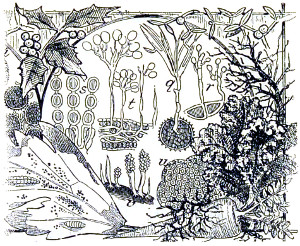
Fig. 276.—Fungi, Moulds.
p. Spores of Tilletia caries; q. Spores of Tilletia caries, when germinating, produce a fœtid olive-coloured spore in cereal grains; r. Telentospores of Puccinia graminis; s. Crystopus candidus, spores growing in chains; t. Petronospora infestans, mildew of turnips, &c.; u. A transverse section of ergot of rye, showing spores in masses; v. Claviceps purpuræ, associated with ergoted rye.
Ergot of rye is used in medicine, but if not used with care it will produce a dangerous disease. This parasitic fungi consists of minute microscopic masses of spores, which cover the young flower of the rye with a white flocculent mass, formerly termed sphacelium. The mycelium formed spreads over the ear of corn in thick felt-like masses, termed sclerotis. The sphacelium changes its form in the following spring. Other changes are brought about, and it seems to pass through a cycle of alternations of generations.
Bread made from rye so infested is known to produce grave consequences, soon to become fatal if not detected in time. The disease is termed ergotism, and gangrene of the extremities takes place among people of the north of France and Russia, who consume bread made from rye flour. Ergot of maize will also cause similar diseases. Fowls and other animals fed upon this cereal become in a short time poisoned, and the cause of death is not rightly suspected. There is another fungus belonging to the same group of Ascomycetes, known as Eurotium repens, which appears upon leather when left in a damp place, and also upon vegetable or animal substances if badly preserved, and gradually destroys it. This mould is of a darkish green colour.
The minute spores display themselves as rows of beads when fully ripe on the erect mycelium. Aspergillus glaucus represents the white exogenous spores of the sphacelium of the ergot of rye; and those subsequently produced in the yellow balls correspond with the asci developed in sclerotis, the endogenous species. Many of the parasitic species belonging to the genera Erysiphe, Sphæria, Sordaria, Penicillium, &c., have a similar mode of propagation, and affect a large number of plants.
Parasitic Fungi of Men and Animals.
In the microscopical examinations especially given to the elucidation of parasitic diseases of the skin, previously referred to, I discovered more varieties of spores and filaments of certain cryptogamic plants associated with a larger number of specific forms of fungi than any previous observer. I did not, however, feel justified in concluding, with Küchenmeister, Schœnlein, and Robin, that these fungoid growths were the primary cause of the diseases referred to. Indeed, the foremost dermatologists of the period utterly refused to entertain the specific germ theory of the German investigators. Nevertheless, I contended, “the universality of their distribution is in itself a fact of very considerable importance, and one pointing to the belief that they are scavengers ever ready to fasten on decaying matter, and, on finding a suitable soil, spread out their invisible filaments in every direction in so persistent a manner as to arrest growth and overwhelm the plant in destruction.”53
Special forms of fungi are given in Plate I., Nos. 10-14, and those of the ascomycetes in Nos. 17-21.
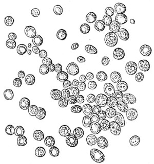
Fig. 277.—Healthy fresh Yeast, from a large Brewery, in an active stage of formation, × 400.
Oïdium albicans affects both animals and plants. It often attacks the mucous membrane of the mouths of young children. The spores become elongated and converted into hyphæ, and ramify about in all directions, producing a troublesome form of disease. This parasitic fungus is better known under another name, Saccharomyces mycoderma. Oïdium resemble algæ in their mode of life, as they are mostly found in a liquid media. The structure of all ferments is very simple: each plant is composed of a single cell, either of a spherical, elliptical, or cylindrical form, varying in size, and filled with protoplasmic and nucleated matter. This grows, and is seen to bud out and divide into two or more parts, all resembling the mother cell.
Fig. 277 represents the healthy cells of yeast, Saccharomyces cerevisiæ, freshly taken from a brewer’s vat, and in an active stage of growth. The mode of multiplication continues as long as the plant remains in a liquid favourable to its nutrition.
The changes from one stage to another are rapid, as will be noticed on reference to the consecutive formative processes the cells are known to pass through, Fig. 278 (1859).
If the development of the plant is arrested by want of a saccharine or nitrogenous substance, and the liquid dries up, the protoplasm contained in the cell contracts, and the spores, or endogenous reproductive organs, of the plant will remain in a state of rest, become perfectly dry, and yet retain life. They are not easily killed, even when subjected to a very high or low temperature, they do not lose the power of germination when favourable conditions present themselves, and at once take on a new birth.
There are, however, many other ferments besides that of beer-yeasts, such as alcoholic and wine ferments, the commonest of which, according to Pasteur, is Saccharomyces ellipsoideus.
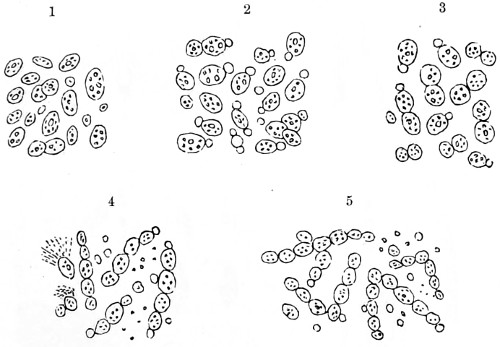
Fig. 278.—Development of Yeast Cells.
1. When first taken; 2. One hour after introducing a few cells into sweet-wort; 3. Three hours after; 4. Eight hours; 5. Forty-eight hours, when the cells become elongated.
But yeast-fungi and mould-fungi, like bacteria or fission-fungi, are micro-organisms, belonging to two specific orders, the Saccharomycetes and the Hyphomycetes, which are intimately related to each other, but quite distinct from bacteria. Their germs occur widely distributed in air, soil, and water. Many species are of hygienic, while others are of pathological interest and importance in being either accidentally associated with, or the cause of, disease processes, while others are fermentations of very essential service in various industrial processes. The making of beers, wines, and spirits, as we understand them, constitutes but a small part of the province of fermentation. The life activities of ferments open out a study of vast importance to mankind, and while they have only been regarded in their worst aspect—that of a bane—they are, nevertheless, a boon to mankind. The first clear view we obtained of this was that of Reess, who in 1870 showed there were several species or forms of the yeast-fungus. Hansen followed up this discovery in 1883, and, taking advantage of the strict methods of culture introduced by bacteriologists, found that by cultivating yeast on a solid media from a single spore it was quite possible to obtain constant types of pure yeasts, each possessing its own peculiar properties. One consequence of Hansen’s labours was that it now became possible for every brewer to work with a yeast of uniform type instead of with haphazard mixtures, in which serious disease forms might predominate and injure the beer. Among other things made clear was that a true yeast may have a mycelial stage of development. Furthermore, there is the influence exercised by the nucleus of the yeast cell. Many other points of interest arose out of these investigations; one was, that many higher fungi can assume a yeast-like stage of development if submerged in fluids, as, for instance, various species of Mucor, Ustilago, Exoascus, and numerous others. Ascomycetes, and Basidiomycetes as well, are known to form budding cells, and it was thought that the yeasts of alcoholic fermentation are merely reduced forms of these higher fungi, which have become habituated to the budding condition—a conclusion supported by Hansen’s discovery that a true Saccharomyces can develop a feeble, but a true, mycelium.
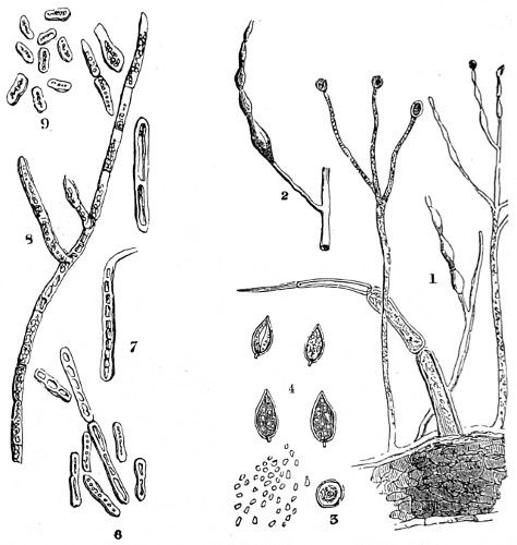
Fig. 279.—Saccharomyces and Moulds.
1. Section from a tomato, showing spores growing from cuticle; 2. Portion detached to show budding-out process; 3. Lateral view of spore sac with oospores issuing forth; 4. Apiculated ferment spores; 6 and 7. Mycoderma cerivisiæ in different stages of growth, as seen on wine bottles; 8 and 9. Torulæ diabeticæ, torulæ and fission spores.
“This view has been entirely confirmed by an inquiry into the mode of brewing saké by the Japanese, by the aid of the Aspergillus fungus. Further researches established the fact that other forms of fungi, e.g., those on the surface of fruits, developed endogenous spores, which cause alcoholic fermentation. More recently, and by further experimental inquiry, partly by pure cultures of separate forms, and partly by well-devised cultures on ripening fruits still attached to the plant but imprisoned in sterilised glass vessels, it has been found that yeast and moulds are separate forms, not genetically connected, but merely associated in nature, as are so many other forms of yeasts, bacteria, and moulds. Further, Hansen has discovered that several yeasts furnish quite distinct races or varieties in different breweries in various parts of the world, so that we cannot avoid the conclusion that their race characteristics have been impressed on the cells by the continued action of the conditions of culture to which they have so long been exposed—they are, in fact, domesticated races.”
The environments of yeasts are peculiar. Sauer found that a given variety of yeast, whose activity is normally inhibited when the alcohol attains a certain degree of concentration in the liquid, can be induced to go on fermenting until a higher degree is attained by the addition of a certain lactic acid bacterium. The latter, indeed, appears to prepare the way for the yeast. It has been shown, also, that damage may be done to beers and wines by allowing plant germs to gain access with the yeast; there are, too, several forms of yeast that are inimical to the action of the required fermentation. Other researches show that associated yeasts may ferment better than any single yeast, and such symbiotic action of two yeasts of high fermenting power has given better results than either alone. English ginger-beer furnishes a curious symbiotic association of two organisms—a true yeast and a true bacterium—so closely united that the yeast cells become imprisoned in the gelatinous meshes of the bacterium; and it is a curious fact that this symbiotic union of yeast and bacterium ferments is far more energetic than either when used alone, and the product is different, large quantities of lactic and carbonic acids being formed, and little or no alcohol.
Many years ago I gave an account of similar curious symbiotic results obtained by introducing into a wort-infusion a small proportion of German yeast, an artificial product composed of honey, malt, and a certain proportion of spontaneously-fermented wheat flour. This, to my astonishment, produced ten per cent. more alcohol than any of its congeners, and did not so soon exhaust itself as brewer’s yeast.54
In the hephir used in Europe for fermenting milk, another symbiotic association of yeast and a bacterium, it is seen that in this process no less than four distinct organisms are concerned. I have already instanced the fermentation of rice to produce saké, which is first acted upon by an Aspergillus that converts the starch into sugar and an associated yeast, and this is also shown to be a distinct fungus, symbiotically associated in the conversion. “Starting, then, from the fact that the constitution of the medium profoundly affects the physiological action of the fungus, there can be nothing surprising in the discovery that the fungus is more active in a medium which has been favourably altered by an associated organism, whether the latter aids the fungus by directly altering the medium, or by ridding it of products of excretion, or by adding gaseous or other body. It is not difficult to see, then, that natural selection will aid in the perpetuation of the symbiosis, and in cases like that of the ginger-beer plant it is extremely difficult to get the two organisms apart, a difficulty similar to that in the case of the soredia of lichens.”
Buchner discovered that by means of extreme pressure a something can be extracted from yeast which at once decomposes sugar into alcohol and carbon-dioxide. This something is regarded as a kind of incomplete protoplasm—a body, as we have already seen, composed of proteid—and in a structural condition somewhere between that of true soluble enzymes like invertin and a complete living protoplasm. This reminds me of an older experiment of mine, the immediate conversion of cane-sugar into grape-sugar. If we take two parts of white sugar and rub it up in a mortar with one part of a perfectly dry solid, the German yeast before spoken of, it is immediately transformed as if by magic into a flowing liquid mass—a syrup. This process of forming “invert sugar” can be watched under the microscope; the liberation of carbonic acid gas in large bubbles is seen to go on simultaneously with the assimilation of the dextrose, and the breaking up of the crystals of sugar; the cell at the same time increasing in size as well as in refractive power; a curious state of activity appears to be going on in the small mass, which is very interesting to watch throughout.
However, the enzymes of Buchner are probably bits off the protoplasm, as it were, and so the essentials of the theory of fermentation remain, the immediate agent being not that of protoplasm itself, but of something made by or broken off from it. Enzymes, or similar bodies, are known to be very common in plants, and the suspicion that fungi do much work with their aid is abundantly confirmed. It seems, indeed, that there are a whole series of these bodies which have the power of carrying over oxygen to other bodies, and so bringing about oxidations of a peculiar character. These curious enzymes were first observed owing to studies on the changes which wine and plant juice undergo when exposed to the action of the oxygen of the air.
The browning of cut apples is known to be due to the action of an oxydase, that is, an oxygen carrying ferment, and the same is claimed for the deep colouring of certain lacs obtained from the juice of plants, such as Anacardiaceæ, which are pale and transparent when fresh drawn, but which gradually darken in colour on exposure to the air. Oxydases have been isolated from beets, dahlia, potato-tubers, and several other plants. This fact explains a phenomenon known to botanists, and partly explained by Schönbein as far back as 1868, that if certain fungi (e.g., Boletus beridies) are broken or bruised, the yellow or white flesh at once turns blue; this action is now traced to the presence in the cell sap of an oxydase.
It is the diastatic activity of Aspergillus which is utilised in the making of saké from rice, and in the preparation of soy from the soja bean in Japan. Katz has recently tested the diastatic activity of Aspergillus, of Penicillium, and of Bacterium megatherium, in the presence of large and small quantities of sugar, and found all are able to produce not only diastase, but also other enzymes; as the sugar accumulates the diastase formed diminishes, whereas the accumulation of other carbo-hydrates produces no such effect. Harting’s investigation on the destruction of timber by fungi derives new interest from the discovery of an emulsion-like enzyme in many such wood-destroying forms, which splits up glucosides, amygdalin, and other substances into sugar, and that hyphæ feed on other carbo-hydrates. The fact, also, that Aspergillus can form inverts of the sucrase and maltase types, as well as emulsin, inulate, and diastase, according to circumstances of nutrition, will explain why this fungus can grow on almost any organic substance it may happen to alight upon. The secretion of special enzymes by fungi has a further interest just now, for recent investigations promise to bring us much nearer to an understanding of the phenomena of parasitism than it was possible when I was at work upon them some forty or fifty years ago.
It was De Bary who impelled botanists to abandon older methods, and he who laid the foundation of modern mycology. Later on he pointed out that when the infecting germinal tube of a fungus enters a plant-cell, two phenomena must be taken into account, the penetration of the cell-walls and tissues, and the attraction which causes the tips of the growing hypha to face and penetrate these obstacles, instead of gliding over them in the lines of apparent least resistance. The further development of these two factors shows that in the successful attack of a parasitic plant on its victim or host these fungi can excrete cellulose-dissolving enzymes, and that they have the power of destroying lignine. Zopf has also furnished examples of fungi which can consume fats. There is, however, one other connection in which these observations on enzymes in the plant-cell promise to be of considerable importance, viz., the remarkable action of certain rays of the solar light on bacteria. It has been known for some time past that if bacteria in a nutrient liquid are exposed to sunlight they quickly die. The further researches of Professor Marshall Ward and other workers in the same direction have brought out the fact that it is really the light rays, and not high temperatures, that it is especially the blue-violet and ultra-violet rays, which exert the most effective bactericidal action. This proof depended upon the production of actual photographs in bacteria of the spectrum itself. Apart from this, the Professor demonstrated that just such spores as those of anthrax, at the same time pathogenic and highly resistant to heat, succumb soonest to the action of these cold light-rays, and that under conditions which preclude their being poisoned by a liquid bath. It is in all probability the action of these rays of light upon the enzymes, which abound in the bacterial cells, that bring about their death.
The sun, then, is seen to be our most powerful scavenger, and this apparently receives confirmation in connection with Martinaud’s observations, that the yeasts necessary for wine-making are deficient in numbers and power on grapes exposed to intense light, and to this is due that better results are obtained in central France as contrasted with those in the south. “When we reflect, then, that the nature of parasitic fungi, the actual demonstration of infection by a fungus spore, the transmission of germs by water and air, the meaning and significance of polymorphism, heteræcism, symbiosis, had already been rendered clear in the case of fungi, and that it was by these studies in fermentation, and in the life-history of the fungus Saccharomyces, that the way was prepared for the ætiology of bacterial diseases in animals, there should be no doubt as to the mutual bearings of these matters.”
Industrial uses of Fungi and Saccharomycetes.
There are many industrial processes which are more or less dependent for success on bacterial fermentations. The subject is young, but the results already obtained are seen to be of immense importance from a scientific point of view, and to open up vistas of practical application already being taken advantage of in commerce, while problems are continually being raised by the forester, the agriculturist, the gardener, the dairyman, the brewer, dyer, tanner, and with regard to various industries, which will eventually confer great advantages in their economic application.
The remarkable discovery made by Alvarez of the bacillus, which converts a sterilised decoction of the indigo plant into indigo sugar and indigo white, the latter then oxidising to form the valuable blue dye, whereas the sterile decoction itself, even in presence of oxygen, forms no indigo, plainly proves how these minute organisms may be turned to a good account. There are, however, important points to be determined as to the action of the fermentation brought about by these enzymes, and the appearance of certain mysterious diseases in the indigo vats. Again, certain stages in the preparation of tea and tobacco leaves are found to depend upon very carefully regulated fermentations, which must be stopped at the right moment, or the product will be spoilt. Regarding the possible rôle of bacteria, the West Indian tobacco has a special bacterium, which has been isolated and found to play a very important part in its flavour. Every botanist knows that flax and hemp are the best fibres of Linum and Cannabis respectively, separated by steeping in water until the middle lamella is destroyed and the fibres isolated; but it is not so well known that not every water is suitable for this “retting” or steeping process; and for a long time this was as much a mystery as why some waters are so much better than others for brewing. Quite recently Fribes has succeeded in isolating the bacillus upon which the dissolution of the middle lamella depends. This investigation brought out other interesting details as to the reaction produced by living micro-organisms, and which can be utilised in deciding questions of plant chemistry too subtile for testing with ordinary re-agents. One other important fact connected with these researches is that botanists have now discarded the view that the middle lamella of the plants referred to is composed of cellulose, and know that it consists of pectin compounds. Fribes’ anærobic bacillus is found to dissolve and destroy pectins and pectinates, but does not touch cellulose or gum. It is well known that the steeping of skins in water in preparation for tanning involves bacterial action, owing to which the hair and epidermal coverings are removed, but it also appears that in the process of swelling the limed skins, the gases evolved in the substance of the tissues, and the evolution of which causes the swelling and loosens the fibre so that the tanning solutions may penetrate, are due to a particular fermentation caused by a bacterium, which, according to some investigators, is identical with a lactic ferment introduced by the pine bark, and which is responsible for the advantageous acidification of the tanning solutions.
Hay is made in different ways, and in those where a “spontaneous” heating process is resorted to the fermentation is no doubt dependent upon the presence of thermogenic bacteria. But probably no other subject has attained to so much importance as the bacteriology of the dairy: the study of the bacteria found in milk, butter, and cheese in their various forms.
Of milk, especially, much has been written and said as a disease-transmitting medium, and with every good reason, and, if the statement of a Continental authority may be accepted that each time we eat a slice of bread and butter we devour a number of bacteria equal to the population of Europe, we have sure grounds for seeking for further information as to what these bacteria are and what they are doing. And similarly so with cheese, which teems with millions of these minute organisms.
“Some few years ago it was found that the peculiar aroma of butter was due to a bacterium. There are two species of bacteria, one of which develops an exquisite flavour and aroma, but the butter keeps badly, while the other develops less aroma, but the butter keeps better. In America, however, they have isolated and distributed pure cultures of a particular butter bacillus which develops the famous ‘June’ flavour, hitherto only met with in the butter made in a certain district during a short season of the year. This fine-flavoured butter is now constantly manufactured in a hundred American dairies; and the manufacture of pure butter with a constant flavour has become a matter of certainty.
“Properly considered, the manufacture of cheese is a form of ‘microscopic gardening’ even more complex and more horticultural in nature than the brewing of beer. From the first moment, when the cheesemaker guards and cools his milk, till his stock is ready he is doing his best to keep down the growth of micro-organisms rushing about to take possession of his milk. He therefore coagulates it with rennet—an enzyme of animals, but also, as we have seen, common in plants—and the curd thus prepared is simply treated as a medium, on which he grows certain fungi and bacteria, with every needed precaution for favouring their development, and protecting them against the inroads of other pests and against unsuitable temperature, moisture, and access of light. Having succeeded in growing the right kind of plants on his curd, his art then demands that he shall stop their growth at the critical moment, and his cheese is ready for market.
“Furthermore, the particular flavour and peculiar odours of cheeses, as Camembert, Stilton, and Roquefort, have to be obtained, and this is secured, for instance, by cultivating a certain fungus, Penicillium, on bread, and purposely adding it to Roquefort. This is found to destroy the lactic and other acids, and so enables certain bacteria in the cheese to set to work and further change the medium; whereas in another kind of cheese the object is to prevent this fungus paving the way for these bacteria. Another kind of bacillus has been discovered which gives a peculiar clover aroma to certain cheeses.
“It is thought that more definite results will be obtained by the investigation of the manufacture of the vegetable cheeses of China and Japan, which are made by exposing the beans of the leguminous plant, Glycine—termed soja-beans—to bacterial fermentations in warm cellars with or without certain mould-fungi. Several kinds of bean-cheeses are made in this way, known by special names. They all depend upon the peculiar decompositions of the tissues of the cotyledons of the soja-beans, which contain 35 to 40 per cent. of proteids and quantities of fatty matter. The softened beans are first rendered mouldy, and the interpenetrating hyphæ render the contents accessible to certain bacteria, which peptonise and otherwise alter them. There is the further question of the manufacture of vinegar by fermentation, of the preparation of soy from a brine extract of mouldy and fermented soja-beans, of bread-making, and other equally interesting manufactures.”
Results of De Bary’s Investigations in Parasitism.
“When the idea of parasitism was rendered definite by the fundamental distinction drawn by De Bary between a parasite and a saprophyte, it soon became evident that some further distinction must be made between obligate facultative parasites and saprophytes respectively. De Bary, when he proposed these terms for adoption, was clearly alive to the existence of transitions which we now know to be numerous and so gradual in character that we can no longer define any such physiological groups. Twenty years ago penicillium and mucor would have been regarded as saprophytes of the most obligate type, but we now know that under certain circumstances these fungi can become parasites, and the borderland between facultative parasites and saprophytes on the one hand, and between the former and true parasites on the other, can no longer be recognised.”
In 1866 the germ of an idea was sown which has taken root and extended. De Bary pointed out that in the case of lichens we have either a fungus parasite on an algæ, or else certain organisms hitherto accepted as algæ are merely incomplete forms.
“In 1879 the same observer definitely launched the new hypothesis of symbiosis. The word itself is due to Frank, who, in a valuable paper on the biology of the thallus of certain lichens, very clearly set forth the existence of various stages of life in common among all the lower forms of plants. The details of these matters are now principally of historical interest. We now know that lichens are dual organisms, composed of various algæ, symbiotic with Ascomycetes, with Basidiomycetes, and, as Massee has shown, even with Gastromycetes. The soil contains also bacterio-lichens. Hence arose a new biological idea—that a fungus may be in such nicely-balanced relationship with the host from which it derives its sustenance, that it may be attended with nearly equal advantage to both.
“In the humus of forests we find the roots of beeches and other Cupuliferæ (willows, pines, and so forth) clothed with a dense mantle of hyphæ, and swollen into fleshlike masses of mycorhiza. In similar soils, and in moorlands, which abound in the slowly decomposing root-fibres and other vegetable remains so characteristic of these soils, the roots of orchids, heaths, gentians, &c., are similarly provided with fungi, the hyphæ of which penetrate further into the tissues, and even send haustoria into the living cells, but without injuring them. As observations multiplied it became clear that the mycorhiza, or fungus-root, was not to be dismissed as a mere case of roots affected by parasites, but that a symbiotic union, comparable to that of the lichens, exists, and we must assume that both tree and fungus derive benefit from the connection.
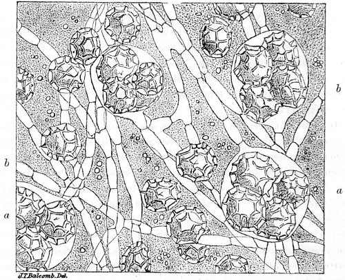
Fig. 280.—Fine Section through Truffle.
a. Asci filled with spores; b. Mycelia, × 250.
“Frank stated, as the result of his experimental research, that seedling forest-trees cannot be grown in sterilised soil, where their roots are prevented from forming mycorhiza; and he concluded that the fungus conveys organic materials to the roots, which it obtains by breaking down the leaf-mould and decaying plant remains, together with water and minerals from the soil, and plays the especial part of a nitrogen-catching apparatus. In return for this import service the root pays a tax to the fungus by sparing it certain of its tissue contents. It is a curious fact then that the mycorhiza is only formed where humus or vegetable mould abounds.”
These instructive investigations offer an intelligible explanation of the growth of that well-known subterranean fungus, the truffle (Tuber cibarium), the microscopic appearances of a section of which formed the subject of a paper I contributed to “The Popular Science Review” some years ago (1862). The fungus, as will be seen by the fine section cut through a truffle, Fig. 280, consists of flocculent filaments, which in the first instance cover the ground at the fall of the leaf in autumn, under oak or beech trees, the hyphæ of which penetrate the ground, through the humid soil to the root-hairs of the tree. Filaments (mycelia) are again given off which terminate in asci or sacs filled with minute spores of about 1⁄2500th of an inch in size, while the interspaces are filled up by mycelia, that become consolidated into a firm nut-like body.
What happens, then, is this: Trees and plants with normal roots and root-hairs, when growing in ordinary soil, can adapt their roots to life in a soil heavily charged with humus only by contracting symbiotic association with the fungus and paying the tax demanded by the latter in return for its supplies and services. If this adaptation is impossible, and no other suitable variation is evolved, such trees cannot grow in such soils. The physiological relations of the root to the fungus must be different in details in the case of non-green, purely saprophytic, plants, Neottia, Monotropa, &c., and in that of green plants like Erica, Fagus, and Pinus. It is, however, a well-known fact that ordinary green plants cannot utilize vegetable débris directly, and forest trees do so in appearance only, for the fungi, yeasts and bacteria there are actively decomposing the leaves and other remains. A class of pseudo-symbiotic organisms are, however, being brought into the foreground, where the combined action of two symbionts results in the death of or injury to a third plant, each symbiont alone proving harmless. Some time ago Vuillemin showed that a disease in olives results from the invasion of a bacillus (B. oleæ), which can, however, only obtain its way into the tissues through the passages driven by the hyphæ of a fungus (Chætophoma). The resulting injury is a sort of burr. This observer also observed the same bacillus and fungus in the canker burrs of the ash.
Among many similar cases well worth further attention are the invasion of potato-tubers by bacteria, these making their way down the decaying hyphæ of pioneer fungi. Professor Marshall Ward has seen tomatoes infected by similar means, and other facts show that many bacteria which quicken the rotting of wood are thus led into the tissues by fungi.
Probably no subject in the whole domain of cryptogamic botany has wider bearings on agricultural science than the study of the flora and changes on and in manure and soil. Nitrifying bacteria play a very important part by providing plant life with a most necessary food. They occur in the soil, and two kinds have been described—the one kind converting ammonia into nitrous acid, and the other changing nitrous into nitric acid. We are principally indebted to Winogradsky for our knowledge of these bacteria; he furnishes instances of the bearing of bacteriological work on this department of science, and explains, not only the origin of nitre-beds and deposits, but also the way the ammonia compounds fixed by the soil in the neighbourhood of the root-hairs are nitrified, and so rendered directly available to plant life. The investigations of other observers show that the nitrifying organism is a much more highly-developed and complex form than had been suspected; that it can be grown on various media, and that it exhibits considerable polymorphism—i.e., it can be made to branch out and show other characteristics of a true fungus. “I have,” writes Professor Ward, “for some time insisted on the fact that river water contains reduced forms of bacteria—i.e., forms so altered by exposure to light, changes of temperature, and the low nutritive value of the water, that it is only after prolonged culture in richer food media that their true nature becomes apparent.” Strutzer and Hartleb show that the morphological form of the nitrifying organism can be profoundly altered by just such variations of the conditions described by Ward, and that it occurs as a branched mycelial form; as bacilli or bacteria; or as cocci of various dimensions, according to the conditions.
“These observations, and others made on variations in form (polymorphism) in other fungi and bacteria, open out a vast field for further work, and must lead to advancement in our knowledge of these puzzling organisms; they also help us to explain many inconsistencies in the existing systems of classification of the so-called ‘species’ of bacteria as determined by test-tube culture.”
Algæ.—The algals have a special charm for microscopists. I am free to confess my interest in these organisms, and for several reasons. In this humid climate of ours they are accessible during the greater part of the year; they can be found in any damp soil, in bog, moss, and in water—indeed, wherever the conditions for their existence seem to be at all favourable for development. Should the soil dry up for a time, when the rain returns algæ are seen to spring into life and give forth their dormant spores, which once more resume the circle of formation and propagation. In the earliest stage of development the spore or spore cell is so very small when in a desiccated state, that any number may be carried about by the slightest breath of air and borne away to a great distance. To all such organisms I originally gave the name of Ærozoa; now recognised as ærobic and anærobic organisms (Fig. 281).