Part 4 — MORPHOLOGY OF THE HUMAN CÆCUM AND VERMIFORM APPENDIX.
Reading edition
Not only is the anatomy of this portion of the alimentary tract of great interest in relation to the evolution of the human structure, but in addition the pathological and surgical importance of the region warrants a very careful study of the cæcum and appendix. This is more especially the case since a number of variations in the arrangement of the structures are encountered. These departures from what we consider the normal human type have an important bearing on the development and progress of the pathological conditions prone to involve the appendix. We may consider the subject under the following subdivisions:
I. DEVELOPMENT OF THE CÆCUM AND APPENDIX.
Much light is thrown on the adult anatomy of the parts and on the origin of the variations observed by the study of their embryonic history. In considering the factors which determine the variations in the position, size, and shape of the appendix it must be remembered that the rudimentary character of this structure is responsible for many of the aberrant conditions encountered.
As a part of the general cæcal pouch which persists in an early developmental stage and which we can regard as destined for further reduction and ultimate elimination in the course of evolution, the appendix shares with other vestigial structures a wide range of variation. Consequently the study of the development of this portion of the alimentary tract enables us to gain a clearer view of the primary arrangement of the structures and to trace the causes which are active in determining the adult conditions most frequently encountered.
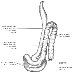
Fig. 486.—Alimentary canal and appendages of human embryo of 12.5 mm. × 12. (Kollmann, after His.) Fig. 487.—A, schematic representation of alimentary canal, with umbilical loop and mesenteric attachments in human embryo of about six weeks. B and C, stages in the intestinal rotation.
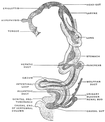
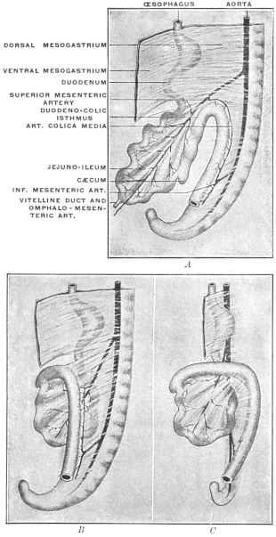
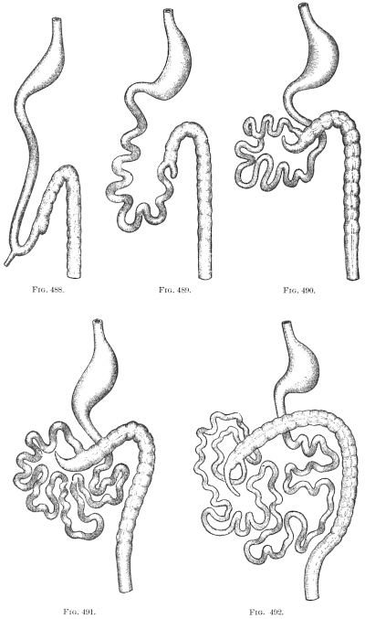
At the time when the umbilical loop of the intestine has formed and has begun to protrude into the cavity of the umbilical cord (fifth to sixth week), the first indication of the future cæcum appears as a circumscribed thickening of the returning or ascending limb of the intestinal loop a short distance from the apex (Figs. 486-488). This rudiment indicates the derivation of the future definite intestinal segments from the elements of the loop. The descending limb, apex (site of embryonic vitelline duct, Meckel’s diverticulum of adult) and a short succeeding portion of the ascending limb furnish the ileum and jejunum. The rest of the ascending limb develops into cæcum and appendix, ascending and transverse colon. The increase in the length of the intestine is not uniform. The formation of convolutions begins in the seventh week in the apex and subsequently in the descending limb. By the eighth week a considerable number of jejuno-ileal coils have resulted from the growth in length of these parts of the original umbilical loop, while the growth of the segment which furnishes the colon is at this time still inconsiderable (Fig. 489). In the meanwhile the thickening of the tube which forms the first rudiment of the cæcum has developed into a small sac-like enlargement of the gut, budding from the left and dorsal aspect of the ascending limb, crescentic in shape, turning its concavity toward the parent tube. In the majority of instances examined the small outgrowth is packed closely between the incipient ileal convolutions, lying under cover of the more prominent bulging coils of the umbilical protrusion, between them and a single coil of larger arc situated dorsally and belonging to the jejunal or proximal portion of the small intestine (Fig. 490). Fig. 497, taken from an embryo of 11 mm. cervico-coccygeal length, represents this stage in the development of the umbilical loop. The arrangement of the cæcum which we can assume as the typical condition at this stage and which determines in part the subsequent final arrangement of the structures, is illustrated by this relation of the cæcal bud to the surrounding incipient convolutions of the small intestine, with the larger part of these coils situated ventrad of the cæcum and only a single coil of larger curve placed dorsally; the cæcal pouch, derived from the ascending limb of the umbilical loop, is situated between these two divisions, turning its concave border to the right and embracing the parent tube. At the time when the human cæcum first appears as a distinct structure it forms a small conical pouch with blunt extremity whose shape is well illustrated by the cæcum of some of the new-world monkeys, as Mycetes fuscus, the brown howler monkey (Figs. 449 and 450). The outgrowth develops rapidly in length and very soon assumes a distinct crescentic shape, gradually tapering toward the extremity, a type which is found reproduced in the cæcum of Ateles ater, the black-handed spider monkey (Fig. 443). There is as yet no constriction or demarcation separating the distal segment (future appendix) from the proximal part (cæcum proper) but the entire pouch gradually narrows funnel-like to its termination.
Figs. 488-492.—Series of schematic figures illustrating stages in the rotation of the intestinal canal.
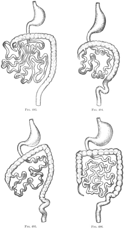
Figs. 493-496.—Series of schematic figures illustrating stages in the rotation of the intestinal canal.
II. CHANGES IN THE POSITION OF THE CÆCUM AND APPENDIX DURING NORMAL DEVELOPMENT, DEPENDING UPON THE ROTATION OF THE INTESTINE AND THE SUBSEQUENT DESCENT OF THE CÆCUM.
The primary cause leading to the rotation of the intestinal canal and inaugurating the successive stages which produce the adult disposition of the tube is to be found in the rapid increase in length of the small intestine. Numerous convolutions of this tube succeed to the few primary coils noted in the first stages. This condition is illustrated in Fig. 498, taken from an embryo of 4.4 cm. cervico-coccygeal measure, and the arrangement of the intestine is indicated in schema, Fig. 491. The cæcum is found nearly in the median line imbedded among the surrounding coils of the small intestine, which by their rapid increase have pushed the pouch cephalad nearly into contact with the caudal surface of the liver.
Fig. 497.—Human embryo of 11 mm. cervico-coccygeal measure. Enlarged view of ventral and left aspect of intestinal canal. (Columbia University, Study Collection.) Fig. 498.—Human embryo of 4.4 cm. cervico-coccygeal measure. Intestinal canal; Liver removed. (Columbia University, Study Collection.)
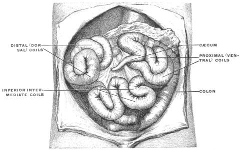
Three main divisions of the convolutions of the small intestine can be made out, slightly separated from each other in the figure to exhibit the cæcum between them. The proximal (jejunal) set of these convolutions occupy the upper and left part of the abdominal cavity. They are the product of the single larger coil which in the earlier stage (Fig. 497, schema Fig. 490) appeared dorsad of the cæcal diverticulum. The distal (ileal) division of small intestinal convolutions has become greatly augmented and lies to the right of the cæcum. The concavity of the pouch is still, as in the earlier stages, directed to the right and the entrance of ileum into colon takes place from right to left. The caudal part of the abdominal cavity is occupied by an intermediate set of transition convolutions which join the proximal and distal divisions. In the two stages just described (Figs. 497 and 498, Schema Figs. 490 and 491), the initial step in the intestinal rotation has been taken, i. e., the beginning of the colon has been displaced cephalad from its original position in the caudal and left part of the abdominal cavity by the pressure of the rapidly growing coils of the small intestine and now lies transversely ventrad of the duodenum, having crossed the duodeno-colic neck or isthmus of the primitive umbilical loop (cf. Fig. 487, C).
At first the distal coils of the small intestine occupy a position behind as well as to the right of the cæcum, forming a dorsal retro-cæcal division connected by intermediate convolutions with the ventral division occupying the lower and left portion of the abdominal cavity. The apex of the cæcum is frequently imbedded among these terminal coils of the ileum. With the continued growth of the small intestines a further displacement of the cæcum cephalad and to the right takes place, while at the same time the terminal ileal coils pass downwards and to the left, from a retro-cæcal into a subcæcal position, thus permitting a direct apposition of the cæcum to the dorsal parietal (prerenal) peritoneum. The last steps in this process of withdrawal of the original voluminous dorsal (retro-cæcal) division of ileal convolutions are well seen in the preparation shown in Fig. 499, taken from an embryo of 6.7 cm. vertex-coccygeal measure, and corresponding to the schematic stages represented in Figs. 490 and 491. The cæcum in this preparation has not yet completed its rotation and still turns its concavity upwards and to the right, with the apex imbedded among the terminal convolutions of the ileum.
Fig. 499.—Human embryo of 6.7 cm. vertex-coccygeal measure. Liver removed. (Columbia University, Study Collection.)
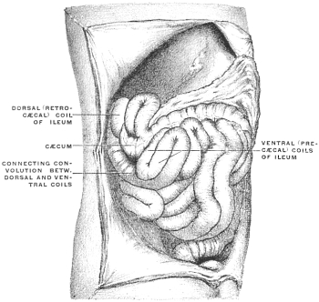
Fig. 500.—Human embryo of 4.9 cm. vertex-coccygeal measure. Ventral view of abdominal cavity, with liver partially removed. (Columbia University, Study Collection.)
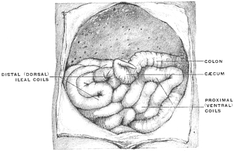
Fig. 501.—The same embryo represented in Fig. 500. The colic coil further depressed and turned to the left; seen from the right side.
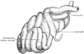
The ileo-cæcal junction takes place from right to left in a downward direction. Nearly the entire mass of the small intestine is situated below and to the left of cæcum and colon, but a terminal ileal coil still occupies, although evidently in the process of withdrawal, the retro-cæcal position, separating the cæcum from direct contact with the dorsal parietal peritoneum. The withdrawal of this terminal coil of the small intestine is accompanied, or immediately followed, by a further turn of the colon cephalad and to the right, which brings it into contact with the caudal surface of the liver and completes the rotation, producing a change in the relative positions of the terminal ileal coils and the cæcum. In the stages illustrated in Figs. 498 and 499 and shown schematically in Figs. 490 and 491, the terminal coils of the ileum pass from right to left behind the cæcum to enter the colon, and the concavity of the cæcal pouch is directed upwards and to the right. After the final rotation has occurred (schema, Fig. 492) the ileum enters the large intestine from the left and from below, and the concave border of the cæcum is directed caudad and to the left. This change in relative position has been accomplished by a revolution of the colon and cæcum through an arc of 180° around its own long axis carrying the cæcum above and behind the small intestine and bringing it into contact with the dorsal prerenal parietal peritoneum. At the same time the terminal coils of the ileum turn downwards and to the left. If this final step in the rotation of the large intestine fails to occur, with otherwise normal development of the parts, the ileum will persist in entering the large intestine from right to left after the cæcum has obtained its final lodgment in the right iliac fossa. We have had occasion to refer previously to the significance of these instances of partially arrested development (cf. p. 61, Figs. 123, 127 and 128).
In Figs. 500 and 501, taken from an embryo of 4.9 cm. vertex-coccygeal measure, the final rotation of the cæcum from the position occupied in Fig. 498 has occurred and the concavity of the pouch is directed caudad and towards the left. At the same time the escape of the terminal ileal coils from behind the cæcum and beginning of the colon has not yet taken place and hence the colon is still kept by these coils from direct opposition to the dorsal prerenal parietal peritoneum. The condition presented by this preparation can be schematically indicated by Figs. 492 and 493. The rotation has carried the beginning of the colon (Fig. 500), with the cæcal bud and appendix curved on itself and turning its concavity to the left, into the subhepatic position. The greater part of the small intestinal coils lie now below and to the left of the cæcum, but the terminal ileal convolutions (Fig. 500) still occupy a retro-cæcal position, separating the pouch and the colon from the dorsal parietal peritoneum. In Fig. 501 the right lateral view of the same embryo is shown with the cæcum and colon depressed and turned to the left. The termination of the ileum reaches the ileo-colic junction by passing behind the cæcum, and the immediately adjacent ileal coils are still retro-cæcal, intervening between the pouch and the dorsal parietal peritoneum.
In the next succeeding stage (schema, Fig. 494) these coils of the ileum turn downward and to the left so as to lie below and mesad to the cæcum and colon, thus permitting the direct apposition of the large intestine to the parietal prerenal peritoneum. The terminal ileum now passes from below and to the left upwards and to the right to its junction with the colon. This freeing of the dorsal surface of cæcum and colon from contact with the coils of the small intestines, and the consequent direct apposition of the same to the dorsal parietal peritoneum influences to a great extent the subsequent arrangement of the parts, because it affords the conditions necessary to the fixation of the colon and mesocolon by adhesion to the parietal peritoneum (cf. p. 81).
Fig. 499, taken from an embryo of 6.7 cm. vertex-coccygeal measure, illustrates this stage, which is encountered in the majority of instances and during which the retro-cæcal coils of the terminal ileum are withdrawn (schema, Fig. 493). The convolutions of the small intestine have greatly increased in size and number. The retro-cæcal ileal coils, compared with Fig. 500, have shifted their position caudad and to the left, so as to lie below and ventrad of the beginning of the colon. Only a single coil remains behind the cæcum and appendix, intervening between these structures and the ventral surface of the right kidney, and this coil is in the process of withdrawal from the dorsal position as indicated by the superficial and short course of the coil which connects it with the remaining ventral convolutions. As soon as the withdrawal of this single remaining dorsal coil is completed the entire mass of the small intestines will occupy a position ventrad, caudad and to the left of the cæcum and colon (Fig. 494), which will then rest directly against the dorsal parietal peritoneum investing the ventral surface of the right kidney.
Fig. 502.—Human embryo, 6.6 cm. vertex-coccygeal measure. Liver removed. (Columbia University, Study Collection.)
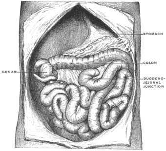
This stage is illustrated in Fig. 502, taken from an embryo of 6.6 cm. vertex-coccygeal measure. The cæcum and appendix here occupy the subhepatic position, well to the right of the median line and in the background of the abdominal cavity. The terminal retro-cæcal ileal coils of the embryo shown in Figs. 500 and 501 have descended caudad and to the left, thus freeing the dorsal surface of cæcum and colon and permitting direct contact with the prerenal parietal peritoneum.
In the succeeding stages the cæcum gradually descends along the background of the right lumbar region from the subhepatic position to the right iliac fossa, producing by this descent the ascending colon as a distinct segment of the large intestine.
It will be observed that in the stage shown in Fig. 502 (schema, Fig. 494) the large intestine passes from the cæcum to the splenic flexure transversely from right to left across the upper part of the abdominal cavity, caudad and ventrad of the stomach and cephalad of the coils of the small intestine.
In the following stages the disproportionately large size of the embryonic liver compels the colon, as the cæcum descends, to assume an oblique position. When the cæcal descent is completed the colon traverses the abdominal cavity in contact with the caudal surface of the liver passing from the right iliac fossa obliquely cephalad and to the left to the splenic flexure where it becomes continuous with the descending colon, which segment has early assumed its definite position in the background of the abdominal cavity on the left side (Fig. 495). This oblique position of the colon is seen in Figs. 503 and 504. During this stage the increase in the length of the colon may lead to the arrangement seen in Fig. 505, where the future transverse segment of the large intestine is bent caudad in form of an arch whose summit extends nearly to the pelvis. This condition at times persists in the adult, in cases of unusually long large intestine, and recalls the normal arrangement found in many of the cynomorphous monkeys in whom the transverse colon forms an extensive V-or U-shaped loop, with the apex directed caudad toward the pubic symphysis (Fig. 506). In other instances in the human fœtus this part of the large intestine is thrown into a number of shorter irregular coils (Fig. 507).
Fig. 503.—Human embryo, 7.6 cm. vertex-coccygeal measure. Liver and small intestine from the duodeno-jejunal to the ileo-colic junction removed. (Columbia University, Study Collection.) Fig. 504.—Human fœtus, 10.6 cm. vertex-coccygeal measure. Liver and greater part of small intestine removed. (Columbia University, Study Collection.)
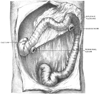
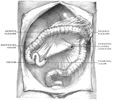
Fig. 505.—Human fœtus, 20.4 cm. vertex-coccygeal measure. (Columbia University, Study Collection.) Fig. 506.—Abdominal viscera of Macacus rhesus, rhesus monkey, hardened in situ. (Columbia University Museum, No 1817.)
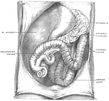
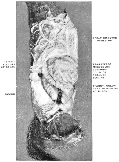
Fig. 507.—Abdominal viscera of human fœtus at term, hardened in situ; hepatic flexure formed, and ascending and transverse colon differentiated. (Columbia University Museum, No. 1816.)
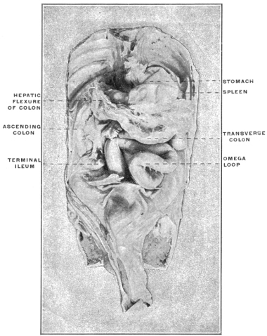
Fig. 508.—Human fœtus of 10.7 cm. vertex-coccygeal measure. Liver and small intestine from the duodeno-jejunal to the ileo-colic junction removed. The colon already presents an ascending, transverse and descending segment. The appendix is retro-cæcal, curved, with the tip turned down, under cover of the ileo-colic junction and mesentery. (Columbia University, Study Collection.)
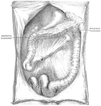
Normally, however, in the process of further development and with the relative decrease in the size of the liver, the hepatic flexure (Fig. 505) becomes defined and passes cephalad and to the right, taking up the slack of the bent segment and establishing the typical ascending and transverse colon as seen in Fig. 508 (schema, Fig. 496).
III. VARIATIONS OF ADULT CÆCUM AND APPENDIX.
The study of the variations of the adult cæcum and appendix involves the consideration of the following points:
(a) Shape of cæcum and origin of appendix. (Type of adult cæcum.)
(b) Position, direction and peritoneal relations of the appendix.
(c) Arrangement of the vascular and serous ileo-cæcal folds.
The peculiarities encountered in any individual case usually depend upon the combination of all three of these factors, which together influence and determine the arrangement of the structures in the adult. Hence the examination of each case should be made with reference to these three points, which we will now consider in detail.
A. SHAPE OF CÆCUM AND ORIGIN OF APPENDIX. TYPES AND VARIATIONS OF ADULT CÆCUM AND APPENDIX.
The various forms of the adult cæcum are all derived by modifications from the fœtal type of the pouch.
In the embryo the cæcum is funnel-shaped, narrowing gradually and symmetrically in caliber to the root of the appendix, at which point the three colic tænia or longitudinal muscular bands of the large intestine meet. The appendix arises from the apex of the funnel, the lateral walls of which are equally and symmetrically developed. The entire pouch is of a crescentic shape, the concavity of the curve turned to the left and directed toward the caudal margin of the terminal ileum. Two subdivisions of the fœtal type are found:
Fig. 509.—Schematic table of types of human cæca.
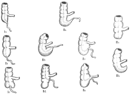
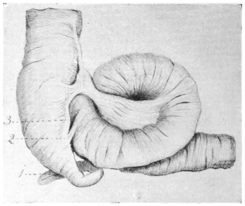
Fig. 510.
—Human fœtus at term. Cæcum and ileo-colic junction; ventral view. (Columbia University, Study Collection.)
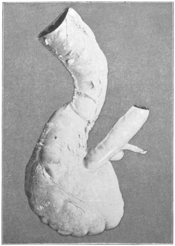
1. Appendix. 2. Reduced intermediate non-vascular fold. 3. Ventral vascular fold.
I. The crescentic curve of the cæcum is only slightly marked; the appendix arises from the most pendent part of the pouch and hangs downward (schema, Fig. 509, I, a).
This form, which is encountered only occasionally in the fœtus and infant, is illustrated by the preparation shown in Fig. 510, taken from a fœtus at term.
II. In the majority of cases the inherent crescentic shape of the cæcal pouch is pronounced and carries the termination of the funnel with the root of the appendix cephalad and to the left toward the caudal margin of the ileo-colic junction (schema, Fig. 509, II, a).
At birth this typical arrangement of the cæcum frequently places the pouch in a nearly transverse position, with the apex and the root of the appendix turned to the left, in contact with, or under cover of the terminal piece of the ileum at its junction with the large intestine.
Figs. 511 and 512 represent the parts in the ventral view in the fœtus at term.
Fig. 511.—Human fœtus at term. Cæcum and ileo-colic junction; ventral view. (Columbia University, Study Collection.) 1. Appendix, coiled spirally behind terminal ileum. 2. Non-vascular intermediate fold. Fig. 512.—Human fœtus at term (negro). Cæcum and ileo-colic junction; ventral view. (Columbia University Museum, No. 692.)
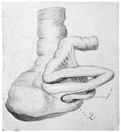
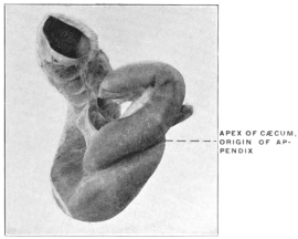
Fig. 513.—Human fœtus at term. Cæcum and ileo-colic junction; dorsal view. (Columbia University, Study Collection.) Fig. 514.—Human fœtus at term. Cæcum and ileo-colic junction; dorsal view. (Columbia University Museum, No. 1715.)
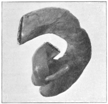
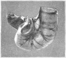
Figs. 513 and 514, also taken from the fœtus at term, show the cæcum from the dorsal aspect and illustrate well the sharp character of the curve which carries the apex of the pouch up and to the left.
All the variations observed in the adult cæcum are derived from these two fœtal types by a subsequent and usually asymmetrical enlargement and dilatation of the pouch.
We can consider the derivatives of each form separately.
I. Adult Cæca Derived From Type I. (schema, Fig. 509, Ia, Fig. 510).
1. Further development leads to an enlargement of the cæcal pouch and a sharper demarcation between the same and the appendix. The resulting cæcum is symmetrical, with equally developed lateral sacculi, between which the termination of the longitudinal muscular bands and the root of the appendix is situated (schema, Fig. 509, Ib).
Fig. 515.—Human fœtus at term. Cæcum and ileo-colic junction; ventral view. (Columbia University Museum, No. 1510.) Fig. 516.—Human fœtus at term. Cæcum and ileo-colic junction; ventral view. (Columbia University Museum, No. 1548.)
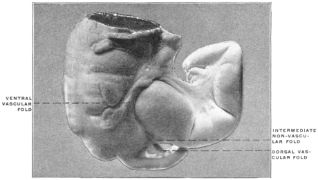
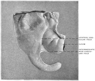
In Figs. 515 and 516 two infantile cæca are shown which illustrate this form. The narrow and pointed apex of the fœtal conical cæcum is replaced by the capacious pouch which is differentiated sharply from the appendix. Among the anthropoid apes the same type is seen in the cæcum of the gibbon (Figs. 455 and 456), and of the young chimpanzee shown in Fig. 460.
2. An increased development of the cæcal pouch in the adult leads to the protrusion caudad of two symmetrical sacculations on each side of the root of the appendix which appears between them. The original apex of the cæcal pouch is still marked by the implantation of the appendix and by the termination of the longitudinal muscular bands, but the lowest level of the pouch is found on each side of this point at the fundus of the secondary lateral sacculi (schema, Fig. 509, Ic). Treves, to whom belongs the credit of first accurately describing and classifying the forms of the adult cæcum based on the development, found this type in three of a series of 100 cases examined.
Fig. 517.—Adult human cæcum and ileo-colic junction. (Columbia University, Study Collection.) Fig. 518.—Adult human cæcum and ileo-colic junction. (Columbia University Museum, No. 234.)
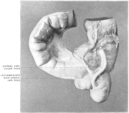
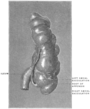
Figs. 517 and 518 illustrate this form of the pouch, which, in our experience, is frequently associated with the retro-cæcal erect position of the appendix (cf. infra, p. 251). Fig. 472 shows this type in the adult with pendent appendix.
II. Adult Cæca Derived from Type II. (schema, Figs. 509 and 511).—From this more commonly observed type of fœtal cæcum the following adult forms are developed:
1. The general shape and trend of the fœtal cæcum is preserved. The pouch turns sharply to the left, carrying the apex with the root of the appendix upward toward the ileum, the appendix itself being frequently placed under cover of the terminal coil of the small intestine (schema, Fig. 509, IIb).
Fig. 519.—Human adult (Smith’s Sound Eskimo). Ileo-colic junction and cæcum. (Columbia University Museum, No. 59/1483.)
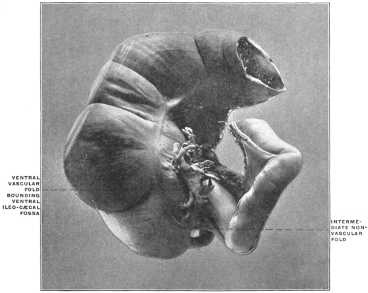
Fig. 520.—Human adult. Ileo-colic junction and cæcum. (Columbia University, Study Collection.)
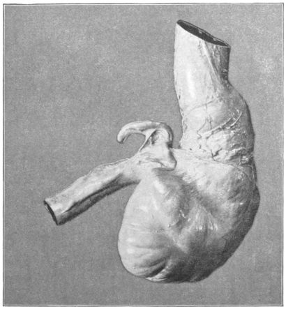
Fig. 521.—Human adult. Ileo-colic junction and cæcum. (Columbia University, Study Collection.) Fig. 522.—Human adult (Smith’s Sound Eskimo). Ileo-colic junction and cæcum. (Columbia University Museum, No. 56/1571.)
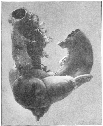
The apex of the cæcal pouch is either conical, narrowing gradually toward the root of the appendix (Figs. 520 and 521), or blunt and more sharply defined against the appendix (Fig. 522). Mr. Treves encountered this “persistent fœtal type” in two per cent. of his series.
Fig. 523.—Human adult. Ileo-colic junction and cæcum; dorsal view; dried preparation. (Columbia University Museum, No. 200.)
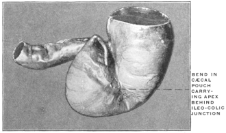
Fig. 524.—Human fœtus at term. Ileo-colic junction and cæcum; ventral view. (Columbia University Museum, No. 1717.) Fig. 525.—Same preparation as Fig. 524; dorsal view.
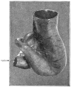
The cæcum is frequently sharply bent on itself in making the turn upward and to the left, resulting in a deep indentation of the concave border and producing a corresponding projecting fold in the interior of the pouch (Fig. 523). The ventral longitudinal muscular band follows the crescentic sweep of the cæcum to the root of the appendix.
Figs. 524a and 525b, representing the cæcum of a fœtus at term in the ventral and dorsal view respectively, show very clearly the arrangement of the fœtal pouch from which the adult type with sharp angular bend is derived. This type of adult cæcum is found in certain of the anthropoid apes.
In the orang (Figs. 458 and 459) the cæcum turns sharply upward and to the left, gradually narrowing in caliber to the root of the appendix which is coiled behind the termination of the ileum.
The same type is seen in Figs. 462 and 463, taken from a preparation of the adult chimpanzee. Fig. 463 shows especially well the sharp bend between the cæcum and colon by means of which the apex of the pouch is carried cephalad behind the ileo-colic junction.
Fig. 431, taken from another specimen of the same animal, shows the characteristic crescentic curve of the cæcum and the corresponding course of the longitudinal muscular band. The apex of the pouch in this preparation is more rounded and blunt.
The same blunt termination of the cæcum of this type, with a corresponding sharper demarcation of the appendix, is seen in the gorilla (Fig. 457) recalling the conditions found in certain instances in the human subject (Fig. 522).
2. In by far the larger proportion of cases (ninety per cent. in Treves’ series) the adult cæcum obtains its characteristic form by an unequal development of the walls of the intestine. The right segment between the ventral and dorso-lateral muscular bands dilates, forming a sacculation which projects caudad and constitutes the secondary caput coli, while the segment between the lower border of the ileum and the original apex, marked by the origin of the appendix, remains stationary or is further reduced. This unequal development produces a relative displacement of the root of the appendix upward and to the left toward the ileo-colic junction.
In some cases the primitive crescentic curve of the cæcum, as indicated by the direction of the ventral longitudinal muscular band, is still perceptible.
The right wall of the fœtal cæcum, forming the most pendent portion of the pouch, dilates uniformly and thus constitutes the adult caput coli. The left wall appears as a small sacculation separating the root of the appendix from the ileo-colic junction (schema, Fig. 509, II, c). This type of the adult cæcum is illustrated by the preparations shown in Figs. 526-528. In other cases part of the right wall of the cæcum between the ventral and dorso-lateral colic tænia, dilates abruptly forming a very prominent rounded sacculation which carries the lowest part of the pouch caudad in a sharper curve than in the preceding form as indicated by its deviation from the direction of the longitudinal muscular band (schema, Fig. 509, II, d).
Fig. 526.—Human adult (Smith’s Sound Eskimo). Ileo-colic junction and cæcum; dorsal view. (Columbia University Museum, No. 61/1461.)
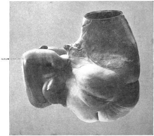
Fig. 527.—Human adult. Ileo-colic junction and cæcum; ventral view. (Columbia University, Study Collection.)
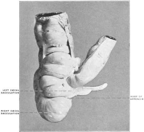
Fig. 528.—Human adult. Ileo-colic junction and cæcum; ventral view. (Columbia University, Study Collection.)
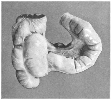
Figs. 529-531 afford examples of this type, while Fig. 532, taken from an infantile preparation, shows that the same may begin to develop at a very early age.
Fig. 529.—Human juvenile. Ileo-colic junction and cæcum; dorsal view. (Columbia University, Study Collection.) Fig. 530.—Human adult. Ileo-colic junction and cæcum; dorsal view. (Columbia University Museum, No. 115.)
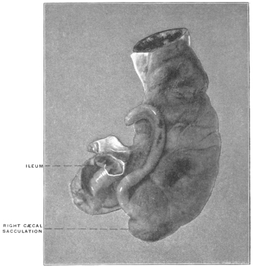
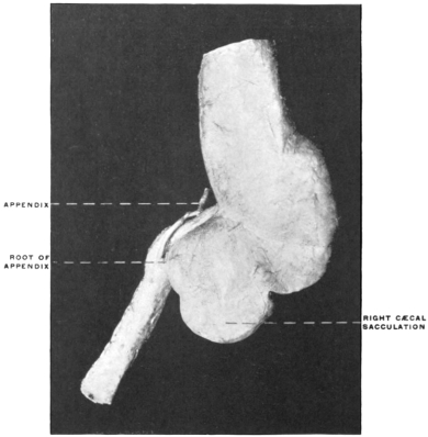
Fig. 531.—Human adult. Ileo-colic junction and cæcum; dorsal view. (Columbia University, Study Collection.) Fig. 532.—Human infant, Ileo-colic junction and cæcum, with secondary terminal sacculation. Human infant, Ileo-colic junction and cæcum, with secondary terminal sacculation. (Columbia University Museum, No. 1632.)
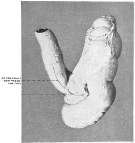
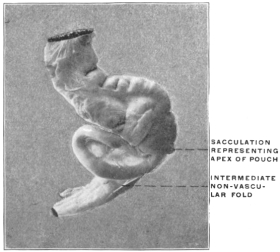
3. Finally, in about four per cent. to five per cent., adult cæca, the reduction of the wall to the left of the root of the appendix, between this point and the ileo-colic junction, is complete. The entire cæcal pouch is formed by the dilated right wall between the ventral and dorsolateral muscular bands. The ventral band terminates at the lower border of the ileo-colic junction, from which the appendix appears to arise, indicating the original apex of the fœtal cæcum (schema, Fig. 509, IIe).
This type is illustrated in the specimens shown in Figs. 533 and 534.
Fig. 533.—Human adult. Ileo-colic junction and cæcum; dorsal view; dried preparation. (Columbia University Museum, No. 124.) Fig. 534.—Human adult. Ileo-colic junction and cæcum; ventral view; dried preparation. (Columbia University Museum, No. 14.)
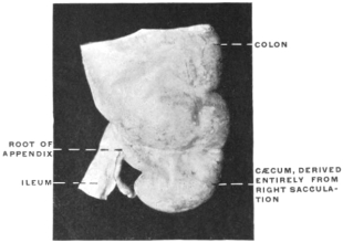
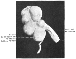
III. Adult Cæca in Cases of Absence of the Appendix.—A few instances of congenital absence of the appendix have been observed.
Fig. 535.—Human adult. Ileo-colic junction and cæcum; absence of appendix. (Columbia University Museum, No. 1077.)
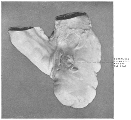
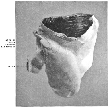
Fig. 536.—Human adult. Ileo-colic junction and cæcum, hardened in situ; absence of appendix. (Columbia University Museum, No. 715.)
A. v. Haller9 describes the condition in the following words: “Defuisse visa est in homine appendicula, ut tuberculum minimum superesset.”
Fr. Arnold,10 without describing any individual case, states that “very rarely the appendix is entirely wanting.”
E. Zuckerkandl,11 reports having observed one case of absence of the appendix.
J. D. Bryant,12 reports a case in which he operated for appendicitis but found “absolutely no appendix.” “The point of tenderness was found to be a glandular growth located posterior to the usual site of the appendix.”
Two instances of this variation are shown in Figs. 535 and 536, taken from preparations in the Morphological Museum of Columbia University. In both careful examination of the external as well as of the mucous surface of the cæcum demonstrated the entire absence of the appendix, and the subjects from which they were obtained presented no scars or other evidences of operative removal or of pathological processes. They are both, therefore, authentic instances of complete congenital absence of the appendix, not of so-called retro-peritoneal or hidden appendix.13
The two examples differ from each other in some details. In the first case (Fig. 535, schema, Fig. 509, IIIa) the cæcum is rounded and globular. The ventral longitudinal muscular band is vertical and continued to the lowest point of the pouch, which greatly resembles the cæcum of a typical cynomorphous monkey.
In the second case (Fig. 536, schema, Fig. 509, IIIb) the cæcum turns upwards and to the left, terminating in a sharp point, to which several lobes of epiploic fat are attached.
We must assume that in these cases the embryonic portion of the cæcal bud was developed just sufficiently to yield the required adult pouch with nothing to spare, so to speak, which could remain rudimentary in the form of an appendix.
Instances of exceedingly rudimentary and reduced appendix are also encountered.
In the case illustrated in Fig. 537 the appendix formed a small conical elevation without distinct lumen, measuring only 0.5 cm. in length.
Fig. 537.—Human adult. Ileo-colic junction and cæcum, with rudimentary appendix. (Columbia University Museum, No. 1655.)
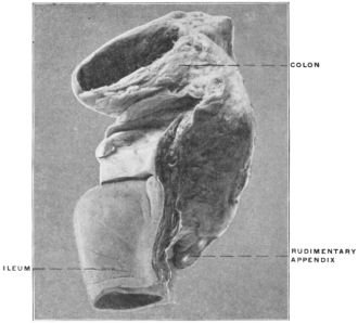
B. Position and Peritoneal Relations of the Appendix.—Statistical records of the position of the appendix indicate a wide range of variation. In general the results obtained by different observers show that certain positions of the appendix are encountered in a sufficiently large percentage of the cases to enable us to adopt a classification, but that a very extensive series of records are required in order to determine even approximately the preponderant relations of the appendix. The following are the most frequently observed positions:
1. The appendix is directed upward, inward and to the left, the terminal portion being frequently coiled under cover of the ileum and mesentery. This position of the appendix is largely due to the normal crescentic curve of the cæcum, which carries the apex of the pouch and the root of the appendix upward and to the left. Its production is, moreover, favored by the tendency of the adult cæcum to develop by dilatation of the ventral and right wall at the expense of the left side of the pouch, thus relatively shortening the interval between the origin of the appendix and the ileo-colic junction.
Examples of this commonly encountered position of the appendix are given in Figs. 512, 513, 514, 520, 521, 523 and 526.
Fig. 538.—Human juvenile. Cæcum in situ lifted up to show vertical course of appendix, situated behind cæcum and ascending colon. The large intestine has a free peritoneal dorsal surface, and the appendix is held in position by adhesion to the large intestine. (Columbia University, Study Collection.)
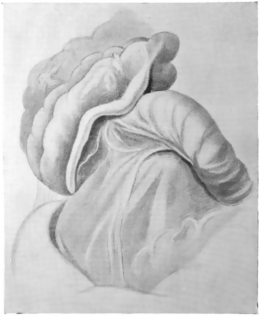
2. The appendix is erected vertically behind the cæcum and ascending colon and closely attached to the dorsal wall of the large intestine. In some instances the cæcum and colon, with the adherent vertical appendix, possess a free serous dorsal surface, not adherent to the parietal peritoneum (Figs. 529, 538, 539 and 540). In other cases the ascending colon is fixed and the greater part of the retro-colic appendix is buried in the connective tissue which attaches the large intestine to the abdominal parietes (Fig. 517). Even in these cases, however, the dorsal surface of the cæcum and the root of the appendix retain their free serous investment.
Fig. 539.—Human adult. Ileo-colic junction and cæcum; dorsal view. (Columbia University Museum, No. 1594.)
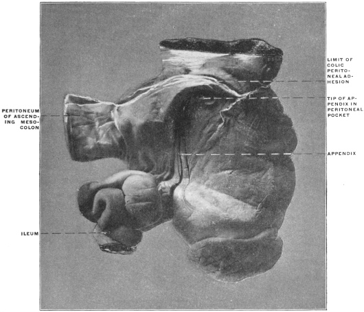
Fig. 540.—Human adult. Ileo-colic junction and cæcum; dorsal view. (Columbia University Museum, No. 1850.)
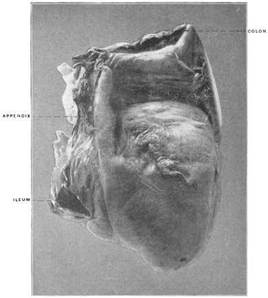
3. The proximal part of the appendix turns upward and to the left in continuation of the cæcal curve, but the distal portion is directed downward and inward, hanging over the brim of the pelvis (Figs. 505, 541 and 542).
Fig. 541.—Human infant. Ileo-colic junction and cæcum; ventral view. (Columbia University, Study Collection.) Fig. 542.—Human infant. Ileo-colic junction and cæcum; dorsal view. The dorsal surface of cæcum as far as root of the appendix is adherent to the parietal peritoneum of the iliac fossa. (Columbia University Museum, No. 394.)
4. The appendix is directed downward, pendent from the lowest point of the conical cæcal pouch, and hangs free over the pelvic brim.
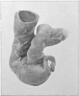
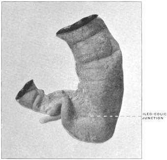
This type is encountered at times in fœtal and infantile subjects (Figs. 516 and 543).
Fig. 543.—Human fœtus at fifth month. Abdominal cavity and viscera; liver and greater part of small intestine removed. (Drawn from preparation in Columbia University Museum, No. 1814.) Fig. 544.—Human fœtus at term. Abdominal cavity and viscera; greater part of small intestine removed. (Drawn from preparation in Columbia University Museum, No. 1813.)
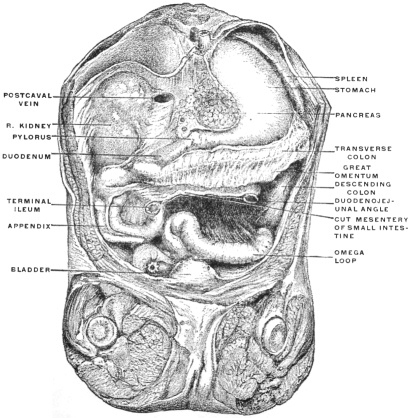
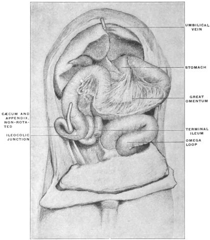
Fig. 545.—Human fœtus at term. Ileo-colic junction and cæcum. (Columbia University Museum, No. 998.)
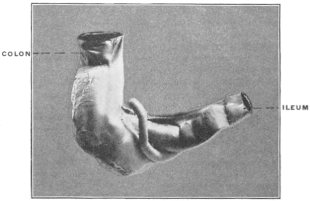
5. The position of the appendix is variant and abnormal, as e. g. placed to the right of cæcum and colon (Fig. 544) or turned up ventrad of the ileo-colic junction (Fig. 545).
These variations in the position of the appendix and the resulting peritoneal relations of the structure depend upon the following factors.
1. The influence of peritoneal adhesions established during the descent of the cæcum from the subhepatic position to the iliac fossa.
2. The inherent curve of the cæcal pouch.
3. The subsequent alterations in the caliber of the intestine and the unequal development of the pouch leading to the formation of the types of adult cæca above considered.
In determining the causes which lead to the establishment of any given position of the appendix all three of the factors above enumerated must be taken into account, although their influence is not exerted in every case to an equal degree.
We have seen that normally, after completed rotation of the intestine, the cæcum with the appendix and the beginning of the colon are lodged in the upper and right part of the abdomen, below the liver and in contact with the prerenal parietal peritoneum (schema, Figs. 493, 502). During the subsequent stages the cæcum descends into the right iliac fossa, thus producing the ascending colon. It is immaterial whether this change in position is regarded as an actual descent of the pouch over the ventral surface of the right kidney, which seems more probable, or as a growing away from the iliac region of the remainder of the abdominal wall, with a concomitant relative reduction in the size of the liver, producing a relatively lower position of the cæcum, or as a combination of these processes. In either case during this period the dorsal surface of the ascending colon and mesocolon normally becomes adherent to the dorsal parietal peritoneum, connective tissue developing between the opposed serous areas and leading to the usual fixation of the ascending colon and obliteration of the free ascending mesocolon. If this process of adhesion is inaugurated at an early stage, i. e., before the descent of the cæcum has been accomplished, it will act as a drag on the dorsal surface of the colic tube during the subsequent change in position, which carries the cæcum downward into the iliac fossa. This leads to a backward bend of the cæcum and appendix which parts will in the ventral view appear under cover of the protruding free ventral and lateral walls of the colon. Hence in many late embryos and fœtus at term the lowest point of the large intestine in the right iliac fossa is formed by the proximal part of the cæcum or by the adjacent segment of the colon, while the original termination of the pouch, with the root of the appendix, is turned backward and upward, and, as we have seen, by reason of the inherent shape of the pouch, also to the left, carrying the beginning of the appendix frequently behind the terminal ileum and the ileo-colic junction.
Two of the more common positions of the appendix, viz., backwards, upwards and inwards behind the ileo-colic junction, and directly backward, erected vertically behind cæcum and colon, can therefore in part be referred to the mechanical conditions obtaining normally during the descent of the cæcum. Of course the shape of the cæcal pouch and the later development of the adult type of cæcum will modify this influence in individual cases. We have seen that this early adhesion and the resulting effects on the position of cæcum and appendix depend on the direct apposition of the colic tube and mesocolon to the dorsal parietal peritoneum. Any condition which will prevent or delay this apposition will likewise perpetuate the original embryonal condition of the tube, completely invested by peritoneum and with a free mesocolon.
Such an element is found in the persistence of the dorsal set of ileal convolutions in the original retro-cæcal position beyond the usual period, as indicated in the schematic Fig. 492, IV, a. If the turn downward and to the left of these coils is for any reason delayed beyond the usual time the cæcal extremity of the colon will descend from the subhepatic to the iliac position without coming directly into contact with the dorsal parietal peritoneum, and therefore without the usual peritoneal adhesion and obliteration of the apposed serous surfaces. The cæcum under these conditions descends without making the backward bend, and the origin of the appendix is found at the lowest point of the pendent funnel-shaped pouch, causing it finally to hang downward or downward and inward over the pelvic brim. The resulting form of the cæcum and the position of the appendix is the one above described as type Ia, Ib and Ic (Fig. 509).
Fig. 510 from a fœtus at term, and Figs. 515 and 516 representing infantile cæca, illustrate this form of the pouch, while the parts are shown in situ in Fig. 543 taken from a preparation of a five-month fœtus.
Fig. 546.—Human embryo, 6.5 cm. cervico-coccygeal measure. Abdominal cavity, with liver removed, seen from the right side. (Columbia University, Study Collection.)
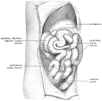
Fig. 547.—Human embryo, 5.9 cm. vertex-coccygeal measure. (Columbia University, Study Collection.)
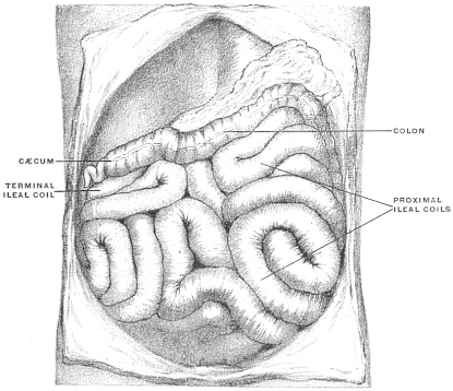
Fig. 548.—Human fœtus at term. Ileo-colic junction and cæcum. Early colic and cæcal adhesion with retroverted appendix. (Columbia University Museum, Study Collection.)
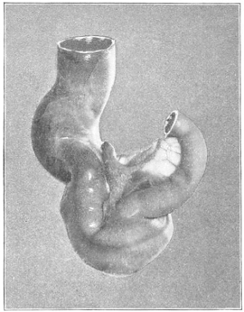
Fig. 546 exhibits the condition obtaining during the development of this type in the more exceptional instances of delayed apposition of the colon to the parietal peritoneum and of increased development of the terminal ileal coils in the original retro-cæcal position. In this embryo, measuring 6.5 cm. in vertex-coccygeal length, the development has progressed sufficiently to establish a distinct transverse colon and to bring the cæcum and appendix into the subhepatic position. But in place of lying in contact with the dorsal parietal peritoneum, as in the embryo, shown in Fig. 502, over the ventral surface of the right kidney, the increased mass of the retro-cæcal ileal coils keeps the cæcum, already in the process of descent, in contact with the ventral abdominal wall. When the final rotation of the retro-cæcal small intestinal coils downward and to the left occurs, placing the ileo-colic junction (C) to the left of the large intestine (schema. Fig. 494), the ascending colon and cæcum are not yet fixed by adhesion to the dorsal parietal peritoneum, and the appendix will present downward and to the left, affording the necessary conditions for the establishment of the permanent pendent position of the tube or causing the same to be directed downward and inward over the brim of the pelvis.
In contrast with the preceding is the condition shown in Fig. 547, taken from an embryo of 5.9 cm. vertex-coccygeal measure. The transverse colon in this preparation has already begun to assume an oblique position, passing down and to the right from the splenic flexure. The cæcum and appendix are in contact with the dorsal prerenal parietal peritoneum. The escape of the dorsal set of ileal convolutions from the retro-cæcal position, by rotation downwards and to the left, is accomplished. The cæcum and appendix are placed in the position which they would have occupied in the embryo shown in Fig. 546 if the dorsal ileal coils had not prevented, in the latter preparation, the apposition of the colon to the dorsal parietal peritoneum.
In considering the effect of these variant conditions on the adult arrangement of the structures it is necessary to bear in mind the second of the above-mentioned factors, namely, the inherent shape of the cæcal pouch and appendix and the resulting direction of its axis.
As previously stated the normal type of the human embryonal cæcum is represented by the pouch of some of the new-world monkeys, as Ateles (Fig. 443) or of certain lemurs, of which Nycticebus (Fig. 420) furnishes an excellent example. The cæcum is distinctly crescentic, turning its concave margin, after completed intestinal rotation, upwards and to the left, toward the lower margin of the ileum. The distal diminished segment of the pouch in Ateles has already assumed the character of a cæcal appendage in Nycticebus and becomes by further reduction the typical appendix in man and the anthropoid apes, while the proximal portion develops into the capacious sacculated cæcum proper. Consequently the initial curve of the cæcum tends to carry the root of the appendix upward and to the left toward the ileo-colic junction. This curve of the pouch, combined with the mechanical effects produced by the adhesion of the colon during the cæcal descent, accounts for the frequency with which the cæcum in the later months of fœtal life and at birth is found curved backward, upward and to the left, placing the root of the appendix under cover of the terminal ileal convolutions (Fig. 548). We have seen that this disposition of the structures accounts for the preponderance of that type of adult cæcum which results from the further and unequal development and dilatation of the segment of the pouch situated to the right of the origin of the appendix.
Bearing in mind the three elements just considered, viz., the effect of adhesion during the cæcal descent, the inherent shape of the pouch and the unequal alterations in caliber in the development of the adult type, we can at once take up the resulting variations in the peritoneal relations of the adult cæcum and appendix which have an important influence on the progress of pathological processes in this region. It should be remembered that in the following schematic figures the colon, cæcum and appendix are represented in the profile view in a straight line, without indicating the characteristic turn of the crescentic cæcal pouch upwards and to the left.
Fig. 549 shows the arrangement in unimpeded cæcal descent without adhesion of colon and mesocolon to the parietal peritoneum. This disposition of the structures, if carried into adult life, would produce the permanently free ascending colon and mesocolon which we encountered exceptionally in the human subject (cf. p. 82) and normally in certain of the cynomorphous monkeys (p. 83). The ascending colon and mesocolon can, under these conditions, be turned mesad, lifting them away from the primary parietal peritoneum investing the ventral surface of the right kidney. Cæcum and appendix have, of course, a complete serous investment.
Figs. 549-554.—Schematic series illustrating the variations in the arrangement of the cæcal and colic peritoneum.
Fig. 549. Fig. 550.
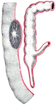
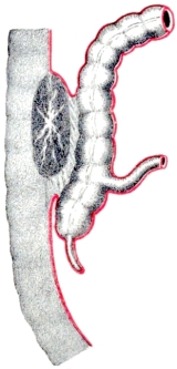
Fig. 551. Fig. 552.
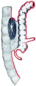
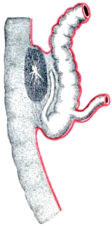
Fig. 553. Fig. 554.
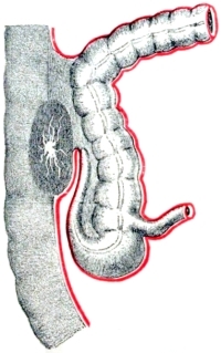
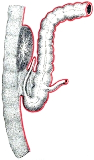
Normally, however, in the human subject, even if the obliteration of the apposed serous surfaces and the resulting fixation of the ascending colon has been delayed beyond the usual period, as above indicated, adhesion takes place subsequently, involving the dorsal surface of the ascending colon between the ileo-colic junction and the hepatic flexure (schema, Fig. 550). The dorsal surface of the cæcum usually retains its free serous surface in whole or in greater part. The appendix is pendent, entirely invested by peritoneum and hangs free in the abdominal cavity, directed toward the pelvic brim, illustrating the effect of delayed fixation of the colon on the position of the appendix.
Examples of this condition are not frequent, and are confined almost exclusively to fœtal and juvenile subjects. Illustrations are afforded by Figs. 515 and 516.
We have already noted (p. 246) the resulting fœtal type of pendent cæcum (Fig. 510).
More commonly colic adhesion before the cæcum obtains its final iliac position results in imparting a backward turn to the pouch, leading to the peritoneal disposition shown in schema, Fig. 551, in which the root of the appendix is involved in the area of obliteration, while the terminal segment remains free. An example of this condition is furnished by the embryo shown in Fig. 508 (10.7 cm. vertex-coccygeal measure). The colon is already segmented into an ascending, transverse and descending portion. The cæcum is retroverted and its apex with the appendix is placed under cover of the terminal ileum which enters the large intestine in the direction from below upward and to the right. In the side-figure the divided end of the ileum is displaced upward to show cæcum and appendix and their relation to the ileal mesentery.
The disposition of the structures illustrated by this example probably depends upon delayed adhesion of the colic embryonal tube to the dorsal parietal peritoneum. The cæcum and appendix appear to have descended freely until the final position in the right iliac fossa has been nearly attained, adhesion and fixation of the colon taking place just before the descent is completed, and thus producing the backward turn of the cæcal end of the tube. Further development of the cæcum to form the adult caput coli in these cases leads to the unequal and exaggerated expansion of the ventral and lateral walls of the pouch, as compared with the fixed and adherent dorsal wall. The former are distended and pushed downwards, producing a relative recession of the root of the appendix upward and to the left, until it comes into relation with, or even under cover of, the ileo-colic junction and of the terminal ileal coil entering the colon at this point.
The resulting characteristic adult position of the appendix in these cases is as follows:
The termination of the cæcum proper, and the root of the appendix are under cover of the terminal ileum and frequently adherent to the parietal peritoneum of the iliac fossa (Fig. 555). The distal portion of the appendix remains free, either hanging down and in over the brim of the pelvis (Fig. 542), or turned upwards and to the left and coiled in several turns (Figs. 504, 555 and 556).
Fig. 555.—Human fœtus at term. Ileo-colic junction and cæcum; dorsal view. The area of peritoneal adhesion is seen to involve the dorsal aspect of the cæcum as far as the root of the appendix. (Columbia University Museum, No. 1549.) Fig. 556.—Human infant. Ileo-colic junction and cæcum, with extensive adhesion to parietal peritoneum. (Columbia University Museum, No. 301.)
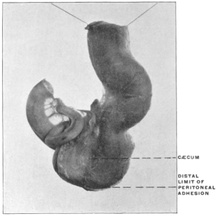
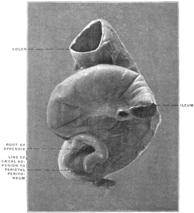
Finally the erect vertical retro-cæcal position of the appendix presents several important variations in the disposition of the peritoneal investment. In Fig. 503, taken from an embryo of 7.6 cm. vertex-coccygeal length, the early complete recession of the retro-cæcal ileal convolutions has probably permitted an early apposition and adhesion of the beginning of the colon to the dorsal prerenal parietal peritoneum. The subsequent descent into the iliac fossa produces a bend in the ventral wall of the colic tube, with a marked convexity directed downwards and forwards, the apex of the bend situated at or near the level of the ileo-colic junction, while the dorsal colic wall is held by the adhesion to the parietal peritoneum, thus giving a backward inclination to the entire cæcum and appendix. During the subsequent descent of the cæcum proper this bend in the colon is gradually diminished and the tube becomes straightened but the apex of the cæcum remains turned back and the appendix is placed in a more or less vertical erect position behind cæcum and ascending colon.
Fig. 557.—Human adult. Ileo-colic junction and cæcum; ventral view. (Columbia University Museum, No. 1612.)
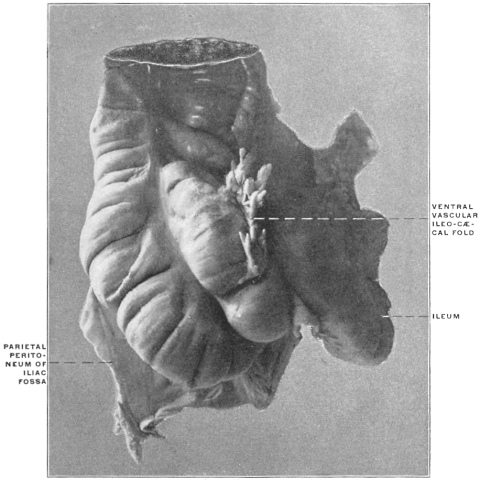
Fig. 558.—Same preparation as Fig. 557; dorsal view. The appendix, erected vertically between cæcum and colon, is completely imbedded in connective tissue.
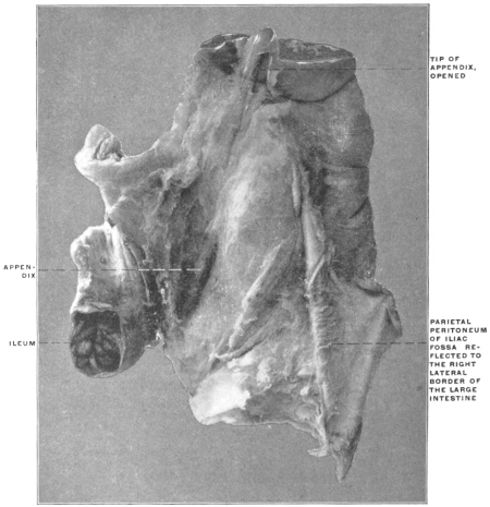
As regards the disposition of the peritoneal membrane in this type of appendix the following conditions are to be noted:
(a) (Schema, Fig. 552.)—The apex of the cæcum and the entire appendix are extraperitoneal, imbedded in the loose connective tissue which occupies the area of serous obliteration. The line of peritoneal reflection from the dorsal wall of the secondary caput coli to the parietal peritoneum of the right iliac fossa is placed transversely below the true apex of the fœtal cæcum and the root of the appendix. The latter tube, imbedded in connective tissue, passes vertically upwards behind the ascending colon, its tip frequently reaching the ventral surface of the right kidney. A well-marked example of this arrangement in the adult is shown in Figs. 557 and 558 (ventral and dorsal view, with peritoneal reflection and vertical retro-colic appendix).
(b) (Schema, Fig. 553.)—In other cases, with the same position of the appendix, the entire cæcum and greater part of the ascending colon remains free. The vertically erected appendix is closely attached to the dorsal surface of the ascending colon, included within the serous investment of the large intestine. The adhesion of the latter is confined to a limited area near the hepatic flexure. Consequently cæcum and greater part of ascending colon can be turned up, away from the parietal peritoneum of the iliac fossa, and the dorsal surface of the appendix shows the free serous covering of the adjacent large intestine.
We may assume that this type of the peritoneal relations of the appendix is produced in one of two ways:
1. Either the retro-colic appendix has become early attached to the adjacent large intestine, whose dorsal surface in large part remains free, or
2. The arrangement of the peritoneum indicated in schema, Fig. 552, may be subsequently changed into that shown in schema, Fig. 553, by a continued downward displacement of the cæcum, producing a secondary serous investment of the dorsal surface of appendix and part of ascending colon.
Examples of this type are found both in infantile and adult subjects.
In Fig. 538, taken from an infant three years of age, the cæcum is lifted up to show the vertical position of the appendix behind the cæcum and ascending colon, the dorsal surface of the large intestine retaining its free serous covering. Another illustration of this arrangement in a juvenile subject is shown in Fig. 529. The same condition in the adult subject is illustrated in Figs. 539 and 540.
Fig. 559.—Human infant. Ileo-colic junction and cæcum; dorsal view. Retro-colic appendix, adherent to the free dorsal serous surface of the large intestine, with intermediate extraperitoneal segment. (Columbia University Museum, No. 1638.)
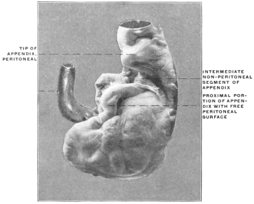
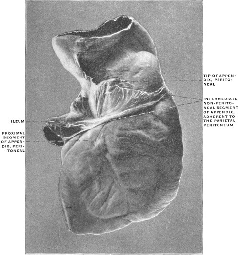
(c) (Schema, Fig. 554.)—Occasionally, with the appendix erected vertically behind the ascending colon, the apex of the cæcum and the proximal portion of the appendix are invested by peritoneum for a short distance and the tip of the appendix likewise obtains a free serous investment, while the intermediate greater portion of the appendix and the corresponding segment of the dorsal surface of the ascending colon are extraperitoneal, adherent to the abdominal parietes. Examples of this peritoneal relation of the appendix in an infant are shown in Figs. 559 and 560, while Fig. 509 represents the same arrangement in an adult specimen. The condition is produced from the arrangement of schema, Fig. 554, by secondary adhesion and obliteration of the serous surfaces over the intermediate portion of the retroverted appendix and the adjacent dorsal surface of the ascending colon.
Fig. 560.—Human adult. Ileo-colic junction and cæcum; dorsal view. Appendix with intermediate non-peritoneal segment, while the proximal portion and the tip are covered by serous investment. (Columbia University Museum, No. 1615.)
C. ILEO-CÆCAL FOLDS AND FOSSÆ.
Certain peritoneal folds, either mesenteric in character, i. e., containing blood vessels, or non-vascular, pass between the terminal ileum and the cæcum and appendix, modifying in some instances very markedly the position and peritoneal relations of the structures.
In considering the influence which these vascular mesenteric and non-vascular serous folds exert in producing further changes in the shape, position and relations of the human appendix it is necessary to remember that in the early embryonal stages these bands and folds of the peritoneum appear only slightly marked, but that they gain their importance and influence on the final adult configuration of the cæcal pouch and appendix in the course of the further development of these structures.
For this reason the comparative study of the corresponding parts in other vertebrates, especially in certain mammalia, is of the utmost value, if we seek to explain and understand the derivation, significance and typical arrangement of these folds. We have seen that the cæcum as found in the large majority of mammalian forms is equivalent to the cæcum and appendix of the human subject and anthropoid apes; that in other words the vermiform appendix represents the distal segment of a cæcal pouch, originally uniform in caliber, which has remained undeveloped, while the proximal portion has progressed evenly with the general development of the alimentary canal to form the cæcum proper. We have seen that this tendency to retain the distal portion of the pouch in a rudimentary condition, i. e., the production of an appendage to the cæcum proper, is encountered in several of the lower forms, as certain Marsupials, Carnivores, Ungulates and Lemurs. The morphology of the ileo-cæcal folds is hence best understood by considering these structures as they appear in connection with the various cæcal types presented by the lower mammalia. Their arrangement and significance can here be readily made out. On the other hand, in studying these structures in the human appendix we are following lines which are already becoming indistinct on account of the rudimentary character of the organ, which we must regard as undergoing an exceedingly slow process of reduction, with a view to its ultimate elimination from the body. We have seen that the structural uncertainty impressed on cæcum and appendix by this evolutionary influence finds its expression in the wide range of variation in size and arrangement which these parts present. Necessarily, of course, this tendency to variation is shared, and even exhibited to a more marked degree, by what we can term the accessory structures connected with cæcum and appendix, viz., the mesenteric vascular and non-vascular serous folds passing to them from the ileum.
We can most profitably begin our consideration of these folds in a form in which they are preserved in their entire and original development, and then successively trace the changes leading up to the normal disposition in the human subject. Such a type is presented by the cæcum of Ateles ater, the black-handed spider monkey (Figs. 444 and 445). The cæcum of this animal presents a uniform crescentic curve, with the concavity directed upward and to the left, and the gradual diminution in the caliber of the pouch, from the ileo-colic junction to the apex, denotes the tendency to retain the distal segment in a rudimentary condition, foreshadowing the eventual formation of a vermiform appendix.
In the ventral view, with the terminal ileum lifted up, the following arrangement of folds passing between ileum and cæcum is noted (Figs. 444 and 445).
(a) Vascular Mesenteric Folds.—The peritoneal vascular folds, carrying the blood vessels to supply the cæcum, are two in number, a ventral (1) and dorsal (3). They are of nearly equal size and extent, passing from the ventral and dorsal aspect of the ileo-colic junction nearly to the apex of the cæcum. Each contains a branch of the ileo-colic artery, which forks in the ileo-colic mesentery, in the angle between ileum and large intestine. The ventral branch continues in the ventral mesenteric fold (Fig. 445) downward across the ventral surface of the ileo-colic junction to supply the ventral part of the cæcum, while the dorsal branch descends behind the ileo-colic junction, preserving a similar course in the dorsal mesenteric fold. The dorsal arterial branch is somewhat larger than the ventral and its distribution extends a little further down to the actual apex of the cæcum.
Fig. 561.—Human adult ileum with Meckel’s diverticulum. Ileo-diverticular serous fold and persistent omphalo-mesenteric artery. (Columbia University Museum, No. 1803.)
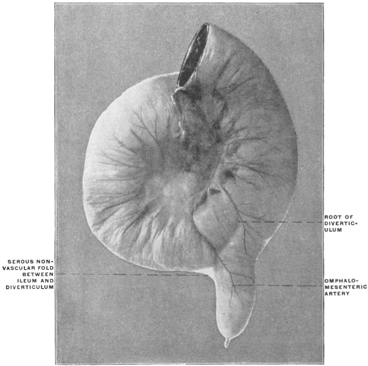
(b) Non-vascular Ileo-cæcal Serous Reduplication.—Between the two vascular mesenteric folds a third serous reduplication, carrying no blood vessels, is found passing between the ileum and cæcum. This fold begins, in the preparation from which the figure is taken, on the ileum opposite the attached mesenteric border, 2.7 cm. from the ileo-colic junction, and passes for exactly the same distance down on the adjacent left concave surface of the cæcum. It is placed a little nearer to the dorsal than to the ventral vascular fold, so that it passes, if the distance between the two vascular folds on the cæcum be divided into three parts, at the junction of the dorsal third with the ventral two thirds. The production of this intermediate non-vascular ileo-cæcal reduplication, which is of very constant occurrence in the mammalian series, is to be led back to the development of the cæcum. When the pouch protrudes from the smooth surface of the embryonic intestine opposite the mesenteric border, it extends backward along the future small intestine and lifts off the serous investment of the gut in the form of a small peritoneal plate filling the interval between itself and the adjacent ileum. A very perfect illustration of this process can be seen in the instance of Meckel’s diverticulum shown in Fig. 561. The proximal portion of the diverticulum is here still closely connected to the small intestine along which it extends, both being surrounded by the common visceral peritoneum. The distal part of the diverticulum has separated more completely from the intestine, and in so doing has drawn out the serous investment in the form of the triangular fold which is seen to pass between the free margin of the intestine and the adjacent surface of the pouch. The same process can be followed in its different stages in certain normal mammalian cæcal types.
Fig. 562.—Human adult. Ileum with Meckel’s diverticulum, 131.5 cm. from ileo-colic junction; a distinct vascular fold is prolonged from the ileal mesentery to the margin of the diverticulum. (Columbia University Museum, No. 1849.)
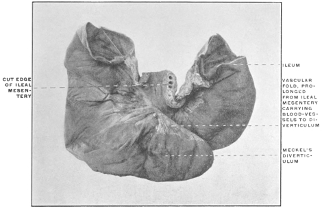
In this connection it may be noted that the production of the cæcal vascular folds and their relation to the mesentery is also very perfectly illustrated in some forms of Meckel’s diverticulum. Thus in the preparation shown in Fig. 562, a broad triangular serous fold passes from the ileal mesentery to the margin of the diverticulum, carrying the blood vessels which supply the pouch. If the section of the intestine to the left of the figure is regarded as representing the terminal ileum, that to the right the colon, and the diverticulum the cæcal pouch, the formation of the fold and its relation to the mesentery, blood vessels and intestine will correspond closely to the ileo-cæcal vascular folds.
Fig. 350 shows the ileo-colic junction and cæcum of Halmaturus derbyanus, the rock kangaroo. The cæcum here extends backwards along the free border of the ileum to which it is closely bound by the common investing visceral peritoneum for the greater part of its extent. In another marsupial form, a small species of opossum from Trinidad (Fig. 349), the cæcum has separated itself more completely from the adjacent small intestine—thus drawing out the peritoneum into a narrow connecting fold. Finally, in the Virginia opossum (Fig. 348), the ileum has attained the usual position at right angles to cæcum and colon. The former pouch is separated from the small intestine by a considerable interval and the angle between the two is filled out by a well-developed triangular serous fold, connecting the free margin of the terminal ileum and the adjacent left border of the cæcum.
This is the “intermediate non-vascular” ileo-cæcal fold.
Passing now from the condition presented by Ateles, with three fully developed and distinct ileo-cæcal folds, to the next stage leading up to the normal human arrangement, we find the same illustrated in the cæcum of another new-world monkey, Mycetes fuscus, the brown howler monkey, shown in the ventral and dorsal views in Figs. 449 and 450. The ventral vascular fold (Fig. 449, 1) is still well developed, the contained ventral branch of the ileo-colic artery descending over the ventral wall of the ileo-colic junction and cæcum and supplying both. The dorsal vascular fold (Fig. 450, 2), on the other hand, is nearly completely fused with the intermediate non-vascular reduplication (Figs. 449 and 450, 3), the approximation between these structures exhibited by Ateles having in Mycetes reached the point of actual union, so that the larger dorsal branch of the ileo-cæcal artery descends to the apex of the cæcum in the following manner: The main post-cæcal artery passes over the dorsal surface of the ileo-colic junction included in a short serous fold which corresponds to the dorsal vascular fold of Ateles. Beyond the lower border of the ileo-colic junction this fold fuses with the intermediate non-vascular fold, one arterial branch descending along the line of attachment of this fold to the cæcum, the other distributed over the dorsal surface of the pouch.
A third type, also taken from the lower Primates, is presented by the cæcum of a cynomorphous monkey, Cercopithecus sabæus, the African green monkey, shown in Fig. 432, in the ventral and left aspect with the terminal ileum lifted up. The cæcum of this animal is comparatively short, somewhat conical, terminating in a blunt apex. The vascular supply is arranged on the same type as in Ateles and Mycetes, i. e., a trunk of the ileo-colic artery divides at the ileo-colic notch, one branch descending ventrad, the other dorsad of the ileo-colic junction. The slightly larger size of the dorsal vessel, noted in Ateles and Mycetes, has been increased in Cercopithecus until the ventral artery (1) supplies merely the front of the ileo-colic junction and the upper part of the adjoining ventral wall of the cæcum, while the larger dorsal vessel (2) descends behind the ileo-colic junction, supplying the same and the entire dorsal and apical portions of the cæcum. The relation of these cæcal arteries to the peritoneum is moreover different from that encountered in Ateles. In place of running in distinct mesenteric folds, as in the latter species, the vessels pass close to the surface of the intestine, merely covered and partly surrounded by slightly redundant visceral peritoneum containing numerous pads of epiploic fat, which bead the course of the vessels at regular intervals. Between the two arteries the intermediate non-vascular fold (2) is seen, presenting much the same arrangement as in Ateles and passing between the left border of the cæcum and the adjacent margin of the ileum, nearer to the dorsal larger than to the ventral smaller cæcal artery.
We have, therefore, in the three types just considered, the following variations in the arrangement of the vascular and non-vascular folds:
1.(a)Ventral and dorsal vascular folds distinct and free. Ventral and dorsal cæcal arteries of nearly equal size. Ateles.
(b)Intermediate non-vascular fold free on both surfaces, placed nearer to the dorsal than to the ventral vascular fold.
2.(a)Ventral vascular fold distinct. Ventral cæcal artery somewhat further reduced in size. Dorsal vascular fold distinct only over the dorsal surface of the ileo-colic junction. At the lower border of the ileo-colic junction the dorsal vascular fold fuses with the intermediate non-vascular fold. Mycetes.
(b)Intermediate non-vascular fold free only on ventral surface, the dorsal surface below the ileo-colic junction being fused with the dorsal vascular fold.
3.(a)Dorsal and ventral vascular folds reduced. Dorsal artery much larger than ventral. Cercopithecus.
(b)Intermediate non-vascular fold well developed, free on both surfaces.
We may judge from this series that the following factors are capable of materially modifying the definite arrangement of the structures:
1. The vascular folds are capable of reduction until the vessels run close to the intestinal surface, merely covered by somewhat redundant peritoneum containing epiploic appendages. (Cercopithecus.)
2. The dorsal cæcal artery tends to assume in all three forms the greater share in the cæcal vascular supply. This tendency is slightly developed in Ateles, becomes more pronounced in Mycetes, and is well marked in Cercopithecus, in which animal the dorsal vessel nearly replaces the ventral branch, the latter confining itself to the ventral surface of the ileo-colic junction and the adjacent ventral parts of the cæcal wall.
3. The intermediate non-vascular fold is placed nearer to the dorsal larger than to the ventral smaller cæcal artery. This condition, present in both Ateles and Cercopithecus, foreshadows the fusion of the intermediate and dorsal vascular folds at the lower border of the ileo-colic junction, as seen in Mycetes.
4. This fusion of the two folds named in Mycetes results in giving different values to the dorsal vascular fold in its proximal and distal segments. The proximal segment descends from the ileo-colic notch behind the ileo-colic junction to its lower border as a distinct fold. Beyond this point its fusion with the distal (cæcal) segment of the intermediate fold rounds out a fossa, the inferior or posterior ileo-cæcal, which is consequently bounded in front by the intermediate vascular fold, behind by the proximal segment of the dorsal vascular fold, to the right side by the inner wall of the cæcum, between the intermediate and dorsal vascular folds, above by the lower border of ileum and ileo-colic junction, and below by the fusion of the two folds.
This pocket or fossa which is the most important and constant of the peritoneal recesses in the neighborhood of the cæcum, opens upward and to the left.
5. A superior or anterior ileo-cæcal fossa, formed in cases of well-developed ventral vascular fold between the same and the ventral wall of the ileo-colic junction, is of small size and shallow.
The cause of the greater development of the dorsal as compared with the ventral cæcal artery is probably to be sought in the adhesion of the colon to the dorsal parietal peritoneum. In Cercopithecus the dorsal surface of the ascending colon is adherent to the parietal peritoneum down as far as the iliac region and beginning of the cæcum, whereas in Mycetes the entire cæcum, as well as the ascending colon, are free and non-adherent to the abdominal parietes. The influence of this adhesion on the arrangement of the vascular supply of the lower portion of the ascending colon and cæcum appears to be important. Some of the departures from the Ateles type presented by Cercopithecus become still better developed in the human subject, where the adhesion of the ascending colon and the obliteration of the apposed serous surfaces of ascending mesocolon and parietal peritoneum is normally complete, even if the cæcum remains entirely free, or only adheres to the iliac parietal peritoneum in the proximal part of its dorsal surface. Comparison with forms presenting non-adherent colic and cæcal tubes indicates that the adhesion determines the relative size and arrangement of the ileo-colic vessels.
Thus the partially adherent colon and cæcum of Cercopithecus presents, compared with the free tube of Ateles and Mycetes, a marked reduction of the ventral and a corresponding enlargement of the dorsal cæcal artery. Further progress in the same direction is noted in the human subject where normally the ascending colon and at times the proximal portion of the cæcum are adherent to the dorsal parietal peritoneum.
It appears that in the adhesion of the colic tube to the parietal peritoneum the dorsal ileo-colic vessels find an element favorable to their more complete development and extension, replacing in part or entirely the ventral cæcal artery which becomes limited in distribution to the region of the ileo-colic junction. The adhesion and fixation of the dorsal wall of the intestine seems to afford an advantage to the dorsal vessel, whereas the greater mobility and the alternating conditions of distension and contraction, with variations of intracæcal pressure, depending upon the contents of the pouch, appear to operate unfavorably upon the development of the ventral vessel.
This view is borne out by the conditions observed in the exceptional instances in which in the human subject the ventral artery assumes the large share in the supply of cæcum and appendix (cf. p. 276). In all the cases observed the type of the cæcum indicated delayed or imperfect colic adhesion, and the ascending mesocolon remained partially free.
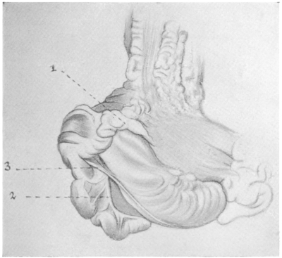
Fig. 563.
—Human; child one year old. Cæcum and ileo-colic junction; ventral view. (Columbia University, Study Collection.)
1. Ventral cæcal artery, surrounded by epiploic appendages.
2. Dorsal vascular fold, forming appendicular mesentery.
3. Intermediate non-vascular fold.
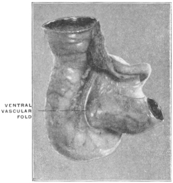
If we now compare the conditions above described for Ateles, Mycetes, Cercopithecus with those usually found in man and in the anthropoid apes, we may appreciate the significance of the structures encountered by beginning the investigation with a type in which the derivation of the different parts is still quite evident. Such a condition is presented by the preparation shown in Fig. 563, taken from a child one year of age. Here the descent of the cæcum has evidently been quite rapid and uniform without dorsal adhesion. The cæcum and ascending colon remain free and can still be lifted away from the ventral facies of the right kidney and turned toward the median line to a point somewhat beyond the renal hilus. The cæcum hangs downward vertically and the appendix arises from the funnel-shaped apex of the pouch.
The ventral cæcal branch of the ileo-colic artery is slightly developed, (1) as a small vessel descending in an epiploic fold over the ventral surface of the ileo-colic junction as far as the root of the appendix. The intermediate non-vascular fold (3) is well marked, measuring 2.9 cm. in length, extending from the free border of the terminal ileum to the cæcum and appendix and crossing over the well-developed dorsal vascular fold (2), which descends, as the appendicular mesenterolium, to the tip of the appendix, carrying the dorsal artery. In studying the conditions presented by this specimen, it is not difficult to trace the analogous structures in the cæca of Cercopithecus, Ateles and Mycetes. The same vascular and non-vascular serous reduplications are found passing between the ileum and cæcum. In accordance with the type presented by Cercopithecus the ventral artery is much reduced and runs in a short serous fold loaded with epiploic appendages. The dorsal artery, on the other hand, is well developed and the intermediate non-vascular fold is distinct. In their relative arrangement these folds follow the Ateles type. The dorsal vascular fold forms the true mesentery of the appendix, and, although close to and crossed by the intermediate non-vascular reduplication, remains still quite separable and distinct from the same; consequently the lower limit of the usual posterior ileo-cæcal fossa, produced by the fusion of the dorsal vascular and the intermediate non-vascular fold, is absent.
A very perfect illustration of this type of the human ileo-cæcal fold is presented by the preparation of Gorilla savagei shown in Fig. 457. The ventral fold and artery appear reduced in this animal. The dorsal vascular fold forms a broad triangular plate of serous membrane carrying the dorsal artery in its free border and extending to the tip of the appendix.
The intermediate non-vascular fold is narrow but distinct, continued for a considerable distance along the ileum, opposite to the attached border, but only for a short extent along the left border of the cæcum below the ileo-colic junction. It crosses the ventral surface of the broad dorsal vascular fold in passing to the cæcum, but remains entirely free and is not adherent to the same.
Consequently here again the dorsal or posterior ileo-cæcal fossa loses its distal limitation. The usual arrangement of the parts, as found in the human subject and derived from the preceding, is well illustrated by another anthropoid ape, Hylobates hoolock. Fig. 455 shows the ileo-cæcum of this animal in the ventral view and the homologous parts, as compared with Gorilla, are readily recognized. On turning the terminal ileum ventrad and cephalad (Fig. 456), it is, however, seen that the intermediate non-vascular fold does not merely cross the dorsal vascular reduplication, as in Gorilla, but that it has begun to adhere to the same at the point of intersection. Consequently a well-marked and clearly limited posterior or dorsal ileo-cæcal fossa is formed, bounded ventrally by the intermediate fold at its accession to the cæcum, dorsally by the proximal part of the dorsal vascular fold, to the right by the left wall of the cæcum, behind by the attachment of the intermediate fold, below by the confluence of the two folds, and above by the lower border of ileum and ileo-colic junction.
The open mouth of the fossa looks to the left. Fig. 464, taken from an adult specimen of the chimpanzee, Troglodytes niger, shows the extent of the dorsal vascular fold and of its connection with the mesentery of the terminal ileum.
The intermediate non-vascular fold extends from the ileum downwards along the entire left border of the cæcum to the root of the appendix, fusing with the dorsal vascular fold and rounding out a deep posterior ileo-cæcal fossa.
The typical arrangement, as encountered in the human subject, corresponds closely to the conditions presented by these anthropoid apes.
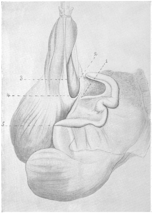
Fig. 564.
—Human adult. Cæcum and ileo-colic junction. (Drawn from preparation in Columbia University, Study Collection.)
1. Dorsal vascular fold at the beginning of the distal free portion, forming the appendicular mesentery.
2. Proximal segment of dorsal vascular fold, fusing with
3. Intermediate non-vascular fold.
4. Rounded edge of union of dorsal vascular and intermediate folds bounding the ileo-cæcal fossa caudad.
5. Point of accession to appendix of proximal branch of appendicular artery derived from posterior ileo-cæcal artery.
In Fig. 564, taken from an adult male human subject, the dorsal surface of the ascending colon and of the ileo-colic junction is adherent to the parietal peritoneum. The distention of the cæcum is nearly uniform, the right sacculation being only slightly larger than the left. The appendix, measuring 18.4 cm. in length, arises from the dorsal surface of the caput coli, 1.7 cm. from the point where the ventral longitudinal muscular band turns around the caudal end of the pouch between the two sacculations, and 3.7 cm. below the caudal margin of the ileo-colic junction.
The dorsal vascular fold (2), forming the broad appendicular mesentery (1), is well developed and free in its distal portion, extending, with gradually diminishing width, to the apex of the appendix. The proximal segment of this fold (between 1 and 2) descends over the dorsal surface of the ileo-colic junction and meets (at 4) the intermediate non-vascular fold (3) which extends between the ileum and cæcum, rounding out a crescentic ridge (4) which bounds the entrance into the posterior ileo-cæcal fossa (between 2 and 3). The influence of the folds and of the blood vessels on the position and curves of the appendix is quite apparent in this preparation.
The dorsal larger branch of the ileo-colic artery, supplying cæcum and appendix, passes over the dorsal surface of the ileo-colic junction (2) where the same, as well as the adjacent dorsal surface of the colon, is adherent to the parietal peritoneum. At the point where the dorsal vascular fold intersects and fuses with the intermediate non-vascular fold (4) the artery divides into a proximal and distal branch. The former proceeds to the cæcum and root of the appendix, reaching this tube at the point marked 5. The latter continues (from 1 on) in the free border of the appendicular mesentery to the beginning of the distal third of the appendix, from which point on the fold extends as a narrow reduplication to the tip of the tube. The segment of the appendix situated between these two main arterial branches is thrown into several coils, the expression of the continued growth between two points relatively fixed by the accession of the two arterial branches. The pathological significance of these bends is apparent when we consider the effect which the kinking of the tube would have on catarrhal and other inflammations accompanied by distension of the appendix.
Typical examples of the posterior ileo-cæcal fossa and of the mutual relationship of the limiting folds are seen in Figs. 565 and 566, both taken from adult human subjects.
Fig. 565.—Human adult, Cæcum and ileo-colic junction with large intermediate non-vascular fold and deep posterior ileo-cæcal fossa. (Columbia University Museum, No. 1546.)
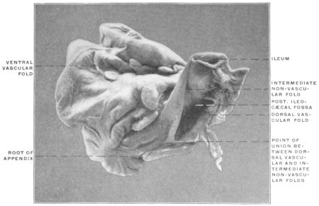
Fig. 566.—Human adult. Ileo-colic junction and cæcum. (Columbia University Museum, No. 1659.)
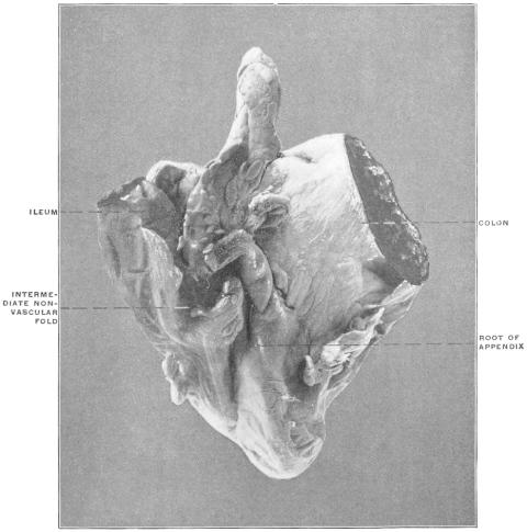
The significance and mutual relations of the folds seen in the preparations just considered—which illustrate the typical adult human arrangement of the structures—will perhaps be best understood by comparison with an adult cæcum in which the infantile condition, as seen in Fig. 563, has become further developed.
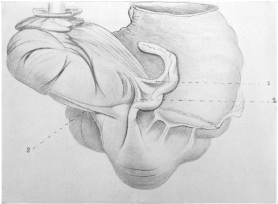
Fig. 567.
—Human adult. Ileo-colic junction and cæcum; dorsal view. (Drawn from preparation in Columbia University, Study Collection.)
1. Dorsal vascular fold, carrying the distal appendicular branch of the dorsal cæcal artery in the mesentery of the appendix.
2. Proximal branch of the same vessel, turning downward to cæcum and root of appendix.
3. Intermediate non-vascular fold.
Fig. 567 shows the dorsal view of such a preparation. The cæcum is funnel-shaped with the apex, carrying the root of the appendix, turned upward and to the left, the sacculation to the right of the ventral muscular band being somewhat dilated. The appendix—7.2 cm. long—turns sharply upward and to the left, closely applied to the left cæcal sacculation, passes dorsad to the ileo-colic junction and lies in its terminal part under cover of the ileo-colic mesentery. The ventral branch of the ileo-colic artery descends over the ileo-colic junction, supplying the ventral wall of the cæcum. The intermediate non-vascular fold (3) is 3.9 cm. long and entirely free.
The dorsal vascular fold contains the large dorsal branch of the ileo-colic artery, dividing into two main branches. The first of these (1) passes distally in the free edge of the fold to the terminal part of the appendix. The other proximal branch (2) turns downward to the root of the appendix and the adjacent wall of the cæcum, aiding materially in holding the proximal upturned segment of the appendix in contact with the left cæcal sacculation.
The intermediate fold, short in its cæcal attachment, does not meet the dorsal vascular fold at any point, consequently the ileo-cæcal fossa is not limited caudad toward the root of the appendix. The conditions presented by this specimen correspond exactly to those found in the gorilla (Fig. 457) and in the human infantile preparation (Fig. 563).
In comparing Figs. 564 and 567 it will be noticed that the line of fusion between the intermediate fold and the dorsal vascular fold (Fig. 564, 4) corresponds to the point where the dorsal ileo-cæcal artery divides into its proximal and distal branches (Fig. 567, angle between 1 and 2). Fig. 567 shows that the proximal arterial twig, even without fusion with the intermediate fold, suffices to influence to a considerable degree the curves and position of the appendix, inasmuch as it serves to hold the proximal segment of the tube closely applied in the erected position to the surface of the left cæcal sacculation. The intermediate segment of the appendix, between the points of accession of the two arterial branches, is most prone to develop spiral twists and bends, especially when the usual fusion of the two folds takes place and still further fixes the parts, while the distal segment, carrying the narrow crescentic terminal appendicular mesentery, remains free.
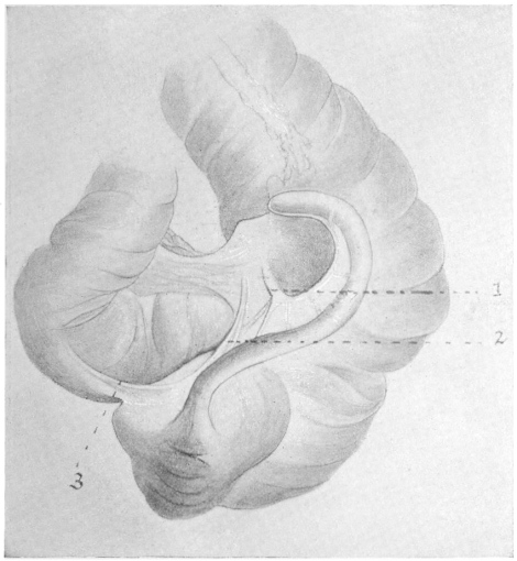
Fig. 568.
—Human adult. Ileo-colic junction and cæcum. (Drawn from preparation in Columbia University, Study Collection.)
1. Distal and
2. Proximal branch of dorsal ileo-cæcal artery running in dorsal vascular fold.
3. Intermediate non-vascular fold fusing with 2 and forming a narrow caudal limit to the posterior ileo-cæcal fossa.
Finally, in a certain number of cases, an intermediate condition between the types presented by Figs. 564 and 567 is encountered. In Fig. 568 the general arrangement of the parts corresponds pretty accurately to that seen in Fig. 566, but the transition from a completely free intermediate non-vascular fold to one which has begun to fuse with the dorsal vascular fold is evident. The cæcum is bent upward and to the left, the caput coli being formed by the right sacculation. The appendix, 7.8 cm. long, takes a wide ≀-shaped curve. The convexity of the proximal curve corresponds to the point where the proximal appendicular artery (2) passes to the tube. The non-vascular intermediate fold (3), measuring 2.2 cm., fuses with the dorsal vascular fold at this point.
The three preparations illustrate serially the share which the peritoneal folds take in the formation of the posterior ileo-cæcal fossa.
In Fig. 566 the failure of the intermediate fold to meet and fuse with the dorsal vascular fold has left the caudal boundary of the fossa (between 2 and 3) incomplete, the ventral and dorsal walls being formed by the folds in question. Fig. 568, in which fusion between the non-vascular and the dorsal vascular folds has commenced, shows the shallow form of the complete fossa under these conditions, while in Fig. 567, with extensive union of the folds, the fossa has correspondingly increased in depth.
Fig. 569.—Human adult. Ileo-colic junction and cæcum. (Columbia University Museum, No. 1610.)
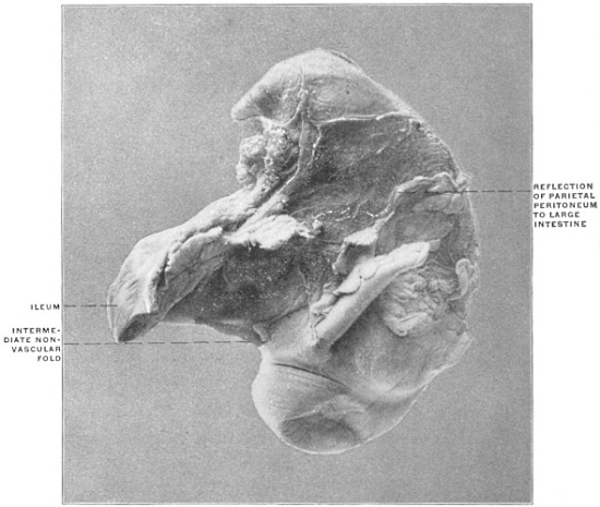
A similar series is shown in Figs. 569, 570 and 571. In Fig. 569, taken from an adult subject, the intermediate non-vascular fold is entirely free, the dorsal branch of the ileo-cæcal artery passes to cæcum and appendix in an area of adhesion between parietal peritoneum and the intestine which includes the dorsal vascular fold. There is consequently no caudal boundary to the ileo-cæcal fossa. Figs. 570 and 571 are both taken from infantile preparations.
Fig. 570.—Human infant, four days old. Ileo-colic junction and cæcum. (Columbia University Museum, No. 879.) Fig. 571.—Human infant. Ileo-colic junction and cæcum. (Columbia University, Study Collection.)
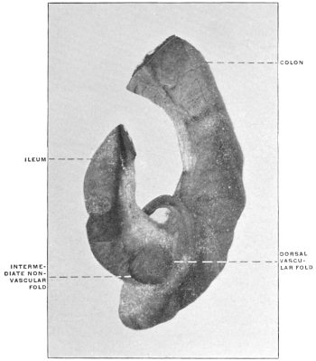
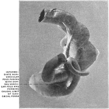
In Fig. 570 the dorsal vascular and the intermediate folds nearly meet at the root of the appendix. They serve to outline the fossa, which appears completed in Fig. 571 by the actual meeting and fusion of the folds.
The Ileo-cæcal Folds in the Anthropoid Apes.—(1) Chimpanzee, Troglodytes niger.
The structures in a juvenile specimen of this animal are shown in Figs. 460 and 461.
The ventral vascular fold (Fig. 460, 3), containing epiploic fat, descends over the ileo-colic junction nearly to the level of the lower ileal margin. The intermediate non-vascular fold (Figs. 460 and 461, 2), derived from the ileum opposite to the mesenteric border, passes to the ventral and left aspects of the cæcum and meets, near the root of the appendix, the dorsal vascular fold (Fig. 461, 3) carrying the dorsal cæcal branch of the ileo-colic artery, which ramifies over the cæcum and supplies the appendix.
The appendix measures 12.3 cm. and presents a terminal hook, slightly dilated.
The appendicular mesentery terminates within the concavity of this hook and measures 1.5 cm. in width at the broadest part, about 4.5 cm. from the root of the appendix.
Figs. 462 and 463 show the cæcum of the adult chimpanzee in the ventral and dorsal view. The ventral vascular fold (Fig. 462, 1) is well developed, heavily fringed with epiploic appendages.
The non-vascular fold is extremely short and tense, fusing with the short appendicular mesentery near the point where in the dorsal view (Fig. 463) the appendix is seen bent at its origin sharply to the right.
Fig. 464, also taken from an adult specimen of the same animal, shows a very well-developed dorsal vascular fold, which fuses with the intermediate fold to limit a distinct ileo-cæcal recess.
The chimpanzee, therefore, agrees closely with the human subject in the arrangement of the folds.
(2) Orang, Simia satyrus.
In Figs. 458 and 459 the arrangement of the folds in an adult specimen of the orang is shown.
The ventral cæcal artery (Fig. 458) is well developed, forming with the peritoneal fold and epiploic appendages surrounding it, a sharp sickle-shaped edge which descends over the ventral surface of the ileo-colic junction following the curve of the left cæcal margin, and turning its concavity to the left toward the entering ileum.
The ventral cæcal artery follows the left margin of the cæcum below the ileo-cæcal junction and passes for 0.5 cm. upon the portion of the pouch which turns up behind the terminal ileum.
The dorsal cæcal artery is a vessel of large size, supplying branches to the narrow appendicular mesentery which extends, with many epiploic appendages, to within 9 mm. of the blunt apex of the appendix. 2.5 cm. beyond the first bend in the appendix the fold is narrowed to a fringe not more than 0.75 cm. wide. Up to this point the dorsal vascular fold measures 1.5 cm. in width, and just where it narrows it is joined by the intermediate non-vascular fold (Fig. 459), which forms a membranous band, 3.3 cm. wide in the middle, spread out in the angle between the lower and dorsal surfaces of the ileum and the dorsal surface of the cæcum which turns up behind the ileo-colic junction. Between this fold and the dorsal vascular fold is seen the deep recess of the posterior ileo-cæcal fossa—which by reason of the sharp curve of the cæcum looks not only to the left but also upward and backward.
Direct comparison of the preparations of these two anthropoid apes just described with the conditions found in many adult human cæca shows the close correspondence in the arrangement of these folds and of their influence on the configuration of the parts.
Figs. 572 and 573—taken from an adult human subject—show a cæcum and appendix which almost reproduces that of the chimpanzee illustrated in Figs. 462 and 463 and closely resembles that of the orang.
Fig. 572.—Human adult. Ileo-colic junction and cæcum; ventral view. (Drawn from preparation in Columbia University, Study Collection.) 1. Ventral vascular fold. Fig. 573.—Dorsal view of the same preparation. 1. Appendix. 2. Intermediate non-vascular fold.
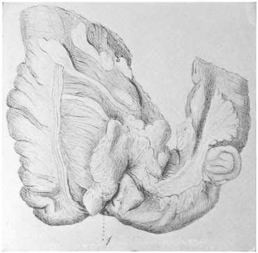
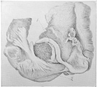
Fig. 572, giving the ventral view, shows, by the course of the ventral longitudinal muscular band, the turn of the cæcum upwards and to the left. The ventral cæcal artery runs in a fold (1) loaded with epiploic appendages.
The non-vascular intermediate fold (Fig. 573, 2) passes to the root of the appendix, joining the proximal segment of the dorsal vascular fold in which the dorsal branch of the ileo-colic artery runs to the tip of the appendix. The distal two thirds of the appendicular mesentery are free.
3. Gibbon, Hylobates hoolock (Figs. 455 and 456).—In the gibbon the folds appear well developed. The intermediate and dorsal vascular folds are quite distinct structures, although fusion (Fig. 456) has begun at one point, thus limiting a typical posterior ileo-cæcal fossa.
4. Gorilla, Gorilla savagei (Fig. 457).—Finally in the gorilla all three folds appear quite distinct and separate from each other, the dorsal vascular fold being especially well developed.
Unusual and Aberrant Types of Ileo-cæcal Folds and Fossæ.—(A) Ventral cæcal artery larger than the dorsal, supplying the greater part of the cæcum and the appendix.
Fig. 574.—Human adult. Cæcum and ileo-colic junction; well-developed ventral vascular fold, carrying appendicular artery. (Columbia University Museum, No. 1613.)
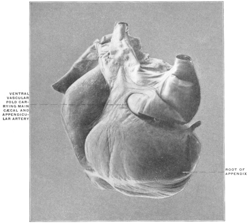
This condition is occasionally encountered. Dr. Martin, in a recent examination of the vascular supply of cæcum and appendix in one hundred subjects, found it to obtain in six instances.
Apparently the dorsal wall of the cæcum and of the proximal segment of the ascending colon remains free in these cases and does not become adherent to the parietal peritoneum. The shape of the pouch, moreover, indicates a free and unimpeded embryonal cæcal descent. The normal relative size of the two vascular folds is reversed. A good example of this variation, in the cæcum of an infant, is seen in Fig. 516. The same arrangement in an adult specimen is seen in Fig. 574.
In the Slow Lemur (Nycticebus tardigradus) (Fig. 420) the ventral artery is normally the larger of the two, extending in the ventral fold to the tip of the reduced appendix of the cæcal pouch.
(B) Fusion of ventral vascular fold with the intermediate fold, resulting in the production of a well-defined superior or ventral ileo-cæcal fossa.
Fig. 575.—Human fœtus at term. Ileo-colic junction and cæcum; ventral view. (Columbia University Museum, No. 1715.)
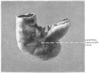
Normally the reduced ventral artery crosses the ileo-colic junction in a slightly developed ventral vascular fold, closely adherent to the intestine, with a very narrow free margin. The superior or ventral ileo-cæcal fossa in these cases is very shallow and confined (Fig. 574) to the ventral surface of the ileo-colic junction. Occasionally the fold is better developed and fuses with the intermediate non-vascular fold, producing a fossa of greater extent, which is bounded dorsad by the ileum, ventrad and cephalad by the ventral fold, caudad by the fusion of this fold with the intermediate reduplication, and to the right by the left wall of the cæcum. Figs. 576, 577, 578 and 579 show this aberrant disposition of the structures in a series of adult human cæca.
Fig. 576.—Human adult. Ileo-colic junction and cæcum; ventral appendicular artery and ileo-cæcal fossa. (Columbia University Museum, No. 1614.)
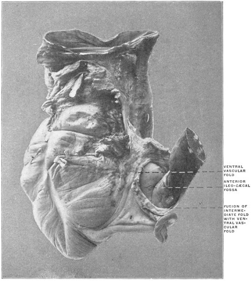
Fig. 577.—Human adult. Ileo-colic junction and cæcum; ventral appendicular artery and ileo-cæcal fossa. (Columbia University Museum, No. 1657.)
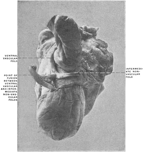
Fig. 578.—Human adult. Ileo-colic junction and cæcum; ventral appendicular artery and ileo-cæcal fossa. (Columbia University Museum, No. 1856.)
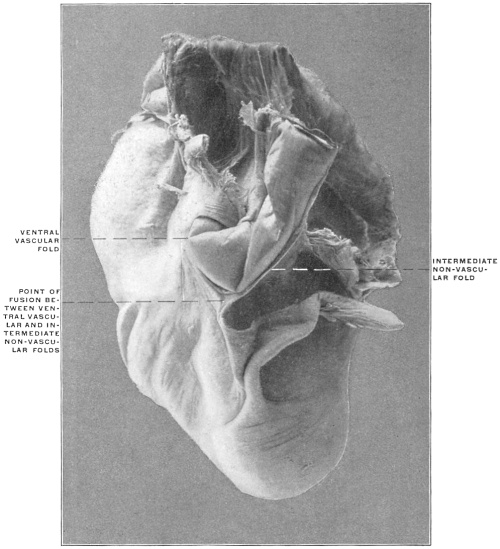
Fig. 579.—Human adult. Ileo-colic junction and cæcum; ventral appendicular artery and ileo-cæcal fossa. (Columbia University Museum, Study Collection.)
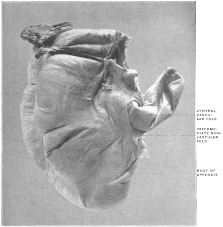
A corresponding arrangement is noted in the preparation of the cæcum of Cercopithecus campbellii (Fig. 433). The large intermediate fold is joined by the ventral vascular fold, thus defining the lower boundary of ventral ileo-cæcal fossa.
(C) Union of both vascular folds with the intermediate non-vascular fold.
Fig. 580.—Human infant. Ileo-colic junction and cæcum; fusion of ventral and dorsal vascular folds, with intermediate fold. (Columbia University Museum, No. 1663.)
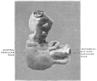
I have encountered one instance of this arrangement in an infant, whose cæcum and ileo-colic junction is shown in Fig. 580. Both the ventral and dorsal arteries in this case were equally developed, and shared equally in the supply of cæcum and appendix. Both vascular folds fused with the intermediate fold, thus producing two typical ileo-cæcal fossæ, one ventral, the other dorsal.
(D) Abnormal positions of the appendix due to variations in the arrangement and tension of the intermediate fold.
Fig. 510 shows a fœtal cæcum in the ventral view. The ventral vascular fold (3) is well developed. The non-vascular fold is short, arising from the ventral surface of the ileum, instead of from the free border of the intestine opposite to the mesenteric attachment. It fuses with the ventral vascular fold a short distance below the ileo-colic junction, thus limiting a small ventral ileo-cæcal fossa. The dorsal cæcal artery in this specimen was large, but the fold carrying it extremely narrow.
The preparation illustrates the type resulting from the reduction in size and extent of the non-vascular and mesenteric folds. The intermediate fold is reduced to a short and narrow band. Compared with the usual infantile type the cæcum lacks the characteristic turn upwards and to the left, possibly in consequence of the slight traction caused by the rudimentary intermediate fold. The pouch occupies a nearly vertical pendent position, which the appendix, arising from the lowest point of the cæcal funnel, shares. The appendix is not drawn into the retro-ileal position by the dorsal vascular fold, which is much reduced.
In Fig. 511, representing the cæcum and appendix of a fœtus at term, the effect of the tense non-vascular intermediate fold (2) is seen in the sharp turn to the left which it imparts to the nearly transversely directed funnel-shaped cæcum. The appendix (1) is coiled spirally for 1¾ turns behind the ileo-colic junction, with the tip directed upward behind the mesentery of the terminal ileum. The non-vascular intermediate fold (2) extends to the rest of the appendix. It appears short in its cæcal attachment, on account of the turn of the cæcum backwards and to the left and the close connection between the adjacent margins of the ileum and cæcum.
Fig. 581.—Human fœtus at term. Ileo-colic junction and cæcum. (Columbia University, Study Collection.) 1. Appendix, terminal portion turned ventrad of ileo-colic junction. 2. Intermediate non-vascular fold. Fig. 582.—Human infant. Ileo-colic junction and cæcum; ventral position of appendix. (Columbia University Museum, No. 693.)
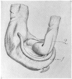
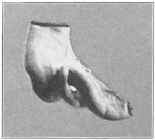
In Fig. 581—a fœtal preparation at term—the cæcum is turned to the left, below and behind the terminal ileum. The non-vascular fold (2) is well developed as regards length of ileal attachment, but is very narrow and tense, passing between ileum and the proximal curve of the cæcum behind the ileo-colic junction, where it merges with the dorsal vascular fold. The appendix takes a sudden turn caudad at this point and then continues up ventrad to the ileo-colic junction, the proximal portion being kept firmly in contact with the dorsal and caudal circumference of the ileum by the tension of the non-vascular band. It is quite evident that this peculiar turn of the appendix is directly due to the confining influence of the non-vascular band—which passes from its ileal attachment almost directly dorsad to the point of fusion with the dorsal vascular fold, causing the sharp downward and forward turn of the proximal segment of the appendix. Similar cases with ventral position of the appendix are shown in Figs. 545 and 582.