Part 2 — ANATOMY OF THE PERITONEUM IN THE SUPRA-COLIC COMPARTMENT OF THE ABDOMEN. (continued 3)
Reading edition
The main facts pertaining to the structure of the lesser peritoneal sac and its connection with the greater peritoneal cavity by means of the foramen of Winslow may be summed up as follows:
Fig. 297.—Schematic sagittal section of the ventral and dorsal mesogastria and epiploic bursa in a human embryo of eight weeks. (Modified from Kollmann.)
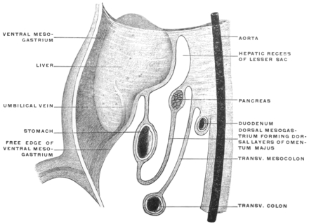
Fig. 298.—Transection of human embryo of 3 cm., vertex-coccygeal measure. (Kollmann.)
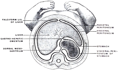
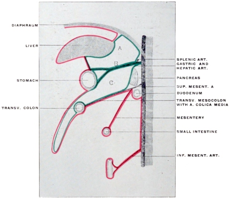
Fig. 299.—Schematic sagittal section of abdomen to illustrate the intestinal branches of the abdominal aorta. The gastric and hepatic arteries are shown for the sake of convenience as arising together from the cœliac axis (B), hence the left and right gastro-pancreatic folds carrying these vessels appear fused at their beginning, separating the hepatic recess of the lesser peritoneal sac (A) from the cavity of the omental bursa (C).
The mesogastrium as a whole, expanding originally in the sagittal plane in a fan-shaped manner between the vertebral column and the ventral abdominal wall, from the level of the umbilicus to the septum transversum (diaphragm), divides the cephalic part of the abdominal cavity into a symmetrical right and left half.
Figs. 172 and 273 represent the membrane as seen in a profile view from the left side. We distinguish the segment dorsad of the stomach as the dorsal mesogastrium, directly continuous with the remaining segments of the common primitive dorsal mesentery, while the portion ventrad of the stomach forms the ventral mesogastrium in which the liver develops. The segment of the ventral mesogastrium between liver and stomach becomes the lesser or gastro-hepatic omentum, while that between liver and ventral abdominal wall forms the falciform or suspensory ligament.
A transection, showing the dorsal and ventral mesogastrium at the level of the fundus of the stomach, is given in Fig. 298. The mesogastria are here seen to be short, while in the schematic Figs. 291 and 292 the membrane is, for the sake of distinctness, represented as being of considerable extent.
The ventral mesogastrium surrounding the liver and stomach extends caudad to include the first portion of the duodenum. Beyond this point it terminates in a thickened free edge which includes the umbilical vein. This vein extends from the umbilicus to the transverse fissure of the liver (Fig. 297), lying within the umbilical fissure on the caudal surface of the gland.
At the point where the vein enters the liver the thickened margin of the ventral mesogastrium is continued, as ligamentum hepato-duodenale, to the upper part of the duodenum and forms the ventral boundary of the foramen of Winslow. Between the layers of the mesogastrium which meet in this margin are situated the portal vein, biliary duct and hepatic artery, together with the nerves and lymphatics of the liver.
The mesogastrium originally divided the abdominal cavity between umbilicus and diaphragm into symmetrical right and left halves of equal size and extent. This early symmetrical arrangement becomes disturbed about the seventh week by the rotation of the stomach and the resulting altered course of the mesogastrium, which render the two original equal halves of the abdominal cavity unequal and asymmetrical. The original right half becomes placed behind the stomach and is converted into a blind sac with its opening directed to the right.
The communication of the general abdominal cavity with the retrogastric space by means of this channel is still wide in the embryo, but gradually becomes narrowed in the course of further development to form the foramen of Winslow. This opening is situated between the hepato-duodenal ligament and the parietal peritoneum covering the vena cava. It is constricted from below by the curve of the hepatic artery as this vessel passes from the cœliac axis to reach the liver at the transverse fissure between the layers of the lesser omentum.
The earlier developmental stages of the higher mammalian embryos are in general well illustrated by the permanent adult conditions found in some of the lower vertebrates, in which development does not proceed beyond the primitive condition.
In reptiles, birds and mammals the epiploic bursa is generally formed, while in amphibia the dorsal mesogastrium is very short and connects the stomach directly to the dorsal midline of the abdominal cavity without forming the sac-like extension of the great omentum.
The dorsal mesogastrium with the stomach, and the ventral mesogastrium including the liver between its layers, divides in these animals the cephalic part of the body cavity into two halves, corresponding to the earlier embryonic stages in man and in the higher mammalia.
The foramen of Winslow of the higher forms appears in the lower vertebrates as the wide-open space leading from below into the right half of the cœlom cavity. The dorsal mesogastrium remains short, not forming the pouch-like extension of the great omentum. The stomach retains more or less its primitive vertical position without rotation or elevation of the pyloric extremity, and the intestinal canal is simple, short and comparatively straight.