Part 3 — LARGE AND SMALL INTESTINE, ILEO-COLIC JUNCTION AND CÆCUM.
Reading edition
In considering the anatomy of the human cæcum and vermiform appendix many structural conditions are encountered which can only be correctly appreciated in the light of the physiology of the digestive tract. The alimentary canal as a whole affords one of the most striking examples of the adaptation of structure to function. The constant renewal of the tissues of the body by the absorption of nutritive material, the necessary concomitant egestion of undigestible remnants, the variety in the quantity and character of the food habitually taken, all serve to explain why the alimentary canal responds structurally in individual forms so completely to the physiological demands made upon it. This will become especially evident if we extend our observations to include, in addition to man, a review of the corresponding structures in representative types of the lower vertebrates. Moreover the human cæcum and appendix are in part rudimentary structures, representing a portion of the alimentary tract which, in accordance with altered conditions of food supply and nutrition, has lost its original functional significance to the organism and which consequently exhibits the wide range of structural variation which characterizes the majority of rudimentary and vestigial organs.
The vermiform process of man and the higher primates is thus one of several indications given in the structure of the alimentary canal (the character of the dentition is another example) which suggests that at one phylogenetic period the forms composing the order or their immediate ancestors were largely or entirely herbivorous, and hence possessed a more extensively developed cæcal apparatus than their omnivorous descendants of to-day. In approaching, therefore, the study of the human cæcum and appendix we will at once meet with conditions which call for the simultaneous physiological and morphological consideration of the adjacent small and large intestine.
Again many of the structural peculiarities which characterize the human cæcal apparatus can only be correctly valued by comparison with the corresponding parts in the lower vertebrates. Our inquiry will, therefore, most profitably include the following subdivisions of the subject:
I. General review of the functional and structural characters of the vertebrate large and small intestine.
II. Systematic consideration of the ileo-colic junction and the connected structures in the vertebrate series.
III. Phylogeny of the types of vertebrate ileo-colic junction and cæcum, and their probable lines of evolution.
IV. Detailed morphology of the human cæcum and vermiform appendix.
I. GENERAL REVIEW OF THE MORPHOLOGY AND PHYSIOLOGY OF THE VERTEBRATE INTESTINE.
We have seen that the intestinal tube of all vertebrates is the product of two of the embryonal blastodermic layers, the entoderm and mesoderm. The former furnishes the characteristic and cardinal elements of the digestive tract, viz., the secretory and absorbing epithelium of the mucous membrane and of the accessory digestive glands, the liver and pancreas.
From the mesoderm, on the other hand, are derived the muscular and connective tissue coats which surround the epithelial tube and contribute to the thickness of the intestinal wall, as well as the blood vessels and lymphatics. The alimentary canal separates from the yolk-sac of the embryo by the development of cranial, caudal and lateral folds, and at an early period communicates with the neural canal by the primitive postanal gut (cf. p. 23). This connection subsequently becomes lost. The oral and anal openings, by means of which the alimentary canal communicates with the exterior, are formed secondarily by entodermal invaginations which finally break through into the lumen of the canal (cf. p. 24).
At an early embryonic stage the alimentary canal appears therefore as a straight cylindrical tube running cephalo-caudad in the long axis of the body-cavity, and suspended by the primitive mesentery from the ventral aspect of the chorda dorsalis.
In Amphioxus, the cyclostomata, certain teleosts, dipnœans and lower amphibians the canal remains permanently in this condition (cf. Fig. 310).
In the remaining vertebrates the uniform non-differentiated tube of the embryo develops further and appears more or less distinctly divided into a proximal segment, the foregut, a central segment, the midgut, and a distal segment, the hindgut, or endgut. This differentiation of the tube into successive segments is closely connected with the character and quantity of the food habitually taken and with the method and rapidity of its elaboration in the process of digestion, absorption and excretion. In general the foregut is formed by the segment which succeeds to the oral cavity, and includes the pharynx, œsophagus and stomach. The midgut is composed of a longer or shorter narrower tube of nearly uniform caliber, the small intestine, which follows the gastric dilatation. Even in forms in which the stomach is not distinctly differentiated (cf. p. 40) the connection of the biliary duct with the intestinal canal serves to separate the fore- and midgut. The hindgut or large intestine is usually separated from the preceding segment by an external circular constriction, with a corresponding annular valve or fold of the mucous membrane in the interior.
The beginning of the large intestine is marked in many forms by the development of an accessory pouch or diverticulum, the cæcum. The hindgut extends from its junction with the midgut to the cloacal or anal opening.
1. Midgut or Small Intestine.
The small intestine is the segment of the alimentary canal in which digestion of the non-nitrogenous food substances takes place, and which affords the necessary area of mucous surface for the absorption of all digested matters. Consequently the character and habitual quantity of the food here elaborated exerts a very marked influence on the length of the small intestine, i. e., on the extent of the digestive and absorbing surface represented by its mucous membrane.
The relative length of the small intestine in any individual form will vary with both the quantity and volume of the food and with the rapidity of the metabolic processes. Animals, in which digestion is rapid and the usual food small in bulk and concentrated in its nutrient qualities, have a relatively short intestine, while the canal is longer in forms subsisting on food which is bulky and which demands considerable time for its elaboration. Hence we find the relatively shortest intestine in carnivora, the longest in herbivora, while the canal in omnivora occupies an intermediate position in regard to its relative length.
The rapidity of tissue-metabolism also exerts a marked influence on the length and development of this portion of the alimentary canal.
In the warm-blooded animals (mammals and birds) the tissue-changes are constant and rapid and call for a large amount of nutrition within a given period, while the metabolic processes in the cold-blooded vertebrates (reptiles, amphibia and fishes) are slow, these animals being able to go without food for long periods. Consequently in the former class the small intestine is relatively much longer than in the latter. Thus in certain birds and herbivorous mammals the small intestine exceeds the total length of the body many times. This influence of the quantity and quality of the food on the length of the intestinal canal is seen, for example, very well during the course of development in the frog.
The increase in the length of the intestine, and the consequent varying degrees of coiling and convolution, are therefore secondary acquired characters, depending for their development upon the habitual kind and volume of the food. Additional provisions for increasing the efficiency of the digestive apparatus are encountered throughout the whole of the intestinal canal. In many forms the digestive secretory and absorbing area is augmented by the development of folds, valves, diverticula, villi and papillæ from the mucous surface of the intestine. Certain valves and folds, moreover, both control the direction in which the contents of the canal move and retain the same for a longer period in the intestinal segment in which they develop. Such folds appear especially well developed in the intestine of certain cyclostomes, selachians and dipnœans (cf. Figs. 203 and 204). In these forms the alimentary canal is usually short and straight, and the fold which has a typical spiral course and projects far into the lumen of the gut, evidently makes up to a very large extent for the shortness of the intestine, serving the threefold purpose of
(a) Increasing the digestive and absorbing surface;
(b) Prolonging the period of retention of the food-substances in the intestine, and thus increasing the time available for elaboration and absorption.
(c) Regulating the direction in which the intestinal contents move.
We will see presently that a similar spiral mucous fold is also encountered in some of the higher vertebrates, especially in the large intestine. Examples are found in the well-developed spiral valve in the cæca of the ostrich (Fig. 341), the similar fold in the large intestine of many rodents (Figs. 387 and 388) and in the crescentic plicæ of the primate large intestine (Figs. 471, 472 and 473).
To the same physiological category belong the digestive diverticula of the intestinal canal, such as the pyloric appendices of the midgut found in many teleosts and ganoids (cf. p. 119) and the varieties of cæca or blind diverticula of the hindgut encountered throughout the vertebrate series. They all function as reservoirs which increase the available digestive and absorbing surface and which in addition are especially adapted to retain substances difficult of digestion until the processes of elaboration have been completed.
Divisions of the Small Intestine.—In the higher forms the segment of the small intestine which succeeds to the pylorus is distinguished as the duodenum. Into it empty the ducts of the liver and pancreas. In some animals a pear-shaped enlargement is found, corresponding to the duodenal antrum of the human intestine, as the dilated proximal portion of the duodenum immediately beyond the pylorus is called. Examples of this condition are furnished by the cetaceans, several rodents, the llama and dromedary and the koala (Phascolarctos).
In the birds and in many mammals (e. g., dog, Fig. 200, and many rodents, as the rabbit) the duodenum is drawn out into a long loop surrounding the pancreas.
Structure of the Small Intestine. 1. Secretory Apparatus.—The glands whose ducts empty into the small intestine and which furnish the digestive secretions, may be divided as follows:
(a) Glands situated in the substance of the intestinal walls.
Two kinds are distinguished:
1. Brunner’s glands, small acinous glands confined to the first part of the duodenum.
2. Glands of Lieberkühn, small cæcal pits distributed not only over the entire small intestine, but also found in the mucous membrane of the large intestine.
These structures furnish the intestinal juice, whose chief function is the conversion of starches into sugar, while aiding in carnivorous animals also the digestion of proteid substances. The glands are hence best developed in herbivora, while in carnivora they are present in diminished numbers since they assist in the digestion of proteid substances.
The size and number of these glands also depends on the amount of food digested within a given period. When a considerable quantity of digestive fluid is required, in order to obtain the nutritive value of the food for the organism rapidly, the glandular apparatus of the intestine will be well developed. Hence mammalia, in whom these conditions exist, possess both the glands of Brunner and of Lieberkühn. In birds the latter structures are still found, but the former are absent, while amphibia and fishes are devoid of both kinds. In these lower vertebrates the typical intestinal glandular apparatus of the higher forms is to a certain extent replaced by small pits and depressions of the mucous membrane bounded by reticular folds.
(b) Glands situated outside the intestinal tube, into whose lumen their ducts empty.
The liver and pancreas fall under this head. The liver functions in the digestion of the fatty substances of the food, while the secretion of the pancreas converts the starches into sugars, and aids in the digestion of albumenoid substances and to a lesser degree in that of the fats.
2. Absorbing Apparatus of Small Intestine.—The mucous membrane of the intestine is provided with villi, containing lymphatics, by whose agency the digested matters are absorbed. These structures are developed in individual forms in direct proportion to the ease and rapidity with which the food is habitually absorbed.
The more rapid and complete the digestion is the greater will be the amount of digested nutritive material at any given time in the intestine, and the greater will be the development of the absorbing structures. Hence the villi of the small intestine are especially large and prominent in the carnivora, while they are small and insignificant in herbivora and omnivora. Intestinal villi are found in nearly all mammals and in many birds. Fig. 300 shows the villi of the intestinal mucous membrane in a carnivore mammal (Ursus maritimus, polar bear) and Fig. 301 the same structures in the cassowary (Casuarius casuarius) in which bird they are very well developed. The villi are not confined to the two highest vertebrate classes, but are encountered also in the mucous membrane of the midgut in certain reptiles, notably the ophidia.
Fig. 302 shows the intestine mucous membrane of the boa constrictor with well-developed and prominent villous projections.
Fig. 300.—Small intestine, of polar bear, Ursus maritimus. Mucous surface. (Columbia University Museum, No. 782.) Fig. 301.—Duodenum, with entrance of pancreatic and biliary ducts and well-developed diverticulum Vateri in the cassowary, Casuarius casuarius (Columbia University Museum, No. 1821.) Fig. 302.—Mucous membrane of midgut of Boa constrictor. (Columbia University Museum, No. 1837.)
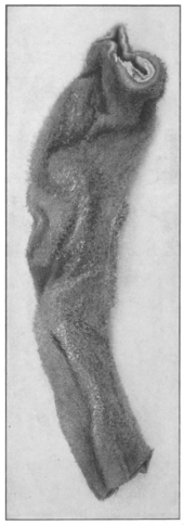
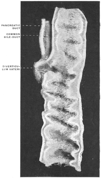
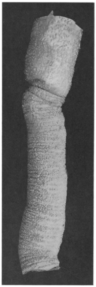
Fig. 303.—Mucous surface of small intestine of a species of African antelope, Cervicapra arundinacea. (Columbia University Museum, No. 1843.) Fig. 304.—Mucous surface of small intestine of Phocæna communis, porpoise. (Columbia University Museum, No. 1057.)
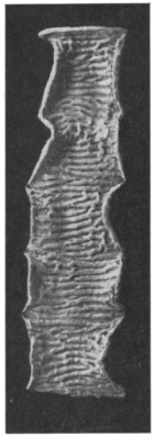
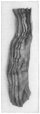
Fig. 305.—Mucous membrane of mid-gut of Lophius piscatorius, the angler, 18 cm. caudad of pylorus. (Columbia University Museum, No. 1838.)
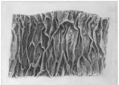
Figs. 306, 307.—Intestinal mucous membrane of logger-head turtle, Thalassochelys caretta. (Columbia University Museum, No. 1839.)
Fig. 306.—Mid-gut. Fig. 307.—End-gut.
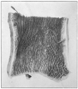
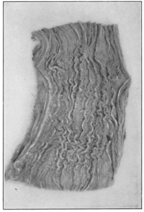
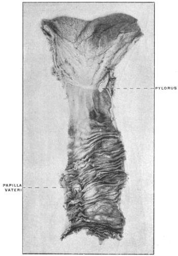
Fig. 308.—Adult human subject. Mucous membrane of pyloro-duodenal junction and of duodenum. (Columbia University Museum, No. 1840.) Fig. 309.—Adult human subject. Mucous membrane of small intestine, showing arrangement of valvulæ conniventes in successive portions of jejunum and ileum. (Columbia University Museum, No. 1841.)
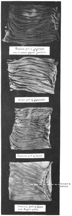
Some birds, such as the snipes, herons and crows, have in place of the intestinal villi projecting folds of the mucosa, often arranged in a reticular manner. This type is prevalent in amphibia and fish (Fig. 112, distal segment of midgut). Collections of lymphoid tissue in the mucous membrane of the small intestine, either aggregated to form Peyer’s patches (Fig. 309) or as solitary follicles, are only found in the two highest vertebrate classes, birds and mammals. In the former they appear scattered over the surface of the mucous membrane, in the latter they may be arranged in aggregations or regular rows. They are not secreting structures, but their exact function in absorption is not known. This lymphoid or adenoid tissue in certain forms is especially well developed at the ileo-colic junction, forming the lymphatic sac of some rodents, as lepus (cf. Fig. 386). It is not confined to the small intestine, but is found in the large intestine as well. At times it appears especially well developed in the terminal portion of the cæcal pouch (appendix), as in Lepus (Fig. 388).
The valvulæ conniventes or valves of Kerkring of the human small intestine serve to very greatly increase the secreting and absorbing mucous surface. They are not found in this complete development in any other mammals, although a very few forms present a transverse reduplication of the intestinal mucosa and the circular layer of muscular fibers. An example of this is found in the intestinal mucous membrane of a species of antelope, shown in Fig. 303.
The complete development of the valvulæ conniventes in man is possibly also associated with a mechanical function in connection with the upright posture. In some mammalia, as in certain rodents and the porpoise (Fig. 304), the mucous membrane of the terminal part of the small intestine is thrown into longitudinal folds.
The mucosa of the midgut in the lower vertebrates may be smooth, or thrown into longitudinal folds, or the longitudinal folds may become connected by oblique and transverse secondary folds, resulting finally in a more or less complicated reticulated pattern of crypts. A very good example of the type-form from which the more complicated conditions are derived is seen in Fig. 305, showing the mucous membrane of the midgut in Lophius piscatorius, the angler. The specimen is taken 18 cm. from the pylorus and shows a ground plan of longitudinal plicæ connected by short oblique cross folds.
Fig. 306, showing the midgut mucosa of the loggerhead turtle (Thalassochelys caretta), exhibits the same arrangement further developed, resulting in a fine reticulated pattern, while in the endgut of the same animal the primitive longitudinal folding is resumed (Fig. 307).
The number and size of the human valvulæ conniventes vary in different parts of the small intestine (Fig. 309). They are not usually found in the beginning of the duodenum (Fig. 308), but commence in the second or descending portion.
They become very large and closely packed immediately beyond the common entrance of the biliary and pancreatic ducts and continue to be well developed and numerous throughout the rest of the duodenum and upper half of the jejunum (Figs. 308 and 309). From here on they become smaller, more irregular and less closely packed, and finally in the terminal two feet of the ileum disappear almost entirely (Fig. 309). This varying development of the valvulæ is the chief reason why a given segment of the ileum weighs less than a corresponding length of the jejunum. This reduction in the fold-formation of the intestinal mucosa toward the terminal portion of the midgut is seen even in the lower vertebrates. Thus in Fig. 112, showing the entire intestinal tract of the conger eel, Echelus conger, in section, the plicæ of the mucous membrane in the proximal segment of the midgut, at and immediately beyond the entrance of the biliary duct, are prominent and numerous. This redundancy continues but slightly reduced in the descending limb of the intestinal loop, while in the ascending limb and up to the ileo-colic junction the folds are reduced to a fine reticulated meshwork. Beyond the ileo-colic valve plate, in the short endgut, the mucosa again presents numerous pointed reduplications.
II. ENDGUT OR LARGE INTESTINE.
In this segment of the intestinal canal the undigested remnants of the food are collected and evacuated from time to time.
In addition, the mucous membrane of the large intestine absorbs all digested material which is passed from the small intestine. While digestion of food-substances will not be inaugurated in the large intestine, material already in the process of digestion and mixed with the intestinal juices of the preceding segment, will be further elaborated in this portion of the canal and the nutritive products absorbed. This is especially the case in herbivora and omnivora, whose food is bulky, containing a large amount of refuse material, and is hence only slowly digested. On the other hand the food of the carnivora is easily and rapidly digested and absorbed. After passing through the small intestine hardly any substances remain which are capable of digestion and absorption. Hence the large intestine of herbivora and omnivora is uniformly longer in proportion to the small intestine than it is in carnivorous animals. In the former this segment of the canal functions as an accessory digestive apparatus and hence, as we will see, often develops accessory structural modifications, such as a large cæcum and spiral colon, while in the latter it acts almost solely as a canal for the evacuation of the indigestible remnants.
Again, the large intestine is better developed in the higher animals, in mammalia and to a lesser degree in birds, in whom the functional demands for nutrition are active and require that a relatively large amount of food should pass through the digestive tract in a given time. On the other hand in the lower cold-blooded vertebrates the metabolism is less active, less food is taken and it is not necessary to secure all the nutrient material contained in the same for the organism. The great differences observed in the vertebrate series in regard to length, width and structure of the large intestine depend upon these physiological conditions. The divisions of the human large intestine into cæcum, ascending, transverse and descending colon, sigmoid flexure and rectum are found only in the primates, and here not uniformly.
In the lower vertebrate classes the endgut is very short, corresponding only to the pelvic segment of the Mammalia (rectum), a colon proper being absent in these forms (cf. Fig. 112, Echelus conger). The human large intestine exhibits a very characteristic structure. Throughout the greater part of the colon the longitudinal muscular layer is mainly disposed in the form of three bands or tænia (ligamenta coli). The canal itself is longer than these bands, thus producing a folding of the walls in the form of three rows of pouches (cellulæ coli), in the intervals between the bands. The pouches of each row are separated from each other externally by constrictions, internally by projecting crescentic folds (plicæ coli) (Figs. 471, 472 and 474).
This arrangement of the large intestine is also found in the monkeys (Fig. 473) and in certain Rodents (Fig. 474).
In other mammals the large intestine is smooth and cylindrical and the longitudinal layer of muscular fibers uniform (Fig. 475).
In general the vertebrate large intestine is wider than the small, usually in the proportion of 5:1 or 6:1.
In some ruminant Herbivora, however, the great length of the colon leads to a reduction of the caliber in certain segments so that the large intestine does not exceed the width of the small, or even falls below the same.
The length of the large intestine, as in man, is usually much less than that of the small intestine. As already stated this disproportion is more marked in Carnivora than in Herbivora.
The ratio in length of the large to the small intestine is very low in the Seals (1:14), and in several Edentates, as Myrmecophaga, Tamandua and Bradypus (1:9-11).
In the carnivorous mammals it ranges 1:5-7.
In some of the ruminant Herbivora, as the cow and sheep, it is 1:4, while in the deer, horse, certain Rodents (as Lepus and Cricetus) it reaches as high as 1:2 or 1:3.
The large intestine is usually relatively short in birds, reptiles, amphibia and fish.
In the Cassowary the length of the large to the small intestine is 1:6.
In some of the birds of prey (eagle) the proportion falls as low as 1:68 or 70.
Exceptions to the general rule are furnished by some of the herbivorous Cetaceans and by the Dugong (Halicore) in whom the large intestine is twice as long as the small. Again in the Ostrich the large intestine in one example measured 40′, while the length of the small intestine was only 22′. This unusual development of the large intestine indicates the necessity of retaining the food, which is bulky and difficult of digestion, until the elaboration is completed. The same significance belongs to the enormously developed cæca of these birds (cf. p. 204).
The separation of the small and large intestine is marked externally by the cæcum, when present, and internally by the valve of the colon. The details of the vertebrate ileo-colic junction will be considered in the following pages.
II. SERIAL REVIEW OF THE ILEO-COLIC JUNCTION AND CONNECTED STRUCTURES IN VERTEBRATES.
I. FISHES.
In the Cyclostomata there is no differentiation between the mid- and hindgut. Fig. 310 shows the entire alimentary canal of Petromyzon marinus, the lamprey, caudad of the pericardium.
In some fishes the midgut is differentiated from the hindgut by an external circular constriction, corresponding to an annular projecting fold of the mucosa in the interior which resembles the pyloro-duodenal valve. There is no cæcum, and the short hindgut empties into the cephalic and ventral aspect of the cloaca. Fig. 311 shows the entire intestinal tract of a Teleost fish, Echelus conger, the conger eel. The midgut, provided at the beginning with a short globular pyloric appendix (cf. p. 119), constitutes the longest individual segment of the canal. The hindgut, separated from the preceding by a constriction, is very short and of large caliber. Fig. 312 shows the broad annular valve with central circular opening which separates mid- and hindgut in the interior, and Fig. 313 the ileo-colic junction in section in the same animal.
Fig. 310.—Petromyzon marinus, lamprey. Entire alimentary canal below pericardium. (Columbia University Museum, No. 1575.) Fig. 311.—Echelus conger, Conger eel. Alimentary canal, stomach, mid- and hindgut, liver, and spleen. (Columbia University Museum, No. 1430.)
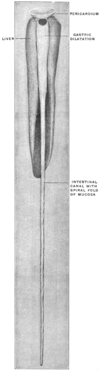
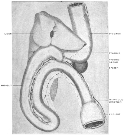
Fig. 312.—Echelus conger, Conger eel. Ileo-colic junction, opened. (Columbia University Museum, No. 1434.)
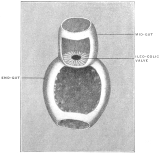
Fig. 313.—Echelus conger, Conger eel. Section of mid- and end-gut, with ileo-colic junction, hardened. (Columbia University Museum, No. 1349.) Fig. 316.—Galeus canis, dog-shark, ♂. Genito-urinary tract and cloaca in situ. (Columbia University Museum, No. 1694.)
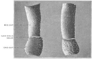
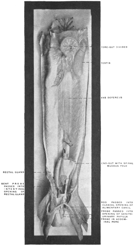
Fig. 314.—Gadus callarias, cod-fish. Ileo-colic junction. Intestine on each side opened, with probe passed through constricted opening of ileo-colic valve. (Columbia University Museum, No. 1260.)
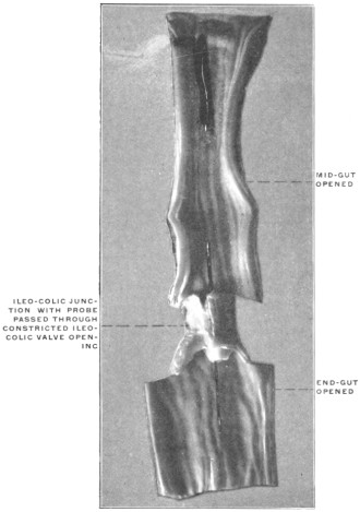
Fig. 315.—Pleuronectes maculatus, flounder. Ileo-colon, opened to show ileo-colic valve. (Columbia University Museum, No. 1493.) Fig. 317.—Accipenser sturio, sturgeon. Alimentary canal. (Columbia University Museum, Nos. 1826, 1827, and 1828.)
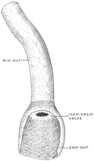
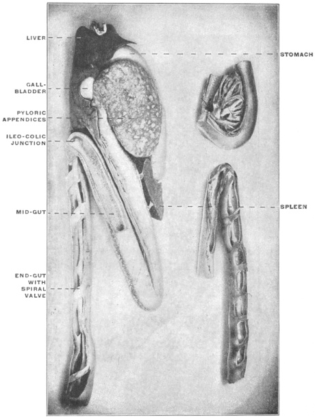
A similar type of ileo-colic junction is seen in other Teleosts, as in Gadus callarias, the cod (Fig. 314), Pleuronectes maculatus, the flounder (Fig. 315), and in some Ganoids, as Accipenser sturio, the sturgeon (Fig. 212). In some Selachians an appendicular diverticulum, the so-called “rectal” or “digitiform gland,” is found connected with the terminal segment of the gut near the entrance of the same into the cloaca (Fig. 316).
II. AMPHIBIA.
The alimentary canal is simple and usually comparatively short. There is no cæcal pouch. Differentiation of mid- and endgut is usually marked externally by a constriction and by the increased caliber of the terminal intestinal segment.
Fig. 318.—Rana catesbiana, bull-frog. Alimentary canal and appendages. (Columbia University Museum, No. 1454.) Fig. 319.—Necturus maculatus, mud-puppy. Alimentary canal and appendages. (Columbia University Museum, No. 1582.) Fig. 320.—Cryptobranchus alleghaniensis, hellbender. Ileo-colic junction. (Columbia University Museum, No. 1711.)
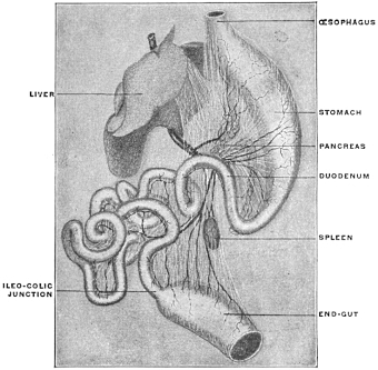
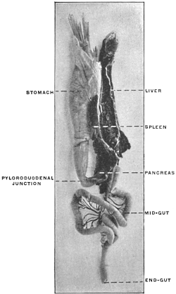
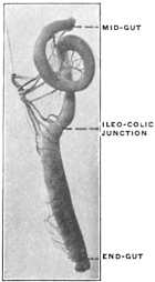
Fig. 318 shows the alimentary canal of the bull-frog, Rana catesbiana, Fig. 319 that of a Urodele Amphibian, Necturus maculatus, and Fig. 320 the ileo-colic junction isolated in Cryptobranchus alleghaniensis, the hellbender.
III. REPTILIA.
In reptiles a well-marked differentiation of small and large intestine is the rule.
Four types of ileo-colic junction are encountered in this class:
1. The transition from small to large intestine is marked by the greatly increased caliber of the latter and by an annular valve in the interior. An example of this type is furnished by Alligator mississippiensis (Fig. 321) and a similar form is encountered in some lizards, as Heloderma suspectum, the Gila monster (Fig. 322).
Fig. 321.—Alligator mississippiensis, alligator. Ileo-colon; dried preparation. (Columbia University Museum, No. 179.) Fig. 322.—Heloderma suspectum, Gila monster. (Columbia University Museum, No. 69/1536.)
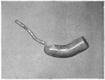
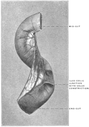
2. The large intestine immediately beyond the ileo-colic junction protrudes along the convex border to form a rudimentary lateral cæcum. This type is found in many Chelonians, e. g., in Pseudemys elegans, the pond turtle (Figs. 323 and 324) and Chelydra serpentaria, the snapping turtle (Fig. 325).
Fig. 323.—Pseudemys elegans, pond-turtle. (Columbia University Museum, No. 1069.) Fig. 325.—Chelydra serpentaria, snapping turtle. Intestinal canal, pancreas, and spleen. (Columbia University Museum, No. 1369.)
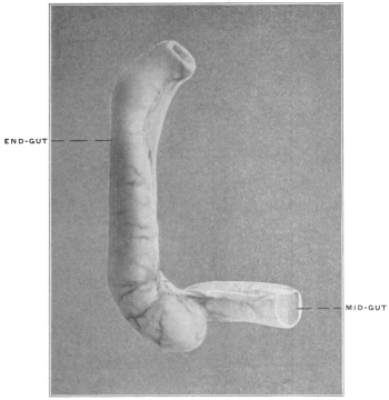
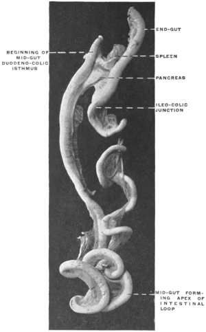
Fig. 324.—Pseudemys elegans, pond-turtle. Ileo-colic junction, opened. (Columbia University Museum, No. 1524.)
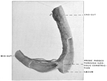
Fig. 326.—Iguana tuberculata, iguana. Ileo-colic junction and cæcum; dried preparation. (Columbia University Museum, No. 243.)
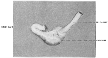
3. The ileo-colic junction is provided with a well-developed sacculated cæcal pouch derived from the proximal segment of the colon and divided in the interior by folds into several secondary compartments.
This type is found in some of the phytophagous lizards, as Iguana tuberculata (Figs. 326 and 327). The small intestine of this animal is of considerable length and of uniform caliber from the pylorus to the ileo-colic junction. The cæcum is a large sacculated pouch developed chiefly along the convex border of the large intestine opposite to the mesenteric attachment.
Fig. 327.—Iguana tuberculata, iguana. Mid-gut, ileo-colic junction, cæcum, and end-gut; dried preparation. (Columbia University Museum, No. 178.) Fig. 329.—Drawing taken from same preparation (No. 1321) to elucidate more clearly internal structure of cæcal pouch.
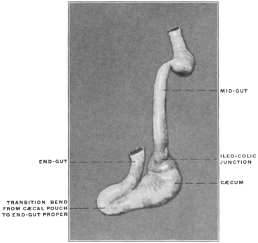
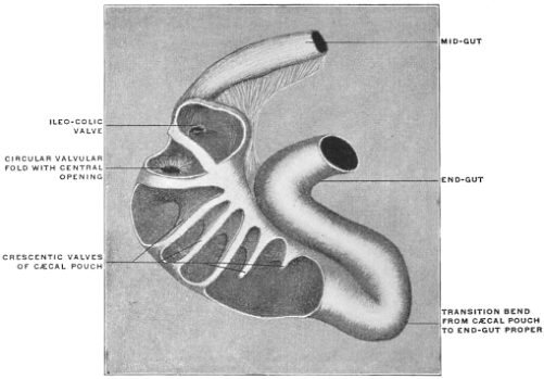
Fig. 328.—Iguana tuberculata, iguana. Ileo-colic junction and cæcum in section. (Columbia University Museum, No. 1321.) Fig. 330.—Cyclura teres, smooth-backed cyclura. Ileo-colic junction and cæcum in section. (Columbia University Museum, No. 1523.)
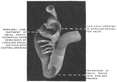
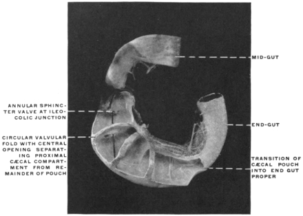
The examination of the interior of this pouch reveals a complicated structure (Figs. 328 and 329). Fig. 330 shows the same structures in a closely allied form, Cyclura teres. The entrance of the small intestine is guarded by an annular sphincter valve, whose central circular opening leads into a proximal compartment of the cæcum. This compartment is in turn separated from the remainder of the cæcal pouch by a second circular valvular fold with central opening. Beyond this valve the interior of the pouch carries a number of crescentic mucous folds, corresponding to the external constrictions between the cæcal sacculations. The entire pouch gradually diminishes in caliber and finally passes with a sharp angular bend into the terminal portion of the endgut. At this point the lumen of the canal is slightly diminished by a sphincter-like thickening of the muscularis, producing an annular projection of the mucous membrane. The entire cæcal pouch appears as a specialized segment of the large intestine interposed between the termination of the midgut and the terminal portion of the endgut, which latter is characterized by uniform caliber and increased thickness of the muscular walls.
The highly developed and complicated structure of the cæcal apparatus in Iguana and allied forms exemplifies very clearly the influence of vegetable food on the development of this segment of the alimentary tract when compared with the simple type of ileo-colic transition found in carnivorous lizards, as Heloderma (Fig. 322). Iguana subsists on leaves, fruits and other vegetable matter and the cæcal pouch is invariably found filled with the firmer and less digestible portions of this food. These are undoubtedly retained in the pouch by the series of valves and folds until digestion and absorption of all available nutritive material forwarded from the small intestine is completed. On the other hand Heloderma lives almost entirely on bird eggs, a concentrated and easily digested food. Consequently the ileo-colic junction in this lizard is exceedingly simple and rudimentary, marked merely by a slight external constriction, with an annular valve in the interior, and an increase in the caliber of the short hindgut, resembling the form found in many teleost fishes.
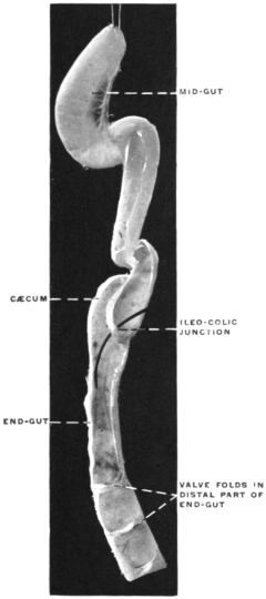
4. Finally in some Ophidians a typical lateral cæcal pouch of considerable dimensions is found connected with the endgut immediately beyond the ileo-colic junction.
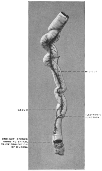
An example of this reptilian type, closely resembling the corresponding structure in many Mammalia, is presented by Eunectes marinus, the anaconda, shown in Figs. 331 and 332.
Fig. 331.—Eunectes marinus, anaconda. Mid- and end-gut, with ileo-colic junction and cæcum. (Columbia University Museum, No. 72/1535.) Fig. 332.—Eunectes marinus, anaconda. Mid- and end-gut, with ileo-colic junction and cæcum laid open. (Columbia University Museum, No. 1709.)
IV. ILEO-COLIC JUNCTION IN BIRDS.
In the birds the length of the intestine is subject to great variations. The canal is short in species subsisting on fruits and insects, long in those feeding on seeds, plants and fish. The large intestine, immediately beyond the ileo-colic junction, is provided typically with two symmetrical lateral cæca which extend in some forms for a considerable distance cephalad on each side of the small intestine to which they are bound by peritoneal connections.
Fig. 333.—Buteo harloni, black hawk. Ileo-colic junction and cæca. (Columbia University Museum, No. 1502.) Fig. 334.—Phalacrocorax dilophus, double-crested cormorant. Ileo-colic junction and cæca. (Columbia University Museum, No. 67/1534.)
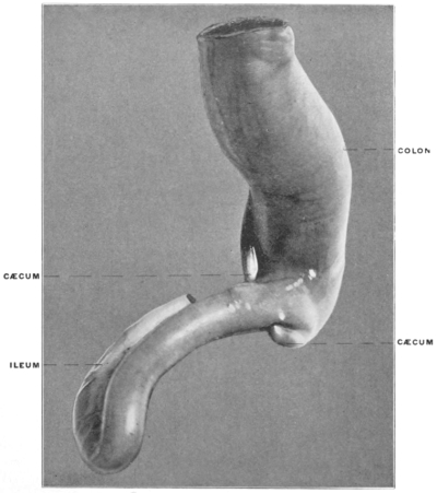
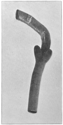
As a rule carnivorous birds have short and rudimentary pouches (Figs. 333 and 334), whereas they are long in herbivorous forms (Figs. 335 and 336). Some carnivorous birds, as Corvus, Strix, etc., have fairly long cæca (Fig. 337). In the passerine birds living on seeds and insects, the cæca are of considerable length as they are also in some of the piscivorous divers (Figs. 338 and 339). They are long in the Ratitæ, and in the Lamellirostra, who feed chiefly on plants (Fig. 340).
Fig. 335.—Gallus bankiva, hen. Ileo-colic junction and cæca. (Columbia University Museum, No. 1486.) Fig. 337.—Bubo virginianus, great horned owl. Ileo-colic junction and cæca. (Columbia University Museum, No. 672.)
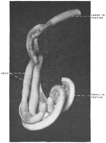
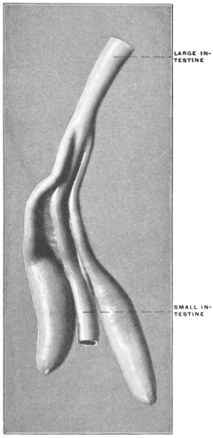
Fig. 336.—Chen hyperborea, Canada snow-goose, Ileo-colic junction and cæca. (Columbia University Museum, No. 47/1448.) Fig. 338.—Urinator lumme, red-throated loon. Ileo-colic junction and cæca. (Columbia University Museum, No. 1001.)
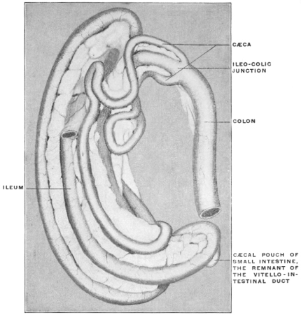
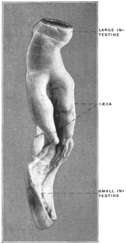
Fig. 339.—Merganser serrator, red-breasted merganser. Ileo-colic junction and cæca. (Columbia University Museum, No. 1798.) Fig. 340.—Casuarius casuarius, cassowary. (Columbia University Museum, No. 1799.)
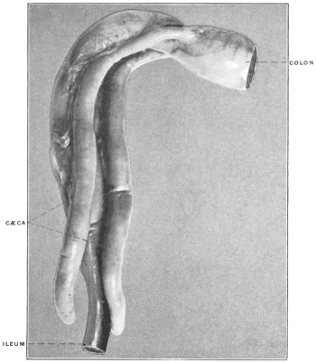
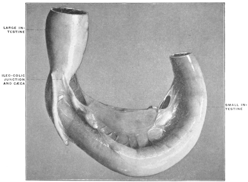
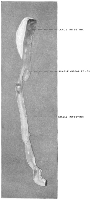
Fig. 341.—Struthio africanus, African ostrich. Ileo-colic junction and cæca. (Columbia University Museum, No. 48/1573.) Fig. 342.—Ardea virescens, green heron. Ileo-colic junction and cæcum. (Columbia University Museum, No. 1132 a.)
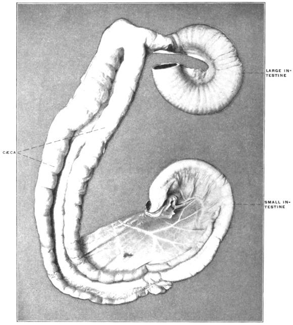
The enormously elongated cæca of the African ostrich contain a spiral fold of the mucous membrane in the interior (Fig. 341).
In place of the usual double avian cæcum a single pouch is found in a few forms, namely in the Herons (Fig. 342).
Fig. 343.—Urinator lumme, red-throated loon. Small intestine with cæcal pouch; the remnant of the vitello-intestinal duct. (Columbia University Museum, No. 997.) Fig. 344.—Urinator imber, great northern diver. Small intestine with cæcal pouch; the remnant of the vitello-intestinal duct. (Columbia University Museum, No. 78/1573)
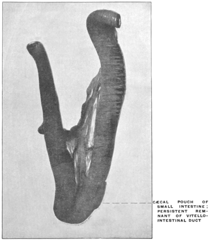
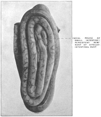
In some birds the small intestine is also provided with a cæcal pouch, the remnant of the vitello-intestinal duct corresponding in its significance to the occasional mammalian diverticulum of Meckel (Figs. 343 and 344). (cf. p. 35.)
V. ILEO-COLIC JUNCTION, CÆCUM AND VERMIFORM APPENDIX IN THE MAMMALIA.
I. Subclass: Ornithodelphia.
I. Order: Monotremata.
In many particulars the anatomical structure of these animals reveals a close relationship to the Sauropsida. They represent the mammalian class in its lowest stage of evolution.
The ileo-colic junction in all the existing forms is direct, without angular bend at the entrance of the small into the large intestine. The cæcum is a long narrow pouch, slightly dilated at the extremity, derived from the beginning of the colon and extending backward along the free margin of the small intestine. The cæcum resembles in its general shape and structure the pouches seen in many birds, except that it is unilateral, while the birds normally have two symmetrical cæca. The cæcum of Ornithorhynchus anatinus, the platypus or duck bill, is shown in Figs. 345 and 346, and that of Echidna hystrix, the spiny ant-eater, in Fig. 347. These two animals represent the two genera into which the order is divided.
Fig. 345.—Ornithorhynchus anatinus, duck mole. Ileo-colon and cæcum. (Columbia University Museum, No. 1499.) Fig. 346.—Ornithorhynchus anatinus, duck mole. Ileo-colon and cæcum. (Columbia University Museum, No. 1500.)
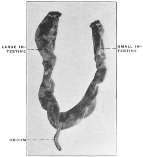
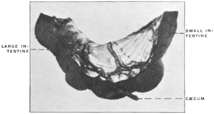
Fig. 347.—Echidna hystrix, spiny ant-eater. Ileo-colon and cæcum. (Columbia University Museum, No. 1501.)
II. Subclass: Didelphia.
II. Order: Marsupialia.
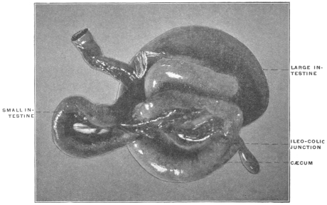
Fig. 348.—Didelphis virginiana, opossum. Ileo-colic junction and cæcum. (Columbia University Museum, No. 1533.)
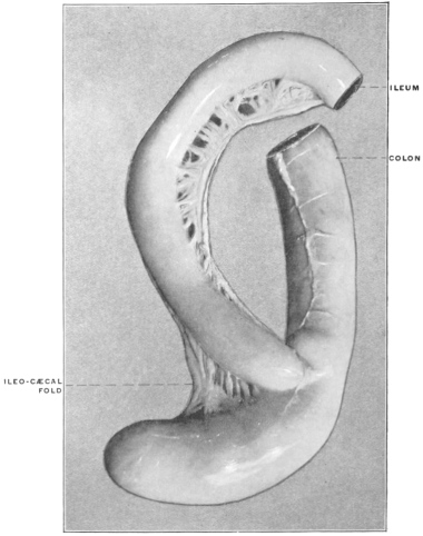
The Didelphia are represented by numerous species, which are united by certain common anatomical characters of the reproductive organs and dentition to form the order of the Marsupialia. The individual species included within this order differ widely in abit, food, mode of locomotion, etc., and consequently exhibit great diversity in the structure of the skeletal and muscular systems and of the alimentary canal. With the exception of the Opossums inhabiting the new world, the families composing the order are confined to the Australian continent and the adjacent islands. In respect to the alimentary tract in general and the ileo-colic junction in particular, we are evidently dealing with a group of animals which, while they retain the common characters above indicated as uniting them in the marsupial order, yet have in the structure of their digestive canal adapted themselves to widely divergent conditions of food supply and environment. Consequently within the confines of this single and largely isolated order, we encounter nearly all the types of cæcum and ileo-colic junction which are found among the remaining mammalia. The group in its individual representatives has passed, so to speak, through the different stages of development and evolution which, on a very much larger scale, are exhibited by the remaining mammalian orders.
We can, independently of the systematic zoölogical classification, arrange the forms composing the order under the following types:
1. Forms with large well-developed simple cæca, of uniform caliber, with rounded globular termination.
This type is encountered among the herbivorous Marsupials, such as the opossums, kangaroos and wallabys. Fig. 348 shows the structures in Didelphis virginiana, the common opossum, Fig. 349 in a small species of opossum from Trinidad, and Fig. 350 the same parts in Halmaturus derbyanus, the rock wallaby.
Fig. 349.—Didelphis sp.? opossum. Ileo-colic junction and cæcum. (Columbia University, Study Collection.) Fig. 350.—Halmaturus derbyanus, rock kangaroo. Ileo-colic junction and cæcum. (Columbia University Museum, No. 727.)
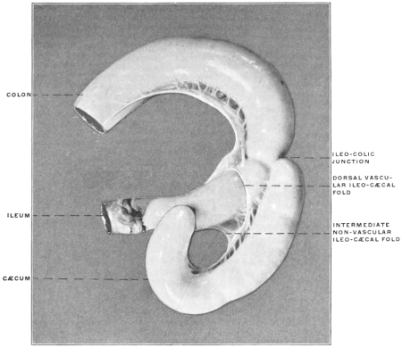
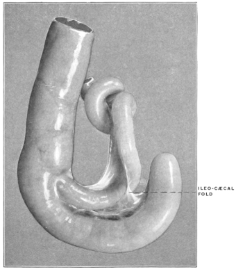
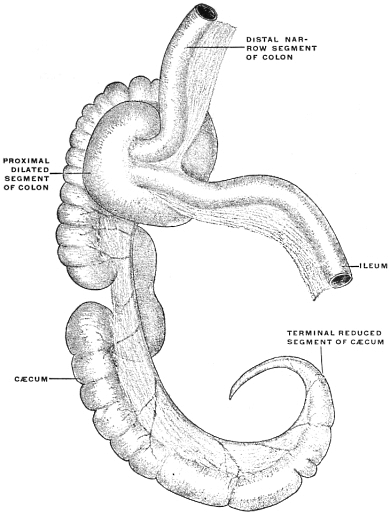
2. Forms with enormously developed sacculated cæca, coiled spirally, with or without additional convolutions of the proximal colon; the terminal portion of the cæcal pouch diminishes in caliber to form a pointed appendage.
This type of cæcum characterizes the Phalangeridæ or Phalangers and the Phasolarctidæ.
Examples are shown in Figs. 351 and 352, representing the structures in Trichosurus vulpinus, the vulpine phalanger, and Phascolarctos cinereus, the koala.
Fig. 351.—Trichosurus vulpinus, vulpine phalanger. Ileo-colic junction and cæcum. (Columbia University Museum, No. 1800.) Fig. 352.—Phascolarctos cinereus, koala. Ileo-colic junction and cæcum. (Drawn from preparation.) (Columbia University Museum, No. 1528.)
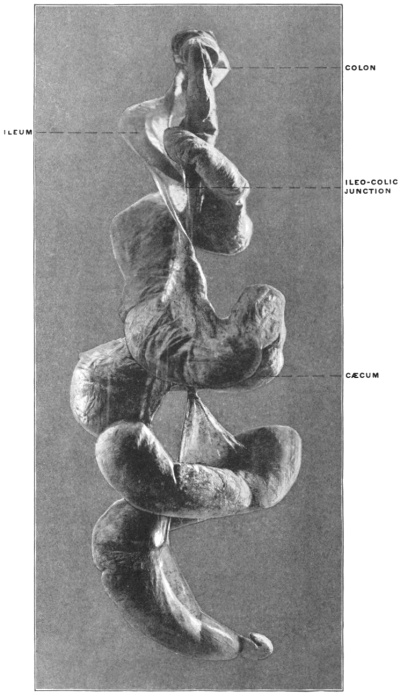
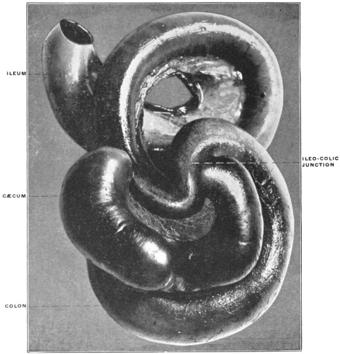
3. Forms with simple cæca of moderate size.
The Peramelidæ or bandicoots.
Fig. 353 shows the ileo-colic junction, cæcum and proximal segment of the colon in Perameles nasuta, the bandicoot.
Fig. 353.—Perameles nasuta, bandicoot. Ileo-colic junction and cæcum. (Columbia University Museum, No. 1481.)
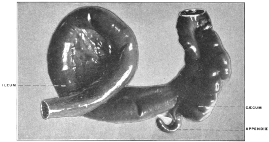
4. Forms with sacculated short cæca, whose terminal portion is reduced to constitute a typical vermiform appendix.
The cæcum of the Phascolomyidæ or wombats, resembles, in its general structure and in the presence of a typical vermiform appendix, very closely the corresponding parts of the alimentary canal in man and the anthropoid apes. Fig. 354 shows these structures in Phascolomys wombat, the common wombat.
Fig. 354.—Phascolomys wombat, wombat. Ileo-cæcum and appendix. (Columbia University Museum, No. 1508.)
5. Forms with simple direct ileo-colic junction without cæcum.
In the purely carnivorous Marsupials, comprising the family of the Dasyuridæ, the reduction of the cæcal apparatus, foreshadowed by the appearance of the distal rudimentary segment as a vermiform appendix in the wombats, has been carried to the complete elimination of the pouch. The ileo-colic junction in these animals is simple, marked externally by a circular constriction and internally by an annular valve. The colon forms a very short terminal segment of the alimentary canal. The parts are shown in Fig. 355 in a typical representative of the family, Dasyurus viverinus, the Tasmanian devil.
Fig. 355.—Dasyurus viverinus, dasyurus, Tasmanian devil. Intestinal canal. Ileo-colic junction. (Columbia University Museum, No. 1463.)
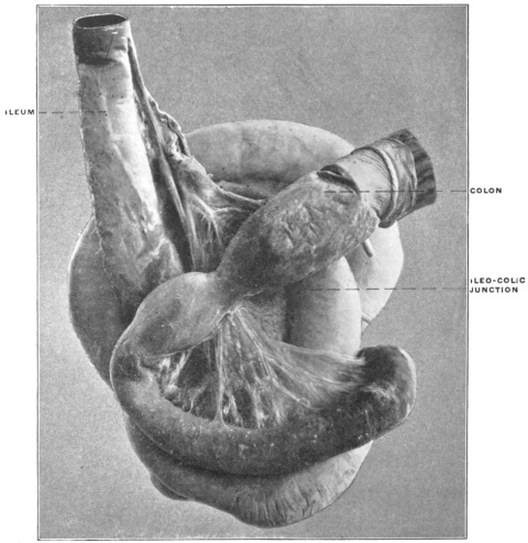
The structural modifications encountered in the digestive tract of these carnivorous Marsupials can properly be regarded as the result of their habitual diet, and we will meet with analogous and identical examples of cæcal reduction in the typical Carnivores among the placental mammals (cf. p. 212).
III. Subclass: Monodelphia.
III. Order: Edentata.
In all probability the Sloths, Ant-eaters and Armadillos composing this order represent a highly specialized remnant of an ancient group now largely extinct. In respect to the ileo-colic junction the Edentates may be arranged in two groups which offer, within the limited number of existing species, a very complete transitional series.
I. Symmetrical Type of Ileo-colic Junction.
1. Differentiation in caliber, with direct funnel-like transition of small into large intestine. No cæcum.
Beyond the ileo-colic junction the caliber of the large intestine increases gradually. The terminal ileum is thus implanted into the apex of a funnel formed by the proximal segment of the colon.
Examples of this type are furnished by Myrmecophaga jubata, the great ant-eater (Fig. 356), and by Cholœpus didactylus, the two-toed sloth (Fig. 357).
Fig. 356.—Myrmecophaga jubata, great ant-eater. Ileo-colic junction. (Columbia University Museum, No. 1519.) Fig. 357.—Cholœpus didactylus, two-toed sloth. Ileo-colic junction. (Columbia University Museum, No. 714.)
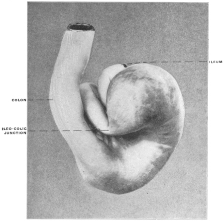
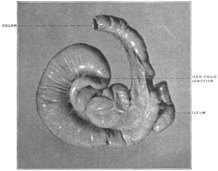
Fig. 358.—Tatusia novemcincta, nine-banded armadillo. Ileo-colic junction. (Columbia University Museum, No. 176.)
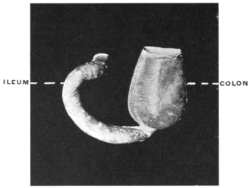
2. Abrupt demarcation of small and large intestine, with median transition of ileum.
The caliber of the intestine enlarges rapidly immediately beyond the ileo-colic junction. This form is derived from the preceding by the substitution of the abrupt ileo-colic transition for the gradual funnel-shaped development of the large intestine.
The type is illustrated by Tatusia novemcincta, the nine-banded armadillo (Fig. 358), and is also found in two other armadillos, Tolypeutes and Xenurus.
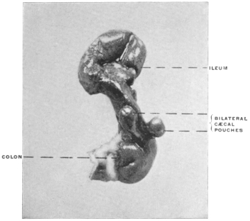
3. The colon on each side of the ileo-colic junction is prolonged backward along the small intestine, forming two symmetrical lateral globular colic cæca.
This type, which is to be regarded as a further development of the preceding form, is also found in the armadillos. Fig. 359 represents the structures in Dasypus sexcinctus, the six-banded armadillo, and a similar arrangement of the parts exists in Chlamydophorus, another species of armadillo.
Fig. 359.—Dasypus sexcinctus, six-banded armadillo, Ileo-colic junction and cæca. (Columbia University Museum, No. 1478.)
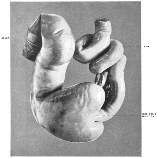
4. The cæcal pouches are more completely differentiated, communicating with the colon by a constricted neck.
This results in an arrangement which recalls the structure of many avian cæca (cf. Fig. 337) and is seen in the double cæcal pouches of Cyclothurus didactylus, the little ant-eater (Fig. 360).
Fig. 360.—Cyclothurus didactylus, little ant-eater. Ileo-colic junction and cæca. (Columbia University Museum, No. 1512.)
II. Asymmetrical Type of Ileo-colic Junction.
The second general group of the Edentates is characterized by the gradual development of a single lateral asymmetrical cæcum, in place of the median symmetrical ileo-colic transition found in the forms just considered. The species composing this group thus form a link leading up to the right-angled accession of ileum to large intestine and the lateral cæcum characteristic of most other mammalia.
Fig. 361.—Manis longicauda, long-tailed pangolin. Ileo-colic junction; dry preparation. (Columbia University Museum, No. 95.) Fig. 362.—Manis longicauda, long-tailed pangolin. Ileo-colic junction. (Columbia University Museum, No. 328.)
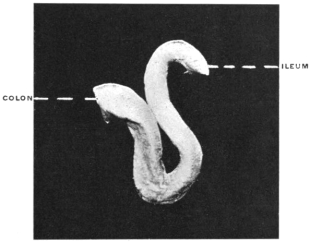
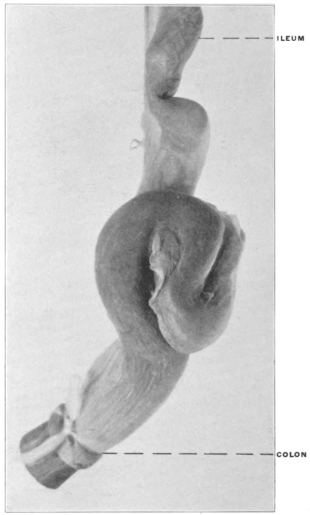
Fig. 363.—Arctopithecus marmoratus, three-toed sloth. Ileo-colic junction. (Columbia University Museum, No. 1479.)
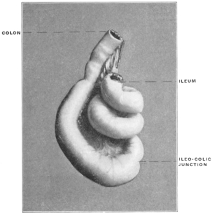
1. This type may be considered as being inaugurated by the form of ileo-colic junction found in the Manidæ or Pangolins, as illustrated by Figs. 361 and 362, taken from the long-tailed pangolin, Manis longicauda. There is no cæcum and only a slight differentiation in caliber between the small and large intestine. The gut in all the forms examined shows a very characteristic bend at the ileo-colic junction, being twisted into a figure of 8 and held in place by mesenteric folds.
2. The second stage, illustrated by Arctopithecus (Bradypus) marmoratus, the three-toed sloth (Fig. 363), reveals a distinct increase in the caliber and convexity of the large intestine opposite the mesenteric border immediately beyond the ileo-colic junction.
3. This leads in the third stage, represented by Tamandua bivittata, the Tamandua ant-eater (Figs. 364 and 365), to the development of a distinct lateral cæcal pouch. I have had no opportunity of examining the structures in Orycteropus, but from the published descriptions8 the large cæcum of this animal would form the final link in this series.
Fig. 364.—Tamandua bivittata, Tamandua ant-eater. Ileo-colic junction and cæcum. (Columbia University Museum, No. 1590.)
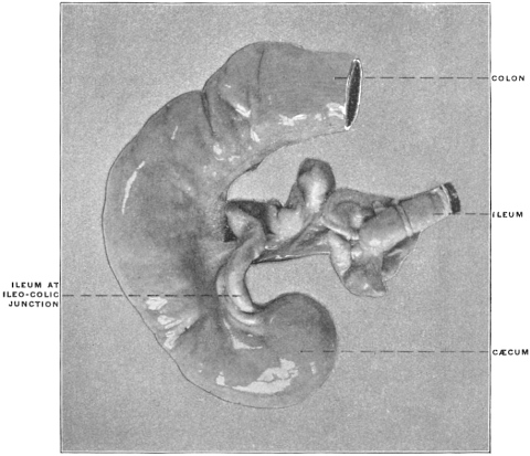
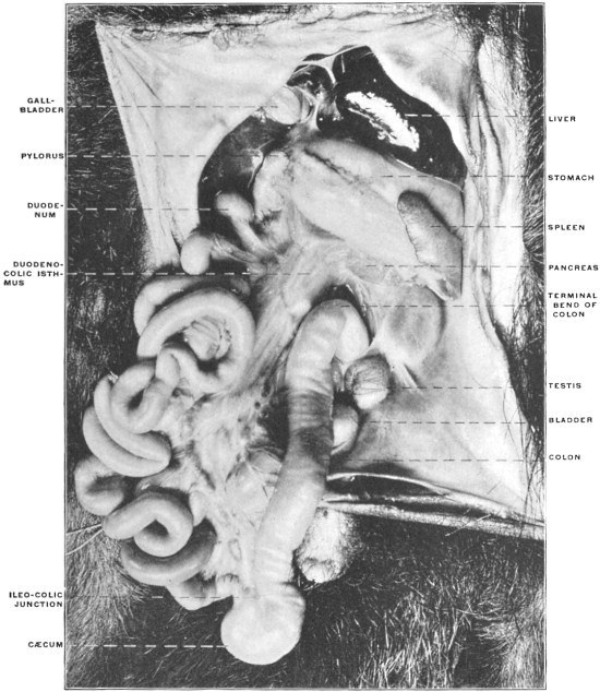
Fig. 365.—Tamandua bivittata, Tamandua ant-eater. Ventral view of abdominal viscera from the left side. (Study Collection, Columbia University.)
IV. Order: Sirenia.
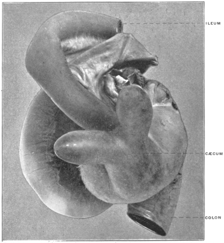
Of the two living representatives of this remarkable mammalian order the dugong (Halicore) is described as possessing a single cæcum, while the cæcal pouch of Manatus americanus, the manatee, is symmetrically bifid at the extremity (Fig. 366).
Fig. 366.—Manatus americanus, American manatee. Ileo-colic junction and bifid cæcum. (Columbia University Museum, No. 673.)
V. Order: Cetacea.
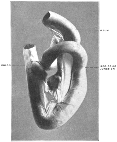
In the majority of the whales the ileo-colic junction is simple without cæcum, as in Physeter, Delphinus, Monodon and Phocæna (Fig. 367).
Fig. 367.—Phocæna communis, porpoise. Ileo-colic junction. (Columbia University Museum, No. 1007.)
A few forms have a small cæcal pouch.
VI. Order: Ungulata.
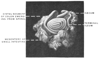
The intestinal canal, in conformity with the herbivorous habit of the group, is uniformly provided with a large cæcum, and in many forms the proximal segment of the colon immediately beyond the ileo-colic junction is more or less extensively coiled in a spiral manner. This arrangement is, without doubt, to be regarded as being functionally accessory to the cæcal apparatus, in the sense of increasing very much the area of the secreting and absorbing surface and of prolonging the period during which food-substances, which are slow and difficult of elaboration, are retained in this segment of the alimentary canal.
1. Suborder: Artiodactyla.
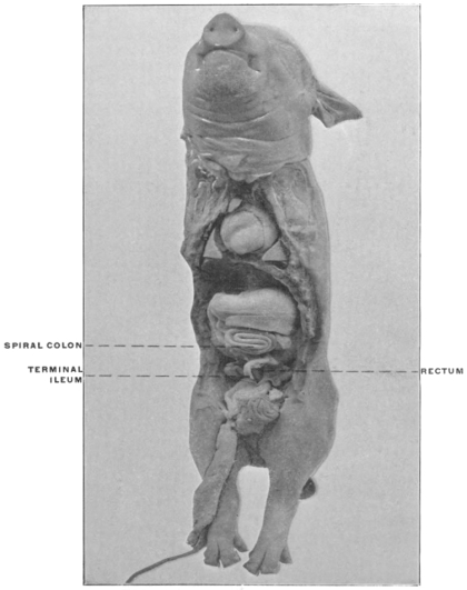
A. Non-Ruminantia.—In the Suidæ the cæcum is large and the spiral colon well developed (Fig. 368).
Fig. 368.—Sus scrofa fœt, fœtal pig. Ileo-cæcum and spiral colon in situ. (Columbia University Museum, No. 1111.)
In the peccaries (Dicotyles) the terminal portion of the cæcal pouch is reduced, constituting a centrally implanted appendage.
Fig. 369.—Dicotyles torquatus, collared peccary. Ileo-colic junction, cæcum, and spiral colon. (Columbia University Museum, No. 60/1462)
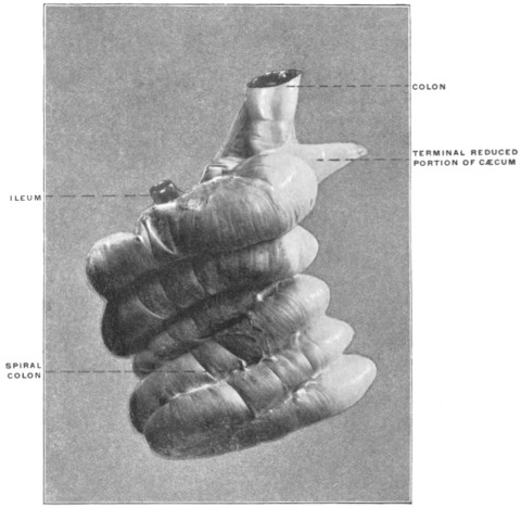
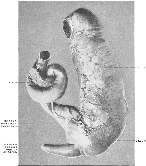
Fig. 369 shows the ileo-colic junction and spiral colon in Dicotyles torquatus, the collared peccary, and Fig. 370 the cæcum and appendix of the same animal detached from the spiral colon. In the hippopotamus, on the other hand, the cæcum is said to be absent. If this is the case the animal forms an isolated exception among the Ungulates.
Fig. 370.—Dicotyles torquatus, collared peccary. Ileo-colic junction and cæcum, isolated. (Columbia University Museum, No. 1464.)
B. Ruminantia.—The cæcum is very large and the spiral coil of the colon extensive.
Fig. 371.—Capra ægagrus, Bezoar goat. Ileo-colic junction and cæcum, isolated; dried preparation. (Columbia University Museum, No. 194.)
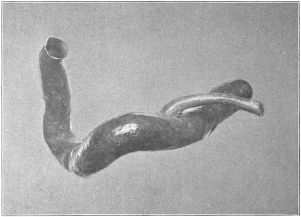
Fig. 371 shows the cæcum of Capra ægagrus, the Bezoar goat, detached from the adjacent intestine, and illustrates the type of the ruminant pouch, of considerable length and caliber, without terminal reduction. The same parts in a preparation of Boselaphus tragocamelus, the nilghai, are shown in Fig. 372.
Fig. 372.—Boselaphus tragocamelus, nilghai. Ileo-colic junction and cæcum, isolated. (Columbia University, Study Collection.)
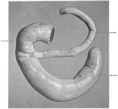
Fig. 373.—Bos indicus, zebu. Ileo-colic junction, cæcum, and spiral colon. (Columbia University Museum, No. 676.)
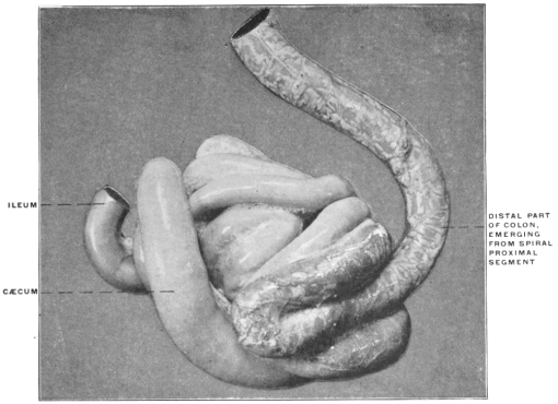
Fig. 373 shows the cæcum and ileo-colic junction, together with the spiral coil of the colon, in Bos indicus, the zebu, and Fig. 374 the same structures with a typical example of the spiral colon from Cervus sika, the Japanese deer; Fig. 375 is taken from a preparation of the parts in a fœtal sheep, while Fig. 376 shows the spiral colon isolated in Oryx leucoryx, the oryx.
Fig. 374.—Cervus sika, Japanese deer. Ileo-colic junction, cæcum, and spiral colon. (Columbia University, Study Collection.) Fig. 376.—Oryx leucoryx, oryx. Spiral colon, isolated. (Columbia University, Study Collection.)
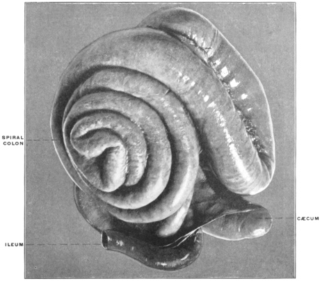
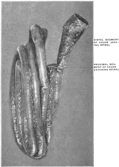
Fig. 375.—Oris aries fœt, fœtal sheep. Ileo-colic junction, cæcum, and spiral colon. (Columbia University Museum, No. 1379.)
2. Suborder: Perissodactyla.
In the horse and the rhinoceros the cæcum is very large and of uniform caliber.
Fig. 377.—Tapirus americanus, American tapir. Ileo-colic junction, cæcum, and colon. (Columbia University Museum, No. 624.)
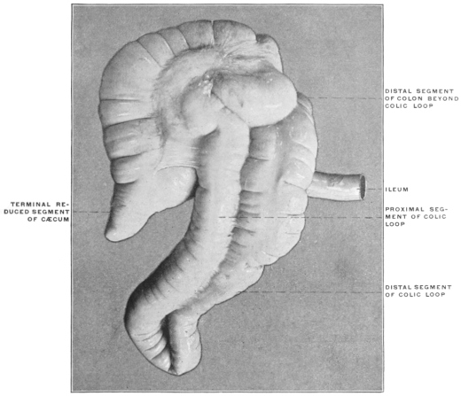
In the American tapir (Fig. 377) the large cæcum tapers at its extremity, to form a species of rudimentary appendix, resembling somewhat the corresponding structure in Dicotyles (cf. Figs. 369 and 370). The proximal segment of the colon is bent on itself in the form of an extensive loop with closely adherent limbs, illustrating an early stage in the development of the ruminant spiral colon (cf. p. 233).
3. Suborder: Hyracoidea.
Fig. 378.—Hyrax syriacus, hyrax or coney. Intestinal canal, with ileo-colic junction, proximal ileo-colic cæcum, and distal paired colic cæca. (Columbia University, Study Collection.) Fig. 379.—Elephas indicus, Asiatic elephant. Ileo-colic junction and cæcum. (Columbia University Museum, No. 995.)
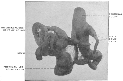
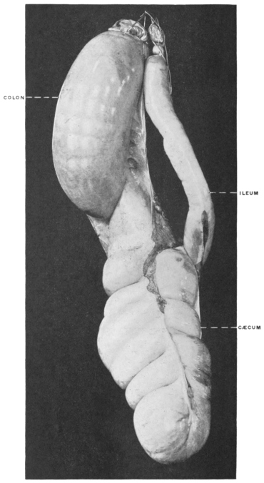
Fig. 380.—Myoxus avellanarius, common dormouse. Alimentary canal. (Columbia University Museum, No. 1466.)
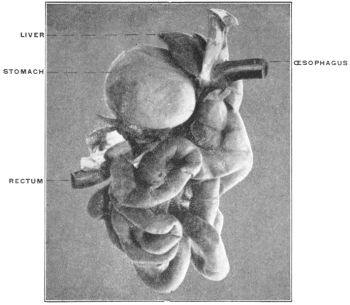
This suborder is formed by the single family of the Hyracidæ. In addition to their other isolated and puzzling structural peculiarities the members of this small group present a most unusual arrangement of the intestinal canal, which is unique among living mammalia. In addition to a large sacculated cæcal pouch, situated in the usual position at the beginning of the colon, the large intestine is provided further on with two supplementary elongated pointed conical pouches (Fig. 378).
This unique arrangement, which is not found in any other known vertebrate, may possibly be led back to a type-form encountered in certain saurians (see p. 234).
4. Suborder: Proboscidea.
The cæcum of the elephant is a very large sacculated pouch with rounded termination, illustrated in Fig. 379, taken from the Asiatic elephant.
VII. Order: Rodentia.
With the exception of a single group, the dormice (Myoxus) (Fig. 380), the rodents possess a well-developed cæcal apparatus.
In some forms the terminal portion of the pouch is reduced so as to constitute an appendix. Many of these animals, in addition to the cæcum proper, have the proximal colon elongated and coiled in a spiral, and in some this part of the large intestine is provided in the interior with a spiral mucous fold. This latter structure functions again to increase the extent of the mucous absorbing surface and to prolong the retention of substances undergoing slow digestion and absorption.
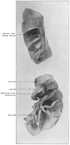
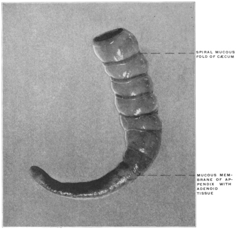
Typical examples of the capacious sacculated rodent cæcum, with a terminal pointed reduced segment, are afforded by Castor fiber, the beaver (Figs. 381 and 382) and by Erethizon dorsatus, the Canadian porcupine (Figs. 383 and 384). Figs. 385 and 386 show the ileo-colic junction, cæcum and appendix in Lepus cuniculus, the rabbit. The interior of the cæcal pouch and of the proximal segment of the colon is provided with a complete spiral valve (Fig. 387), while the appendix is differentiated by the histological character of its mucous membrane which is studded with closely packed adenoid follicles (Fig. 388). A similar aggregation of lymphoid tissue is found in this animal at the ileo-colic junction forming the s. c. saccus lymphaticus (Fig. 387).
Fig. 381.—Castor fiber, beaver. Ileo-colic junction, cæcum, and proximal colon; ventral view. (Columbia University Museum, No. 1607.)
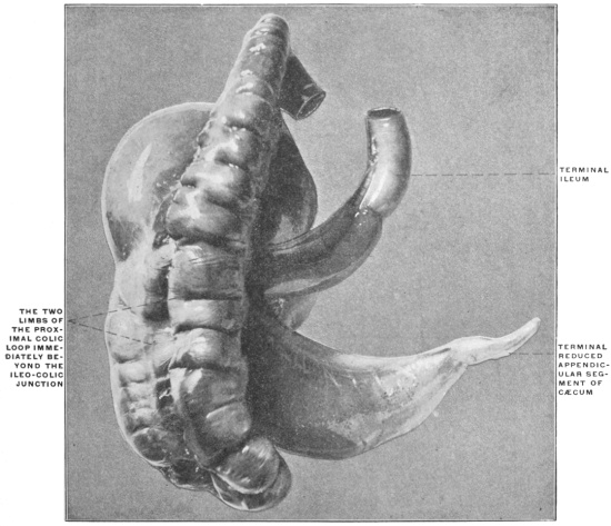
Fig. 382.—Castor fiber, beaver. Ileo-colic junction, cæcum, and proximal colon; dorsal view. (Columbia University Museum, No. 1607.)
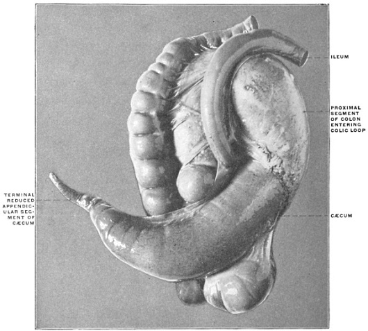
Fig. 383.—Erethizon dorsatus, Canadian porcupine. Ileo-colic junction, cæcum, and colon; ventral view. (Columbia University, Study Collection.)
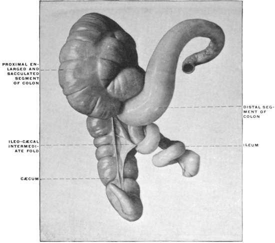
Fig. 384.—Erethizon dorsatus, Canadian porcupine. Ileo-colic junction, cæcum, and colon; dorsal view. (Columbia University, Study Collection.)
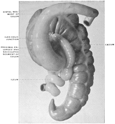
Fig. 385.—Lepus cuniculus, rabbit. Ileo-colic junction and cæcum. (Columbia University Museum, No. 1568.)
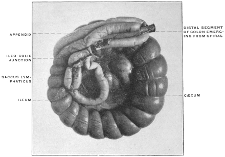
Fig. 386.—Lepus cuniculus, rabbit. Ileo-colic junction with saccus lymphaticus, isolated, (Columbia University, Study Collection.)
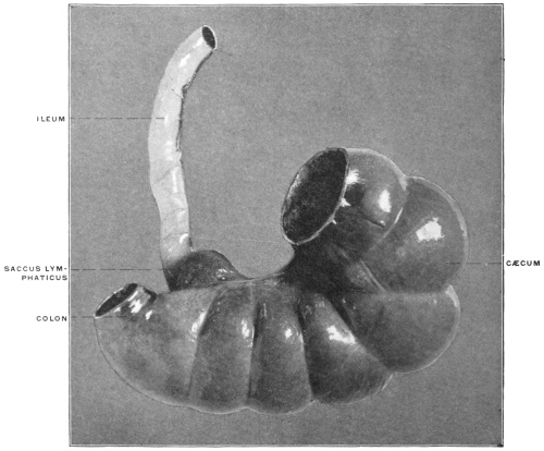
Fig. 387.—Lepus cuniculus, rabbit. Ileo-colic junction with saccus lymphaticus. Cæcum and proximal segment of colon opened to show spiral mucous fold in interior. (Columbia University Museum, No. 1587.) Fig. 388.—Lepus cuniculus, rabbit. Cæcum and appendix inverted to show spiral fold and structure of mucosa. (Columbia University Museum, No. 1588.)
The coils of the proximal colon encountered in many rodents are well seen in Dasyprocta agouti, the agouti (Figs. 389 and 390), which animal also illustrates a type of cæcum found in several members of the order. The pouch here is large, sacculated, uncinate, without reduction of the terminal portion.
Fig. 389.—Dasyprocta agouti, agouti. Ileo-colic junction, cæcum, and colon. (Columbia University Museum, No. 24/1576.)
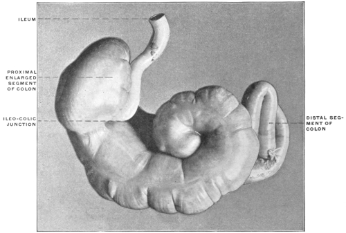
Fig. 390.—Dasyprocta agouti, agouti. Ileo-colic junction, cæcum, and colon. (Drawing based on preparation shown in Fig. 388.)
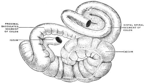
Fig. 391.—Lagomys pusillus. Ileo-colic junction, cæcum, and colon. (After Pallas, from Oppel, “Lehrbuch d. Vergl. mikrosk. Anat. d. Wirbelthiere,” II., Jena, 1897, p. 577, Fig. 314.)
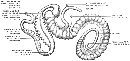
Fig. 392.—Arvicola pennsylvanicus, field mouse. Alimentary canal. (Columbia University Museum, No. 815.) Fig. 393.—Mus decumanus, white rat. Ileo-colic junction, cæcum, and colon. (Columbia University Museum, No. 1574.)
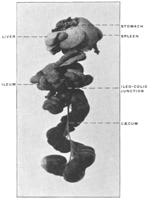
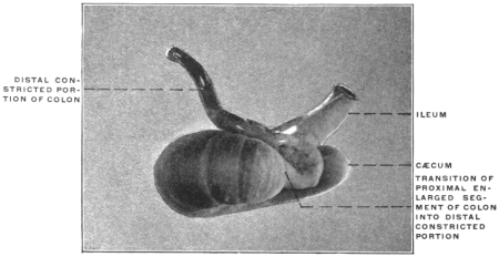
The relatively enormous size of the cæcum in the Muridæ is shown in Fig. 392, representing the entire visceral tract of Arvicola pennsylvanicus, the meadow mouse. The pouch in these animals is large, smooth and of uniform caliber (Fig. 393).
In some the colon beyond the entrance of the small intestine is provided with a spiral mucous valve (Fig. 394).
Fig. 394.—Arvicola riparius, meadow mouse. Ileo-colic junction, cæcum, and colon. (Columbia University, Study Collection.)
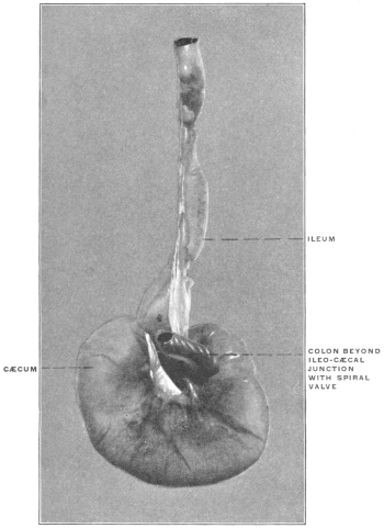
In the single instance of Myoxus among the rodents, the ileo-colic junction is simple, without any cæcal pouch (Fig. 380).
VIII. Order: Carnivora.
A. Pinnipedia.—In the seals and walrus the cæcum is very small with a blunt termination. Fig. 395 shows its structure in Zalophus gillespiei, Gillespie’s sea-lion, and Fig. 396 in Phoca vitulina, the harbor seal.
Fig. 395.—Zalophus gillespiei, Gillespie’s sea-lion. Ileo-colic junction and cæcum; dried preparation. (Columbia University Museum, No. 90.) Fig. 396.—Phoca vitulina, harbor seal. Ileo-colic junction and cæcum. (Columbia University Museum, No. 762.)
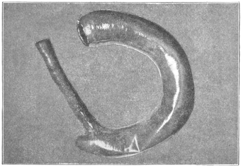
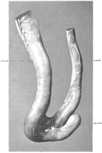
Fig. 397.—Vulpes fulvus, red fox. Ileo-colic junction and cæcum; dried preparation. (Columbia University Museum, No. 114.)
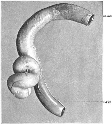
B. Fissipedia.—The Cynoidea, including the dogs, jackals, wolves and foxes, form a well-marked central group with well-developed convoluted cæca placed laterally to the ileo-colic junction (Figs. 397-399).
Fig. 398.—Canis familiaris, dog. Ileo-colic junction and cæcum, Type I. (Columbia University Museum, No. 1550.) Fig. 399.—Canis familiaris, dog. Ileo-colic junction and cæcum, Type II. (Columbia University Museum, No. 1551.)
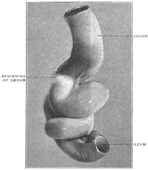
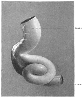
Fig. 400.—Genetta vulgaris, genet. Ileo-colic junction and cæcum. (Columbia University Museum, No. 1625.) Fig. 401.—Felis concolor, puma. Ileo-colic junction and cæcum; dried preparation. (Columbia University Museum, No. 119.)
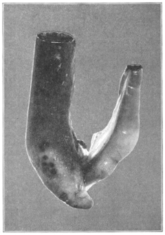
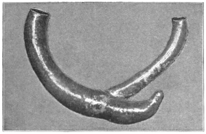
Fig. 402.—Felis borealis, var. rufus, red lynx. Ileo-colic junction and cæcum; dried preparation. (Columbia University Museum, No. 177.) Fig. 404.—Herpestes sp.?, ichneumon. Ileo-colic junction and cæcum; dried preparation. (Columbia University Museum, No. 120.)
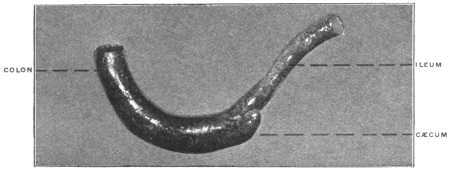
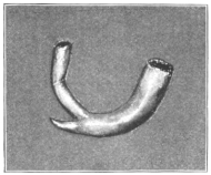
Fig. 403.—Paradoxurus typus, paradoxure. Ileo-colic junction and cæcum; dried preparation. (Columbia University Museum, No. 112.) Fig. 405.—Herpestes griseus, mongoose ichneumon. Ileo-colic junction and cæcum; dried preparation. (Columbia University Museum, No. 149.)
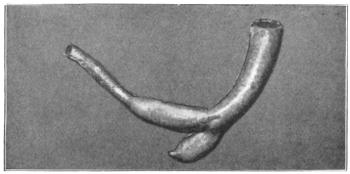
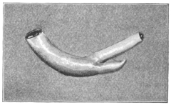
From this type depart on the one hand the Ailuroidea, including the civets, ichneumons and true cats, with the cæcum uniformly present, but short and markedly pointed, suggesting the degeneration of a formerly better developed structure (Figs. 400-406), while on the other the Arctoidea, including the bears, weasels and raccoons, constitute a group united by many common fundamental peculiarities of structure, among which is the entire absence of a cæcal pouch (Figs. 407-415).
Fig. 406.—Proteles lalandii, aard-wolf. Ileo-colic junction and cæcum. (Columbia University Museum, No. 1520.) Fig. 407.—Nasua rufa, brown coati-mundi. Ileo-colic junction. (Columbia University Museum, No. 1089.)
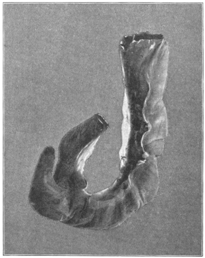
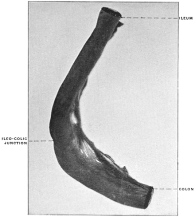
Fig. 408.—Nasua rufa, brown coati-mundi. Ileo-colic junction, opened, showing pyloric-like ileo-colic valve. (Columbia University Museum, No. 1581.) Fig. 409.—Bassaris astuta, raccoon-fox. Ileo-colic junction; dried preparation. (Columbia University Museum, No. 289.)
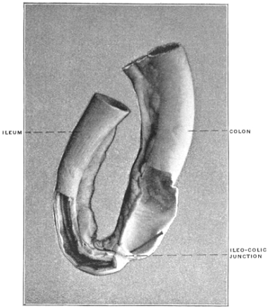
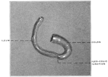
Fig. 410.—Mustela sp.?, marten. Ileo-colic junction; dried preparation. (Columbia University Museum, No. 199.) Fig. 411.—Taxidea americana, American badger. Ileo-colic junction; dried preparation. (Columbia University Museum, No. 180.)
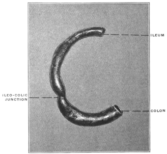
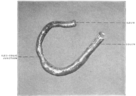
Fig. 412.—Procyon lotor, raccoon. Ileo-colic junction; dried preparation. (Columbia University Museum, No. 230.) Fig. 414.—Ursus americanus, black bear. Ileo-colic junction; dried preparation. (Columbia University Museum, No. 226.)
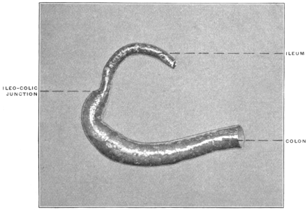
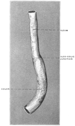
Fig. 413.—Cercoleptes caudivolvulus, kinkajou. Ileo-colic junction; dried preparation. (Columbia University Museum, No. 295.)
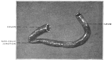
Fig. 415.—Ursus maritimus, polar bear. Ileo-colic junction. (Columbia University Museum, No. 748.)
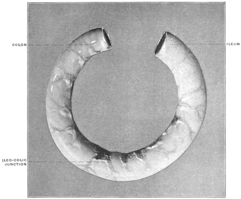
Fig. 416.—Hyæna striata, striped hyena. Ileo-colic junction and cæcum; dried preparation. (Columbia University Museum, No. 56.)
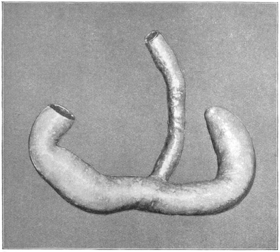
Fig. 417.—Felis leo, lion, Ileo-colic junction and cæcum. (Columbia University Museum, No. 1516.)
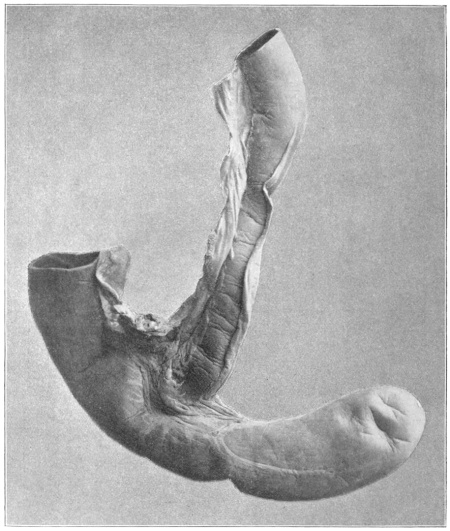
Among the ailuroid carnivora, the hyæna and the lion occupy an isolated position in regard to the cæcum. Both of these animals possess a well-developed long cæcal pouch with blunt extremity (Figs. 416 and 417). They probably afford examples of a persistent ancestral common type from which the remaining carnivorous forms are derived by reduction of the cæcal apparatus in conformity with the food-habits of these animals. The cæcum of both the lion and hyæna resembles very closely the pouch of the herbivorous marsupials, such as Halmaturus or Didelphis (cf. Figs. 348 and 350, p. 205).
IX. Order: Cheiroptera.
In the bats the alimentary canal is uniformly simple without cæcum and scarcely any differentiation between small and large intestine (Fig. 418).
Fig. 418.—Pteropus medius, Indian fruit-bat. Ileo-colon; dried preparation. (Columbia University Museum, No. 533.)
X. Order: Insectivora.
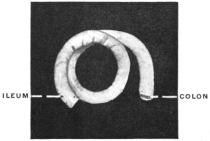
In the true Insectivora the cæcum is also absent and the alimentary canal a simple non-differentiated tube.
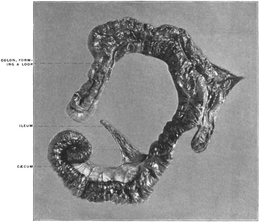
In certain herbivorous animals included in this group on the other hand, such as Galeopithecus (Fig. 419), the cæcum is present as an enormous sacculated pouch with spiral convolutions.
Fig. 419.—Galeopithecus volans, colugo. Ileo-colic junction, cæcum, and colon. (Columbia University Museum, No. 1844.)
XI. Order: Primates.
The cæcum is uniformly present. In certain of the Lemuroidea the terminal portion of the pouch is reduced, forming a species of appendix. A typical vermiform appendix is regularly found in man and in the anthropoid apes, orang, gibbon, chimpanzee and gorilla.
1. Suborder Lemuroidea.
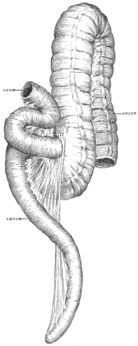
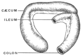
In the typical lemurs the cæcum is long, frequently terminating in a pointed appendage. The proximal segment of the colon is looped and coiled, resembling the spiral colon of the Ungulates and Rodents. Fig. 420 shows the cæcum of Nycticebus tardigradus, the slow lemur, with the typical appendage, and Fig. 421 shows the spiral arrangement of the proximal colon immediately beyond the ileo-colic junction in the same animal. Fig. 422, taken from another specimen of the same animal shows the cæcum, appendix and spiral colon. Figs. 423, 424, 425 illustrate the structure of the parts in three other members of the group, Lemur macaco, Lemur mongoz and Otolicnus crassicaudatus, all showing terminal reduction of the cæcal pouch and tendency to spiral coiling of the proximal colon. In Tarsius spectrum (Fig. 426) the cæcum is relatively well-developed, but forms a simple pouch of uniform diameter, without terminal reduction.
Fig. 420.—Nycticebus tardigradus, slow lemur. Ileo-colic junction, cæcum, appendix, and colon; dorsal view. (Columbia University Museum, No. 20/1468.)
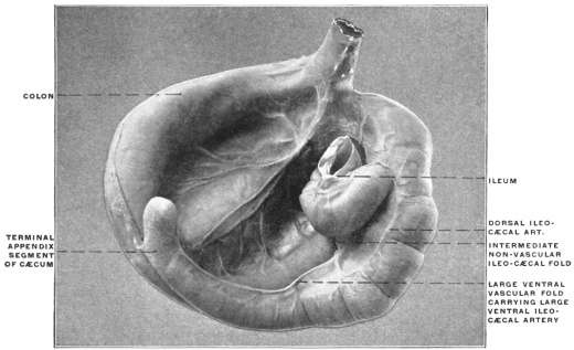
Fig. 421.—Nycticebus tardigradus, slow lemur. Same preparation as Fig. 420; ventral view, showing spiral coiling of proximal colon. (Columbia University Museum, No. 20/1468.) Fig. 422.—Nycticebus tardigradus, slow lemur. Ileo-colic junction, cæcum, appendix, and spiral colon. (Columbia University, Study Collection.)
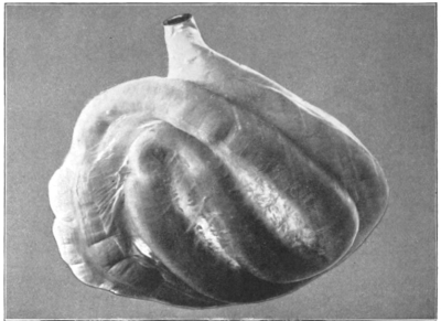
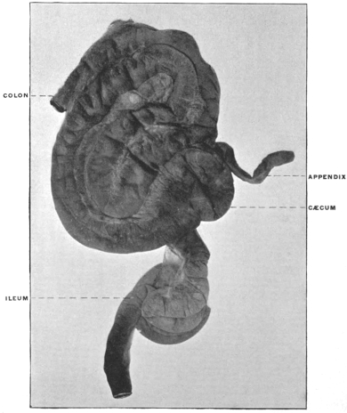
Fig. 423.—Lemur macaco, lemur. Ileo-colic junction and cæcum. (Drawn from preparation.) (Columbia University Museum, No. 1623.) Fig. 424.—Lemur mongoz, lemur. Ileo-colic junction and cæcum. (Drawn from preparation.) (Columbia University Museum, No. 1473.)
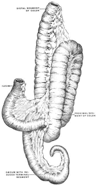
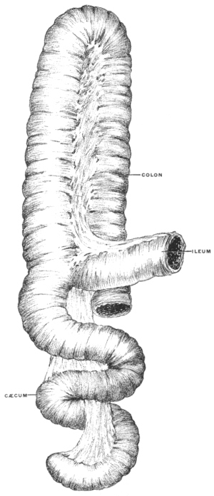
Fig. 425.—Otolicnus crassicaudatus, galago. Ileo-colic junction and cæcum. (Drawn from preparation.) (Columbia University Museum, No. 1626.) Fig. 426.—Tarsius spectrum, spectre lemur. (Drawn from preparation.) (Columbia University Museum, No. 1521.)
2. Suborder Anthropoidea.
A. Cynomorpha.
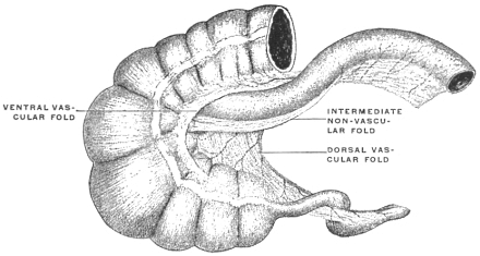
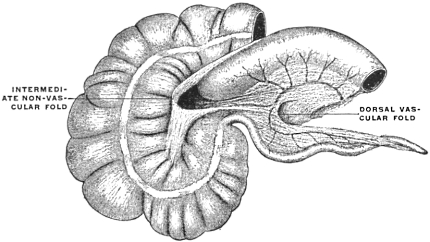
1. Cynocephalus.—The baboons have a well-developed capacious cæcum. The apex of the pouch is usually blunt and rounded, or only slightly pointed. The cæcum is sacculated, conforming in structure to the rest of the large intestine. Two low vascular folds or ridges, a ventral and a dorsal, carry the ventral and dorsal cæcal branches of the ileo-colic artery. The intermediate non-vascular fold is large, frequently fused with the dorsal vascular fold (cf. p. 264).
Figs. 427-433 show the structures in Cynocephalus sphinx, porcarius, babuin, anubis and in Cercopithecus pogonias, sabæus and campbellii.
Fig. 427.—Cynocephalus sphinx, Guinea baboon. Ileo-colic junction and cæcum. (Columbia University Museum, No. 1082.)
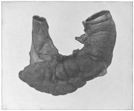
Fig. 428.—Cynocephalus porcarius, Chacma baboon. Ileo-colic junction and cæcum. (Columbia University Museum, No. 1071.) Fig. 429.—Cynocephalus babuin, yellow baboon; dried preparation. (Columbia University Museum, No. 89.)
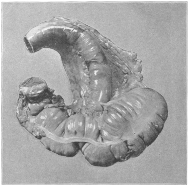
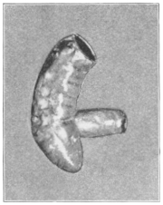
Fig. 430.—Cynocephalus anubis, olive baboon. (Columbia University Museum, No. 51/1618.)
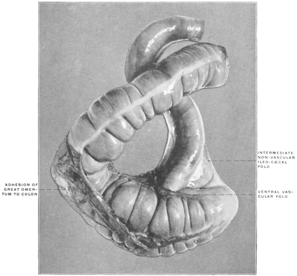
Fig. 431.—Cercopithecus pogonias, bearded monkey. Ileo-colic junction and cæcum; dried preparation. (Columbia University Museum, No. 228.) Fig. 432.—Cercopithecus sabæus, green monkey. Ileo-colic junction and cæcum. (Drawn from preparation.) (Columbia University Museum, No. 746.) 1. Ventral ileo-cæcal vascular fold. 2. Dorsal ileo-cæcal vascular fold. 3. Intermediate ileo-cæcal non-vascular fold.
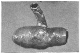
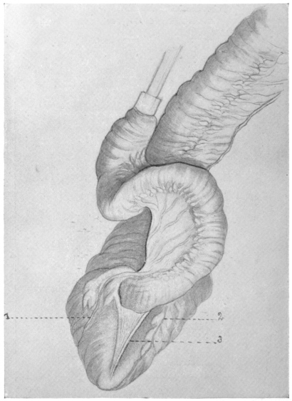
Fig. 433.—Cercopithecus campbellii, cercopithecus monkey. Ileo-colic junction and cæcum. (Drawn from preparation.) (Columbia University Museum, No. 55/1542.)
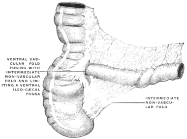
2. Macacus.—The cæcum is of large caliber, blunt, or in some forms slightly pointed at the apex, sacculated like the colon.
The two vascular folds are narrow and low, studded with epiploic appendages. The intermediate non-vascular fold is large, placed nearer to the dorsal than to the ventral vascular fold.
Figs. 434-439 show the structures in Macacus cynomolgus, ochreatus, rhesus and pileatus.
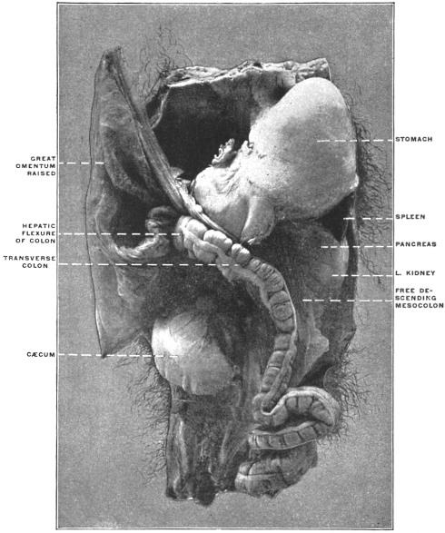
Fig. 439 is from a formaline hardened situs preparation of the abdominal viscera in Macacus cynomolgus, the Kra monkey.
Fig. 434.—Macacus cynomolgus, Macaque monkey. Ileo-colic junction and cæcum; dried preparation. (Columbia University Museum, No. 19.) Fig. 436.—Macacus rhesus, rhesus monkey. Ileo-colic junction and cæcum. (Columbia University Museum, No. 1126.)
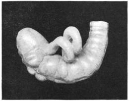
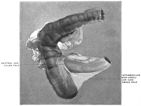
Fig. 435.—Macacus ochreatus, ashy-black macaque. Ileo-colic junction and cæcum; dried preparation. (Columbia University Museum, No. 11.)
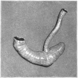
Fig. 438.—Macacus sinicus, bonnet macaque. Ileo-colic junction and cæcum. (Columbia University Museum, No. 1072.)
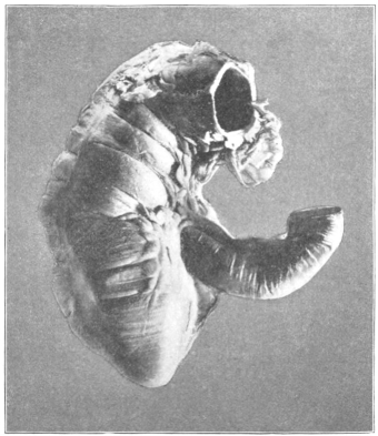
Fig. 437.—Macacus pileatus, macaque. Ileo-colic junction and cæcum. (Columbia University Museum, No. 719.)
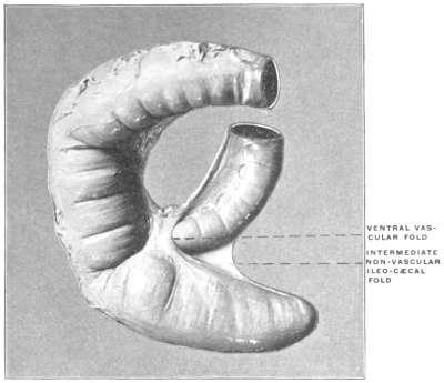
Fig. 439.—Macacus cynomolgus, kra monkey. Abdominal viscera, hardened in situ. (Columbia University Museum, No. 1801.)
B. Arctopithecini.
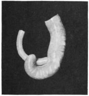
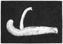
The marmosets have a long crescentic-shaped cæcum, turning the concavity of the curve upwards and to the left, terminating in a blunt point.
Typical forms are shown in Fig. 440, Hapale jacchus, Fig. 441, Midas ursulus, and Fig. 442, Midas geoffrei.
Fig. 440.—Hapule jacchus, common marmoset. Ileo-colic junction and cæcum. (Columbia University Museum, No. 975.)
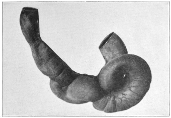
Fig. 441.—Midas ursulus, negro tamarin. Ileo-colic junction and cæcum; dried preparation. (Columbia University Museum, No. 235.) Fig. 442.—Midas geoffrei, Geoffrey’s marmoset. Ileo-colic junction and cæcum; dried preparation. (Columbia University Museum, No. 197.)
C. Cebidæ.
1. Ateles and other howlers have a large cæcum, crescentic in shape, narrowed at the apex, separated from the colon by a sharp and deep constriction, opposite the wedge-shaped ileo-colic junction.
The ileo-cæcal folds are well-developed and symmetrical, two equal vascular folds, and a free intermediate non-vascular reduplication.
Types: Ateles ater (Figs. 443-445), Chrysothrix sciureus (Fig. 447) and Nyctipithecus commersonii (Fig. 446). In Mycetes (Figs. 448-450) the pouch is shorter, less curved, with a slight reduction toward the less distinctly pointed apex.
Fig. 443.—Ateles ater, black-faced coaita. Ileo-colic junction and cæcum; dried preparation. (Columbia University Museum, No. 240.)
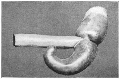
Fig. 444.—Ateles ater, black-faced coaita. Ileo-colic junction and cæcum, with ileo-cæcal folds. (Columbia University Museum, No. 720.) Fig. 445.—Ateles ater, black-faced coaita. Ileo-colic junction and cæcum, with ileo-cæcal folds. (Drawn from preparation.) (Columbia University Museum, No. 300.) 1. Ventral vascular ileo-cæcal fold. 2. Intermediate non-vascular ileo-cæcal fold. 3. Dorsal vascular ileo-cæcal fold.
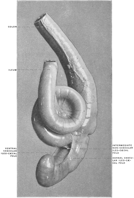
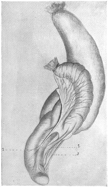
Fig. 446.—Nyctipithecus commersonii, Vitœ monkey. Ileo-colic junction and cæcum; dried preparation. (Columbia University Museum, No. 238.) Fig. 447.—Chrysothrix sciureus, Viti monkey. Ileo-colic junction and cæcum. (Columbia University Museum, No. 1624.) Fig. 448.—Mycetes cavaya, black howler. Ileo-colic junction and cæcum. (Columbia University Museum, No. 1136.)
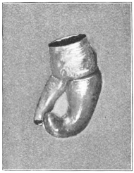
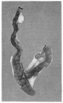
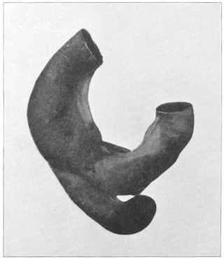
Fig. 449.—Mycetes fuscus, brown howler. Ileo-colic junction and cæcum, with ileo-cæcal folds; ventral view. (Columbia University Museum, No. 674.) 1. Ventral vascular ileo-cæcal fold. 3. Intermediate non-vascular ileo-cæcal fold. Fig. 450.—Drawn from the same preparation as Fig. 449; dorsal view. 2. Dorsal vascular ileo-cæcal fold. 3. Intermediate non-vascular ileo-cæcal fold.
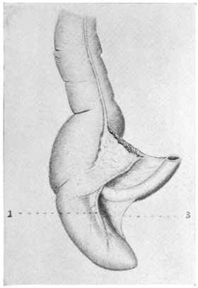
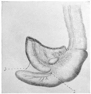
2. Lagothrix.—The cæcum is very capacious and long, bent at a sharp angle upwards and to the left toward the ileo-colic junction.
Type: Lagothrix humboldtii (Fig. 451).
Fig. 451.—Lagothrix humboldtii, Humboldt’s lagothrix. Ileo-colic junction and cæcum. (Columbia University Museum, No. 1511.)
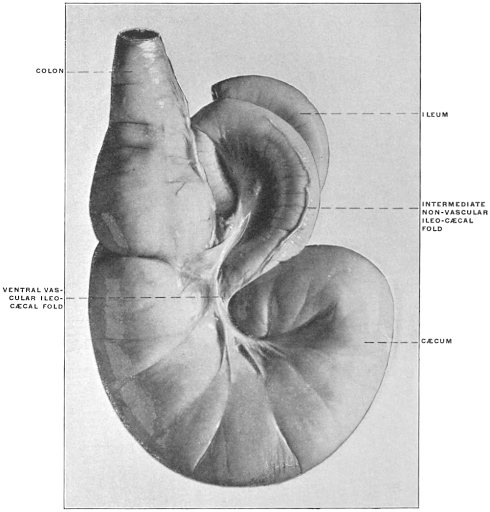
3. Pithecia.—The cæcum resembles in general the type presented by Ateles, but is less curved and less reduced and pointed at the termination.
Type: Pithecia satanas (Fig. 452).
Fig. 452.—Pithecia satanas, black saki monkey. Ileo-colic junction and cæcum. (Columbia University Museum, No. 641.)
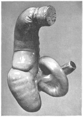
In general the Arctopithecini and Ateles, Mycetes, Lagothrix and Pithecia among the Cebidæ form a group containing a series of cæcal transition types which lead up to the anthropomorphous type, illustrating the following conditions:
(a) The inherent crescentic curve of the cæcum, with the concavity directed toward the left, and carrying the apex of the pouch upward toward the lower border of the ileum and the ileo-colic junction. (Hapalidæ, Ateles, Lagothrix.)
(b) The reduction in caliber of the terminal part, foreshadowing by the pointed and narrow extremity of the pouch the appearance of the appendix in the anthropomorphous group. (Hapalidæ, Ateles.)
(c) The constriction at the level of the ileo-cæcal junction, with the corresponding well-marked differentiation between cæcum and colon in the interior. (Ateles.)
(d) The sharp bend in the pouch as it makes its turn upward and to the left, repeated in certain types of adult human cæca (cf. p. 247). (Lagothrix.)
(e) Pithecia forms a transitive type between the blunt sacculated cæca of the Cynomorpha and the curved pointed pouches of the Cebidæ, partaking of the characters of both.
(f) The same character is seen in the cæcum of Mycetes fuscus the brown howler monkey (Figs. 449 and 450).
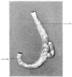
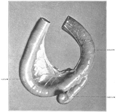
4. Cebinæ.—In the typical genus Cebus the cæcum is placed laterad to the small intestine which is in direct linear continuity with the colon. The pouch is slightly convoluted toward its termination, resembling in this respect and in its position relative to the lumen of the intestinal canal, the disposition of the parts in the cynoid carnivora. Figs. 453 and 454 show the structures in two typical species, Cebus monachus and C. leucophæus.
Fig. 453.—Cebus monachus, capuchin monkey. Ileo-colic junction and cæcum; dried preparation. (Columbia University Museum, No. 26.) Fig. 454.—Cebus leucophæus, capuchin monkey. Ileo-colic junction and cæcum. (Columbia University Museum, No. 1467.)
D. Anthropomorpha.
The cæcum is large, sacculated, provided uniformly with a vermiform appendix.
The pouch of the four anthropoid apes agrees in curve, direction, implantation of the appendix and the general arrangement of the vascular and peritoneal folds with the structure in the human subject.
1. Hylobates hoolock, Gibbon.—Figs. 455 and 456 represent respectively the ileo-cæcum of this animal in the ventral view, and from the left side with the ileum turned forward. The cæcum is a globular rounded pouch of nearly uniform diameter, only slightly enlarged to the right of the root of the appendix which arises from its lowest part and is pendent.
Fig. 455.—Hylobates hoolock, hoolock gibbon. Ileo-colic junction and cæcum; ventral view. (Drawn from Columbia University Museum preparation No. 1530.) Fig. 456.—Drawn from same preparation as Fig. 455; view from left side, showing formation of posterior ileo-cæcal fossa.
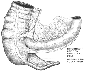
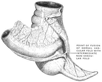
Fig. 457.—Gorilla savagei, gorilla. Ileo-colic junction and cæcum, with ileo-cæcal folds. (Drawn from Columbia University Museum preparation No. 1543.)
(For arrangement of the ileo-cæcal folds and fossæ in this form see p. 269.)
2. Gorilla savagei, Gorilla (Fig. 457).—The cæcum is large, distinctly sacculated, presenting a decided curve with the concavity directed toward the left. The appendix is implanted at the center of the blunt apex of the pouch, the cæcal sacculations on each side of the root of the appendix being of nearly equal size (folds and fossæ, cf. p. 269).
Fig. 458.—Simia satyrus, orang. Cæcum and ileo-colic junction; ventral view. (Drawn from Columbia University Museum preparation No. 716.) 1. Appendix. 2. Ventral vascular fold.
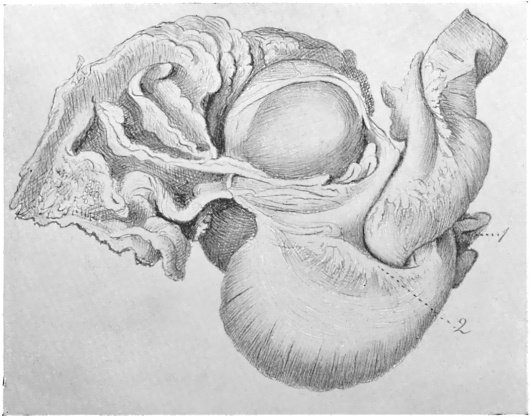
Fig. 459.—Simia satyrus, orang. Cæcum and ileo-colic junction; dorsal view. (Drawn from Columbia University Museum preparation No. 716). 1. Appendix. 2. Intermediate non-vascular fold.
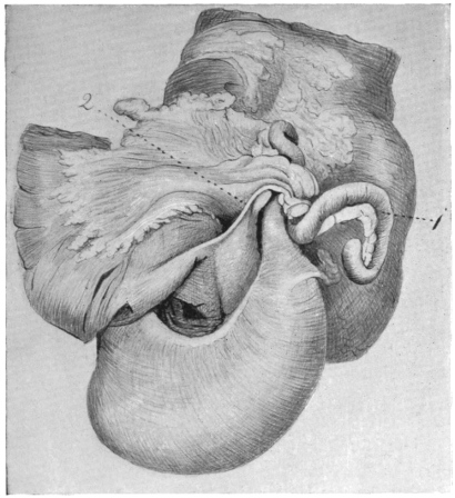
3. Simia satyrus, Orang-outang.—Figs. 458 and 459 represent respectively the ventral and dorsal views of the cæcum and ileo-colon in a nearly adult male specimen of orang, about 4½ feet high.
The cæcum is funnel-shaped, gradually narrowing to the origin of the appendix from its apex, which is carried upwards to the left by the well-marked crescentic curve of the pouch. The sweep of the funnel to the left and upwards is characterized by the curved course of the ventral longitudinal muscular band (Fig. 458), whose fibers spread out over a surface 3 cm. wide. The apex is thus placed behind the terminal ileum close to its entrance into the large intestine.
At the level of the upper margin of the ileo-colic junction the narrow pointed termination of the cæcum passes gradually into the beginning of the appendix (Fig. 459).
The appendix measures along its free border 22.6 cm. It follows the direction of the cæcal curve for 2.7 cm., at which point it appears somewhat constricted and takes an abrupt bend downwards for 4.3 cm.; curving again upwards for 7.5 cm., it turns downward a second time for 5.4 cm. and terminates in a hook-like extremity 2.7 cm. long (Fig. 459).
Fig. 460.—Troglodytes niger, chimpanzee. Ileo-colic junction and cæcum; ventral view. (Drawn from Columbia University Museum preparation No. 675.) 1. Appendix. 2. Intermediate non-vascular ileo-cæcal fold. 3. Colon. Fig. 461.—Troglodytes niger, chimpanzee. Dorsal view. (Drawn from Columbia University Museum preparation No. 675.) 1. Appendix. 2. Intermediate non-vascular ileo-cæcal fold. 3. Dorsal vascular fold.
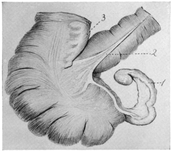
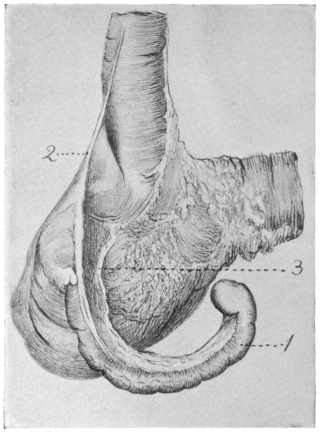
4. Chimpanzee, Troglodytes niger.—Figs. 460 and 461 represent the ventral and dorsal view respectively of the cæcum and ileo-colon in a young specimen.
The cæcum is curved to the left and the lowest point of the pouch is formed by the right lateral and ventral wall of the gut, but the extreme crescentic bend which carries the origin of the appendix up and to the left behind the ileo-colic junction is not yet developed in the young animal; on the other hand this character of the cæcum is typically apparent in Figs. 462 and 463, taken from an adult individual of the same species.
Fig. 462.—Troglodytes niger, chimpanzee. Ileo-colic junction and cæcum; ventral view. (Drawn from Columbia University Museum preparation No. 1083.) 1. Ventral vascular ileo-cæcal fold. Fig. 463.—Troglodytes niger, chimpanzee. Ileo-colic junction and cæcum; dorsal view. (Drawn from Columbia University Museum preparation No. 1083.) 1. Appendix.
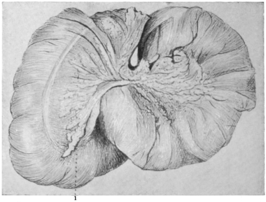
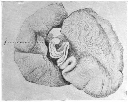
Fig. 464.—Troglodytes niger, chimpanzee. Ileo-colic junction and cæcum. (Drawn from Columbia University Museum preparation No. 1525.)
This extreme curve is well seen in the ventral view in Figs. 462 and 464, the latter taken from a large adult specimen. Seen from behind in Fig. 463 the sharp bend or kink in the lumen of the cæcal pouch produced by this curve is striking and resembles the arrangement of certain types of adult human cæca (p. 247).
II. PHYLOGENY OF THE TYPES OF ILEO-COLIC JUNCTION AND CÆCUM IN THE VERTEBRATE SERIES.
The segments of the alimentary canal illustrate very clearly the adaptation of structure to function. Diversity of kind and quantity of food habitually taken and variations in the rapidity of tissue metabolism produce marked morphological modifications in different forms. This is more especially the case with the junction of the mid- and hindgut, the site of development of the cæcal apparatus and of structural alterations of the large intestine possessing a similar physiological significance. No other portion of the visceral tract, with the possible exception of the stomach, illustrates more completely the result of physiological demand on the development of anatomical structure and the morphological possibilities of departure, progressive and retrograde, from a common primitive type in accordance with varying conditions of alimentation.
In coördinating, from the morphological standpoint, the structural differences encountered in this segment of the alimentary canal, two facts become apparent.
1. In the first place the serial study of the ileo-colic junction, as we can briefly define the region in question by borrowing the terminology of anthropotomy, reveals a limited number of principal structural types from which by successive gradations the vast variety of individual forms may be derived.
Fig. 465.—Schematic table of the vertebrate types of ileo-colic junction.
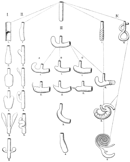
(In the schematic Fig. 465 the fundamental types and their derivatives are indicated. In the following the individual forms illustrating these types are referred to this schema in brackets.)
2. The observer will be impressed by the fact that representatives of all the main types of ileo-colic junction are found within a very limited zoölogical range, as within the confines of a single order. Examples of this are furnished by the Marsupialia and, to a lesser extent, by the Edentata. The members of these zoölogical groups, while united by certain common anatomical characters, such as the reproductive system and dentition, differ widely in habit and in the kind and quantity of the food normally taken. These differences in the method of nutrition have impressed their influence on the structure of the alimentary canal and have led to the evolution of varying and divergent types of ileo-colic junction. The study of this segment of the intestinal tract can therefore elucidate the mutual relationship of the vertebrate groups only to a limited degree and in special cases. On the other hand, it renders very clear the fundamental structural ground-plan common to all vertebrates and accentuates the specialized modifications of this plan which develop in response to the physiological environment. Moreover, such a review serves to reveal the significance of rudimentary and vestigial structures, such as the human vermiform appendix and the serous and vascular folds connected with the same. Throughout the entire vertebrate series the alimentary canal is found to respond with great readiness in its structure to varying grades of functional demand. This fact becomes still more apparent if the inquiry is not limited strictly to the region of the ileo-colic junction but takes into account likewise the structural modifications of similar physiological significance in other segments of the alimentary tract.
A cæcal pouch or diverticulum in some form at the junction of mid- and hindgut is a very common and widely distributed mammalian character. The activity of the tissue-changes in warm-blooded animals, and the consequent necessity for a rapid and complete digestive process, account for the structural modifications of the alimentary tract so commonly encountered among these forms. On the other hand, in the lower cold-blooded vertebrates, notably in fishes and amphibians, the metabolism is slow and the alimentary canal usually simple.
Specifically, the cæcum appears as a pouch or diverticulum in which food-substances, already partially digested and mixed with the secretions of the small intestine, are retained until their elaboration is completed and the nutritive value of the food ingested is secured for the organism. Consequently the most complicated and highly developed cæcal apparatus is found among mammalia in the Herbivora, such as the Ungulates and Rodents, whose food contains a comparatively small amount of nutriment in ratio to its bulk, and hence requires considerable elaboration before absorption. On the other hand the cæcum appears as a reduced or even rudimentary organ, or defaults entirely, in Carnivora whose food is concentrated and easily assimilated, containing only a small amount of non-nutritive material.
The function of the cæcal apparatus may be defined as follows:
Fig. 466.—Squalus acanthius, dog-fish. Alimentary tract, spleen, pancreas. (Drawn from Columbia University Museum preparation No. 1405.)
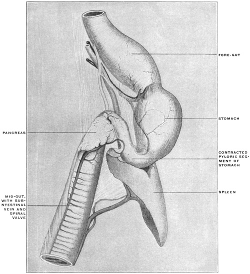
1. It provides space for the retention of partly digested substances, and of such as are difficult of digestion, mixed with the secretions of the preceding intestinal segment, until the digestive elaboration is completed.
2. It increases the intestinal mucous surface for absorption, and may develop, in certain cases, special localized areas of lymphoid tissue.
These two functional characters may be shared by other segments of the intestinal tract, which undergo corresponding structural modifications. It is only necessary to refer in this connection to the extreme morphological variations encountered in the stomach. The intestinal canal proper, however, in many instances exhibits structural peculiarities which possess the functional significance of the cæcal apparatus. Thus the projection into the lumen of the canal of a series of mucous folds, or the development of a continuous spiral mucous valve, evidently serves the double purpose of prolonging the period during which the intestinal contents are retained, and of increasing the intestinal mucous surface for absorption.
Fig. 467.—Galeus canis, dog-shark. Alimentary tract opened, showing spiral intestinal valve. (Drawn from Columbia University Museum preparation No. 1429.) Fig. 468.—Ceratodus forsteri, Australian lung-fish. Intestinal canal with spiral valve. (Columbia University Museum, No. 1645.)
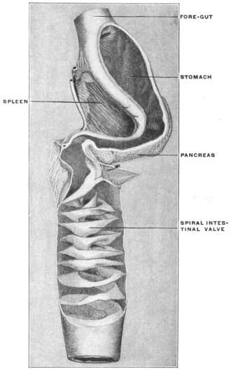
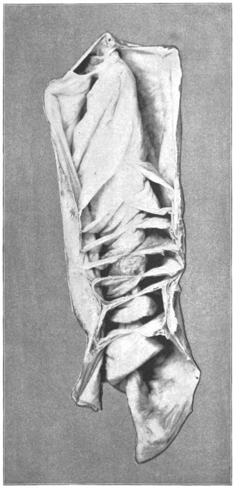
This spiral mucous fold is encountered in the straight intestinal canal of the Cyclostomata (Fig. 465, IV, 1, and Fig. 310), Selachians (Figs. 466 and 467) and Dipnœans (Fig. 468). Phylogenetically it is a very old structure, for evidences of its existence are found in the fossil remains of some Elasmobranchs. In the Ostrich (Fig. 341) the enormously developed cæca possess the same spiral mucous fold in the interior. The direct combination of the cæcum and spiral fold is again seen in certain mammalia, as in Lepus (Fig. 387). In some Ophidians the same physiological purpose is served by the manner in which the convolutions of the long intestine are bound together by a subperitoneal arachnoid membrane. The lumen of the canal is thus made to assume a spiral course (Figs. 331 and 469). The mucous folds of the human intestine, both the valvulæ conniventes and the crescentic folds of the large intestine, represent the same spiral valve, perhaps modified and influenced by the erect posture of man (Figs. 470-475).
Fig. 470.—Human small intestine, opened to show valvulæ conniventes. (Columbia University Museum, No. 1841.) Fig. 469.—Python molurus, Indian python. Mid-gut, distended and fenestrated to show spiral course of lumen. (Columbia University Museum, No. 725.) Fig. 472.—Human large intestine, opened and in section, showing colic plicæ. (Columbia University Museum, No. 1847.)
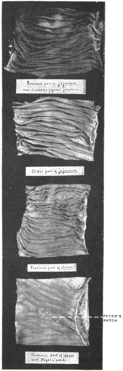
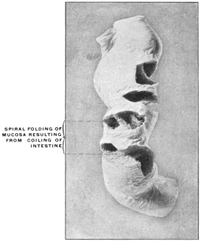
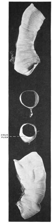
Fig. 471.—Human large intestine, showing colic tænia and plica. (Columbia University Museum, No. 1848.)
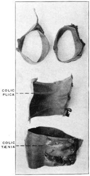
Fig. 473.—Cynocephalus anubis, olive baboon. Large intestine, with cross-section showing colic tænia and plicæ. (Columbia University Museum, No. 26/1168.) Fig. 474.—Comparison of portion of human transverse colon with distal segment of rabbit’s large intestine, showing same arrangement of longitudinal muscular bands (colic tænia) and colic sacculations. (Columbia University Museum, No. 1589.)
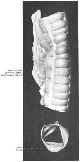
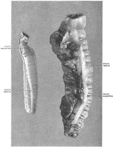
Fig. 475.—Felis leo, lion. Large intestine, with transverse section, showing smooth carnivore lumen, without sacculations or plicæ. (Columbia University Museum, No. 1600.) Fig. 476.—Pyloric cæca of Gadus callarias, codfish. (Columbia University Museum, No. 1825.) A. Bound together by connective tissue and blood-vessels. B. Dissected to show confluence of cæca to form a smaller number of terminal tubes of larger calibre entering the intestine.
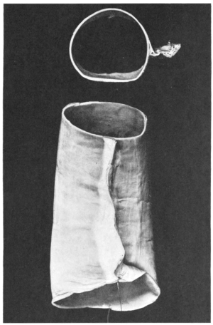
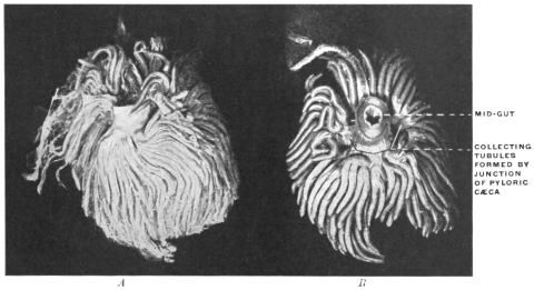
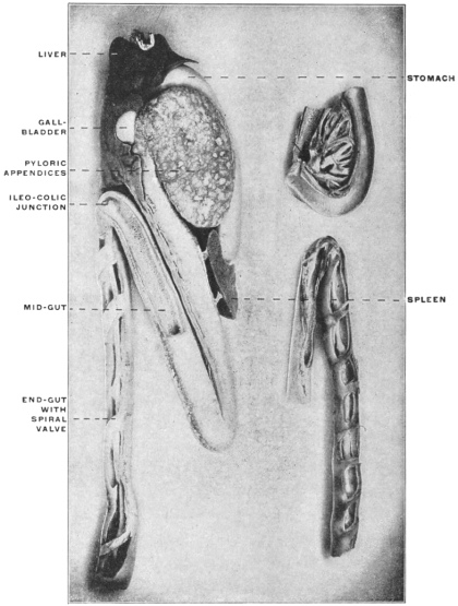
Fig. 477.
—Alimentary canal of
Accipenser sturio
, sturgeon. Numerous pyloric cæca are bound together to form a gland-like organ.
In the smaller upper figure on the left the stomach, mid-gut, and pyloric cæca are seen in section, showing the lumen of the latter and their openings into the mid-gut.
The lower left-hand figure shows the mid- and end-gut in section, the latter provided with a spiral mucous valve. (Columbia University Museum, Nos. 1826, 1827, and 1828.)
A second modification of the intestinal canal, suggesting the same physiological interpretation as the ileo-colic cæcum, is presented by the so-called pyloric cæca or appendices of many Teleosts and Ganoids already referred to (p. 119). While these structures in some forms very probably have assumed a secretory function (Figs. 476 and 477), they evidently act in others as diverticula in which material undergoing digestion is retained, while they increase at the same time the intestinal mucous secretory and absorbing surface (Figs. 478 and 479). They thus correspond physiologically to the ileo-colic cæcum. In this connection it is interesting to note that in Ganoids, which possess both the pyloric appendices and the spiral valve, the two structures develop in inverse ratio to each other, indicating their functional identity. In the serial review of the structure and significance of the vertebrate cæcum and ileo-colic junction these functionally allied modifications of other segments of the intestinal canal deserve notice.
The study of the vertebrate ileo-colic junction proper begins both ontogenetically and phylogenetically with the consideration of the primitive type in which the alimentary tube is not differentiated into successive segments and in which consequently no distinction between mid- and hindgut is found (Fig. 465). An example of this primitive condition is presented by the Cyclostomata, in whom the alimentary canal traverses the cœlom cavity as a straight non-differentiated cylindrical tube. Fig. 310 shows the alimentary canal of the Lamprey, Petromyzon marinus, and it will be observed that the intestine is provided with the spiral mucous fold above mentioned.
From this fundamental type the following main groups are to be derived:
Fig. 478.—Stomach, duodenum, and pyloric cæca of Lophius piscatorius, angler. (Columbia University Museum, No. 1824.) Fig. 479.—Pleuronectes maculatus, window-pane. Stomach and mid-gut with pyloric cæca and hepatic duct. (Columbia University Museum, No. 1432.)
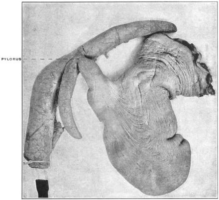
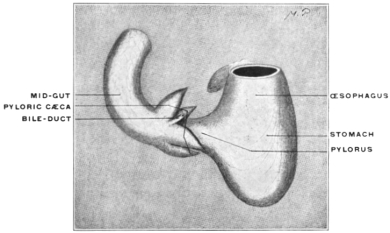
I. Symmetrical Form of Ileo-colic Junction. Mid- and Endgut in Direct Linear Continuity. (Fig. 465, I.)
1. Ileo-colic junction marked externally by an annular constriction, corresponding to a ring-valve with central circular opening in the interior (Fig. 465, I, 1).
This form is encountered in many Teleosts. The projecting annular mucous fold resembles the pyloro-duodenal valve.
Figs. 311-315 illustrate the structures in representative Teleosts.
Among the higher forms this type of ileo-colic junction is encountered in the simple alimentary canal of many Amphibians (Figs. 318-320). Among Reptiles it is found in certain lizards, as in Heloderma suspectum, the gila monster (Fig. 322). This animal lives almost entirely upon bird’s eggs, and its simple and reduced ileo-colic junction contrasts strongly with the highly developed and complicated cæcal apparatus of the phytophagous lizards, as Iguana (Figs. 326-330), affording one of the most striking illustrations of the effect which the character of the food habitually taken has on the structure of the alimentary canal in forms otherwise closely allied.
The same type of ileo-colic junction, as a reduction form, occurs in the arctoid group of Carnivora among Mammalia (cf. p. 212).
2. Differentiation in caliber of large and small intestine. Funnel-shaped ileo-colic transition.
This type, compared with the preceding, is characterized (Fig. 465, I, 2) by the greatly increased caliber of the large intestine, resulting in a funnel-shaped transition between mid- and hindgut, the small intestine continuing into the colon at the apex of the funnel.
Examples of this type are presented by several Edentates, Myrmecophaga jubata, the great ant-eater (Fig. 356), and Cholœpus didactylus, the two-toed sloth (Fig. 357).
3. Abrupt demarcation of small and large intestine with caliber differentiation (Fig. 465, I, 3).
The small intestine is still central at the ileo-colic junction, i. e., the axis of its lumen is continuous with the central axis of the colic lumen. In place of the gradual funnel-shaped transition of the preceding type the demarcation is abrupt.
An example of this form is furnished by another Edentate, Tatusia peba, the nine-banded armadillo (Fig. 358).
Among reptiles a similar well-marked ileo-colic transition is encountered in Alligator mississippiensis (Fig. 321).
4. Colic pouch prolonged back on each side of the ileo-colic junction, producing symmetrical colic cæca (Fig. 465, I, 4).
A growth of the colic tube cephalad, on each side of the junction with the midgut, leads to the formation of this type, characterized by the presence of two symmetrical globular cæcal pouches. In its simplest form this condition is illustrated by the double colic cæca of another armadillo, Dasypus sexcinctus (Fig. 359).
The bifid cæcal apparatus of the American manatee (Fig. 366) belongs to the same group.
5. Cæcal pouches of the birds (Fig. 465, I, 5).—A continuation of the backward extension of the bilateral colic pouches leads to the production of the typical double avian cæca in a greater or lesser degree of development. Frequently the cæca differentiate more completely from the colon, appearing as pouches of varying capacity joined to the large intestine by a narrower neck.
Figs. 334-341 show the well-developed pouches as they appear in representative avian types, while Fig. 333 illustrates the reduction of the cæcal apparatus encountered in many carnivorous birds.
6. Among mammalia Cyclothurus didactylus (Fig. 360), the little ant-eater, furnishes an example of double symmetrical globular cæca, connected with the colon by a narrow neck (Fig. 465, I, 6). Reference to the schema given in Fig. 465 will show that the types heretofore examined all have the following common character:
They appear derived from the primitive type by a differentiation in the caliber of the gut and by the gradual development of symmetrical bilateral cæcal pouches, resulting in central median implantation of the small intestine and its direct continuity with the colon.
II. Asymmetrical Development of a Single Cæcal Pouch, Lateral to the Ileo-colic Junction, Mid- and Endgut Preserving Their Linear Continuity. (Fig. 465, II.)
In the second general group the symmetry of the ileo-colic junction is disturbed. The following types are encountered, forming a series of successive stages:
1. The increase in the caliber of the large intestine is chiefly marked along the border opposite to the mesenteric attachment, resulting in a greater degree of convexity in this part of the intestinal wall (Fig. 465, II, 1). Among Reptilia this condition is found in the ileo-colic junction of some of the pond-turtles, as Pseudemys elegans (Fig. 323), while a mammalian example is furnished by the three-toed sloth, Arctopithecus marmoratus (Fig. 363).
2. An increase of this lateral extension of the colon leads to the formation of a single lateral cæcal pouch (Fig. 465, II, 2) such as is seen in another Edentate, Tamandua bivittata (Fig. 364), among Mammalia, and in certain Ophidians among Reptiles, as in the Anaconda (Figs. 331 and 332).
3. Prolongation of the pouch and reduction in caliber lead to the formation of the slender lateral cæcum found in all the Monotremes (Figs. 345-347, Fig. 465, I, 3). In its general appearance the cæcum of these singular animals bears a close resemblance to the cæcal pouches of many birds.
4. Direct continuity of small and large intestine, with lateral colic cæcum, extending along the convex free border of the terminal ileum and slightly convoluted at the extremity (Fig. 465, II, 4), characterizes the entire group of the Cebidæ among the new-world monkeys. The cæcum in these animals is a comparatively long pouch, nearly equalling in caliber the remainder of the intestine, occupying a distinctly lateral position, with the terminal portion rounded and slightly recurved (Figs. 453 and 454).
5. The Cynoid group of Carnivora, including the dogs, wolves, jackals and foxes, presents a similar relative position of small and large intestine and cæcum (Fig. 465, II, 5). The cæcum, compared with that of Cebus, is longer and more highly convoluted (Fig. 397). Variations encountered in certain forms indicate reversions to a more primitive type. Thus Fig. 398 shows the usual form in the dog, while Fig. 399 exhibits an occasional type in the same animal. The cæcum here is less twisted and indicates the probable derivation of the more commonly encountered type.
III. Rectangular Ileo-colic Junction with Direct Linear Continuity of Cæcum and Colon. (Fig. 465, III.)
The third general group, to which the large majority of Mammalia belong, is characterized in its typical form by a right-angled entrance of ileum into large intestine and by the direct caudal prolongation of the colon into a cæcal pouch of nearly uniform caliber with globular termination. The axes of the small and large intestine are not in the same line as in the two former groups, but are placed nearly at right angles to each other. With this change in the direction of the main intestinal segments the cæcum ceases to be a lateral appendage to the canal and appears as a caudal prolongation of the colon beyond the ileo-colic junction (Fig. 465, III). The type-form of this group is encountered among the herbivorous Marsupialia, such as the kangaroos and opossums. Fig. 350 shows the ileo-colic junction and cæcum in the rock wallaby, Halmaturus derbyanus, and Fig. 348 the same structures in our common opossum, Didelphis virginiana. The majority of the remaining mammalian forms depend upon modifications of this type, either in the direction of reduction of the cæcal apparatus, or of increased development with concomitant structural changes of similar physiological import in the proximal portion of the colon.
The following subdivisions of the general group may be established.
A. 1. The cæcum is long, markedly curved or uncinate, with the crescentic medial margin turned toward the free border of the terminal ileum. The entire pouch usually diminishes gradually in caliber to its termination (Fig. 465, III, A, 1). This type is encountered in a large group of new-world monkeys, including the marmosets and howlers.
Fig. 440 shows the structures in Hapale jacchus, one of the marmosets, and Fig. 443 illustrates the typical cæcum of this form in Ateles ater, the black-handed spider monkey.
2. The cæcum and appendix of man and of the anthropoid apes can be regarded as a reduction form of this type (Fig. 465, III, A, 2). Arrest of development of the terminal portion converts the distal segment of the cæcal pouch into an appendix whose relation to the apex of the funnel-shaped proximal segment or cæcum proper is seen in its pure form in the human embryo (Figs. 512 and 525). With the further development of the cæcum the sharper demarcation between it and the appendix results (Figs. 517 and 518). The displacement of the root of the appendix cephalad and to the left, toward the lower margin of the ileo-colic junction, as it is usually seen in adults, is due to the relatively greater growth of the right terminal sacculation of the cæcum compared with the left (cf. types of cæca, p. 248). Throughout these changes the initial crescentic curve of the cæcum, turning its concavity upwards and to the left, can be recognized by tracing the course of the longitudinal colic muscular bands. The cæca and appendices of the anthropoid apes present the same characters. The structures in the orang, chimpanzee, gorilla and gibbon are shown in Figs. 455-464.
B. The Æluroid and Arctoid groups of the Carnivora and the Pinnipedia constitute a very complete and instructive series illustrating the gradual reduction of the cæcum from the capacious pouch of the primitive type and its final complete elimination from the organism (Fig. 465, III, B).
In Hyæna (Fig. 416), the large cæcum with undiminished caliber of the terminal portion persists in its full development, as seen in the Marsupials furnishing the fundamental type (Fig. 465, III). The same type of cæcum is found in the lion (Fig. 417), the only true cat in which the cæcal apparatus has not undergone extensive reduction. Phylogenetically the presence of a capacious and uniform cæcal pouch in these two animals is exceedingly important and indicates that this type of cæcum represents the ancestral form common to the æluroid carnivore group, which, in the remaining living representatives, has become reduced in response to the influence which the character of the food has on the structure of this portion of the intestinal canal. The two instances of persistence of the primal type are all the more important as exceptions to the rule which is otherwise universal throughout the group.
1. The first example of this reduction (Fig. 465, III, B, 1) is encountered in the Aard-Wolf, Proteles lalandii, a near relative of hyæna (Fig. 406). The cæcum in this animal is considerably shortened, although still of fairly large and uniform caliber.
A similar type of cæcal reduction is encountered in the Pinnipede Carnivora. Fig. 396 shows the ileo-colic junction and the short blunt cæcum of the harbor seal, Phoca vitulina.
2. The cæcum of the typical Felidæ, other than the lion, is short and the terminal portion much reduced in caliber, constituting in many forms a species of pointed rudimentary appendix (Fig. 465, III, B, 2). Fig. 401 represents the typical feline cæcum as seen in the puma, Felis concolor. Among the smaller Æluroid Carnivora related to the true cats, as the civets and ichneumons, the terminal reduction of the short cæcum is still more marked, as seen for example in Herpestes griseus (Figs. 404 and 405).
3. In the Arctoid group of Carnivora (Fig. 465, III, B, 3 and 4) the reduction of the cæcal apparatus has been carried to the complete elimination of the pouch, restoring the primitive type of a straight intestinal tube without diverticulum as encountered above in some of the Edentates (Figs. 356 and 357).
In some forms allied to the true bears, such as Procyon, Bassaris, Cercoleptes, Taxidea and Nasua, the ileo-colic junction is marked externally by a slight constriction and internally by the projection of an annular pylorus-like valve (Figs. 407-409). The transition from the thin-walled ileum to the thick muscular walls of the large intestine is abrupt. The latter is very short and usually increases in caliber as it approaches the anal orifice. The mucosa of the terminal ileum presents very commonly one or two large oval areas of agminated follicles near the ileo-colic junction. The mucous membrane of the large intestine is thrown into prominent longitudinal folds. Fig. 408 shows the intestine of the brown coati, Nasua rufa, opened on each side of the ileo-colic transition.
In some of the Arctoidea, as Procyon and Nasua, the beginning of the colon just beyond the ileo-colic valve is bowed out opposite the mesenteric border indicating the original site of the eliminated cæcum, and recalling the arrangement of the intestine encountered above in Arctopithecus among the Edentates (Figs. 363, 407, 412, and 465, III, B, 3). Moreover, in the same forms rudimentary vascular and serous folds around the ileo-colic junction, corresponding to similar structures found in connection with a well-developed cæcal apparatus in other mammalia, point to the former existence of a cæcum.
4. In the typical Ursidæ even these remnants and traces of a cæcal pouch have disappeared and the intestinal canal preserves a uniform caliber, without any differentiation of large and small intestine (Figs. 414 and 415, Fig. 465, III, B, 4).
C. The last subdivision of the third main group contains forms in which the large uniform pouch of the primal type appears moderately reduced in length and sacculated, terminating either in a blunt extremity or carrying a distal constricted and rudimentary segment as an appendage.
1. The first of these types is encountered in the Old World cynomorphous monkeys. In all of these animals the cæcal pouch is wide but comparatively short, of nearly uniform caliber and sacculated like the rest of the colon, of which it forms the direct caudal continuation (Fig. 465, III, C, 1). The terminal portion of the pouch is usually blunt, globular and rounded (Figs. 428, 430 and 431), in a comparatively small number of forms slightly pointed (Figs. 427 and 437).
2. In the second group the terminal reduced portion persists either as a fairly distinct appendage, or in the form of a tapering pointed extremity into which the cæcal pouch proper is continued (Fig. 465, III, C, 2). This type is encountered in certain non-ruminant Ungulates. An example of the first condition is furnished by the cæcal apparatus of the peccary (Dicotyles torquatus) (Fig. 370), while the structures in Tapirus americanus (Fig. 377) illustrate the second form.
IV. Cæcal Apparatus Combined with Structural Modifications of the Proximal Colon of Similar Physiological Significance. (Fig. 465, IV.)
The fourth general mammalian group comprises forms in which the cæcal pouch is large, with or without terminal appendage, while in addition the large intestine develops structural modifications which possess the general functional significance of the cæcal apparatus. This highly developed and complicated structure of the alimentary canal indicates that the habitual food of these animals is bulky and difficult of digestion. Accordingly we find the group composed in main of the majority of the Ungulates and Rodents (with the exception of Myoxus), forms in which the diet under natural conditions is purely herbivorous. Other mammalian orders, however, also furnish representatives of this type of cæcal apparatus, the conditions as regards character and quantity of food habitually taken corresponding to those encountered among the Ungulates and Rodents. Thus the Phalangers among Marsupials (Fig. 352), Galeopithecus (Fig. 419) as an exceptional form among the Insectivora, and certain lemurs among Primates (Figs. 420-425) present examples of a highly developed and specialized type of cæcal apparatus.
The intestinal tract of these forms must therefore be considered from two points of view:
I. The cæcum proper.
II. The analogous structural modifications of the proximal segment of the colon.
I. CÆCUM PROPER.
The pouch of the Ungulates and Rodents, taking these forms as the typical representatives of the entire group, is usually of very large size compared with the rest of the alimentary canal. Two types are found:
1. Large capacious smooth cæcal pouch of uniform caliber (Fig. 465, IV, 2). This form is met with in the Muridæ among Rodents and is illustrated in Fig. 393 showing the cæcum of Mus decumanus, var. albinus, the white rat. Fig. 392 represents the entire alimentary canal of the meadow mouse, Arvicola pennsylvanicus, and indicates the proportion which the cæcal apparatus bears to the remainder of the intestinal tract. The typical cæcum of the Ungulates is shown in Fig. 371, taken from Capra ægagrus, the bezoar goat, and in Fig. 372, taken from a preparation of Boselaphus tragocamelus, the Nilghai.
2. The cæcal pouch is large, markedly crescentic in shape, sacculated, or provided in the interior with a more or less complete spiral valve, and reduced in caliber in the terminal segment, forming at times a pointed appendix (Fig. 465, IV, 3). This form is encountered typically among certain Rodents, as in Castor fiber, the beaver (Figs. 381 and 382), and Erethizon dorsatus, the Canadian porcupine (Figs. 383 and 384), but is not confined to this order. Thus cæca of very similar structure are found among the Marsupials, as in Phascolarctos and Cuscus (Fig. 352). In some of these forms the terminal reduction of the cæcum is very marked, resulting in a long narrow segment of the pouch tapering to a sharp point. It is significant to note in this connection that in one member of the marsupial order, the wombat (Phascolomys), this tendency to terminal reduction of the pouch has led to the development of a cæcum and appendix identical in structure and arrangement with the corresponding parts of man and the anthropoid apes (Fig. 354). This is merely another illustration of the fact, evidenced throughout the entire vertebrate series, that a primal type-form of cæcal apparatus, in responding to the conditions which influence the development of structural modifications, will produce identical specific types in animals otherwise widely separated in the zoölogical series.