Part 1 — ANATOMY OF THE PERITONEUM AND ABDOMINAL CAVITY. (continued 2)
Reading edition
Figs. 152-154.—Schematic representation of peritoneum in fixation of descending duodenum and formation of transverse colon and mesocolon.
Fig. 152.—Sagittal section through right kidney and descending duodenum before adhesion of latter to parietal peritoneum.
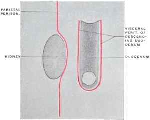
Fig. 153.—Adhesion of descending duodenum to primitive parietal peritoneum. Colon and mesocolon after rotation of the intestine, but before adhesion.
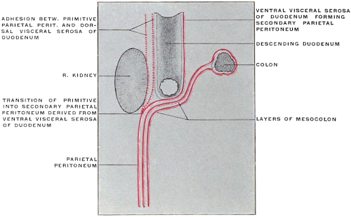
Fig. 154.—Adhesion of mesocolon to duodenum and primitive parietal peritoneum, resulting in formation of root of transverse mesocolon.
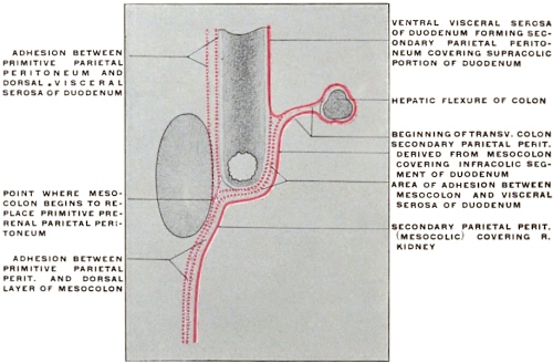
Now fix the transverse mesocolon firmly against the background of the abdomen and place the ascending and descending colon as far as possible over to the right and left side respectively. We will assume a line of secondary adhesion between the transverse mesocolon and the parietal peritoneum investing the dorsal abdominal wall. Along this line the upper or cephalic surface of the transverse mesocolon would become continuous with the dorsal parietal peritoneum, while the lower or caudal layer would still be continuous with the left leaf of the ascending and the right leaf of the descending mesocolon. We have already seen that the duodenum and mesoduodenum become anchored in the subhepatic region and that the visceral ventral peritoneum of the gut and the original left leaf of the mesoduodenum appear then as secondary parietal peritoneum. Hence a sagittal section through the right lumbar region, right kidney and descending duodenum would, immediately after rotation and establishment of the transverse mesocolon, show the peritoneal arrangement indicated in Fig. 153. After adhesion of the transverse mesocolon continuity would be established between its upper or cephalic layer and the secondary parietal peritoneum investing the supra-colic portion of the descending duodenum (Fig. 154) while its caudal layer becomes continuous with the secondary parietal peritoneum covering the infra-colic segment of the duodenum and the lower portion of the ventral surface of the right kidney.
Reference to the schematic Figs. 152, 153 and 154, will show that the adult duodenum becomes fixed to the posterior parietes of the abdomen by adhesion of its visceral serous covering and of the dorsal layer of the mesoduodenum to the primitive parietal peritoneum. The supra-colic segment of the adult descending duodenum lies under cover of a single peritoneal layer, derived from its own visceral investment and appearing as secondary parietal peritoneum by continuity laterad along the line of adhesion with the primitive parietal peritoneum covering the upper part of ventral surface of right kidney, while mesad, the layer covering this segment of the duodenum, is continued into the secondary parietal peritoneum derived from the left or ventral leaf of the mesoduodenum and covering the ventral surface of the pancreas (cf. Figs. 138-140).
On the other hand, the infra-colic segment of the descending duodenum, as well as the lower and mesal angle of the ventral surface of right kidney, between ascending and transverse colon, is covered by a layer of secondary parietal peritoneum derived from the ventral layer of the ascending mesocolon and continuous with the caudal layer of the transverse mesocolon. Beneath this secondary parietal peritoneum are two obliterated layers, on the one hand the dorsal layer of the mesocolon, on the other the visceral infra-colic duodenal serosa and the primitive prerenal parietal peritoneum.
In the further development of the adult human arrangement the changes below the level of the transverse colon and mesocolon result in the fixation of the ascending and descending colon to the background of the right and left lumbar regions. The opposed serous surfaces of the ascending and descending mesocola and of the dorsal parietal peritoneum adhere and the process also usually involves the dorsal visceral peritoneum of the ascending and descending colon, so that these portions of the gut obtain a fixed position.
Adhesion of the mesocolon to the dorsal body wall (parietal peritoneum) does not occur at all points at the same time. Usually adhesion proceeds from the midline laterad. The fixation of the ascending colon in the human embryo begins about the fourth month.
In the descending segment by the same time adhesion has usually proceeded nearly up to the descending colon, but the intestine itself is as yet free. In the fifth month the descending colon has usually become fixed between the splenic flexure and the beginning of the sigmoidea. In the latter region a free mesocolon usually persists throughout life.
Differences in the rate of growth between the length of the body wall and the length of the mesocolon may play an important part in the production of peritoneal fossæ, small pouches which in some regions of the abdomen may assume considerable proportions. Such fossæ are found around the duodeno-jejuneal angle, the cæcum and appendix, and the sigmoid flexure. They will be considered more in detail with these respective regions, especially in reference to their relation to retro-peritoneal hernia.
In a certain proportion of cases adhesion between the parietal peritoneum and the ascending and descending mesocolon is incomplete or entirely wanting, resulting in the formation of a more or less completely free ascending and descending mesocolon. Treves, in an examination of 100 bodies, obtained the following figures:
In 52 subjects there was neither an ascending nor a descending mesocolon, the intestine being fixed in the manner which is regarded as normal.
In 22 there was a descending, but no trace of an ascending mesocolon.
In 14 a mesocolon was found in both the ascending and descending segments of the large intestine.
In 12 there was an ascending mesocolon, but no corresponding fold on the left side. Hence from this series a mesocolon may be expected on the left side in 36 per cent., on the right side in 26 per cent.
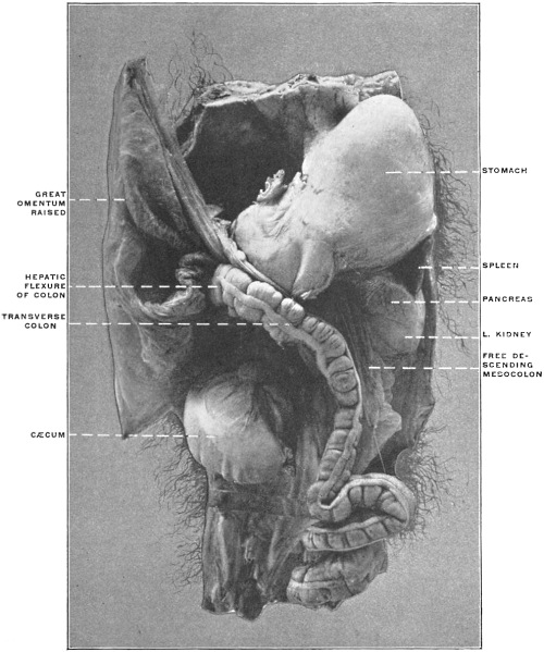
Fig. 155.—Abdominal viscera of Macacus cynomolgus, Kra monkey, hardened in situ. (Columbia University Museum, No. 1801.)
Both development and comparative anatomy would lead us to expect that the descending mesocolon would be found more frequently than the ascending.
In the lower animals the descending mesocolon is always an extensive and conspicuous membrane. It is well developed in all monkeys and the anthropoidea, as the remains of the primary vertical fold of the dorsal mesentery, while the ascending mesocolon is a secondary production, acquired during the development of the bowel by rotation.
In most of the lower monkeys the ascending mesocolon is also largely or entirely free. The descending mesocolon can always in these animals be reflected to the median line (cf. Fig. 155).
The line of attachment in man of the descending mesocolon is usually along the lateral border of the left kidney and vertical, while the line of attachment of the ascending mesocolon is usually less vertical, crossing the caudal end of the right kidney obliquely from right to left and with an upward direction (Fig. 156).
Fig. 156.—Schema of visceral and peritoneal relations of ventral surface of right kidney.
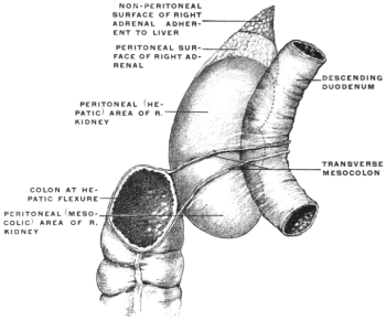
In like manner when both the ascending and descending mesocola are absent as free membranes the left or descending colon is adherent along the lateral border of the kidney to the abdominal parietes, while the ascending colon is fixed at the hepatic flexure a little obliquely across the ventral surface of the caudal end of the corresponding gland ascending toward the medial margin.
Treves found in the cases of persistent ascending mesocolon in the adult that the membrane varied in breadth from 1″ to 2″ while the persistent fold on the left side varied between 2″ and 3″ in breadth.
In the fœtus, up to 5″–6″ in length, the descending mesocolon is usually an extensive fold. Its attachment is vertical, but nearer to the median line than in the adult, usually along the medial border of the left kidney. It is at times found attached along this line in the adult.
An ascending mesocolon is rare even in the fœtus. The cæcum and beginning of the ascending colon are completely invested by peritoneum, but above the parts so invested the colon is usually adherent along an oblique line to the ventral and medial aspect of the right kidney.
In the fœtus at full term, if the cæcum is still undescended and in contact with the liver, it is not uncommon to find the cephalic portion of the descending colon provided with a mesocolon, while the caudal part of the descending colon is fixed by adhesion to the ventral surface and lateral border of the left kidney. This free membrane is then really a part of the transverse mesocolon. Where the cæcum descends to the iliac fossa the portion of the fœtal descending colon so invested is drawn over to the right and incorporated in the transverse colon.
Treves in two out of 100 bodies found the cæcum in the right iliac region, but both it and the whole of the ascending colon were entirely free from any peritoneal connections with the dorsal parietes of the abdomen.
The gut from the tip of the cæcum to the hepatic flexure was entirely invested by peritoneum continuous with the mesentery. The ascending colon was covered in the same manner and by the same fold as the small intestine. The segment of large intestine thus free measured 8″ in both instances.
The mesentery lacked the usual attachment to the dorsal abdominal wall and its root was represented by the interval between the duodenum and the transverse colon. The membrane had no other than its original primary attachment, and small intestine and ascending colon formed together a loop that practically represented the condition of the great primary intestinal loop. (Compare p. 73.)
The arrangement presented in these two subjects corresponds to that met in many animals, such as the cat.
A cross-section of the cat’s abdomen arranged as above would show the following disposition of the peritoneum, corresponding to the stage in the human development preceding the fixation of the two vertical colic segments (Fig. 157). It will be seen that the right and left mesocola can be reflected to the median line where they become continuous ventrad of the vertebral column and aorta with the mesentery of the small intestine. The ventral surfaces of both kidneys are seen to be covered by the primitive parietal peritoneum of the abdominal cavity.
Figs. 157, 158.—Schema showing peritoneal arrangement in transection of infra-colic compartment of abdomen before and after fixation of ascending and descending colon.
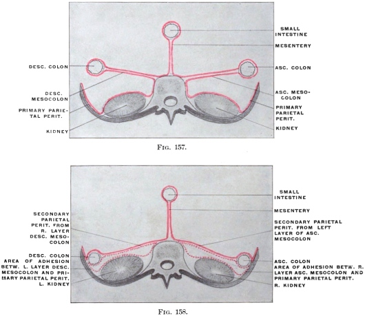
Fig. 158 shows the adult human arrangement of the same parts, after fixation of the vertical colic segments by adhesion of the opposed surfaces of their mesocola and the primitive parietal peritoneum. The background of the abdomen is now seen to be covered by a layer of secondary parietal peritoneum, viz., the original left leaf of the ascending and right leaf of the descending mesocolon, continuous above with the lower or caudal layer of the transverse mesocolon.
This adhesion is so complete that the original condition is disregarded in adult descriptive anatomy. The layer which has adhered to the parietal peritoneum can no longer be recognized and the other has assumed the rôle of parietal peritoneum.
The connection of the transverse mesocolon with the dorsal lamella of the great omentum will be considered below.
The course of the vessels in the ascending and descending mesocola is not altered by the secondary adhesions. These vessels are in the adult situated behind the secondary parietal peritoneum derived from the mesocola.
The origin of the transverse mesocolon obtains by the fixation of the hepatic and splenic flexures high up in the abdomen a transverse course, and the transverse growth of the abdomen holds the membrane in this position cephalad of the duodeno-jejunal flexure, so that on elevating the transverse colon the mesocolon appears as separating the upper from the lower abdominal compartment. This posterior line of attachment or so-called “root of the transverse mesocolon,” is nothing more than the upper limit of the area of adhesion between the primitive parietal peritoneum and the opposed surfaces of the ascending and descending mesocola. Reference to the abdominal cavity of the cat after complete rotation (Fig. 146) will show the original continuity of the three mesocola very clearly. A secondary connection is established along the lateral border of ascending and descending colon (Fig. 158), between the primitive parietal peritoneum and the ventral visceral peritoneal investment of the large intestine. Both of the vertical segments of the colon now appear fixed. Their dorsal surfaces are uncovered by peritoneum and can be reached in the lumbar region, laterad of the kidney, without opening the peritoneal cavity (lumbar colotomy).
The caudal portions of both kidneys are covered, beneath the secondary parietal peritoneum, by a layer of loose connective tissue representing the result of obliteration by adhesion of the first and second of the original three layers of prerenal peritoneum, viz., the primitive parietal (1) and the two layers of the mesocola (2 and 3).
Line of Attachment of the Mesentery of the Jejuno-ileum.—Examination of the caudal surface of the transverse mesocolon in the cat, with the parts in the above outlined position, will show how and why in the adult human abdomen the duodeno-jejunal angle appears to dip out from beneath the transverse mesocolon, becoming gradually more and more free until complete transition to the mobile jejunum is obtained. From this point, situated to the left of the second lumbar vertebra, the dorsal attachment of the adult human mesentery of the jejuno-ileum extends somewhat obliquely caudad and to the right to terminate in the right iliac fossa at the ileo-colic junction.
Returning to the conditions presented by the cat’s intestines to obtain an explanation of this line of fixation we must recall the fact that in the peritoneum included within the limits of the umbilical loop, after differentiation of small and large intestine, but before rotation, we have both the elements of the mesentery of the small intestines and of the ascending and transverse mesocolon combined (Fig. 141). For it will be seen that this membrane carries at this time vessels both to the jejuno-ileum and to the segments of the large intestine (cæcum, ascending and transverse colon). This fact will be at once recognized if the cat’s intestines are arranged to correspond to the primitive condition (Fig. 136) and the mesentery examined.
After rotation and differentiation of the colic segments and after the adhesion of the ascending and descending colon in man, the course of the main trunk of the superior mesenteric artery passes, after crossing the third portion of the duodenum, down and to the right to terminate near the ileo-colic junction by anastomosis with its ileo-colic branch. The adhesion of the right and left mesocola to the dorsal parietal peritoneum proceeds mesad as far as this line, leaving free the mesentery of the small intestines, which contains the vasa intestini tenuis derived from the left side of the main vessel. The secondary line of attachment of the mesentery to the abdominal background is therefore along this line. To obtain a clear idea of these processes of development in man assume that in the cat, after rotation and establishment of the three divisions of the colon, the two vertical (ascending and descending) mesocola become adherent to the dorsal parietal peritoneum, leaving the mesentery of the small intestine free.
Fig. 159.—Schematic figure to show lines of mesocolic adhesion, formation of root of transverse mesocolon and root of mesentery of jejuno-ileum in human subject.
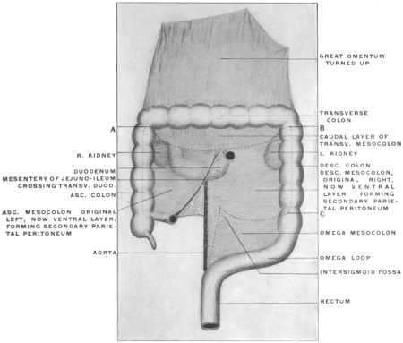
Fig. 159 illustrates schematically the area of mesocolic adhesion in the human subject after complete rotation, and the line of the mesentery of jejuno-ileum.
Fixation of the ascending and descending cola and of their mesocola proceeds cephalad as far as the line AB, which thereby constitutes the root of the free transverse mesocolon.
The secondary parietal peritoneum derived from the ventral layer of the ascending mesocolon covers the lower and inner portion of the ventral surface of the right kidney, the infra-colic division of the descending and the dextro-mesenteric segment of the transverse duodenum, while along the root of the jejuno-ileal mesentery it becomes continuous with the right layer of that membrane. The secondary parietal peritoneum derived from the ventral layer of the descending colon covers the lower part of the ventral surface of the left kidney and the sinistro-mesenteric segment of the transverse duodenum and becomes continuous along the mesenteric radix with the left layer of the jejuno-ileal mesentery.
Caudad the adhesion of the descending colon and mesocolon to the parietal peritoneum proceeds only to the point C, following the dotted line mesad and resulting in the formation of the free mesocolon of the sigmoid flexure.
Résumé of the Adult Arrangement of the Human Peritoneum in the Lower Compartment of the Abdomen, Below the Level of the Transverse Colon and Mesocolon.—We should now consider the arrangement of the human peritoneum in the adult below the dorsal attachment of the transverse mesocolon in the light of the embryological and comparative anatomical facts just stated. In doing this it will be advisable to study both the actual conditions encountered and their significance in the sense of determining the derivation of the peritoneal layers from the primitive dorsal mesentery. Open the abdominal cavity in the usual manner by a cruciform incision.
Turn the great omentum up on the chest wall, exposing the underlying intestines. This manipulation, as already stated, will cause the omentum to carry the transverse colon with it, on account of the adhesion, in the adult, of the gut to the dorsal layer of the omentum. Hence the cephalic or upper layer of the transverse mesocolon will not be seen at this stage because the omental adhesion just referred to prevents us from passing between the greater curvature of the stomach and the transverse colon without tearing peritoneal layers. It will, however, be possible to trace on the right side the duodenum from the pylorus down ventrad of the right kidney until the descending portion disappears behind the hepatic flexure of the colon. With the omentum and transverse mesocolon turned up, as stated, and the transverse mesocolon put upon the stretch, it will be seen that the abdominal space now overlooked is bounded cephalad by the lower layer of the transverse mesocolon and its attachment to the dorsal abdominal wall. The lateral limits of the space are given by the ascending and descending colon respectively. The attachment of the mesentery of the small intestine to the oblique line extending from the left of the vertebral column at about the level of the second lumbar vertebra to the right iliac fossa subdivides the entire space into a secondary right and left compartment.
Begin by following the caudal layer of the transverse mesocolon dorsad on the right side. In the angle between ascending and transverse colon (hepatic flexure) pressure will locate the caudal portion of the ventral surface of the right kidney. Remember that the peritoneum touched in these procedures appears in the adult as parietal prerenal peritoneum, but that in reality it is the left leaf of the originally free ascending mesocolon, whereas the original right leaf of this membrane and the primitive parietal peritoneum have, by adhesion of their serous surfaces, been converted into the loose subserous connective tissue covering the ventral aspect of the kidney beneath what now appears as parietal peritoneum.
Mesad of the resistance offered to the finger by the right kidney the caudal (infra-colic) portion of the descending duodenum and the angle of transition between it and the third or transverse portion will be found, invested in the same way by secondary (mesocolic) parietal peritoneum. It will be seen, especially if the duodenum is injected or inflated, that the hepatic flexure of the colon lies ventrad of the vertical descending second portion of the duodenum, so that one part of this intestine is situated cephalad the other caudad of the colon. (Supra- and infra-colic segments of descending duodenum.)
Individual differences are observed in the area of colic attachment to the duodenum. Usually the two intestines are in contact with each other and adherent over a considerable surface. Exceptionally the transverse mesocolon extends across to the right so as to include the hepatic flexure. In this latter case the uncovered non-peritoneal surface of the descending duodenum is small, represented by the interval between the layers of the transverse mesocolon, and the hepatic flexure is then not directly adherent to the gut.
If we now trace the transverse duodenum from right to left we will encounter the right layer of the root of the jejuno-ileal mesentery. The caudal layer of the transverse mesocolon, the right leaf of the mesentery and the secondary parietal peritoneum investing the ventral surface of the transverse duodenum all meet at this point. Surround the mesentery of the free small intestine with the fingers of one hand so that the entire mass of intestinal coils can be swung alternately from side to side.
Turning them over to the left, as already stated, the proximal portion of the transverse duodenum can be traced from right to left as far as the root of the mesentery. Here the peritoneum investing the ventral surface of the duodenum becomes continuous with the right leaf of the mesentery. Now swing the whole mass of small intestines over to the right, exposing the parietal peritoneum in the space to the left of the vertebral column, between the attachment of the mesentery to the median side, the root of transverse mesocolon cephalad and the descending colon to the left. Remember that the same significance attaches to this secondary parietal peritoneum as on the right side. It appears in the adult as parietal peritoneum, but is in its derivation the original right leaf of the descending mesocolon. Close to the root of the mesentery the continuation from the right side of the transverse duodenum will be seen, crossing the median line from right to left ventrad of aorta and vertebral column and usually turning cephalad on the left side of the lumbar vertebræ, as the fourth or ascending duodenum, to reach the caudal surface of the transverse mesocolon near its attachment, where the gut turns ventrad to form the duodeno-jejunal angle and become continuous with the free small intestine.
From the fact that the transverse duodenum is thus seen on each side of the root of the mesentery it will be recalled that after rotation of the primitive intestine the superior mesenteric artery crosses the transverse portion of the duodenum to reach its distribution between the leaves of the mesentery. Hence this portion of the small intestine consists of a dextro- and sinistro-mesenteric segment. This intersection of mesentery and duodenum marks the site of the primitive duodeno-colic isthmus through which the superior mesenteric artery passed to reach its distribution to the gut composing the embryonic umbilical loop.
To the left of the ascending duodenum a portion of the caudal surface of the pancreas will be seen, covered by the continuation of the caudal leaf of the transverse mesocolon into the parietal peritoneum. The consideration of this relation of peritoneum and pancreas will profitably be deferred until we have studied the developmental changes in the region of the dorsal mesogastrium and great omentum.
In the angle between termination of the transverse colon and proximal part of descending colon (splenic flexure) the caudal part of the ventral surface of the left kidney will be felt. The disposition of the peritoneum and its significance is the same as on the right side. Inasmuch as we have already seen that the secondary parietal peritoneum covering the dorsal abdominal wall on each side of the small intestine’s mesenteric attachment is derived from the primitive ascending and descending mesocolon, it will be readily understood why the blood vessels supplying the ascending and descending colon (arteria ileo-colica, a. colica dextra, a. colica sinistra) are placed behind the parietal peritoneum, while the colica media, supplying the transverse colon, runs between the layers of the transverse mesocolon. Originally the same condition obtained for the two vertical colic segments, but with the anchoring of these portions of the large intestine and the adhesion of their mesocola to the parietal peritoneum the blood vessels which formerly ran between the two layers of the membrane, as long as it remained free, now appear as retroperitoneal vessels placed beneath the parietal peritoneum derived secondarily from the mesocola.
This fact must be borne in mind in studying the arrangement of certain folds and fossæ of the parietal peritoneum which are now to be considered.
Duodenal Fossæ. Fossa of Treitz and Retro-peritoneal Hernia.—The peritoneal cavity of the cat can be used to great advantage in order to obtain a clear idea of the formation of these folds and fossæ, whose relation to the so-called “retro-peritoneal hernia” has led to an exaggerated elaboration of minute detail and a somewhat puzzling terminology in human descriptive anatomy.
Fig. 160.—Abdominal cavity of cat, with intestines everted and elevated to show duodenal fold. (From a fresh dissection.)
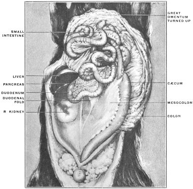
Fig. 161.—Abdominal viscera of Nasua rufa, brown coaiti. (From a fresh dissection.)
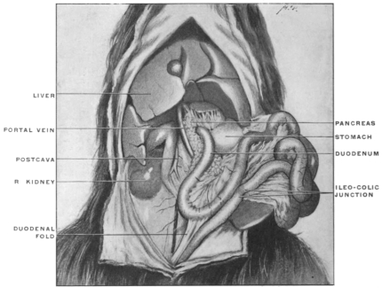
Fig. 162.—Abdominal viscera of Hapale vulgaris, the marmoset. (Columbia University Museum, No. 1818.)
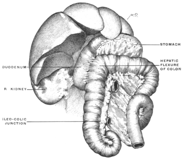
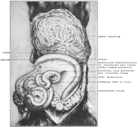
Fig. 163.—Abdominal viscera of cat; intestines rotated and turned to the right to show duodenal fold. (From a fresh dissection.)
Directions for Examining the Folds and the Formation of the Duodeno-jejunal Fossa in the Cat.—Turn the omentum and the coils of the small intestine cephalad out of the abdomen until they rest upon the ventral thoracic wall. Press the large intestine over to the left side, putting the mesocolon on the stretch until the parts are arranged as shown in Fig. 160. The loop of the duodenum with the head portion of the pancreas will be seen caudad of the liver and ventrad of the right kidney. A well-marked peritoneal fold, somewhat sickle-shaped, with the concavity of the free edge directed caudad and to the right, will be seen extending from the convex border of the duodenum, directly opposite the mesenteric or attached margin, to the right leaf of the mesocolon. This fold indicates the beginning adhesion of the duodenum to the mesocolic peritoneum, the first step toward the subsequent complete fixation of the gut as it is found in man.
Fig. 161 shows the abdominal cavity of Nasua rufa, the brown Coati-mundi, a South American arctoid carnivore, with the intestines everted and turned to the left side. In this animal the large intestine is very short, there is no cæcum, the ileo-colic junction is only marked on the surface by a pyloric-like constriction of the tube and in the interior by the projection of a ring-valve (Fig. 408).
The duodenal fold is very well developed, passing between the convex surface of the duodenal loop and the adjacent right leaf of the short mesocolon.
In Primates, in which complete rotation of the intestine, on the plan of the human development, takes place, still further and more extensive agglutination of the serous surface of the duodenum to the peritoneum of the mesocolon occurs. Fig. 162 shows the condition in Hapale vulgaris, one of the marmosets. The ascending and descending mesocola and the mesoduodenum of this animal are still free, but the surface of the duodenum has become fastened to the opposed mesocolon. With fixation of the hepatic flexure and adhesion of the ascending colon, such as occurs in man, the duodenum is carried dorsad against the ventral surface of the right kidney, and now anchoring of the duodenum, by obliteration of the mesoduodenum and adhesion to the prerenal parietal peritoneum, takes place as already detailed above. To return now to the formation of the duodeno-jejunal fossa by means of this fold, as illustrated in the cat. Perform the manipulations already described in rotation of the intestine. The appearance of the parts then will be as shown in Fig. 163. The large intestine is drawn over so as to represent the human ascending and transverse colon in one segment, the descending colon in the other, and the mesocolon appears correspondingly as transverse and descending. In other words the cat’s intestines as arranged in the figure would represent the stage in the human development in which cæcum and beginning of large intestine are still subhepatic in position ventrad of the right kidney, before differentiation of ascending and transverse colon by descent of cæcum into right iliac fossa.
In the human subject, as we have seen, the transverse mesocolon obtains a secondary attachment to the background of the abdominal cavity, its caudal surface remaining free.
The descending mesocolon turns its original right leaf ventrad, its left leaf dorsad, and the latter adheres to the primitive parietal peritoneum covering the left lumbar region and ventral surface of left kidney. This area of adhesion extends up to and usually involves the dorsal surface of the descending colon, anchoring the same in the left lumbar region, down to the point where the sigmoid flexure begins and where the original mesocolon again appears free.
In the cat, therefore, with the intestines arranged to correspond to the course of the human large intestine after rotation has been accomplished, the lines representing the peritoneal human adhesions should be fixed, as shown in the schema, Fig. 159: AB, line of secondary attachment after rotation resulting in the formation of the “root” of a free transverse mesocolon. BC, line of limit of secondary adhesion to the original parietal peritoneum involving the entire left (now dorsal) layer of the descending mesocolon and the dorsal surface of the descending colon, resulting in the fixation of the latter part of the large intestine.
This establishes, as already stated, a secondary parietal peritoneal surface in the left lumbar region derived from the original right leaf of the descending mesocolon. Inasmuch as the inferior mesenteric vessels originally passed to the descending colon between the layers of the mesocolon they will now apparently be placed beneath the (secondary) parietal peritoneum of the left lumbar region.
If now the duodenal fold in the cat be examined after rotation of the intestine it will be found presenting the original relations (Figs. 160 and 163), viz., passing from the convex margin of that portion of the duodenal loop which would correspond to the human fourth or ascending portion, to the original right layer of the mesocolon, which in man becomes secondarily converted into the parietal peritoneum of the left lumbar region. Hence the connections of the fold are as follows:
On the right: ventral surface of the ascending duodenum.
On the left: right layer of mesocolon (secondary lumbar parietal peritoneum in the adult human subject).
Cephalad it abuts against the caudal layer of the transverse mesocolon along the line which would correspond to the root of the mesocolon in the adult human subject.
The concave caudal edge is free and bounds the entrance into a fossa, the “superior duodenal fossa” of anthropotomy. This fossa opens caudad and extends cephalad to the root of the transverse mesocolon. The ventral and left wall of the fossa is formed by the fold in question, its background by the mesocolon (right leaf); to the right the left circumference of the ascending duodenum enters into the formation of the fossa, and its fundus is formed by the confluence of the fold and of the caudal layer of the transverse mesocolon. The inferior mesenteric vessels are found near the left margin of the entrance into the fossa.
Fig. 164.—Abdominal viscera of Nasua rufa, the brown coaiti, showing the position of the duodenal fold after rotation of the intestine. (From a fresh dissection.)
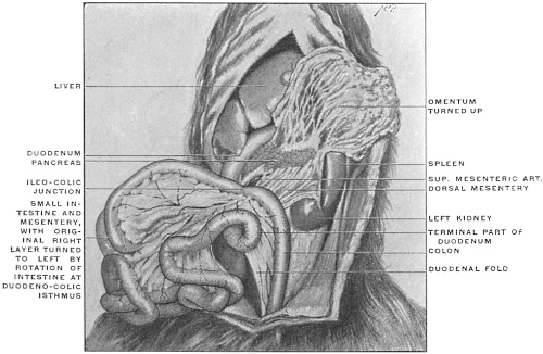
Fig. 165.—Abdominal viscera of human fœtus at term, arranged to show duodenal folds and fossa. The jejuno-ileum, ascending and transverse colon have been removed. (Columbia University Museum, No. 1819.)
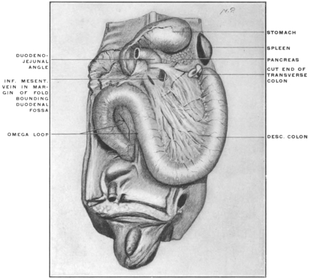
Fig. 164 shows the appearance of the fold in Nasua rufa after rotation of the intestine. The short course of the large intestine in this animal, and the consequent reduction of the mesocolon, brings the fold much below the level which it occupies in the cat.
If we now look for the corresponding structures in man we will find certain modifications depending chiefly upon still closer adhesion between duodenum and the mesocolon which is destined to become the left parietal peritoneum after anchoring of the descending colon. We have already encountered an example of such closer connection in the marmoset shown in Fig. 162.
In all cases the “superior duodenal” fold, corresponding to the fold just encountered in the cat, is the original condition, and the duodenal fossa consequently opens caudad. In many instances this will be the only fold and fossa encountered in the adult human subject. In other instances more extensive duodeno-mesocolic adhesions result in the addition of an “inferior fold,” bounding a fossa the entrance into which is directed cephalad toward the transverse mesocolon. Such a condition is seen in Fig. 165 taken from a fœtus at term. The duodenal fossa in this case is bounded by an “upper” and “lower” duodenal fold continuous with each other on the left side, but separated on the right at their attachment to the duodenum. It will be seen that the inferior mesenteric vein runs in the left margin of the fold, following along the left border of the entrance into the fossa. A segment of the colica sinistra artery may occupy the same position. This position of the vein, or artery, or of both vessels, is not the cause leading to the formation of the duodenal fossa, but is more or less accidental and variable. In many cases the vessels run at some distance from the folds bounding the fossa.
In some subjects the “inferior” fold is the only one found, and the only duodenal fossa then encountered looks cephalad. This condition, when associated with the course of the inferior mesenteric vessels in the free edge of the fold, constituted the classical “fossa duodeno-jejunalis” of Treitz, and is described as “Treitz’s fossa.”
Fig. 166.—Abdominal viscera of human fœtus at term. (Columbia University Museum, No. 1820.)
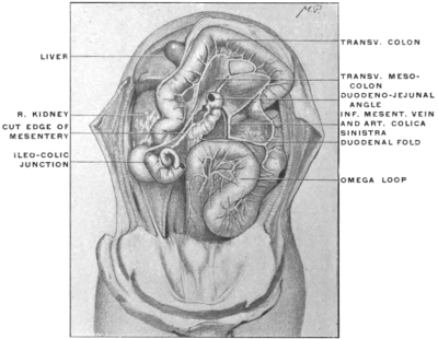
Fig. 167.—Abdominal viscera of adult human subject, showing duodenal folds and fossa. (From a fresh dissection.)
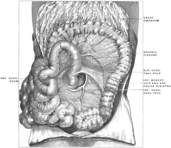
Fig. 168.—Abdominal viscera of adult human subject, showing duodenal folds and fossa. (From a fresh dissection.)
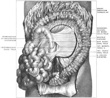
Fig. 166 shows the condition in which only a small inferior fold attaches itself to the termination of the transverse duodenum. There is practically an entire absence of duodenal or duodeno-jejunal folds and fossæ. The inferior mesenteric vessels course under cover of the mesocolic secondary parietal peritoneum, but do not produce a fold.
Fig. 167, from an adult human subject, illustrates the further development of the fossa from the fœtal conditions shown in Fig. 165. The well-marked duodenal fossa is bounded by a superior and inferior duodenal fold, uniting laterally in a crescentic margin containing a segment of the inferior mesenteric vein and colica sinistra artery. The lower division of the peritoneal recess thus produced corresponds to the typical (vascular) “fossa of Treitz.” Mesally the projection of the fourth portion of the duodenum bounds the fossa.
In Fig. 168, also taken from an adult human subject, an extensive duodenal recess is bounded in the same way by a superior and inferior duodenal fold. In the interior of the fossa a third duodenal reduplication of the peritoneum (“intermediate duodenal fold”) is seen, as is also the trunk of the inferior mesenteric vein, while the main trunk of the colica sinistra artery courses laterally behind the secondary mesocolic parietal peritoneum near the margin of the descending colon.
It will be seen that the freedom of the ascending or fourth portion of the duodenum depends largely upon the disposition and extent of these folds. Inasmuch as they are the product of varying degrees of adhesion of this segment of the intestine they are subject to great individual variations and have given rise to an unnecessary and complicated classification of the duodenal folds and fossæ. The close relation maintained between the duodeno-jejunal angle and the caudal layer of the transverse mesocolon near its root at times leads to the production of a peritoneal fold connecting this membrane with the duodeno-jejunal knuckle of intestine (duodeno-jejunal or mesocolic fold) and may result in the formation of a duodeno-jejunal or mesocolic fossa of the peritoneum. An instance of this fold is seen in Fig. 168.
The importance of the duodenal fossæ, and of similar peritoneal recesses in other parts of the abdominal cavity, is founded on the fact that by gradual enlargement they may lodge the greater part of the movable small intestine in their interior, leading to the formation of intra- or retro-peritoneal herniæ.3
Fossa Intersigmoidea.—A second peritoneal pocket or fossa is encountered in the region of the sigmoid flexure and its mesocolon. The formation of this fossa is closely associated with the adult disposition of the sigmoid mesocolon as part of the original primitive vertical dorsal mesentery. In the typical arrangement of the parts the sigmoid or omega loop of the large intestine has a free mesocolon. The adhesion of the descending mesocolon to the parietal peritoneum usually ceases along a line drawn horizontally from the lateral margin of the left psoas at a level with the crest of the ilium to the medial side of the iliac vessels. This line, along which the mesocolon ceases to be adherent to the parietal peritoneum, joins the attachment of the distal portion of the sigmoid mesocolon, which partially retains its primitive vertical origin to the dorsal midline, at a right angle. This angle is the site of the intersigmoid fossa, the entrance into which is seen usually as a round opening of variable size on elevating the sigmoid flexure and putting its mesocolon on the stretch. Fig. 159 shows the area of adhesion between the primitive descending mesocolon and the parietal peritoneum (from C mesad) which results in the formation of a free mesocolon for the sigmoid flexure. Frequently in the angle formed by the horizontal and vertical line of attachment of the sigmoid mesocolon a non-adherent strip of the primitive mesocolon roofs in a more or less extensive intersigmoid fossa, whose fundus is directed upwards and inwards.
Cæcum, Appendix and Ileo-colic Junction.—Several peritoneal fossæ and folds are found in the ileo-colic region in connection with the cæcum, appendix and termination of the ileum. The practical importance of this portion of the intestinal tract and the great morphological interest which attaches to the same make it worth while to consider its anatomy in a separate chapter.