Part 1 — ANATOMY OF THE PERITONEUM AND ABDOMINAL CAVITY.
Reading edition
For the purpose of studying the adult human peritoneum it is in the first place absolutely necessary to obtain a correct appreciation of the disposition of the chief viscera within the abdominal cavity and of their mutual relations. In the second place the visceral vascular supply of the abdomen must be carefully considered in order to correctly appreciate certain important relations of the peritoneal membrane.
A review of the visceral contents of the abdomen shows that we have to deal chiefly with the divisions of the alimentary tract below the œsophagus and the structures directly derived from the same, as liver and pancreas, or associated topographically with the alimentary canal, as the spleen. Portions of the urinary and reproductive systems situated within the abdominal and pelvic cavities will also require consideration.
The digestive apparatus as a whole presents, in the first place, a segment designed to convey the food to the stomach, the œsophagus—supplemented in mammalia by the special apparatus of the mouth and pharynx, in which the food is mechanically prepared for digestion by chewing and mixed with the secretion of the salivary glands.
The digestive apparatus proper, succeeding to the œsophagus, is usually divisible into two sections differing in function and structure.
1. The STOMACH, a short sac-like dilatation, in which chiefly nitrogenous material is digested.
2. The SMALL INTESTINE, a long and usually much convoluted narrow tube, chiefly devoted to the digestion of starches, fats and sugars, and to the absorption of the digested matters.
In some of the lower vertebrates, as the Cyclostomata (Fig. 43), Esox, Belone, etc., among fishes (Fig. 48), Necturus and Proteus among amphibians (Figs. 50 and 51), the separation of the digestive portion of the alimentary tract into stomach and small intestine is not clearly defined (vide infra, p. 43).
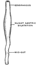
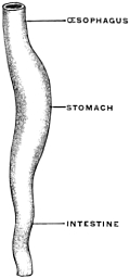
Fig. 43.—Entire alimentary canal of the lamprey, Petromyzon marinus, below the pericardium. (Columbia University Museum, No. 1575.) Fig. 44.—Schematic diagram representing three stages in the differentiation of the mammalian digestive tract: A. Early undifferentiated stage, in which the entire canal appears as a tube of uniform calibre. B. Spindle-shaped gastric dilatation. C. Typical mammalian gastric dilatation.
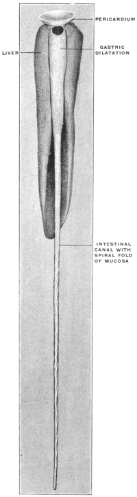
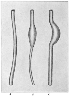
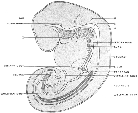
Fig. 45.—Reconstruction of human embryo. 1, 2, 3, 4, Gill-pouches. (After Fol.)
A distinct digestive segment may even be entirely wanting, owing to its failure to differentiate from the œsophagus on the one hand and from the endgut on the other. In such forms the entire digestive canal appears as a tube of uniform caliber extending from mouth to anus. It is necessary to begin with these simple structural conditions in order to obtain a clear conception of the disposition of the viscera in the adult human abdomen. Such simple arrangement of the alimentary tract is found in the embryo of man and of the higher vertebrates, and similar rudimentary types are encountered, as the permanent condition, in some of the lower forms. These latter are especially valuable for purposes of study, because they afford an opportunity of examining directly, as macroscopic objects, structural conditions which are found only as temporary embryonal stages during the development of the higher mammalia (Fig. 43).
In the early stages the alimentary tract of the mammalian embryo consists of a straight tube of nearly uniform caliber (Fig. 44, A), extending from the pharynx to the cloaca, along the median line in the dorsal region of the body cavity, connected with the ventral aspect of the axial mesoderm by a membranous fold forming the primitive common dorsal mesentery. Subsequently differentiation of this simple tube into successive segments takes place, marked by differences in shape and caliber and in histological structure.
The first indication of the future stomach appears early, in human embryos of from 5-6 days (Figs. 44, B, and 45; for later embryonal stomach forms compare also Figs. 33, 35 and 36), as a small spindle-shaped dilatation of a portion of the primitive entodermal tube, placed in the median plane, dorsad of the embryonic outgrowth of the liver, between it and the œsophagus. The appearance of this dilatation marks the separation of the proximal cephalic part (pharynx and œsophagus) from the distal caudal (intestinal) portion of the primitive alimentary canal.
Further growth of the stomach takes place chiefly along the dorsal margin of the dilatation, rendering the same more convex. The ventral border develops to a less degree and in the course of further and more complete differentiation the dorsal margin of the future stomach assumes even at this period the character of the greater curvature, while the opposite ventral margin, the future lesser curvature, following the dilatation of the tube dorsad, becomes in turn concave (Fig. 44, C).
Fig. 46.—Alimentary canal of human embryo of 5 mm. × 15. (Reconstruction after His.)
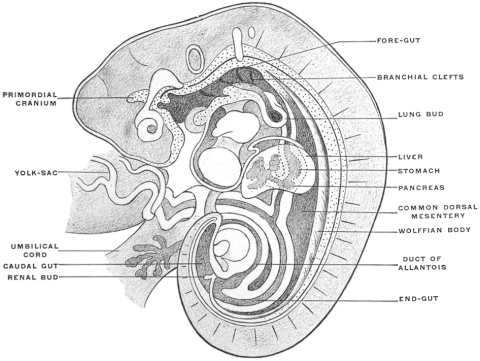
The early spindle-shaped dilatation has therefore assumed the general shape of the adult organ. This differentiation of greater and lesser curvature begins to appear in embryos of 5 mm. (Fig. 46) and is very well marked in embryos of 12.5 mm., Fig. 36, of an embryo of five weeks, indicates the adult form of the stomach clearly.
It will, however, be noted that the œsophageal entrance is still at the cephalic extremity of the rudimentary stomach, while the pyloric transition to the intestine occupies the distal caudal point, under cover of the liver, and turns with a slight bend dorsad and to the right to pass into the duodenum. The future greater curvature is directed dorsad and a little to the left toward the vertebral column, while the concave lesser curvature is turned ventrad and a little to the right toward the ventral abdominal wall. At this time there is but little indication of the subsequent extension of the organ to the left of the œsophageal entrance to form the great cul-de-sac or fundus of the adult stomach.
In this stage of its development the stomach therefore presents ventral and dorsal borders, and right and left surfaces, while the continuity of its lumen with the adjacent segments of the alimentary canal appears as a proximal or cephalic œsophageal and a distal or caudal intestinal opening.
COMPARATIVE ANATOMY OF FOREGUT AND STOMACH.
A serial review of this portion of the alimentary tract in vertebrates forms one of the most interesting and instructive chapters in comparative anatomy.
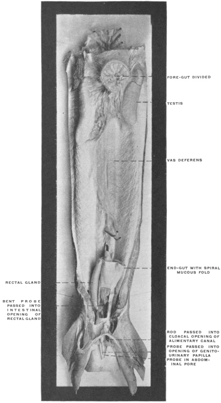
Fig. 47.—Gallus canis, dog-shark, ♂. Genito-urinary tract and cloaca in situ. The foregut has been divided just caudad of the communication with the oral cavity. (Columbia University Museum, No. 1694.)
Not only is every embryonal stage in the development of the higher mammalia represented permanently in the adult structure of some of the lower types, but the far-reaching influence of function and of the physiological demands on the structure of this portion of the digestive tract is strikingly illustrated by the numerous and marked modifications which are encountered.
The foregut, strictly speaking, is in mammals separated from the oral cavity by the musculo-membranous fold of the soft palate and uvula. In all other vertebrates except the crocodile, the oral cavity and foregut pass into each other without sharp demarcation (Fig. 47). In some of the lower vertebrates the alimentary canal never advances beyond the condition of a simple straight tube of nearly uniform caliber. There is no gastric dilatation and hence no differentiation of a stomach properly speaking. Such for example is the case in some teleost fishes, as the pickerel (Fig. 48). In these forms we have to deal with the persistence of the early embryonic pregastric stage of the higher types, before the simple alimentary tube is differentiated by the appearance of the distinct gastric dilatation.
In the Cyclostomata (Fig. 43) the intestinal canal passes through the body in a perfectly straight line and the three segments (mid-, fore- and hindgut) are not clearly differentiated.
In the Ammocœtes the foregut begins behind the wide branchial basket, dorsad of the heart, with a narrow entrance, which is succeeded by a dilated segment. The entrance of the hepatic duct separates fore- and midgut.
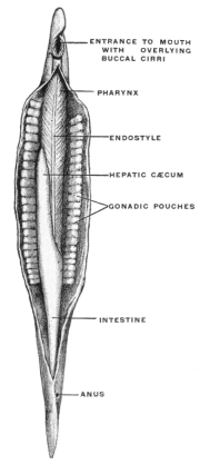
In Amphioxus the branchial pouch passes with a slight constriction directly into the gut which extends through the body-cavity in a straight line.
The narrow segment is usually regarded as the “œsophagus.” This is followed by a slightly dilated segment, the “stomach,” into which a blind pouch enters. This cæcal pouch is usually considered as a hepatic diverticulum (Fig. 49).
But even in these rudimentary forms the point where the liver develops from the entodermal intestinal tube marks the separation of fore- and midgut. The stomach, when it develops, is situated cephalad of the entrance of the hepatic duct into the intestine. The section cephalad of the duct opening may be very short, and the food digested further on in the intestinal tube. Consequently a function which in these lower vertebrates is assigned to the midgut becomes transferred in the higher forms to a specialized segment of the foregut, situated cephalad of the hepato-enteric duct. This segment is the . . .
Fig. 48.—Alimentary canal of Belone, pickerel. (Nuhn.) Fig. 49.—Amphioxus, dissected from the ventral side. The relatively enormous pharynx occupies more than half the length of the body. The walls are separated by the gill-clefts, and the parallel gill-bars abut at the midventral line on the endostyle. (Willey, after Rathke.) Fig. 50.—Necturus maculatus, mud-puppy. Alimentary canal and appendages. (Columbia University Museum, No. 1454.) Fig. 51.—Alimentary canal of Proteus anguineus. (Nuhn.)
STOMACH.
The distribution of the vagus nerve finds its explanation in this derivation of the stomach. The primitive foregut is formed by the passage between the branchial cavity and the midgut, and is within the area supplied by the vagus. Hence when the stomach develops from the foregut, as a specialized segment of the same, it is supplied by vagus branches. The vertebrate stomach varies greatly in size and shape.
The type-form is presented by a longitudinal spindle-shaped dilatation of the foregut, which retains its fœtal vertical position in the long axis of the body. An example of this form, which is encountered among fishes and amphibia, is presented by the alimentary tube of Proteus anguineus and Necturus maculatus (Figs. 50 and 51). Since this condition is common to all vertebrates in the earliest fœtal period it can be designated as the fœtal or primitive stomach form. All others appear as secondary derivatives from this typical early condition.
The influences which bring about such derivations and modifications may be enumerated as follows:
1. The habitual amount of food required by the animal.
2. The volume and digestible character of the food.
Fig. 52.—Alimentary canal of Coluber natrix. (Nuhn.)
3. The size and shape of the abdominal cavity in which the stomach is contained.
4. Structural modifications designed to increase the action of the gastric juice on the food contained in the stomach.
5. The assumption, on part of the stomach, of functions which are usually relegated to other organs.
Most of the individual stomach forms encountered among vertebrates owe their production to several of these influences acting in conjunction.
We may group the main types as follows:
1. Stomach Forms Depending on the Influence exerted by the Habitual Amount of Food required by the Animal.—The greater the activity of tissue changes is, the greater will be the amount of food required and the more pronounced will be the gastric dilatation of the alimentary canal. Hence in the higher vertebrates generally the stomach appears as a large and more sac-like dilatation than in lower forms, such as fishes and amphibia and some reptilia, in which the stomach is usually smaller and fœtal in shape, forming a slight longitudinal dilatation situated in the long axis of the body. An example is seen in the stomach of Coluber natrix (Fig. 52). Frequently this slight dilatation is scarcely differentiated from the œsophagus at the cephalic and from the small intestine at the caudal end. Many batrachians and perennibranchiates possess this form among the amphibia. It is also encountered in the pickerels, the Cyprini, and in Labrus among fishes, and in some saurians and ophidia among reptiles. It constitutes a slight advance in development over the earliest stage represented, as we have seen, by the nearly uniform and undifferentiated alimentary tube of amphioxus and the cyclostomata.
Fig. 53.—Human adult. Mucous surface of œsophageo-gastric junction. (Columbia University Museum, No. 1842.) Fig. 55.—Series of sections showing human pyloric valve and gastro-duodenal junction: 1. Stomach of fœtus at term in section. 2. Adult pyloric valve, gastric surface. 3. Adult pyloric valve and gastro-duodenal junction in section. 4. Fœtal gastro-duodenal junction in section. Entrance of biliary and pancreatic ducts on summit of papilla of duodenum. (Columbia University Museum, No. 1851.)
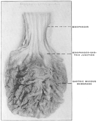
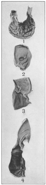
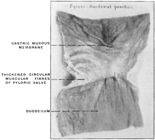
Fig. 54.—Human adult. Pyloro-duodenal junction and pyloric valve in section. (Columbia University Museum, No. 1842.)
This transition of the fœtal form to the more advanced secondary types of the stomach is marked by the development of two important structural features:
(a) The separation in the interior of the canal of the stomach from the intestine by the appearance of a ring-shaped valve, the pyloric valve. This is produced by an aggregation of the circular muscular fibers of the intestine at this point, and causes a projection of the mucous membrane into the lumen of the canal. It begins to appear in the fishes (pickerel, sturgeon, etc.), is found in most amphibia and is regularly present in the stomach of the higher vertebrates. (Figs. 54 and 55.) A good example of the ring-shaped plate of the pylorus with central circular opening produced by the aggregation of the circular muscular fibers is afforded by the view of the interior of the cormorant’s stomach given in Fig. 69. The opposite or œsophageal extremity of the stomach is less well differentiated from the afferent tube of the œsophagus.
There is no aggregation of muscular circular fibers in this situation and no valve. Superficially the external longitudinal muscular fibers of the œsophagus pass continuously and without demarcation into the superficial gastric muscular layer. The separation between œsophagus and stomach is, however, marked on the mucous surface by a well-defined line along which the flat, smooth and glistening œsophageal tesselated epithelium passes into the granular cuboidal epithelium of the gastric mucous membrane. The œsophageo-gastric junction in the adult human subject is shown in Fig. 53.
(b) The pyloric end of the stomach makes an angular bend, while the rest of the organ remains in the original vertical position in the long axis of the body. An example of this condition is presented by the stomach of Scincus ocellatus (Fig. 56; cf. also Fig. 202).
The purpose of both of these provisions is to retain the gastric contents for a longer time within the stomach. Hence this form is encountered especially in those fishes and amphibians in which the nutritive demands require a more complete digestion of the food taken. This is the case, for example, in Gobius (Fig. 57), the plagiostomata (Fig. 58), and many saurians. The same transitory stomach form is even found in some mammals, as the seals. Fig. 59 shows the stomach in Phoca vitulina, the harbor seal. With the further increase in the demand for complete digestion of the food the entire stomach assumes a transverse position to the long axis of the body. This may occur while the stomach still retains its primitive tubular form, as in most chelonians (Fig. 60). In others the change in position occurs after the gastric dilatation has assumed the sac-like form, as in many land-turtles, crocodiles, some batrachians and all higher vertebrates (Figs. 61 and 62). This transverse position, at right angles to the long axis of the body, forms the starting point for the derivation of all secondary types of stomach.
Fig. 56.—Alimentary canal of Scincus ocellatus. Pyloric extremity of the slightly marked gastric dilatation presents an angular bend. (Nuhn.) Fig. 57.—Alimentary canal of Gobius niger. (Nuhn.) Fig. 58.—Alimentary canal of shark. (Nuhn.)
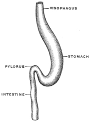
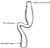
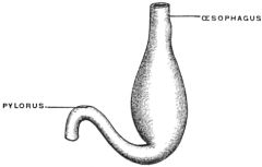
Fig. 59.—Stomach of Phoca vitulina, harbor seal. (Columbia University Museum, No. 600.) Fig. 60.—Stomach of Pseudemys elegans, pond turtle. (Columbia University Museum, No. 1710.)
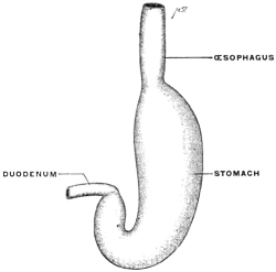
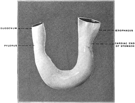
Fig. 61.—Stomach of Chelydra serpentina, snapping turtle. (Columbia University Museum, No. 1852.) Fig. 62.—Same in section.
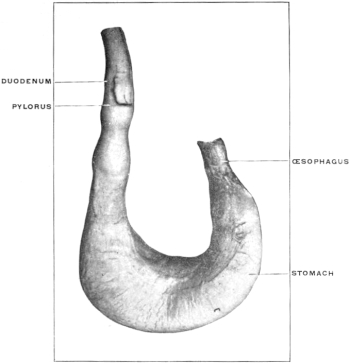
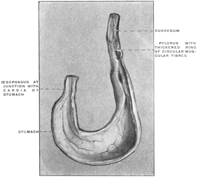
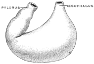
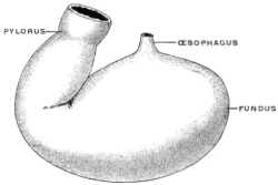
2. Stomach Forms Depending on the Influence Exerted by the Volume and Digestible Character of the Foods.—Vegetable substances usually have a large volume in proportion to the amount of nutritive material which they contain. Meat, on the other hand, contains considerable nutriment in a comparatively small bulk. Hence carnivora (Fig. 63) usually have a smaller stomach than herbivora (Fig. 64).
Fig. 63.—Stomach of Lutra vulgaris, otter. (Nuhn.) Fig. 64.—Stomach of Equus caballus, horse. (Nuhn.)
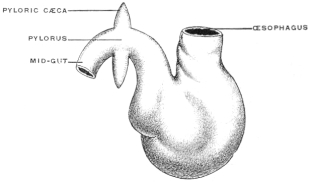
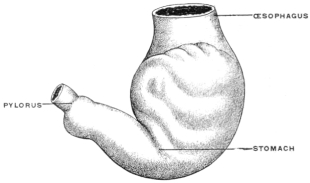
3. Stomach Forms Influenced by Size and Shape of the Abdominal Cavity in which they are Contained.—In animals whose bodies are long and slender, as in snakes (Fig. 52), most saurians (Fig. 56), many tailed batrachians and perennibranchiates (Figs. 50 and 51), many teleosts (Fig. 48), the stomach is likewise usually long and slender in shape, unless special modifying conditions exist. When on the other hand the body is broad and short, as in Lophius (Fig. 65), Pipa (Fig. 66), and most higher vertebrates, the stomach is also broader and more sac-like.
Fig. 65.—Stomach of Lophius piscatorius, angler. (Nuhn.) Fig. 66.—Stomach of Pipa verucosa. (Nuhn.)
4. Stomach Forms Depending on Structural Modifications Designed to Increase the Action of the Gastric Juice on the Food.—This purpose is accomplished:
(a) By increasing the source of supply of the gastric juice.
(b) By increasing the length of time during which the food remains in the stomach.
Fig. 67.—Stomach of Castor fiber, beaver. (Nuhn.) Fig. 68.—Stomach of Manatus americanus, manatee. (Nuhn.)
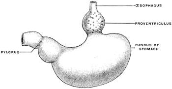
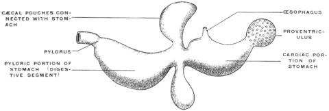
Fig. 69.—Stomach of Phalacrocorax dilophus, double-crested cormorant; section. (Columbia University Museum, No. 67/1804.)
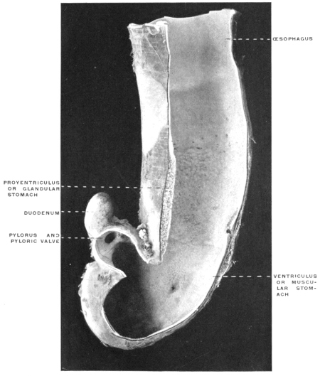
(a) The source of supply of the gastric juice is increased by adding to the usual gastric glands of the stomach a special accessory glandular compartment, either placed at the cardia, where the œsophagus enters, as in Myoxus or Castor (Fig. 67) or attached to the body of the stomach to the left of the cardia, as in the manatee (Fig. 68). The first arrangement is similar to the universal position of the glandular stomach of birds (Fig. 69). In birds, however, the glandular proventriculus is the only source of the gastric juice, while in the above-mentioned mammalia (myoxus and beaver) the accessory glandular stomach is merely an addition to the supply derived from the usual gastric glands situated in the body of the organ.
(b) The increase of the length of time during which the food remains in the stomach subject to the action of the gastric juice can be accomplished in one of several ways.
1. The stomach, while it retains its general tubular form increases considerably in length and assumes the shape and structure found in the human large intestine. It is partially subdivided by folds projecting into the interior and separating compartments resembling the colic cells of the human large intestine. The time required for the passage of food through the stomach is thus increased and the action of the gastric juice is prolonged and rendered more intense.
Such modifications of the structure of the stomach are encountered in Semnopithecus among the monkeys and in the kangaroo, among marsupials (Figs. 70 and 71).
2. The same purpose is accomplished by the development of diverticula from the stomach, in which the food is retained and acted on by the gastric juice for longer periods.
Fig. 70.—Stomach of Halmaturus derbyanus, rock kangaroo. (Columbia University Museum, No. 582.) Fig. 71.—Stomach of Semnopithecus entellus, entellus monkey. (Columbia University Museum, No. 62/1805.)
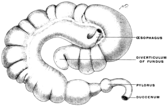
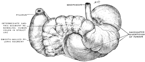
Fig. 72.—Alimentary canal of Anguilla anguilla, eel. (Columbia University Museum, No. 1271.)
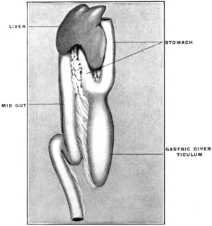
The herbivora, omnivora and such carnivora as live on animal food difficult of digestion furnish examples of this type of stomach. The same is also found in most teleosts. In the latter the cæcal gastric pouch lies in the long axis of the body, opposite the entrance of the œsophagus. A marked example of this arrangement is seen in the stomach of the eel, Anguilla anguilla (Fig. 72).
In other forms, and in the mammalia especially, the blind pouch is developed from the portion of the stomach lying to the left of the œsophageal entrance at the cardia, and is hence placed transversely to the long axis of the body.
This difference in the position of the cul-de-sac is explained by the small transverse measure of the body in teleosts, while the greater amount of available space in the abdominal cavity of mammalia permits of the transverse position of the entire stomach and of the development of the diverticulum from its left extremity.
Most mammals have only a single pouch, whose size varies with the digestibility of the food habitually taken. It is greater in herbivora (Figs. 64 and 73) than in omnivora and carnivora (Figs. 74 and 75). In some of the latter, as Lutra (Fig. 63), the cul-de-sac is almost wanting.
Fig. 73.—Stomach of Lepus cuniculus, rabbit. (Nuhn.) Fig. 76.—Stomach of Erethizon dorsatus, American porcupine. (Columbia University Museum, No. 358.)
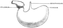
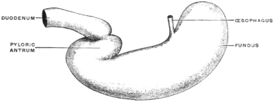
Fig. 74.—Stomach of Nasua rufa, coati. (Nuhn.) Fig. 77.—Stomach of Cercopithecus cephus, moustache monkey. (Columbia University Museum, No. 158.)
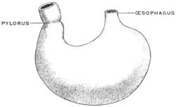
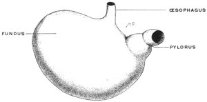
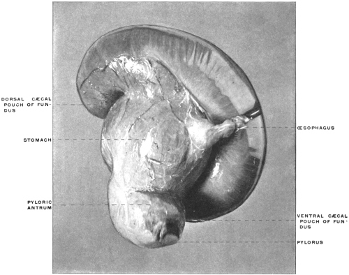
Fig. 75.—Stomach of Felis leo, lion. (Nuhn.) Fig. 78.—Stomach of Sus scrofa, pig. The fundus of the stomach carries a cæcal appendage separated in the interior by a spiral fold of the mucous membrane from the gastric cavity.
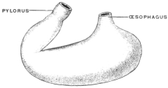
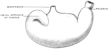
In some forms, as the pig, the left extremity of the stomach carries a cæcal appendix with a spiral valve in the interior separating its lumen from the general gastric cavity (Fig. 78). Others have two such cæcal appendices added to the left end of the stomach (Peccary, Fig. 79). These cæcal pouches may arise from the body of the stomach, instead of from the left extremity. An example of this condition is furnished by the American manatee (Fig. 68).
Fig. 79.—Stomach of Dicotyles torquatus, peccary. The fundus is a capacious pouch prolonged ventrally and dorsally into two cæcal appendages resembling the single appendage of the pig’s stomach. (Columbia University Museum, No. 1806.)
5. Variations in the Form of the Stomach Depending upon the Assumption by the Stomach of Special Functions, which are Usually Relegated to other Organs.—These functions are the following:
Fig. 80.—Macacus nemestrinus, pig-tail macaque monkey; cheek-pouches. (From a fresh dissection.)
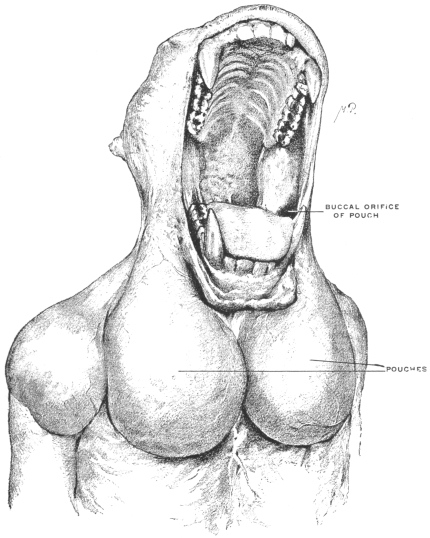
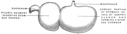
Fig. 81.—Stomach of Cricetus vulgaris, hamster. (Nuhn.)
(a) Storage of food in special receptacles or compartments for subsequent use.
(b) Mastication of the food is in some animals accomplished only partly or not at all in the mouth, and is then performed in the stomach. A portion of the stomach is thus converted into an apparatus for mastication.
(c) The provisions for these two accessory functions may be combined in the same stomach.
(a) Many of the higher vertebrates possess in connection with the alimentary tract additional reservoirs for the storage of food until used. Such reservoirs are found in mammals and birds connected with the oral cavity, as cheek-pouches, or with the œsophagus, such as the crop of the birds (Fig. 88). Fig. 80 shows the development of the cheek-pouches in one of the primates, Macacus nemestrinus.
In many mammals reservoirs of similar import are added directly to the stomach and form an integral part of the organ. Examples are furnished by the compound stomachs of many rodents, ruminants, cetaceans and herbivorous edentates. The peculiar appearance of these stomachs is explained if the additional reservoirs are in imagination removed and the digestive stomach proper restored so to speak to the type-form. The proximal or cardiac portion of the stomach in many rodents is devoid of gastric glands and must be interpreted as a storage chamber for food (Fig. 81). The same significance attaches to the corresponding portion of the manatee’s stomach (Fig. 68).
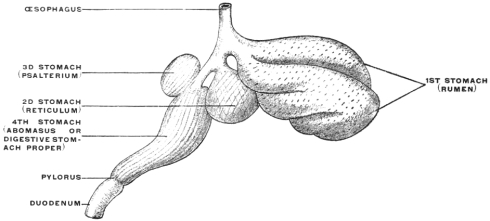
Similar contrivances are found in the ruminant stomach. The first and second divisions (rumen and reticulum) are nothing but sac-like gastric reservoirs or pouches, in which the food is collected, to be subsequently returned to the mouth for mastication. When swallowed for the second time the bolus is carried, by the closure of the so-called œsophageal gutter, past the first and second stomach into the digestive apparatus proper (the abomasum) (Figs. 82 and 83). Many ruminants (e. g., Moschus) only have these three compartments. Most, however, have four, the leaf stomach or psalterium being intercalated between the retinaculum and the abomasum. The psalterium contains no digestive glands. It may possibly serve for the absorption of the liquid portions of the foods.
Fig. 82.—Stomach of Ovis aries, sheep. (Columbia University Museum, No. 1807.) Fig. 84.—Mucous membrane of stomach of Camelus dromedarius, dromedary, showing water-cells. (Columbia University Museum, No. 1123.)
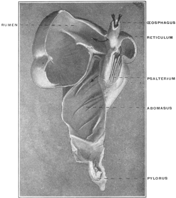
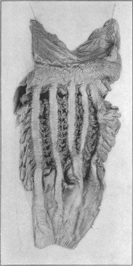
Fig. 83.—Scheme of ruminant compound stomach. (Nuhn.) Fig. 85.—Stomach of Phocæna, porpoise. (Nuhn.)
The rumen or first stomach of the camels and llamas is provided with so-called “water-cells,” for the storage of water. These cells are diverticula lined by a continuation of the gastric mucous membrane. The entrance into these compartments can be closed by a sphincter muscle after they are filled with water (Fig. 84).
Fig. 86.—Stomach of Urinator imber, red-throated loon. (Columbia University Museum, No. 1808.)
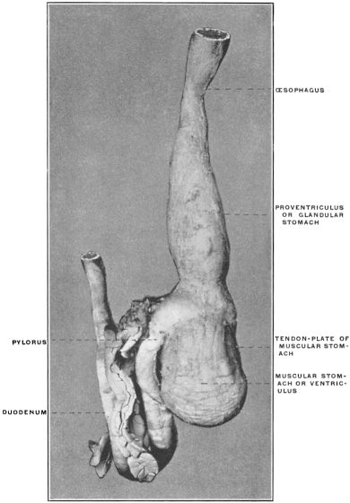
Fig. 87.—Scheme of stomach of granivorous bird. (Nuhn.)
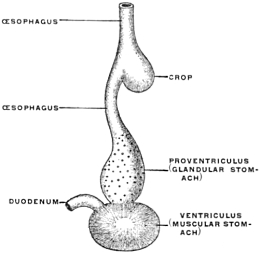
The three stomachs of the cetaceans are similar to those of the ruminants (Fig. 85). The first is a crop-like reservoir for the reception of the food when swallowed. The mucous membrane is entirely devoid of digestive glands. In the dolphins the mucous membrane is provided with a hard horny covering, which serves to break up the food mechanically by trituration. The second stomach and the gut-like pyloric prolongation constituting the third stomach contain gastric glands and are hence digestive in function.
(b) Stomach forms, in which a portion of the organ is converted into an apparatus for mastication, are seen especially in birds, in which animals, on account of the absence of teeth, mastication cannot be performed in the mouth.
The stomach of the bird is usually composed of two segments, one placed vertically above the other.
The first appears like an elongated dilatation of the œsophagus, forming the Proventriculus or glandular stomach.
The second is larger, round in shape, with very strong and thick muscular walls (Figs. 86 and 87).
The proventriculus furnishes the gastric juice exclusively.
The second or muscular stomach, devoid of gastric glands, functions merely as a masticating apparatus for the mechanical division of the food. The thick muscular walls of this compartment may measure several inches in diameter and carry on the opposed mucous surfaces lining the cavity a hard horny plate with corrugated and roughened surface (Fig. 88). These hard plates are designed to crush the food between them, as between two mill stones. The muscle stomach is best developed in herbivorous birds, while both the muscular wall and the horny plate are much weaker and thinner in carnivore wading and swimming birds (Fig. 89).
Fig. 88.—Œsophagus and stomach of Gallus bankiva, hen. (Columbia University Museum, No. 1809.) Fig. 89.—Stomach of Botaurus lentiginosus, bittern. (Columbia University Museum, No. 23/1810.)
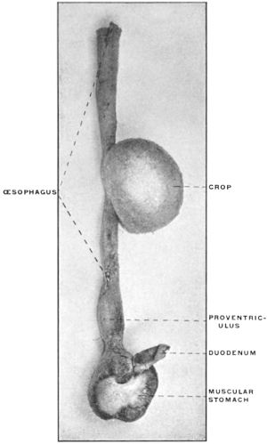
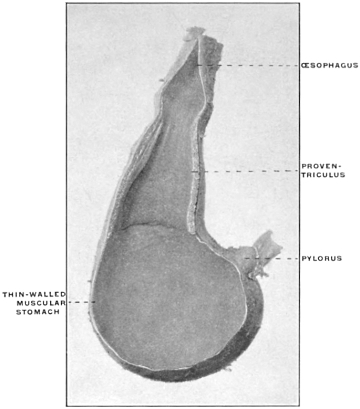
Fig. 90.—Stomach of owl sp. (Nuhn.) Fig. 91.—Stomach of Ardea cinerea, heron. (Nuhn.)
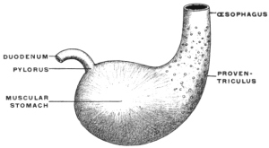
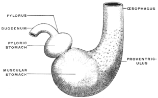
Fig. 92.—Stomach of crocodile. (Nuhn.)
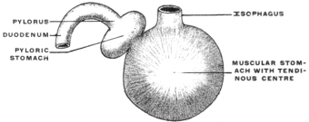
In birds of prey, especially in the owls, the stomach walls are scarcely more massive than in other animals, and the mucous membrane is soft and devoid of a horny covering. The glandular and masticatory stomachs are less sharply divided from each other in these forms, and the entire organ conforms more to the general vertebrate type (Fig. 90).
In some birds (herons, storks, etc.) a small rounded third stomach, the so-called pyloric stomach, is placed between the muscle stomach and the pylorus (Fig. 91). It contains no gastric glands, and possibly may function as an additional absorbing chamber.
Among reptiles the stomach of the crocodile resembles the organ in birds (Fig. 92). It is flat and rounded in shape, the muscle wall carries a tendinous plate, and there is a pyloric stomach. There is, however, no glandular stomach or proventriculus, as in birds, and the mucous membrane is not covered by a horny plate, but is soft and contains the peptic glands. Figs. 93 and 94 show the stomach of Alligator mississippiensis, in the ventral view and in section.
Fig. 93.—Stomach of Alligator mississippiensis. (Columbia University Museum, No. 1811.) Fig. 94.—Same in section. Thin-walled cardiac segment continues into cavity of pyloric ventriculus.
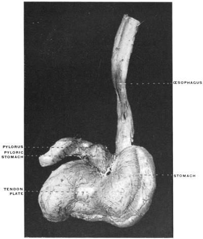
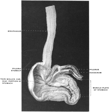
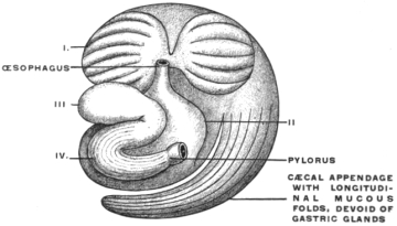
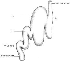
Fig. 95.
—Stomach of
Bradypustridactylus
, three-toed sloth. I. First stomach, devoid of gastric glands, corresponding to rumen of ruminants.
II. Second stomach, the homologue of the ruminant reticulum.
III. Digestive stomach proper, provided with gastric glands connected by a gutter with the œsophagus.
IV. Muscular stomach, the walls formed by a thick muscular plate and provided on the mucous surface with a dense corneous covering for purposes of trituration.
(c) The combination of the two accessory functions just described in the same stomach is found in the three-toed sloth (Fig. 95).
There are here two large reservoirs, which correspond to the rumen and retinaculum of the ruminants, and a digestive compartment containing gastric glands, which corresponds to the ruminant abomasum, and is connected by an œsophageal gutter directly with the œsophagus. At the pyloric extremity the muscle wall is greatly increased and the mucous membrane of this portion carries a thick horny covering, forming a masticatory stomach greatly resembling the corresponding structure in the bird. Its function is evidently to complete the mechanical division of the food which has only been partly masticated in the mouth.
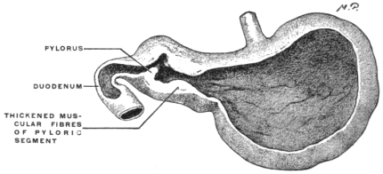
The same significance is probably to be attached to the thickened muscular walls which the pyloric segment of the stomach in Tamandua bivittata, another edentate, presents (Fig. 96), in strong contrast with the thinner walled cardiac segment and fundus.
Fig. 96.—Stomach of Tamandua bivittata, collared ant-eater, (Columbia University Museum, No. 68/1485.)
INTESTINE.
Continuing our consideration of the development of the alimentary canal we find that changes from the simple primitive straight tube below the stomach depend upon two factors:
1. The increase in the length of the intestinal tube, which exceeds relatively the increase in the length of the body cavity in which it is contained.
2. The differentiation into small and large intestine, the development of the cæcum and ileo-cæcal junction, and the development of the accessory digestive glands, liver and pancreas, by budding from the proximal portion of the primitive entodermal intestinal tube.
Fig. 97.—Alimentary canal of human embryo of 5 mm. × 15. (Reconstruction after His.)
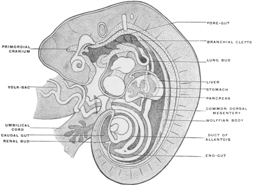
Fig. 98.—Schema of human embryonic intestinal canal, with intestinal umbilical loop, but before differentiation of the large and small intestine.
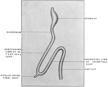
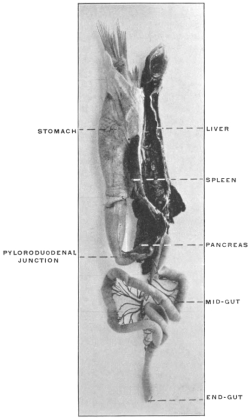
Fig. 99.—Viscera of Necturus maculatus, mud-puppy, in situ. (Columbia University Museum, No. 1175.)
1. In embryos up to 5 mm. cervico-coccygeal measure (Fig. 97) the intestinal tube follows the body curve without deviation. Subsequently the elongation of the intestine causes a small bend, with the convexity directed ventrad, to appear in the umbilical region. This bend gradually increases until the gut forms a single long loop, beginning a short distance below the pylorus and directed ventro-caudad. The apex of the loop, to which the vitello-intestinal duct is attached (Fig. 98) (cf. p. 34) projects beyond the abdominal cavity into the hollow of the umbilical cord, constituting the so-called “umbilical or embryonal intestinal hernia.” This entrance of the apex of the intestinal umbilical loop into the umbilical cord begins in embryos of about 10 mm. During the succeeding weeks—up to the tenth—the segment of the intestine thus lodged within the hollow of the umbilical cord increases. After this period the intestinal coils are gradually withdrawn within the abdomen. The explanation of this temporary extrusion of the intestine into the umbilical cord is probably to be found in the strain produced by the yolk-sac which is attached by the vitello-intestinal duct to the apex of the umbilical loop. As we have seen (p. 35) the site of the original apex of the loop may still be indicated in the adult by the persistence of a portion of the vitello-intestinal duct as a “Meckel’s diverticulum.”
In its simplest primitive condition the loop presents a proximal, descending or efferent limb, an apex, and an ascending, returning or afferent limb (Fig. 98). In the human embryo these segments of the loop furnish the jejuno-ileum and portions of the large intestine, in a manner to be subsequently detailed.
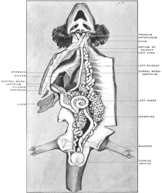
This stage in the development of the higher vertebrate intestine is well illustrated by the alimentary tract of the mud-puppy, Necturus maculatus, shown in Fig. 99, which represents the entire situs viscerum of an adult female animal.
The stomach is tubular, not distinctly differentiated from the œsophagus, placed vertically in the long axis of the body. The pyloric end is marked by a constriction separating stomach from midgut and immediately beyond this point the pancreas is applied to the intestine. The rest of the intestinal canal forms a simple loop, the descending limb presenting one or two primitive convolutions. There is no marked differentiation between large and small intestine, the canal possessing a nearly uniform caliber from pylorus to cloaca.
2. The differentiation of the small from the large intestine, marked by the appearance of the cæcal bud or protrusion (Fig. 100), takes place in the ascending segment of the umbilical loop a short distance from the apex. In the human embryo the cæcal bud appears in the 6th week as a plainly marked protuberance, which grows very slowly in length and circumference. It shows very early an unequal rate of development; the terminal piece, not keeping pace in growth with the proximal portion, is converted into the vermiform appendix, while the proximal segment develops into the cæcum proper. The increase in the length of the loop, which begins to be marked in the 7th week, is not uniform. The apex is the first portion to present the evidences of this growth. Subsequently the descending limb grows in length very rapidly and is early thrown into numerous coils of the future mobile portion of the small intestine (jejuno-ileum). Even before the withdrawal of the apex of the loop within the abdominal cavity a prominent coil of these convolutions is found protruding in the umbilical region (Fig. 544). The ascending limb of the loop from which a portion of the large intestine is developed, grows comparatively slowly at this time.
Fig. 100.—Schema of human embryonic intestinal canal after differentiation of the large and small intestine. Fig. 101.—Human embryo of 2.15 mm., twelve days old. Seessel’s sac is the cephalic blind termination of the embryonic foregut before the communication with the ectodermal invagination of the stomadæum has been formed. (Reconstruction after His.)
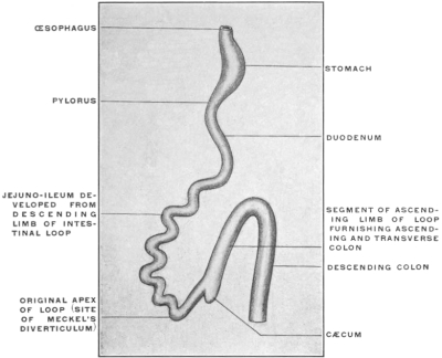
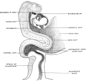
Fig. 102.—Representation of alimentary canal and appendages of human embryo of 4.1 mm.; isolated. × 15. (Kollmann, after His.) Fig. 103.—Alimentary canal and appendages of human embryo of 12.5 mm. × 12. (Kollmann, after His.)
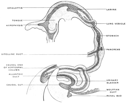
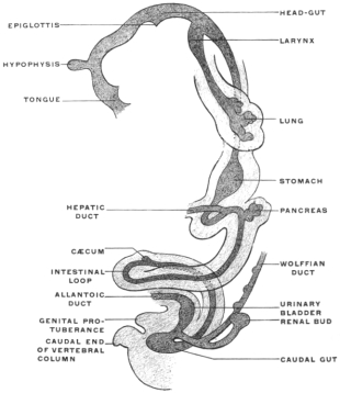
Fig. 104.—A. Schematic representation of alimentary canal, with umbilical loop and mesenteric attachments in human embryo of about six weeks. B and C, stages in the intestinal rotation.
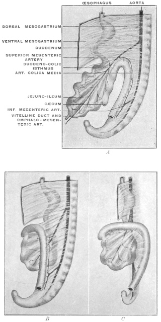
The future portions of the human adult alimentary tract below the stomach may be referred, in reference to their derivation, to this primitive condition of the tube as follows:
1. The segment of small intestine situated between the pylorus and the beginning or point of departure of the proximal or descending limb of the umbilical loop, develops into the duodenum. This portion of the small intestine is indicated early in embryos of 2.15 mm. (Fig. 101), by the origin of the hepatic duct from the intestinal tube. Somewhat later, in embryos of 4.10-5 mm. length, (Fig. 102) it becomes additionally marked by the origin of the pancreatic diverticulum. The duodenum, at first straight, now begins to curve, forming a short duodenal loop or bend. In embryos of 6 weeks the duodenum forms a simple loop placed transversely below the pyloric extremity of the stomach (Figs. 103 and 104).
2. The descending limb, the apex and a small part of the ascending limb of the umbilical loop form the jejuno-ileum.
3. The remainder of the ascending limb forms the cæcum and appendix, the ascending and transverse colon.
4. The distal straight portion of the primitive tube forms the terminal portion of the transverse colon (the splenic flexure), the descending colon, sigmoid flexure and rectum.
The primitive condition of the embryonal mammalian alimentary tract, after differentiation of the large intestine is well illustrated by some of the lower vertebrates in which development never proceeds beyond this stage. Fig. 112 shows the entire alimentary canal of a teleost fish, the conger eel (Echelus conger) isolated.
The preparation forms a good illustration of the embryonal stage of the higher vertebrates in which development has not proceeded beyond the formation of the simple umbilical loop, about corresponding to the schematic Fig. 98. The stomach is differentiated both by its caliber and by the formation of a pyloric ring valve.
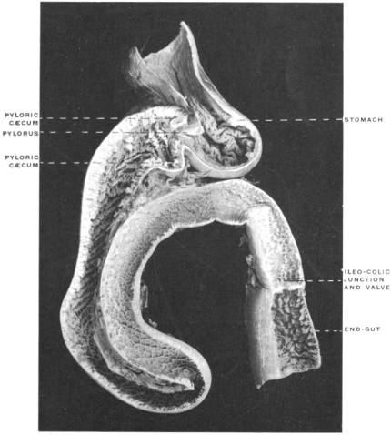
The midgut forms a simple loop with a descending and ascending limb closely bound together by mesenteric attachment. Different from the course of development followed in the human embryo is the situation of the ileo-colic junction. The same appears in the terminal straight segment of the canal—corresponding to the human descending colon—while in the human embryo the differentiation of small and large intestine takes place in the course of the ascending limb of the loop. This condition depends upon the relatively much shorter extent of the teleost endgut compared with the human large intestine. Other examples are afforded by the alimentary tract of some of the Amphibia and Reptilia. Fig. 105 shows the alimentary canal of Rana catesbiana, the common bull frog. The stomach, fairly well differentiated, is succeeded by the small intestine of considerable length and uniform caliber. The proximal portion of the small intestine is characterized as duodenum by its connection with liver and pancreas. In the remaining portion of the intestinal canal it is not difficult to recognize the elements of the umbilical loop of the higher mammalian embryo. The larger mass of the jejuno-ileal coils is developed from the descending limb of the loop; a smaller number of convolutions belong to the returning or ascending limb, which also includes the ileo-colic junction. The very short large intestine of the frog passes straight down to enter the cloaca. Another example, in which the early embryonal stages of the higher mammalia are illustrated by the permanent structure of one of the lower vertebrates, is given in Fig. 106, which shows the alimentary tract of a chelonian, Pseudemys elegans, the pond turtle. The bilobed liver fits over the well-differentiated stomach in the manner of a saddle. The stomach itself, as in chelonians generally, has a markedly transverse position and passes under cover of the right lobe of the liver into the duodenum. The coils of small intestine form a prominent mass, which, however, when unravelled as shown in the figure, permits us to recognize its identity with the mammalian embryonic umbilical loop. The well-marked ileo-colic junction is situated at the termination of the returning limb of the loop, close to the beginning of the descending limb. This close approximation of the duodenum and colon (duodeno-colic isthmus) forms one of the most important factors in the further development of the mammalian intestinal canal and will again be referred to below.
Fig. 105.—Rana catesbiana, bull-frog. Alimentary canal and appendages. (Columbia University Museum, No. 1454.) Fig. 106.—Pseudemys elegans, pond turtle. Alimentary canal. (Columbia University Museum, No. 1437.)
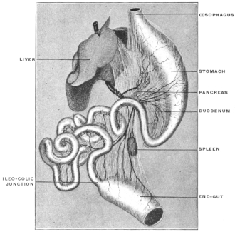
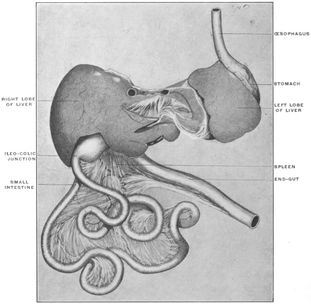
Fig. 107.—Abdominal viscera of Tamandua bivittata, the little ant-eater, seen from the left, with the intestines turned to the right. (From a fresh dissection.)
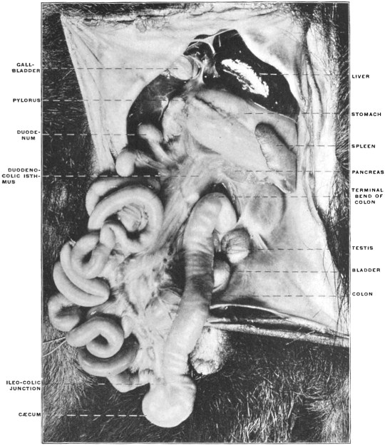
Fig. 108.—The same view, from another specimen. Figures 107 and 108 should be studied and compared together, as each supplements the other.
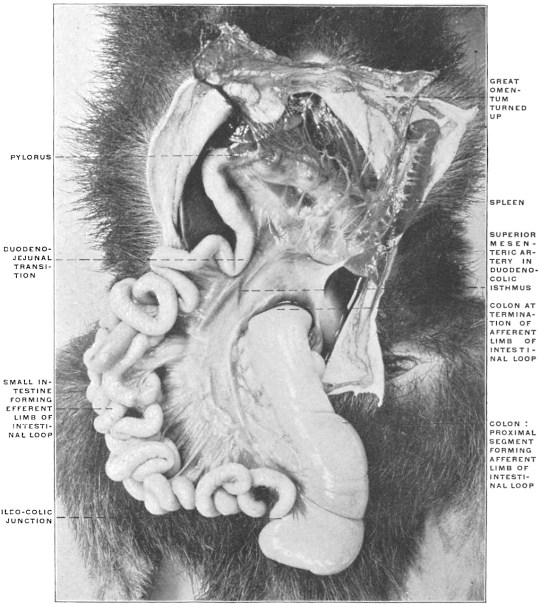
Fig. 109.—Abdominal viscera of Tamandua bivittata, the little ant-eater, seen from the right, with the intestines turned to the left. (From a fresh dissection.)
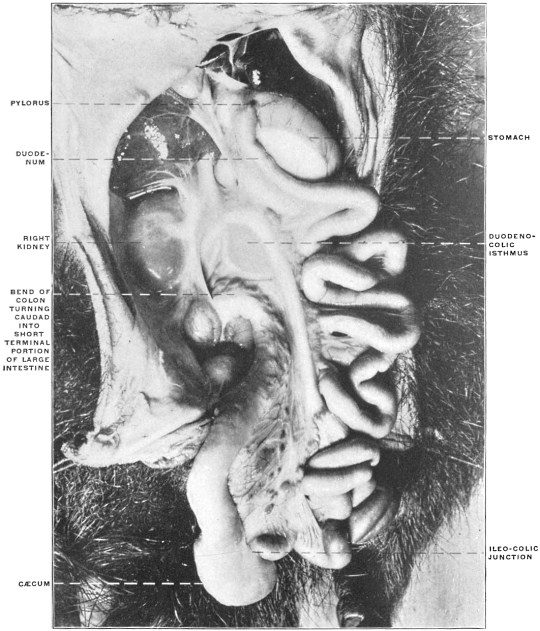
Fig. 110.—The same view, from another specimen.
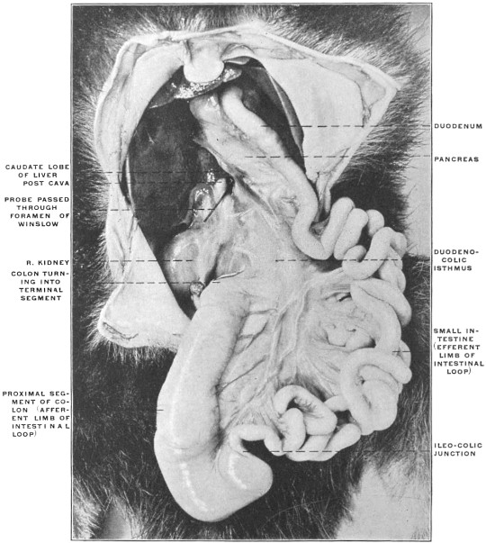
Fig. 111.—Schematic representation of the development of the mesentery of the umbilical loop.
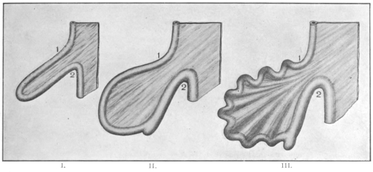
Fig. 112.—Alimentary canal, isolated and in section, of Echelus conger, the conger eel. (Columbia University Museum, No. 1812.) Fig. 113.—Chelydra serpentina, snapping turtle; intestinal canal, pancreas, and spleen, isolated. (Columbia University Museum, No. 1369)
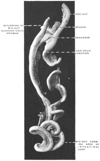
From the ileo-colic junction the large intestine of the turtle continues caudad to the cloaca in a nearly straight line. The same primitive condition of the intestinal canal may be observed in some members of man’s own class, the mammalia—as in certain edentates. Figs. 107 and 108 show the entire abdominal portion of the alimentary tract in Tamandua bivittata, the little ant-eater of Brazil. The stomach is turned cephalad and the great omentum elevated. The intestines are turned over to the right side.
It will be observed that in spite of the numerous coils of the small intestine the general arrangement of the alimentary canal corresponds to the primitive scheme shown in Fig. 98. The entire intestinal canal is attached by a continuous vertical mesentery to the dorsal median line of the abdominal cavity ventrad of the vertebral column and aorta. The growth in length of the small intestine has necessitated a corresponding lengthening of the attached border of the mesentery—consequently the membrane presents a pleated or crenated appearance. The cæcum is well developed, the ileo-cæcal junction being situated within the returning limb of the loop, a little distance from the apex.
In Figs. 109 and 110, taken from the same specimens, the entire mass of the small intestines has been turned to the left so as to exhibit the right leaf of the common dorsal mesentery and the mesoduodenum, the latter containing the head of the pancreas. It will be noted that the mesentery, expanding beyond the duodeno-colic isthmus, is common to the small and to the proximal portion of the large intestine, i. e., to those segments of the alimentary canal which are developed from the two limbs of the umbilical loop. Figs. 107-110 should be studied and compared together, as each supplements the others.
It will be observed, in reference to the change from the primitive loop to the subsequent increase in the length of the tube and the resulting arrangement of the mesentery, that three successive stages are to be considered, represented schematically in Fig. 111. In the earliest stage (Fig. 111, I.) the two segments of the loop are of equal length, parallel to one another, the distance between the beginning and termination of the loop (1-2) being maintained throughout its extent. Hence the mesentery is of equal width in all its parts within the loop, only drawn out, i. e., away from the vertebral column, in accordance with the length of the loop. In the next stage (Fig. 111, II.) the increase in the length of the intestine is accompanied by a corresponding widening of the mesentery. The points 1 and 2 are still approximately the same distance apart as in the earlier stage, but the increase in the length of the tube between these points forces the two limbs of the loop to abandon their early parallel course, and to form curved lines with the concavity turned toward the mesenteric attachment. In this condition the mesentery consequently forms a widely expanded membrane framed by the intestine and narrowing between the points 1 and 2 to a neck or isthmus which effects the transition between the expanded segment surrounded by the intestine and the rest of the dorsal primitive mesentery. Finally in the stage represented in Fig. 111, III., the increase in the length of the small intestine has reached a point where a single curve is no longer sufficient for the accommodation of the growth. Consequently the tube now appears coiled and convoluted, and the mesentery, as it is attached to the gut, of necessity follows all the twists and appears fluted or pleated in its distal attached portion.
If we now carefully examine the conditions presented by the intestine and mesentery in a form like Tamandua (Figs. 107 and 108) we will find that they correspond to the developmental facts thus far considered. The termination of the duodenum (1) and the bend in the colon (2) mark the two points at which in the primitive schema (Fig. 111, I.) the umbilical loop begins and terminates. The proximal of these two points (1) corresponds to the termination of the duodenum, which segment extends from here cephalad to the pyloric extremity of the stomach. The distal point (2) is placed on the colon where the returning limb of the loop resumes the original median vertical course of the large intestine. These two points mark the neck of the loop, which we can describe as the duodeno-colic neck or isthmus.
The same condition is well shown in the intestinal canal of the snapping turtle (Fig. 113). The duodenum and colon approach each other very closely at the isthmus and between these points the convolutions of the intestine extend in a wide circle. We will find this approximation of duodenum and colon a feature which persists throughout all the later developmental stages of the higher vertebrates and has an important bearing on the final arrangement of the intestinal canal in the human adult.
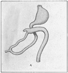
Further Changes in the Development of the Human Alimentary Canal. Rotation of the Intestine. Formation of the Segments of the Colon. Final Permanent Relations of the Segments of the Intestinal Tube.—The next important stage leading up to the final adult disposition of the intestine in man and the higher mammals is the rotation of the portions developed from the two limbs of the primitive loop around an oblique axis drawn from the duodeno-colic isthmus to the apex of the loop. The portion of the large intestine, developed from the ascending limb of the loop, moves in the third month to the middle line, coming into contact with the ventral abdominal wall. From here the large intestine passes, ventrad of the jejuno-ileal coils, toward the cephalic end of the abdominal cavity and lies transversely along the greater curvature of the stomach. The growing coils of the small intestine crowd the colon more and more cephalad. In the fourth month the cæcum turns to the right, coming into contact with the caudal surface of the liver, ventrad of the duodenum, and subsequently reaches the ventral surface of the right kidney. As the result of this rotation the ileo-colic junction, cæcum and succeeding portion of the colon are carried from the original position in the distal and left part of the abdomen cephalad and to the right across the proximal (duodenal) portion of the small intestine, while the coils of the jejuno-ileum, developed from the descending limb and apex of the loop, are turned in the opposite direction, caudad and to the left underneath the preceding (Figs. 114 and 115). This change in the relative position of the parts of the intestinal tract and the resulting altered bearing of the colon to the duodenum will be best appreciated by considering in the first place the effect of the change on the arrangement of the primitive mesentery and the intestinal vessels, and secondly by repeating actually the rotation in the intestinal tract of a mammal (cat) in which the adult arrangement of the intestine and peritoneum permits us to perform the manipulations and note the result.
Fig. 114A.—Intestinal canal in stage of umbilical loop—before rotation. Fig. 114B.—First stage in rotation, colon crossing duodenum.
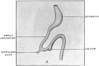
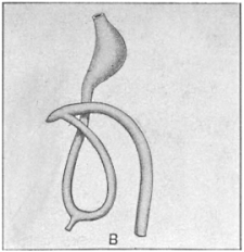
Fig. 115A.—Second stage in rotation—rotation of small intestine. Fig. 115B.—Schema of intestinal canal after complete rotation and descent of cæcum.
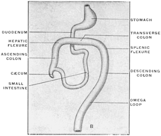
I. Effect of Rotation on the Disposition of the Primitive Mesentery and on the Relative Position of Duodenum and Colon, and Consequent Arrangement of the Intestinal Blood Vessels.—It will be appreciated that in Fig. 111, representing a profile view of the original arrangement, or in Figs. 107 and 108, showing the intestinal canal of Tamandua, the left layer of the primitive mesentery is turned toward the observer. The membrane is seen to pass from the ventral aspect of the vertebral column and aorta, through the narrow neck of the duodeno-colic isthmus, to expand in the manner already indicated toward its intestinal attachment. In the rotation of the intestine the twist takes place at the duodeno-colic neck, carrying, as already stated, the large intestine cephalad and to the right, while the jejuno-ileum is turned in the opposite direction caudad and to the left. During this rotation the duodeno-jejunal angle (Figs. 114, B and 115, A) passes to the left underneath the proximal segment of the colon, which now lies ventrad and to the right of the duodenal portion of the small intestine. The mesenteric peritoneum, occupying the bight of the umbilical loop, will, after the rotation, in the left profile view shown in Fig. 104, A and B, turn its original right leaf toward the beholder, i. e., toward the left, while the original left leaf is turned toward the right.
Observation of the difference in the position of the ileo-colic junction will still further accentuate the change in the relative position of the parts which has been effected by the rotation. In the primitive condition shown in Fig. 104, A, the ileum enters the large intestine from right to left, and the concavity of the cæcal bud turns its crescentic margin ventrad and to the right.
After rotation is accomplished (Fig. 104, B and C, and Fig. 115) the ileo-colic entrance takes place in the opposite direction, from left to right and the cæcum turns its concave margin caudad and to the left.
Fig. 116.—Abdominal viscera of Tamandua bivittata, with the intestine rotated to correspond to the development in the human subject. (From a fresh dissection.)
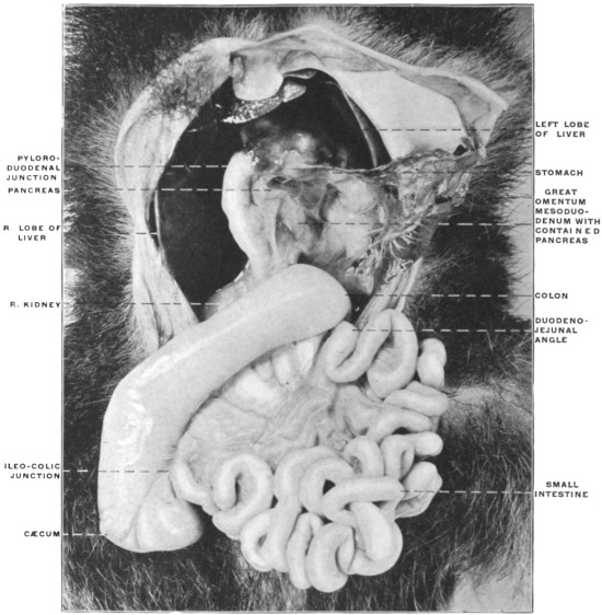
Fig. 117.—The same view as Fig. 116, from another specimen.
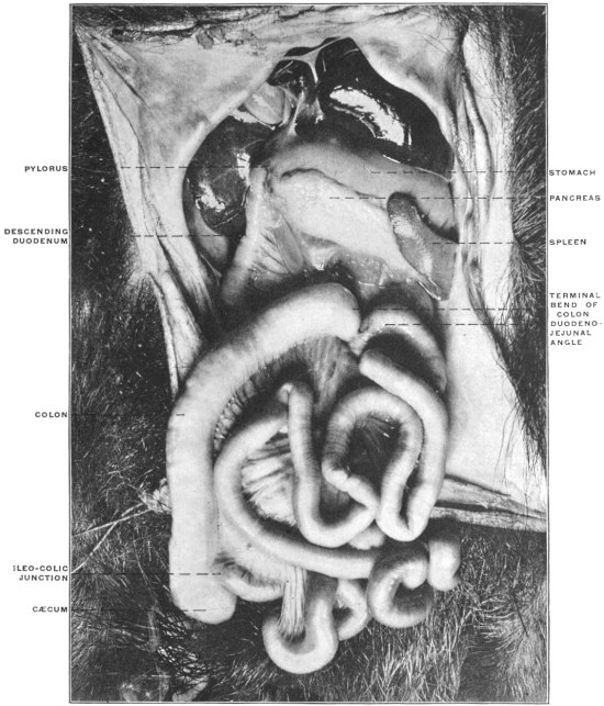
Figs. 116 and 117 show the intestinal tract of Tamandua bivittata arranged so as to correspond to the human embryonic condition after rotation. The cæcum has been brought up and to the right across the proximal duodenal portion of the small intestine, while the jejuno-ileal coils have been turned down and to the left. The rotation has been accomplished by a twist at the duodeno-colic isthmus, and the original right leaf of the mesentery has become the left and vice versa. Comparison with Figs. 107 and 108, representing the condition before rotation in the same animal, will indicate the changes which have been accomplished by imitating the course of development followed in the higher mammals.
Failure of rotation and arrest of development at the primitive stage, with consequent persistent embryonic condition of the mesentery, occurs occasionally in man. Such cases have been reported by W. J. Walsham, in St. Barthol. Hosp. Rep., London, Vol. 16. The following four instances of this condition, taken from the Columbia University museum, will illustrate the disposition of the abdominal contents.
Fig. 118 shows the arrangement of the abdominal viscera in an adult female body. Beginning at the pyloric extremity of the stomach the entire course of the duodenum can be overlooked and its continuation into the jejuno-ileal division traced. The small intestines occupy the ventral and right part of the cavity. The ileo-colic junction is placed in the lower left-hand corner of the abdomen and the small intestine enters the large from right to left, the ascending colon is situated to the left of the median line and at its point of transition into the segment representing the transverse colon is connected by several adhesions with the ventral surface of the duodenum. The transverse colon, folded into several coils bound together by adhesion, occupies the upper left portion of the abdomen.
Fig. 119, taken from the same specimen, shows the entire mass of intestines lifted up and turned to the left, exposing the background of the abdominal cavity lined by parietal peritoneum. The duodenum is still entirely free and non-adherent to the parietal peritoneum. The continuity of the mesoduodenum with the jejuno-ileal mesentery is well shown. The primitive right leaf of the mesentery is turned to the observer. This layer after completed rotation would form the left layer of the adult mesentery of the jejuno-ileum.
Fig. 118.—Abdominal viscera of adult human female, in a case of arrested rotation of the intestines. (Columbia University Museum, Study Collection.) Fig. 119.—The same preparation with the intestinal coils displaced upward and to the left.
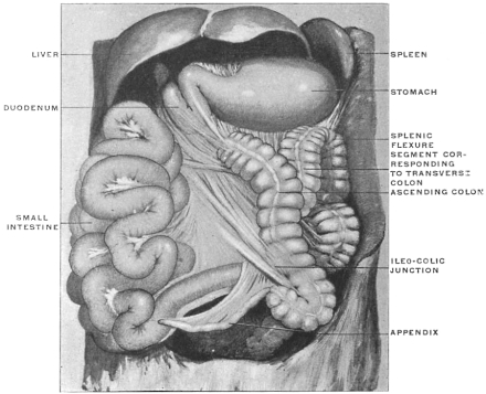
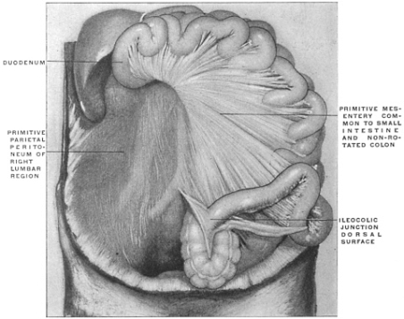
Fig. 120 illustrates another instance of the same condition in the adult. In this case the duodenum was coiled twice upon itself and adherent to the prerenal parietal peritoneum.
Fig. 121, presenting the same adhesion of the duodenum, illustrates very perfectly the persistence of the narrow duodeno-colic isthmus in cases of non-rotation, as well as the development of the different segments of the adult tract from the limbs of the embryonal umbilical intestinal loop.
It will be observed that beyond the duodeno-colic isthmus the coils of the jejuno-ileum have resulted from the increase in length of the descending limb, the apex and the proximal part of the ascending or recurrent limb, carrying the ileo-colic junction and cæcum. The remainder of the ascending limb, terminating in the embryonic condition at the splenic flexure by passing into the descending colon, has in the course of further development in this individual produced a straight segment—the misplaced ascending colon—and a convoluted and bent representative of the normal transverse colon.
The same disposition of the large intestine may be noted in the other preparations.
Fig. 120.—Abdominal viscera of adult human male; non-rotation of intestine. (Columbia University Museum, Study Collection.) Fig. 121.—Abdominal viscera of adult human male; non-rotation of intestine. (Columbia University Museum, Study Collection.)
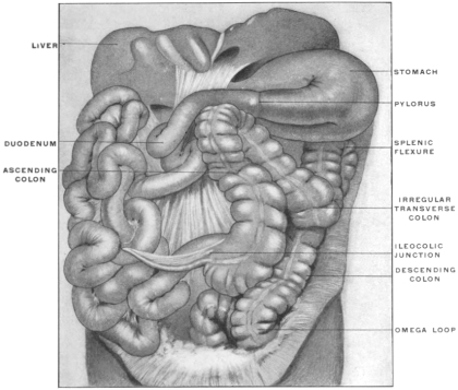
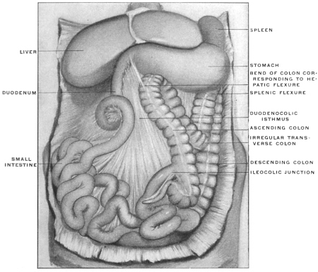
Fig. 122.—Abdominal viscera of child, two years old; non-rotation of intestine. (Columbia University Museum, Study Collection.) Fig. 123.—Human fœtus at term; abdominal viscera, hardened in situ; non-rotation of cæcum. (Columbia University Museum, No. 1813.)
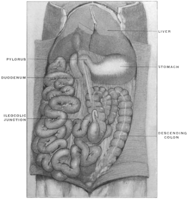
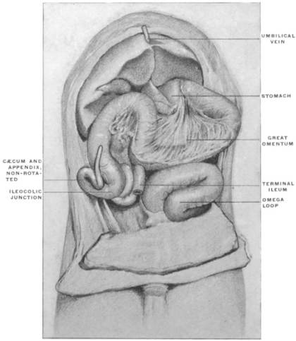
Fig. 122 shows an instance of non-rotation observed in the human infant at two years of age.
Fig. 123, taken from a fœtus at term, shows the result of failure to completely rotate in the region of the cæcum and ileo-colic junction. The rest of the large intestine has rotated as usual and assumed the normal position. The terminal ileum, however, passes behind the cæcum and enters the large intestine on its right side; the cæcum is turned upwards and to the right and the appendix lies ventrad of the beginning of the ascending colon. In order to produce the normal arrangement, shown in Fig. 124, taken from another fœtus at term, it would be necessary to turn the cæcum and ileo-colic junction in Fig. 123 through half a circle. The cæcum would then turn upwards and to the left, the ileum entering the large intestine from left to right, and the appendix would be placed behind the cæcum and ileo-colic junction. Figs. 125 and 126 show the normal and abnormal arrangement presented by these two preparations diagrammatically. The instances in which in the adult the ileo-colic entrance is placed on the right side of the large intestine and in which the appendix is situated laterad of the ascending colon unquestionably find their explanation in the failure of the intestine to completely rotate at the ileo-colic junction.
Fig. 124.—Human fœtus at term; abdominal viscera, hardened in situ; normal position of completely rotated cæcum and appendix. (Columbia University Museum, No. 1814.) Figs. 125, 126.—Schematic representation of final stages in rotation of cæcum and large intestine.
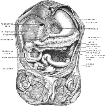
Fig. 125.—Just before final rotation of cæcum and terminal ileum. Concavity of cæcum directed cephalad and to right. Terminal ileum enters colon from right to left. Fig. 126.—Rotation completed. Concavity of cæcum turns caudad and to left. Terminal ileum enters colon from left to right.
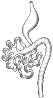
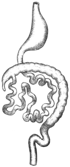
Fig. 127.—Adult human subject with non-rotated cæcum. The terminal ileum turns caudad from right to left to enter right side of colon.
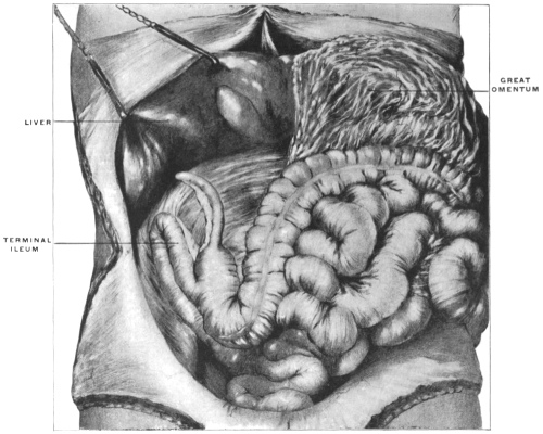
Fig. 128.—Adult human subject with non-rotated cæcum, the ileum entering large intestine from the right and behind, and the appendix placed to the right of the ascending colon. (From a fresh dissection.)
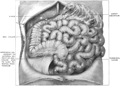
The resulting conditions are shown in Figs. 127 and 128, taken from adult human subjects in which the final stage of rotation of the large intestine has not taken place.
In Fig. 127 the terminal ileum is sharply bent on itself and adherent to the prerenal parietal peritoneum. It passes from right to left and downwards to enter the right posterior circumference of the large intestine. The cæcum is turned cephalad and the appendix is in contact with the right lobe of the liver. The cæcum passes with a sharp bend into the obliquely directed ascending colon.
In Fig. 128 the ileum enters the colon from the right and below. The apex of the cæcum is turned cephalad and to the right and the appendix extends beneath peritoneal adhesions along the lateral border of the proximal segment of the colon.
In the next place it is desirable to clearly understand the vascular supply of the intestine before and after rotation and the final relation of the superior mesenteric artery to the transverse portion of the duodenum.
Development of Aortal Arterial System.
The thoracic and abdominal aortæ are at first double, the first aortic arches continuing as so-called “primitive aortæ” ventrad of the vertebral column to the caudal end of the body.
The cephalic portions of the two vessels unite in the chick on the third day and from this point fusion into a single vessel proceeds slowly caudad.
In the rabbit the fusion of the primitive aortæ begins on the ninth day in the region of the lung-buds and progresses from here caudad until by the sixteenth day a single aorta is formed (Fig. 129).
Fig. 129.—Diagrams illustrating the arrangement of the primitive heart and aortic arches. (After Heisler, modified from Allen Thompson.)
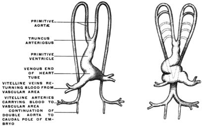
That the entire descending aorta in man results from the fusion of two vessels is shown by the rare cases in which the aorta is divided throughout its entire length by a septum.
The arteries of the allantois are originally the terminations of the primitive aortæ. After fusion of the primitive aortæ to form the abdominal aorta the allantoic arteries, now passing as the umbilical arteries to the placenta, appear as the branches of bifurcation of the abdominal aorta, in the same way as the common iliacs do in the adult.
They furnish branches, which at first are very small, to the budding posterior extremities and the pelvic viscera. In time these rudiments of the future external and internal iliac arteries become larger, but as the umbilical arteries continue to develop throughout the entire intra-uterine period they appear even in the fœtus at term as end branches of the aorta, a condition which is only changed after birth by the obliteration of the umbilical arteries and their conversion into the lateral ligaments of the bladder, while the iliac vessels now appear as the terminal aortic branches. The statement that the umbilical arteries appear as the terminal branches of the embryonal aorta requires to be modified in the following respect:
When the allantois develops its arteries are in fact end-branches of the two primitive aortæ. After their fusion and after the formation of the single aorta this vessel is continued beyond the umbilical arteries as a small trunk, the caudal artery or rudiment of the adult sacralis media. Consequently the umbilical arteries are really lateral branches of a median vessel, viz., aorta abdominalis and arteria sacralis media. But as the umbilical vessels are very large and the caudal aorta very small, the former, even under these conditions, appear as the real terminal branches of the abdominal aorta.
The arteries supplying the yolk-sac and subsequently the intestinal canal are the vitelline or omphalo-mesenteric. At first they are branches derived from the two primitive aortæ, and after the fusion of these vessels they arise from the resulting single abdominal aorta. The omphalo-mesenteric arteries are at first multiple and later are reduced to two. When the primitive intestine loses its original close contact with the vertebral column and the common dorsal mesentery develops, the two omphalo-mesenteric arteries unite to form a single vessel, running between the layers of the mesentery. After a short course this artery divides again into two branches, passing one on each side, around the intestinal tube, which has in the meanwhile become closed. Ventrad of the intestine these branches reunite so that the gut is surrounded by a vascular circle. The left half of this loop becomes obliterated and the trunk of the omphalo-mesenteric artery now passes on the right side of the intestine to the umbilicus. The peripheral segment of the omphalo-mesenteric artery disappears with the cessation of the vitelline circulation. The proximal portion, situated between the layers of the mesentery, gives numerous anastomosing branches to the intestine and is converted into the main trunk of the superior mesenteric artery.
The derivation of the superior mesenteric as the fully developed proximal segment of the embryonic omphalo-mesenteric artery passing to the yolk-sac is responsible for the rare anomaly in the adult of a branch of the superior mesenteric artery continuing beyond the intestine to the umbilicus. I have encountered one instance of this persistence of the intra-abdominal portion of the omphalo-mesenteric artery in a male subject 54 years of age. A connective strand, containing a small artery derived from the superior mesenteric vessels, extended between the right layer of the mesentery, some distance from its attached border, and the ventral abdominal wall at the umbilicus. The vessel which was pervious throughout, was the size of one of the digital arteries.
Hyrtl has observed the same variation. An example of partial persistence of the omphalo-mesenteric artery in the adult is well seen in the case of Meckel’s diverticulum shown in Fig. 37, where the arterial vessel continued upon the diverticulum represents the embryonic omphalo-mesenteric artery.
The remaining intestinal arteries are at first more numerous and paired. In man and most mammals they are early reduced in number, passing from the abdominal aorta to the dorsal or attached border of the intestine, between the two peritoneal layers of the primitive dorsal mesentery (Fig. 104). The arterial blood supply of the intestinal canal then presents three general divisions:
1. Vessels pass from the proximal part of the abdominal aorta to the stomach and pyloric portion of the duodenum. This set of vessels forms the rudiment of the future cœliac axis. With the development of the liver and pancreas by budding from the duodenum, and with the appearance of the spleen in the mesoderm of the dorsal mesentery, branches corresponding to these organs (hepatic and splenic arteries) are added to the gastric and duodenal vessels and the adult arrangement of the cœliac axis is thus obtained (Figs. 130, 131, 132 and 133).
Fig. 130.—Diagrammatic representation of the arteries proceeding to the alimentary canal and appendages prior to rotation of intestine (stage of simple umbilical loop). Fig. 131.—Diagrammatic representation of the arteries of the alimentary canal in the first stage of intestinal rotation, showing relation of superior mesenteric artery to the transverse portion of the duodenum.
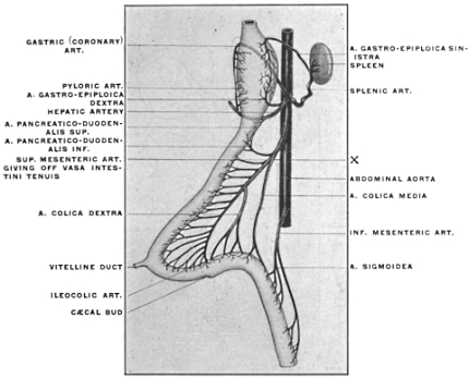
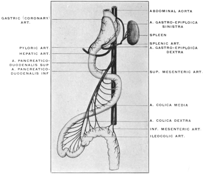
Fig. 132.—Arteries of alimentary canal in the later stages of intestinal rotation. Fig. 133.—Final arrangement of arteries of alimentary canal after completed rotation of the intestines.
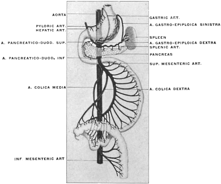
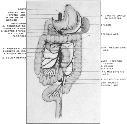
These vessels have an important bearing on the formation of the adult peritoneal cavity in the retro-gastric space, and will be considered in detail below with that portion of the subject.
2. The next vessel in order derived from the aorta and supplying the duodenum, pancreas, the small and a part of the large intestine is the above-mentioned superior mesenteric artery, which arises from the aorta a short distance caudad of the cœliac axis (Figs. 130, 131, 132 and 133).
At the time when the intestine still presents the primitive arrangement of the umbilical loop (Figs. 104 and 130) this vessel passes between the layers of the dorsal mesentery through the narrow duodeno-colic neck to reach the two limbs and the apex of the intestinal loop. In its course it gives off successively branches to the gut from each side. Those from the right side of the main vessel pass to the duodenum, pancreas, jejunum and ileum. Those from the left side of the main vessel accede in succession to the colic angle of the isthmus, the proximal portion of the colon, the cæcum and the ileo-colic junction. The terminal portion of the superior mesenteric artery supplies the ileum near the ileo-colic entrance. After rotation it will be found that the turn has occurred at the point X (Fig. 130), i. e., in that part of the vessel which occupies the duodeno-colic isthmus. Hence it will be found that the first branches derived from the right side of the primitive superior mesenteric artery, supplying the duodenum and pancreas (Art. pancreatico-duodenalis inferior) still arise after rotation from the right side. They are succeeded, beyond the point X, by the original highest left branches passing to colon, cæcum and ileo-colic junction, while all the original right-sided vessels, except the inferior pancreatico-duodenal, appear now as branches from the left side of the main artery, supplying the coils of the jejuno-ileum. Hence in the adult (Fig. 133) the succession of branches derived from the right or concave side of the superior mesenteric artery is as follows:
Fig. 134.—Schematic representation of intestinal arterial supply from superior mesenteric artery in cases of arrested rotation of the intestine.
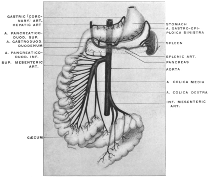
1. Arteria pancreatico-duodenalis inferior. 2. Arteria colica media. 3. Arteria colica dextra. 4. Arteria ileo-colica.
On the other hand, the first branches from what has now become the left or convex side of the vessel are the original lower right-hand vessels to the small intestine developed from the descending limb of the loop. Hence in the adult the left side of the superior mesenteric vessel gives rise to the vasa intestini tenuis.
3. The caudal intestinal arterial branch derived from the aorta is the inferior mesenteric artery supplying parts of the transverse colon, the descending colon, sigmoid flexure and rectum (Figs. 130, 131, 132, and 133).
On the other hand in the cases of non-rotation of the intestine as above described in Figs. 118-122, the embryonic type of the intestinal arterial supply persists, as indicated schematically in Fig. 134. Not only the pancreatico-duodenalis inferior, but all the remaining branches to the small intestine are derived from the right side of the superior mesenteric artery. The terminal branches of the main artery supply the ileo-colic junction, while the arterial supply of the large intestine, A. colica dextra and media, are given off from the left side of the parent vessel.
II. Demonstration of Intestinal Rotation in the Cat.—The changes in the relative position of the different intestinal segments and the final disposition of the mesenteries and blood vessels can best be understood by the direct examination of the abdominal contents in an animal whose permanent adult arrangement corresponds to one of the early embryonal human stages, and in which the necessary manipulations can readily be carried out and their results noted.
It is doubtful if the above detailed developmental stages in man can ever be clearly comprehended unless the student will for himself examine the conditions and perform the manipulations in one of the lower mammals.
The necessity of keeping the three dimensions of space in mind and the fact that certain structures during and after rotation cover and obscure each other, make diagrams and drawings unsatisfactory unless the actual examination of the object itself is combined with their study. Fortunately, among the common domestic animals of convenient size easily obtained the cat answers every purpose of this study admirably. The student is earnestly urged to pursue his study of the development and adult arrangement of the human abdominal viscera and peritoneum in the light which the anatomy of this animal can shed on the complicated and obscure conditions encountered in the human subject. The plan of having the opened abdominal cavity of the cat directly side by side with the human subject, while the arrangement of the abdominal viscera and peritoneum is considered, cannot be recommended too highly.
Directions.—After killing the animal with chloroform the abdominal cavity is to be freely opened by a cruciform incision and the skin flaps turned well back and secured in this position. It is well to select a male animal or an unimpregnated female, as the size of the pregnant uterus in the later stages renders the examination of the abdominal viscera and peritoneum more difficult.
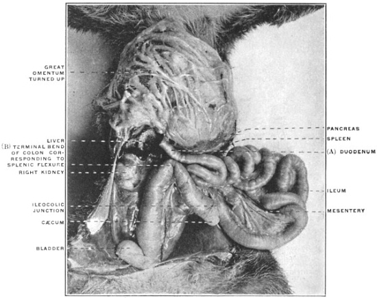
Fig. 135.—Abdominal viscera of cat; great omentum raised; intestines turned down and to left. (From a fresh dissection.)
For purposes of careful study and comparison of the vascular relations of the abdomen, it is highly desirable to inject the animal with differently colored gelatine, starch or plaster of Paris mass. The arterial injection can be made through the carotid artery, the systemic venous injection through the femoral vein, and the portal circulation can be filled after opening the abdomen, by injection through the superior mesenteric or splenic veins. Animals prepared in this manner are especially useful for the study of the upper portion of the abdominal cavity and of the peritoneal relations of liver, stomach, spleen, pancreas and duodenum. They may be kept for permanent reference in a 5 per cent. solution of formaline or 50 per cent. alcohol.
After opening the abdominal cavity turn the great omentum up over the ventral surface of the thorax and secure it in this position, thus exposing the underlying intestines completely (Fig. 135). Trace in the first place the entire course of the intestinal tube from the pyloric extremity of the stomach down. It will be noticed that the first portion of the small intestine (duodenum) is freely movable, completely invested by peritoneum and attached to the dorsal midline by a mesoduodenum between the layers of which a portion of the pancreas is seen.
Following the duodenum caudad it will be observed that the gut can be traced directly continuous with the remaining coils of the small intestine. The ileo-colic junction and the beginning of the large intestine are marked by a short pointed cæcum. The large intestine is short, as it is in all carnivore mammals, and passes from the cæcum almost directly down into the pelvis.
Take the cæcum and the first portion of the large intestine and turn them caudad and over to the left side as far as the peritoneal connections will permit.
Spread out the coils of the small intestine in the opposite direction, i. e., over to the right side.
The arrangement of the intestinal tract after these manipulations should appear as shown in Figs. 136 and 137.
Fig. 136.—Abdominal viscera of cat, hardened; omentum removed to display derivation of intestines from umbilical loop and the relation of the superior mesenteric artery and common dorsal mesentery to the small and large intestines. (Columbia University Museum, No 728.)
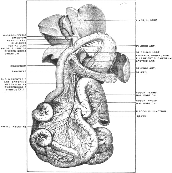
Fig. 137.—Abdominal cavity of cat. (From a fresh dissection.)
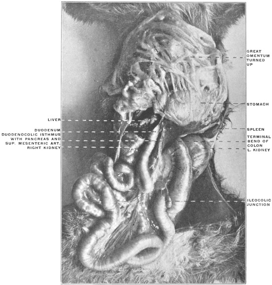
It will be seen that all the essential features described for the corresponding stage in the human embryo (Fig. 104, A) exist here. The proximal portion of the small intestine (duodenum) retains its freedom and mobility, being attached to the ventral surface of the vertebral column by the portion of the primitive mesentery which now constitutes the mesoduodenum. The gut itself forms a bend with the convexity turned to the right.
Observe in the next place that the point (Fig. 136, X), where small intestine and colon approach each other closely, marks the situation of the fœtal duodeno-colic isthmus. The small intestine at this point corresponds to the future duodeno-jejunal angle as will be seen after rotation has been accomplished.
Recalling the development of the jejuno-ileum it will not be difficult to recognize in the numerous coils of small intestine which succeed to the duodeno-colic isthmus the results of the increase in length of the descending or efferent limb of the human embryonal umbilical loop. Tracing these coils it will be found that the terminal portions of the ileum correspond to the apex and to the proximal part of the ascending or recurrent limb of the primitive loop, while the remainder of this limb furnishes the cæcum and the next succeeding segment of the large intestine. Following the tube up to this point the colic boundary of the duodeno-colic isthmus will be reached; from here the short large intestine of the carnivore descends straight into the pelvis, attached to the ventral surface of the vertebral column by a mesocolon which corresponds to the distal part of the original primitive dorsal mesentery.
Now with the parts still in this position examine carefully the arrangement of the mesentery and of the intestinal blood vessels. Starting with the duodenum it will be seen that the primitive sagittal mesentery of this portion of the intestine has followed the gut in its turn to the right, so that the original right layer of the sagittal membrane is now directed dorsad and lies in contact with the parietal peritoneum which invests the background of the abdominal cavity in the right lumbar region below the liver and covers the ventral surface of the right kidney. Beneath this parietal peritoneum the inferior vena cava is seen, receiving the right renal vein and ascending to enter the dorso-caudal aspect of the right lobe of the liver. If now we assume that in the cat the opposed serous surfaces of the original right leaf of the mesoduodenum, now directed dorsad, and of the parietal peritoneum adhere to each other, and that the visceral peritoneum covering the dorsal surface of the descending duodenum likewise becomes obliterated by adhesion to the subjacent parietal peritoneum, we will obtain the arrangement found in the adult human subject, in which the descending duodenum is fixed by adhesion below the right lobe of the liver and ventrad of the medial portion of right kidney, right renal vein and inferior vena cava. During this process of anchoring the head of the pancreas, which is found between the two layers of the free mesoduodenum of the cat, would also become fixed to the abdominal background by adhesion of the original right leaf of the mesoduodenum, investing what has now become the dorsal surface of the pancreas, to the parietal peritoneum. The original left layer of the primitive mesoduodenum would then appear as secondary parietal peritoneum covering what has now become the ventral surface of the transversely disposed head of the gland. The stages may be represented schematically in Figs. 138-140.
Figs. 138-140.—Diagrammatic representation of three stages in the development of the mesoduodenum, duodenum, and pancreas leading to the secondary “retroperitoneal” position of these viscera.
Fig. 138.—Free mesoduodenum in sagittal plane, including head of pancreas between right and left layers.
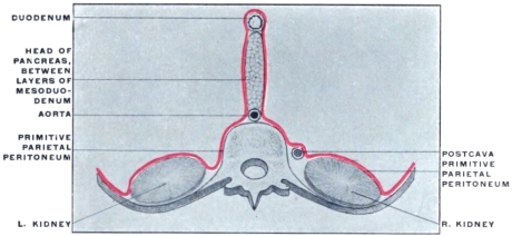
Fig. 139.—Mesoduodenum folded to right; left leaf has become ventral; right dorsal, directed toward primitive prerenal parietal peritoneum.
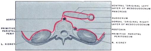
Fig. 140.—Fixation of head of pancreas and duodenum under cover of secondary parietal peritoneum by adhesion of apposed surfaces of mesoduodenum and primitive parietal peritoneum.
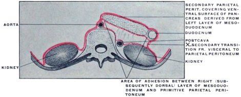
Figs. 138 and 139 shows the arrangement in the cat where a free duodenum and mesoduodenum exists, with the pancreas included between its layers.2
It will be noticed that the duodenum in the cat can be carried over to the median line (Fig. 138) exposing the entire ventral aspect of the right kidney and the inferior vena cava beneath the primary lumbar parietal peritoneum. This manipulation will also expose the dorsal surface of the head of the pancreas, covered by what originally was the right leaf of the mesoduodenum.
Fig. 140 indicates the results of adhesion of the duodenum, pancreas and mesoduodenum to the parietal peritoneum as it normally occurs in the human subject. It will be seen that the primary parietal peritoneum can be traced mesad over the ventral surface of the right kidney as far as the point X, and that from here on to the median line the peritoneum is secondary parietal peritoneum, consisting of the visceral peritoneal investment of the ventral surface of the duodenum and of the original left leaf of the mesoduodenum, beneath which the ventral surface of the pancreas is seen. Pancreas and duodenum occupy in the adult secondarily a “retro-peritoneal” position, i. e., the peritoneum now covering the ventral surface of these viscera appears as a continuation of the parietal peritoneum, the transition between primary and secondary parietal peritoneum occurring along the line marked X in Fig. 140. The opposed peritoneal surfaces indicated by the dotted lines have become adherent and converted into loose connective tissue in which the pancreas and duodenum lie imbedded. In the human embryo this process of adhesion begins in the eighth week, starting at the duodeno-jejunal flexure and ascending gradually toward the pylorus. At the end of the fourth month the union is complete.
Proceeding caudad it will next be observed that the peritoneum of the mesentery occupies the narrow neck of the duodeno-colic isthmus, and that large vessels (the superior mesenteric) pass between its two layers at this point to supply the segments of the intestine forming the loop. In conformity with the greatly increased length of the intestine it will be found that the mesentery expands from the narrow pedicle at the neck in a fan-shaped manner in order to develop a sufficiently long margin for attachment to the intestine. The following points should be carefully borne in mind in studying the mesentery with the intestines in this position:
1. The mesentery presents two free surfaces, right and left. With the coils of the small intestine turned over to the right, the left leaf of the mesentery is turned toward the observer.
Fig. 141.—Schematic representation of mesentery of umbilical loop, common to small intestine and proximal portion of large intestine.
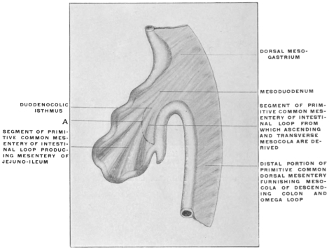
2. Inasmuch as the descending limb of the embryonic loop has developed the greater part of the small intestine, while a portion of the large intestine (cæcum and colon up to the isthmus) is the result of differentiation within the ascending or returning limb of the loop, it will be at once apparent that the double peritoneal layer which extends between the duodeno-colic isthmus and the attached border of the gut is partly mesentery of the small intestine, partly mesocolon passing to the large intestine (cæcum and proximal colon). This condition may be indicated schematically in Fig. 141.
The curved line A may be taken as an arbitrary division between the portion of the membrane which on the right of the figure passes to the small intestine, and the portion which proceeds to the left to be attached to the large intestine. In other words the line will schematically separate the true mesenteric from the mesocolic segment of the primitive membrane.
With the parts in their present position this line might be assumed to indicate a strip along which the opposed serous surfaces of the parietal peritoneum and the right leaf of the primitive mesentery became adherent. In that case an actual division into a mesenteric and mesocolic segment would have been effected.
Ventrad and to the right of this line of adhesion we would trace that portion of the primitive membrane which now passes to the coils of the small intestine as the true mesentery, having an apparent origin in the background of the abdomen to the dotted line of adhesion. In the same manner the peritoneal layers passing to the left to reach the cæcum and beginning of the colon would appear as a free mesocolon with the same line of apparent origin from the background of the abdomen. (cf. p. 80.)
These considerations should be followed out in the dissection of the cat in order to become familiar with the principle of secondary lines of origin for peritoneal layers. As we will see later this factor is of importance in correctly estimating the value of the human adult conditions.
3. A brief consideration of the mechanical conditions and comparison with the earlier stages will show why the peritoneal layers which occupy the bight of the fully developed umbilical loop are especially prone to develop secondary lines and areas of adhesion to other serous surfaces. If we compare the dorsal mesentery in its primitive condition, before the straight intestinal tube has become differentiated into the subsequent segments, and before the umbilical loop has been formed (Fig. 142), with the later stages represented by the intestines of the cat as now arranged (Figs. 143 and 144), it will be seen that the vertical line of attachment to the ventral surface of the vertebral column, between the points a and b corresponds in the advanced stages to the interval ab separating the two points of the duodeno-colic isthmus; also that the entire mesenteric peritoneal surface beyond the isthmus is the result of drawing out and lengthening the intestinal tract. Consequently folding or overlapping of this extensive membrane affords opportunities for adhesions between its own serous surfaces or between it and the remaining visceral and parietal peritoneum of the abdomen.
Figs. 142-144.—Schematic representation of three stages in the development of the mesentery of the umbilical intestinal loop.
Fig. 142.—Early stage before differentiation of intestinal canal. Fig. 143.—Stage of umbilical loop. Differentiation of common dorsal mesentery of earlier stage into dorsal mesogastrium, mesoduodenum, primitive mesentery of umbilical loop, and descending mesocolon.
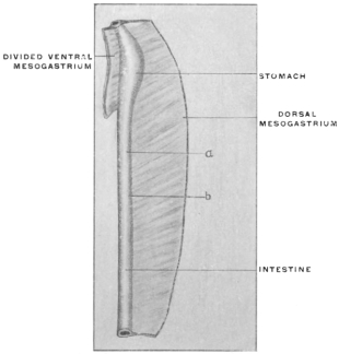
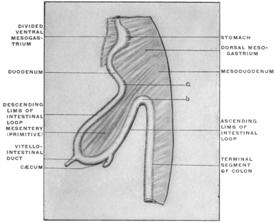
Fig. 144.—Final stage. With complete differentiation of large and small intestine, the primitive mesentery of the umbilical loop contains not only the mesentery of the future jejuno-ileum, but also the mesocola and the ascending and transverse colon, developed from the ascending or afferent limb of the umbilical loop.
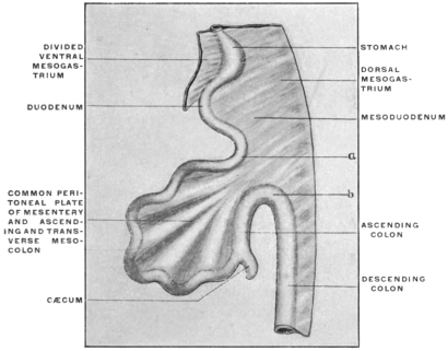
Fig. 145.—Abdominal viscera of cat, with intestines rotated to correspond to the stage in the development of the human canal in which the cæcum has reached the subhepatic position, but before the establishment of the ascending colon. (From a fresh dissection.)
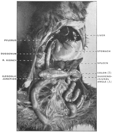
Moreover, it will be appreciated that the entire extensive coil of intestines extending between the two boundaries of the duodeno-colic isthmus (a, b) is suspended from the back part of the abdomen by a narrow pedicle and that consequently rotation will readily occur around the axis drawn through the neck of the isthmus.
Now proceed to illustrate on the cat the result of the rotation as it occurs normally during the development of the primate intestinal tract. Take the cæcum and commencement of the colon and draw the same over to the right across the duodeno-colic isthmus and the duodenum. Twist or rotate the entire mass of small intestines around the isthmic pedicle, so that the original left leaf of the mesentery will look to the right and vice versa (Fig. 145). The conditions thus established will be found to correspond to the schemata shown in Figs. 114 and 115. The main features of the intestinal tract in the rearranged position will be as follows:
1. The two points, a and b, of the duodeno-colic isthmus (Fig. 145) are still close together, but reversed in position, b is in front and to the right, a behind and to the left, whereas before the rotation b was situated below and to the left, a above and to the right (Fig. 135).
2. The direction of the ileo-colic entrance is reversed, the ileum now entering the large intestine from below and the left upwards and to the right, instead of from right to left.
3. The descending duodenum is now situated dorsad to the colon.
4. The original left leaf of the mesentery has become the right, and vice versa.
5. The superior mesenteric artery crosses over the transverse portion of the duodenum, and with the exception of the inferior pancreatico-duodenal artery the original right-sided branches now arise from the left side of the vessel and vice versa.
It is now time to compare the conditions established in the cat by the manipulations just detailed with the arrangement of the adult human intestinal tract and peritoneum below the level of the transverse colon and mesocolon.
I. The shortness of the large intestine in the cat will require careful manipulation in order to produce a disposition in conformity with the arrangement of this portion of the human intestinal tract. By stretching the gut somewhat and pulling it well out of the pelvis sufficient length will be obtained to establish an ascending, transverse and descending colon. Move the cæcum from the subhepatic position which it occupies immediately after rotation (Fig. 145) down to the lower and right-hand corner of the abdomen. Pull the distal portion of the large intestine well out of the pelvis and obtain thus sufficient length to establish an ascending, transverse and descending division each provided with a free mesocolon (Fig. 146). In the formation of the three definite main segments of the human large intestine, ascending, transverse and descending colon, the following stages may be recognized:
Fig. 146.—Abdominal viscera of cat, with the intestines rotated to correspond to the adult human disposition, with ascending, transverse, and descending segments of the colon. (From a fresh dissection.) Fig. 147.—Human fœtus, 6.6 cm., vertex-coccygeal measure; liver removed. (Columbia University Museum, Study Collection.) × 4.
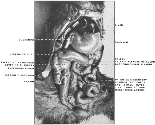
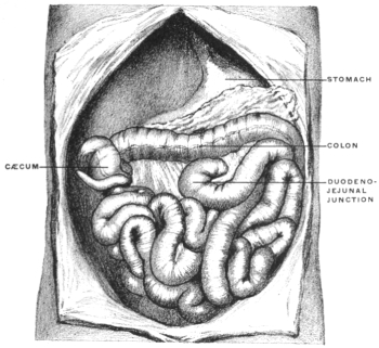
1. Immediately after rotation the large intestine lies transversely along the greater curvature of the stomach, with the cæcum on the right side in front of the duodenum and closely applied to the caudal surface of the right lobe of the liver (Fig. 147).
Persistence of Subhepatic Position of Cæcum in Adult.—The period at which the cæcum descends into the iliac fossa is liable to a considerable range of variation.
Treves found in two fœtus, measuring respectively 4½″ and 5½″, the cæcum on a level with the caudal end of the right kidney, while in several individuals at full term the caput coli was still placed immediately below the liver, with no large intestine in the place of the ascending colon. This condition is well illustrated in the fœtus shown in Fig. 124.
The cæcum may remain undescended throughout life. Treves, in an examination of 100 bodies, found this condition in two subjects, both females, one 41, the other 74 years of age. Both cases presented an identical disposition. There was no large intestine in the place of the ascending colon. The cæcum was placed on the right side, immediately underneath the liver, just to the right of the gall-bladder; it was quite horizontal in position, continuing the long axis of the transverse colon and included between the layers of the transverse mesocolon. From the extremity of the cæcum a horizontal fold was continued to the abdominal parietes and upon it the edge of the liver rested. In one of these instances the colon from the tip of the cæcum to the splenic flexure measured 38″. The great omentum was attached only to the left half of this portion. The descending colon was very long, measuring 15″.
In the other case the distance from the tip of the cæcum to the splenic flexure was 27″, the great omentum commencing 5″ from the former point. The descending colon was of normal length.
In both bodies the remaining viscera were normal.
2. The cæcum next descends ventrad of right kidney to the iliac fossa. The future ascending colon is at this time placed very obliquely on account of the large size of the fœtal liver, and passes without a marked angle into the transverse segment. Thus in Fig. 148, from a fœtus 5″ in length, the descending colon is vertical and the splenic flexure well marked, forming the highest point of the colic arch. There is no hepatic flexure, and no ascending and transverse colon, but instead of these an oblique segment passing upwards and to the left between cæcum and splenic flexure.
This disposition, due to the large size of the liver, is still marked at times in the fœtus at term, and occasionally even in children up to 2 or 3 years of age.
Fig. 148.—Abdominal viscera of human fœtus of 12.5 cm., vertex-coccygeal measure, hardened in situ; transverse and ascending colon not yet differentiated. (Columbia University Museum, No. 1815.) Natural size. Fig. 149.—Abdominal viscera of human fœtus at term, hardened in situ; hepatic flexure formed and ascending and transverse colon differentiated. (Columbia University Museum, No. 1816.)
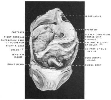
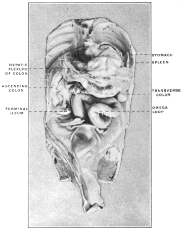
3. The ascending colon is subsequently differentiated from the transverse segment and the hepatic flexure formed consequent upon the diminution of the relative size of the liver, which permits the fœtal oblique segment of the colon extending in the earlier stages between the right iliac fossa and the spleen to become divided by a right-angled (hepatic) bend or flexure into an ascending and a transverse segment (Fig. 149).
4. The splenic flexure develops early and is well marked. It indicates the point of transition of the original ascending limb of the umbilical loop into the remaining vertical median segment of the large intestine, from which the descending colon is formed.
In the adult the ascending and descending portions of the colon are vertical. The transverse colon is not quite horizontal since the splenic flexure is higher and placed more dorsally than the hepatic flexure. In the embryo the rapidly-growing coils of the small intestine push the descending colon to the left and dorsad into close contact with the dorsal abdominal wall.
Fig. 150.—Caudal portion of human embryo of 5 mm., with the end- and caudal gut at the highest stage of its development. × 25. (Reconstruction after His.)
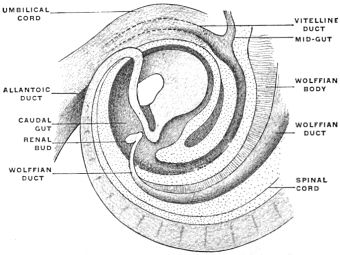
A small bend which appears about the middle of the third month in the left iliac fossa indicates the rudiment of the future sigmoid flexure or omega loop.
The rest of the endgut follows the body wall in a well-marked curve, whose termination lies within the concavity of the caudal portion of the embryo (Fig. 150). From this terminal part the rectum develops after the division of the cloaca and the union of the proctodæum with the entodermal intestinal pouch has taken place as detailed above.
The early position of the colon produced by the large size of the fœtal liver, and before the descent of the cæcum has occurred, is shown in Fig. 124. In Fig. 123, where the liver has regained its normal proportions with reference to the abdominal cavity and viscera, and the cæcum has descended into the right iliac fossa, the hepatic flexure is well marked and the first segment of the colon has acquired the vertical position on the right side, the single obliquely transverse segment of Fig. 124, having become divided into an ascending and a transverse colon.
[Fig. 124. Early stage. Liver relatively large. Proximal portion of the colon extends obliquely between the right lumbar region and the spleen. The cæcum has not yet descended.
Fig. 123. Later stage. The cæcum occupies the right iliac fossa. Relative reduction in the size of the liver allows the colic segment to be divided by the hepatic flexure into an ascending colon and a transverse colon.]
Fig. 151.—Abdominal viscera of Macacus rhesus, rhesus monkey, hardened in situ. (Columbia University Museum, No. 1817.)
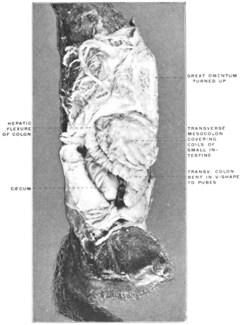
At times the transverse colon, whose normal average length in the adult is 20″, greatly exceeds this measurement and forms an arch which hangs down or makes a well-marked V-shaped bend with the apex directed toward the pubes. This is the normal arrangement of this portion of the large intestine in many of the lower primates. Fig. 151 shows the abdominal viscera of Macacus rhesus, hardened in situ, seen from the front and the right side, with the omentum turned up over the stomach. The transverse colon forms an extensive V-shaped bend, whose apex reaches to the pubes, from which point the large intestine turns again cephalad and dorsad to form the splenic flexure and then descends to the pelvis.
The average length of the ascending colon in the adult, measured from the tip of the cæcum to the hepatic flexure, was found by Treves in his series of 100 bodies to be 8″, while the descending colon, from the splenic flexure to the beginning of the sigmoid loop, measured 8½″.
The descending colon may at times be much longer, up to 15″, and become convoluted.
II. In the next place, in order to understand the arrangement of the peritoneum in this lower larger compartment of the abdomen, disregard for the present the peritoneal connections of the stomach, liver, pancreas and spleen, and the folds of the great omentum entirely. This latter membrane is adherent in the adult human subject by its dorsal surface to the upper margin of the transverse colon, so that in turning the omentum up over the ventral chest wall the transverse colon will be carried with the omentum and the lower layer of the transverse mesocolon will be put upon the stretch. This membrane forms in adult man by its transverse attachment to the abdominal background the cephalic limit of the larger lower compartment of the abdomen, which is framed laterally by ascending and descending colon, continuous below with the pelvic cavity and occupied chiefly by the freely movable coils of the jejuno-ileum.
Remember that the duodenum starting from the pyloric extremity of the stomach first turns cephalad and dorsad in contact with the caudal surface of the right lobe of the liver, forming the first portion or hepatic angle of the duodenum; that in the next place the second or descending portion of the duodenum passes down in front of the medial part of the ventral surface of the right kidney and the inferior vena cava, but behind the right extremity (hepatic flexure) of what after rotation and formation of the ascending colon appears as the transverse colon; that consequently the descending duodenum is divided by its intersection with the transverse colon into a cephalic supra-colic and a caudal infra-colic segment.
Also remember that the second angle of the duodenum (transition between the descending and transverse portions) is consequently situated to the right of the vertebral column below the level of the transverse colon and the secondary attachment presently to be considered of the transverse mesocolon to the background of the abdominal cavity.
The third portion of the duodenum extends from this point more or less transversely—depending upon the type—to the left, across the vertebral column and aorta. This transverse portion, after the rotation of the primitive loop at the duodeno-colic angle, is crossed in the direction caudad and ventrad by the superior mesenteric vessels, which hence divide this portion of the intestine into a right and left segment.
The latter turns cephalad and ventrad on the left side of the vertebral column (4th or ascending portion) to become continuous at the duodeno-jejunal angle with the free or floating small intestine (jejunum).
If we imagine in the cat the duodenum anchored or fixed by adhesion of the dorsal (originally right) leaf of the mesoduodenum and of its own dorsal visceral peritoneum to the abdominal parietal peritoneum in the manner above indicated (p. 70) as far as the duodeno-jejunal angle we will have conditions established which correspond to those found in the human adult abdominal cavity.
III. It is next necessary to study carefully the disposition of the primitive dorsal mesentery connected after rotation with the different segments of the intestinal tube, ascending, transverse and descending colon and free small intestine.
In order to obtain in the cat a cephalic limit to the region now under consideration which will correspond to the arrangement of the adult human peritoneum, we will begin with the peritoneal membrane attached to the portion of the colon which in the rearranged intestinal tract represents the human transverse colon. This transverse segment of the large intestine is now made to extend directly across the abdomen from the liver to the spleen. The two layers composing the transverse mesocolon are an upper or cephalic and a lower or caudal layer.
Now it will be seen in the cat that the upper or cephalic layer of the transverse mesocolon thus established is continuous on each side with the dorsal (originally right) leaf of the ascending and with the dorsal (originally left) leaf of the descending mesocolon, which peritoneal layers are in direct opposition to the parietal lumbar and prerenal peritoneum. On the other hand, the inferior or ventral layer of the transverse mesocolon is continuous on each side of the median line with the ventral (originally respectively left and right) leaves of the same mesocola, while at the site of the duodeno-colic isthmus the two layers of the transverse mesocolon are continuous as originally with the two layers of the mesentery of the jejuno-ileum (Fig. 146).