Part 3 — LARGE AND SMALL INTESTINE, ILEO-COLIC JUNCTION AND CÆCUM. (continued 2)
Reading edition
Thus again the form of cæcum under discussion, found in many Rodents and certain Marsupials, is encountered in the only Insectivore possessing a cæcum (Galeopithecus) (Fig. 419), and in several Lemuroidea among Primates (Figs. 420-425).
II. Structural Modifications of the Proximal Segment of the Colon Analogous in Their Functional Significance to the Cæcal Apparatus.
In these forms, in addition to the cæcal apparatus proper, certain accessory structural modifications of the adjacent large intestine are developed which possess the physiological significance of the cæcal apparatus in general, since they serve to increase the extent of the intestinal mucous surface and to prolong the period during which the contents of the canal are retained for elaboration and absorption. These modifications, which appear most fully developed in certain Rodents and Ungulates, are of two kinds.
1. The development of the colic mucous membrane in the form of a projecting fold or valve usually surrounding the lumen spirally (Fig. 465, IV, 1). The significance and phylogeny of this spiral fold has been considered above (cf. p. 193). Functionally this reduplication must be regarded as in general equivalent to the cæcal apparatus proper, in producing an increased surface for secretion and absorption and in retarding the movement of intestinal contents. The cæcal pouch evidently acts as a reservoir in which partly digested substances, mixed with the secretions of the small intestine, are retained while the slow processes of digestion and absorption, already inaugurated in the antecedent segment of the canal, are completed. It is reasonable to suppose that the system of projecting mucous folds and reduplications encountered in the colon beyond the cæcum have a similar physiological import. Moreover, in certain forms the cæcum itself is provided with a similar spiral mucous fold, as in the instances already mentioned of Lepus among mammalia (Fig. 381) and of the Ostrich among birds (Fig. 341). We have seen above (cf. p. 193) that the spiral intestinal valve is encountered very early in the vertebrate series, in forms in which the alimentary canal is but slightly, or not at all differentiated, short and straight in its course. In these forms the evident purpose of the spiral fold is to retard the movement of the intestinal contents and to increase the area of the secretory and absorbing surface. As a structural modification possessing this character we saw the fold in the Cyclostomata, Selachians and Dipnœans (Figs. 310, 466, 467 and 468) and in certain Ophidians (Python and Anaconda, Figs. 331 and 469). Among Mammals it is found in certain Rodentia in two forms:
(a) In some of the Muridæ, as Arvicola (Fig. 394), the mucous membrane of the large globular cæcal pouch is smooth, but the proximal segment of the colon, immediately beyond the ileo-colic junction, develops the spiral fold (Fig. 465, IV, 2).
(b) In other forms, as in the hares (Fig. 465, IV, 3), the greater part of the cæcum carries a typical spiral fold, continued up to the root of the terminal appendage (Fig. 388), in which segment the mucous membrane is devoid of folds, but studded thickly with lymphoid follicles. Beyond the cæcum proper the spiral fold is continued in the opposite direction into the proximal segment of the colon, which is large and capacious and evidently shares both the physiological and morphological characters of the cæcum proper, forming so to speak an accessory cæcal chamber. Beyond what we thus might term the cæcal division of the colon the large intestine becomes reduced in caliber, and the previously continuous spiral fold becomes broken up into separate semilunar haustral plicæ, corresponding to the superficial constrictions between the colic cells. In structure this distal segment of the rabbit colon closely resembles the human large intestine (Fig. 474).
One of the most marked examples of this secondary modification of the colon is presented by the intestinal canal of another Rodent, Lagomys pusillus (Fig. 391).
The cæcum of this animal is long, curved, provided with a well-developed spiral fold. The terminal segment of the pouch is reduced to an appendix, with smooth mucosa containing adenoid tissue, as in the rabbit. A second adenoid appendix, representing the globular saccus lymphaticus of the rabbit, is derived from the cæcum at the ileo-colic junction. The first segment of the colon beyond the ileo-colic junction is dilated and sacculated, the cæcal mucous fold being prolonged into it. This is succeeded by a narrow smooth-walled second segment. The third division of the colon is again dilated and sacculated, followed by a short fourth smooth-walled section. A fifth stretch is again provided with colic cells, beyond which the terminal segment continues of uniform caliber and with smooth walls to the vent. The colon therefore presents three distinct sacculated portions whose structural modifications suggest that they function in the same sense as the cæcal pouch proper. In man and in other Primates the crescentic colic plicæ are disposed in a more or less evident spiral manner around the axis of the intestine, and it is not difficult to recognize in them the modified remnants of the typical spiral valve of lower forms. On the other hand, in conformity with the general reduction of the cæcal apparatus, the mucous membrane of the large intestine in Carnivora is smooth and devoid of any trace of the spiral fold (Fig. 475).
2. The second structural modification of the large intestine, associated in functional significance with the cæcal apparatus, depends upon the increase in the length of the proximal segment of the colon beyond the ileo-colic junction and the twisting or coiling of this segment in a more or less complicated definite manner, usually in the form of a spiral, the individual turns of the coil being held in place by the peritoneal connections. The proximal colon thus modified is admirably adapted to retard the movement of contents not yet completely digested and to increase the absorbing surface of the intestine, and hence is functionally allied to the cæcal apparatus.
This colic modification is found in its highest degree of development in the ruminant Ungulates, whose cæcal pouch proper is also enormously developed. In these animals the colon immediately beyond the ileo-cæcal junction is arranged in the form of a double spiral, the afferent (cæcal) and efferent (colic) tubes alternating, and continuous with each other in the center of the coil (Fig. 465, IV, 5). Examples of this type of spiral colon are shown in Fig. 373 (Bos indicus), Fig. 374 (Cervus sika), Fig. 375 (Ovis aries), Fig. 376 (Oryx leucoryx). Ontogenetically the complicated spiral colon of the ruminants starts as a simple loop of the proximal colon, which, with the further rapid growth of this segment of the intestine, is bent to produce the turns of the coil as shown in the schematic Figs. 480-482. Phylogenetically the same gradual development can be traced in the vertebrate series. Perhaps the earliest tendency to structurally modify the intestine in the direction named is found in the manner in which the intestinal coils are bound together by the subperitoneal arachnoid in many Ophidians (Fig. 331). Further in the Manidæ among the Edentates there is no cæcal pouch, but the intestine at the ileo-colic junction is twisted into a figure 8 and held in this position by the peritoneal connections (Figs. 362 and 465, IV, 4). In certain Marsupials with well-developed cæcal pouches, such as Phascolarctos and the Vulpine Phalangers (Figs. 351 and 352), the colon immediately beyond the ileo-colic entrance is sacculated and bent in the form of a short loop. In the tapir (Fig. 377), the proximal segment of the colon forms a simple loop, whose afferent and efferent limbs are closely bound together. The arrangement of the large intestine in this animal illustrates the early embryonal stage in the development of the complete ruminant spiral coil (cf. Fig. 480).
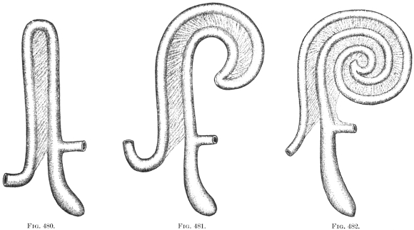
Figs. 480-482.—Schematic representation of three stages in the development of the ungulate spiral colon.
The condition encountered in some Rodents presents a more advanced stage. Thus the large intestine in the agouti (Dasyprocta agouti), shows the development of the spiral coil advanced as far as the second turn of the original loop (Figs. 389 and 390). It is readily seen that continued growth of this segment of the intestine leads to the formation of the complete colic spiral as found in the typical Ungulates.
The same arrangement of the large intestine obtains in certain Lemurs among the Primates. Thus the proximal colon of the Slow Lemur (Nycticebus tardigradus) is seen in Figs. 421 and 422 to present a typical spiral coil, and similar conditions are encountered in other members of the suborder.
V. Cæcal Apparatus and Colon in Hyrax.
Figs. 483-485.—Schematic figures illustrating possible line of derivation of aberrant mammalian type of alimentary canal encountered in Hyrax.
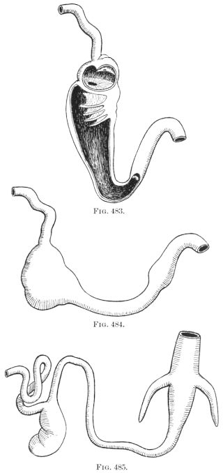
We have left for our final consideration the aberrant and unique mammalian type found in Hyrax (Fig. 378). In this remarkable little animal the large intestine develops a typical mammalian sacculated cæcum at the ileo-colic junction, and in addition is provided further on with two symmetrical pointed lateral colic cæca of large size. It is quite true that this arrangement is unique among Mammalia, confined entirely to the members of the suborder formed by the single family of Hyrax, and that no strictly analogous disposition of the alimentary canal is encountered in the entire vertebrate series. Yet these aberrant structures are possibly capable of explanation, in regard to the method of their development, by reference to the cæcal apparatus of certain phytophagous saurians, as Iguana and Cyclura. In these forms (Fig. 326-330) the small intestine enters the colon somewhat asymmetrically, the opening being guarded by a well developed annular valve.
The proximal segment of the large intestine forms an extensive sacculated pouch. If this is opened (Figs. 328-330) it is seen that the small intestine leads into a compartment which is separated from the remainder of the pouch by a valvular diaphragm with central circular opening. Beyond this primary compartment the colic pouch is incompletely subdivided by a series of gradually diminishing crescentic folds, corresponding to the external constrictions between the sacculations. The entire pouch gradually diminishes in caliber until it passes with a sharp angular bend into the terminal portion of the endgut. This terminal segment is differentiated from the elongated colic pouch by the greater thickness of its muscular walls and by a slight annular projecting fold in the interior. In considering the intestinal tract of Hyrax it is conceivable that the unique condition presented by this animal may be derived from some type conforming in general structure to the reptilian arrangement of the parts just detailed, as indicated in the schematic Figs. 483-485. The proximal typical cæcal pouch of Hyrax would then correspond to the similar colic pouch of Iguana. To explain the supplementary colic cæca it is necessary to suppose that the transition of the colic pouch into the terminal hindgut had become well differentiated, and that on each side of this junction the colic tube had extended backwards, resulting in the production of the supplementary bilateral cæcal pouches of Hyrax.