THE VISCERA. (continued 2)
Reading edition
The Uterus (f, i).—The uterus consists of a median portion or body (i) which is unpaired, and of two horns or cornua (f) which extend from the body to the uterine tubes. The body (i) of the uterus is a tube about four centimeters long which lies in the abdominal cavity, ventrad of the rectum and between it and the bladder. Its caudal end is at the level of the cranial border of the pubis. The cranial end of its cavity is divided by a median dorsoventral partition into lateral halves, while the cavity of its caudal portion is unpaired. The mucosa is thrown up into large longitudinal folds. At its caudal end the uterus projects into the vagina (m), so that when the vagina is opened the end of the uterus is seen projecting into it as a prominent papilla. The portion of the uterus thus enclosed by the vagina is the cervix uteri or neck of the uterus. Its free end is directed ventrocaudad, and a prominent ridge is continued from its caudodorsal side along the mid-dorsal wall of the vagina. The uterine cavity communicates with the vaginal cavity by a V-shaped opening, the os uteri, which looks ventrocaudad and has its apex directed cranioventrad. Each horn (f) of the uterus passes craniolaterad in a nearly straight course from the body (i) to the uterine tube (e). It narrows rapidly and becomes continuous with the tube. The mucosa is thrown into longitudinal folds.
Ligaments of the Uterus.—The Broad Ligament.—The uterus is held in place principally by the broad ligaments. These are two folds of the peritoneum, each of which is attached to the whole length of one of the cornua and the adjacent part of the uterine tube and to the corresponding lateral surface of the body of the uterus. Each ligament ends craniad in a concave free border. Its attached border forms a curved line which begins laterad of the kidney and extends thence to the lateral ligament of the bladder. From the lateral ligament of the bladder the broad ligament extends caudad into the rectovesical pouch of the peritoneum, which lies between the rectum and the bladder. It holds the body of the uterus to the lateral wall of this pouch, and together with the opposite ligament and the body of the uterus forms thus a transverse partition, which divides the rectovesical pouch into dorsal and ventral portions.
The round ligament (h) is a fibrous band which extends from a point of the body wall, which corresponds exactly to the internal inguinal ring of the male, to the cornu of the uterus about two centimeters from the cranial end. It is attached to the broad ligament by an intervening fold of peritoneum.
The Vagina (m).—The vagina extends from the os uteri (at l) dorsad of the symphysis of the pelvis to a point a short distance craniad of the caudal border of the ischiatic symphysis. At this point it joins the neck of the bladder (k) to form the vestibulum or urogenital sinus (n), which is homologous with the urethra of the male.
Urogenital Sinus (n).—The urogenital sinus extends from the caudal end of the vagina (m) to the external opening, which is situated ventrad of the anal opening. It is about a centimeter long and nearly as wide, and is marked off from the vagina by a circular fold of mucosa, while its inner surface presents longitudinal folds. On its ventral wall at its cranial end is the opening of the neck of the bladder, which is enclosed by a ring-like elevation of the mucosa, most prominent at the sides. The external entrance to the urogenital sinus forms the vulva.
The Clitoris.—The clitoris is a minute organ homologous with the penis and lying on the ventral floor of the urogenital sinus. Its distal end lies at the entrance of the urogenital sinus on its ventral border. In adult specimens the prepuce of the clitoris appears as a slight elevation of the integument surrounding a central vascular structure which appears red in the fresh organ. From the clitoris there are two small corpora cavernosa clitoridis (o) passing craniad and then diverging to be attached to the ischiatic rami. The ischiatic portion of each is covered by a muscle (ischiocavernosus). The clitoris is said to contain a bone.
Mammary Glands.—The mammary glands secrete the milk, and lie on the ventral surface of the body beneath the integument. The separate glands are closely gathered into two chief masses, one on each side the ventral middle line. Each of these extends from about the region of the fourth rib to the caudal end of the abdomen, ending over the pubic symphysis. On each side the glands are gathered into five groups, each of which is furnished with a nipple. The nipple is a projection of the integument, having near its distal end numerous fine openings for the ducts of the glands. The first two nipples are on the thorax, the other three on the abdomen, the most caudal ones being about two or three centimeters in front of the cranial edge of the pubis.
Rudimentary mammary glands and nipples are present in the male.
Muscles of the Urogenital Organs, Rectum, and Anus
(Figs. 113 and 114).—The muscles connected with the caudal openings of the alimentary canal and of the urogenital organs are closely interrelated, a single muscle sometimes acting on parts of both systems. For this reason all these muscles are described together.
The region lying between the anus and the external opening of the urogenital organs is known as the perineum. The perineum is formed chiefly by muscles and fascia.
a. Muscles common to the Male and Female.
—M. sphincter ani externus (Fig. 113, i; Fig. 114, a).—This muscle is confounded with the levator scroti (Fig. 113, j) or the levator vulvæ (Fig. 114, b). The two take origin in common from the integument on the dorsum of the root of the tail dorsad of the fifth caudal vertebra. There the fibres from the opposite sides are intermingled. The common muscle passes ventrad about the tail as a flat band close to the integument. Dorsad of the anus the inner fibres of the muscles of the opposite sides are united. They then separate and surround the anus as a band five millimeters wide situated beneath the integument. Ventrad of the anus the fibres are again intermingled. Some fibres on each side then continue to the scrotum as the levator scroti (Fig. 113, j) or to the vulva as the levator vulvæ (Fig. 114, b). Fibres also pass onto the anal pouch and unite with the sphincter ani internus, forming the constrictors of the anal pouch (Strauss-Durckheim).
M. sphincter ani internus (Fig. 113, h; Fig. 114, c).—The sphincter ani internus is a broad and thick band of striated muscle-fibres which surrounds the rectum at the anus. Dorsad the band is about two centimeters broad, while ventrad it is less than one. In the ventral median line some of the fibres pass craniad to help in forming the bulbocavernosus muscle. The muscle surrounds the anal sac.
(The muscle here described under this name is that described under the same name in the cat by Strauss-Durckheim and Mivart; it corresponds, however, to a part of the sphincter ani externus of the dog, as described by Ellenberger and Baum.)
M. levator ani (or pubiocaudalis) (Fig. 162, 11).—This muscle lies in the pelvic cavity. Each muscle forms a nearly vertical sheet, and between the two are the rectum and the urethra.
Origin from the symphysis of the pelvis.
Insertion into the midventral line of the centra of the third, fourth, and fifth caudal vertebræ, close to the muscle of the opposite side. This muscle is frequently continuous with the iliocaudalis (Fig. 162, 11′).
Action.—Bends the tail and compresses the rectum.
M. ischiocavernosus (Fig. 113, m; Fig. 114, e).—A small, flat, spindle-shaped muscle which lies upon the crus of the penis or clitoris. Each has
Origin from the caudal border of the ramus of the ischium, about one centimeter from the median line.
Insertion, in the male, into the whole outer surface of the crus penis, or bulb of the corpus cavernosum penis. In the female the muscle is smaller than in the male, and the insertion is into the ventral surface of the urogenital sinus, at the base of the clitoris.
M. transversus perinei (Fig. 114, i).—A small bundle of fibres which arises from the medial surface of the ischium, just dorsad of the origin of the ischiocavernosus, and passes mediad to join the sphincter ani internus (c).
M. caudoanalis (S.-D.) (Fig. 113, f; Fig. 114, g).—A slender, flat bundle of fibres having origin on the middle line of the ventral surface of the second and third caudal vertebræ. It passes caudoventrad, lying between the levator ani and the caudorectal (Fig. 113, e; Fig. 114, j), and unites with the ventral portion of the sphincter ani internus (Fig. 113, h).
Action.—Draws the anus craniodorsad.
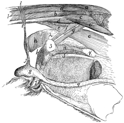
Fig. 113.—Muscles of the Anus, Urogenital Organs, and Tail in the Male (Slightly Schematic).
One side of the pelvis has been removed, a, a′, M. extensor caudæ lateralis; b, M. abductor caudæ externus; c, c′, M. flexor caudæ longus; d, M. flexor caudæ brevis; e, M. caudorectalis; f, M. caudoanalis; g, M. caudocavernosus; h, M. sphincter ani internus; i, M. sphincter ani externus; j, M. levator scroti; k, M. rectocavernosus; l, M. bulbocavernosus; m, M. ischiocavernosus (cut); n, M. compressor urethræ membranaceæ. 1, tail; 2, rectum; 3, bulbourethral or Cowper’s gland; 4, prostate gland; 5, symphysis pubis; 6, penis; 7, glans penis; 8, testis; 9, spermatic cord.
M. caudorectalis (Fig. 113, e; Fig. 114, j).
Origin from the ventral surface of the sixth and seventh caudal vertebræ. A small, at first unpaired band two or three millimeters wide is formed, which passes cranioventrad, soon dividing into two lateral halves. These spread out over the sides of the rectum, forming a broad sheet of fibres which pass into the walls of the rectum, among the transverse fibres of the latter. This muscle is covered by the caudoanalis (Fig. 113, f), caudocavernosus (g) (or caudovaginalis, Fig. 114, h), iliocaudalis, and levator ani.
b. Muscles Peculiar to the Male
(Fig. 113).—M. levator scroti (j).—This is a band of fibres which passes ventrad in the median line from the sphincter ani externus (i) onto the scrotum. Here it spreads out beneath the skin, forming especially a well-marked bundle in the median furrow between the two testes.
M. rectocavernosus, or retractor penis (k).—A small bundle of fibres which arises in two parts from the ventral surface of the sphincter ani internus (h). The two parts unite into a single bundle which passes caudad on the middle line of the ventral surface of the penis; it is inserted into the corpus cavernosum just proximad of the glans. The muscle is covered only by integument, and overlies the ischiocavernosus (m).
M. caudocavernosus (S.-D.) (g).—A slender bundle just craniad of the caudoanalis (f).
Origin on the median ventral line of the first two caudal vertebræ. The muscle passes caudoventrad, lying between the levator ani and the caudorectal (e). It divides into two bands, one of which is inserted into the base of the corpus cavernosum, while the other extends farther caudad and is inserted at the distal extremity of the corpus cavernosum.
Action.—Flexes the penis (bends it backward).
M. bulbocavernosus (accelerator urinæ) (l).—The two muscles cover the ventral surface of the penis.
Origin of each from a median raphe, which passes from the bulbous portion of the urethra toward the anus. The fibres pass toward the distal end of the penis and have their
Insertion into the distal half of the lateral surface of the corpus cavernosum penis.
M. compressor urethræ membranaceæ (n).—A thick layer of striated muscle-fibres which surrounds the urethra between Cowper’s gland (3) and the prostate (4). The fibres have a circular course, and the cranial ones are attached to the crura of the penis. The other fibres have no fixed attachment.
c. Muscles Peculiar to the Female
(Fig. 114).—M. levator vulvæ (Strauss-Durckheim), or constrictor cunni (b).—This is homologous with the levator scroti of the male. It consists of a band of fibres which pass ventrad from the external sphincter ani (a) and surround the vulva (3), lying immediately beneath the integument.
M. constrictor vestibuli, or rectovaginalis (Strauss-Durckheim) (d).
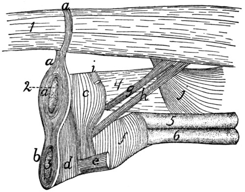
Fig. 114.—Muscles of the Anus and Urogenital Organs in the Female.
a, M. sphincter ani externus; b, M. levator vulvæ; c, M. sphincter ani internus; d, M. constrictor vestibuli; e, M. ischiocavernosus (cut); f, M. urethralis; g, M. caudoanalis; h, M. caudovaginalis; i, M. transversus perinei; j, M. caudorectalis. 1, the tail; 2, anus; 3, vulva; 4, rectum; 5, vagina; 6, neck of the bladder.
Origin from the sides of the sphincter ani internus (c). The muscle forms a distinct bundle two or three millimeters wide, which passes ventrocaudad and is inserted into the ventral surface of the urogenital sinus, caudad of the insertion of the ischiocavernosus (e).
M. caudovaginalis (Strauss-Durckheim) (h).—A slender band just craniad of the caudoanalis (g), and corresponding to the caudocavernosus of the male.
Origin from the median line of the ventral surface of the first two caudal vertebræ. The muscle passes caudoventrad, lying between the levator ani and the caudorectal (j), and is inserted into the ventral side of the urogenital sinus, at the base of the clitoris.
M. urethralis (f).—This consists of fibres surrounding the cranial part of the urogenital sinus and the caudal parts of the vagina and neck of the bladder.
Origin partly on the caudal part of the symphysis of the ischium, partly from the ventral surface of the urogenital sinus, where the fibres are attached to the corpora cavernosa clitoridis. The fibres pass dorsad over the surface of the sinus, and over the surface of the union of the vagina and neck of the bladder, to be inserted into the sides of the vagina and the dorsal surface of the urogenital sinus.