THE CIRCULATORY SYSTEM.
Reading edition
I. THE HEART. COR.
The heart lies in the mediastinum, enclosed in the pericardial sac, and projects rather more toward the left than toward the right. It is an ovoid or pear-shaped organ, with its long axis directed approximately craniocaudad. Its caudal end or apex is, however, directed slightly ventrad and to the left, while the larger cranial end or base faces slightly dorsad as well as craniad.
Laterally and dorsally the heart is largely covered by the lungs. The ventral side and a considerable portion of the lateral surface are, however, not thus covered, so that they lie against the thoracic wall. The heart extends from about the fourth or the fifth to the eighth rib, and its apex touches the diaphragm.
The cavity of the heart is divided by a longitudinal dorsoventral septum into lateral halves—a right and a left side. Each side is again divided by a transverse dorsoventral septum (auriculoventricular) into two chambers, one of which, the auricle, lies at the base; the other, the ventricle, lies at the apex of the heart. There are thus right and left auricles and right and left ventricles. An external groove, partly filled with fat, separates the auricular portion of the heart from the ventricular part; this groove is known as the sulcus coronarius.
Each auricle or ventricle has a single set of blood-vessels either leaving it or entering it. Thus the blood enters the right auricle by the inferior and superior venæ cavæ (Fig. 116, d and e) (præcava and postcava). It passes thence into the right ventricle, and from the right ventricle to the lungs by a single pulmonary artery (Fig. 115, f). It returns to the left auricle by the numerous pulmonary veins (Fig. 116, g, h, i), and passes thence to the left ventricle. From the left ventricle it passes to the body by a single aorta (Fig. 115, g).
When the heart is viewed from the ventral surface (Fig. 115), a considerable blood-vessel, the coronary artery (q), running from the base to a point a little to the right of the apex, indicates the position of the septum (ventricular septum), between the right (a) and left (b) ventricles. The apex thus belongs to the left ventricle. From the cranial end of the right ventricle the pulmonary artery (f) is seen passing obliquely craniad and toward the left. Beneath the pulmonary artery the aorta (g) appears rising from the middle of the base of the heart and passing directly craniad. At the base appears a part of the auricular appendage of the left auricle (d), and at the right a part of the right auricular appendage (c). The former (d) is larger and bent at right angles, so that its free end is directed toward the apex.
In the dorsal view (Fig. 116) the position of the ventricular septum is not indicated. On the surface of the left ventricle a short distance from the ventricular septum and nearly parallel to it are seen branches of the coronary artery and the coronary vein passing toward the apex. Craniad of the left ventricle is seen the left auricle (b) with the pulmonary veins (g, h, i) opening into it. Craniad of the right ventricle (a) is the right auricle (c) with the venæ cavæ (d and e) opening into it near the middle. Coming from beneath the auricles are seen the pulmonary artery (j) and the aorta (f).
The chambers of the heart.
1. The right auricle (atrium dextrum) (Figs. 115 and 116, c). Externally the right end of the right auricle projects so as to form its auricular appendage (Fig. 115, c) which lies at the right of the base of the aorta (Fig. 115, g). The superior vena cava (Fig. 116, e) is seen entering near the cranial end of the auricular septum opposite the base of the aorta. The inferior vena cava (Fig. 116, d) enters the auricle near the coronary sinus and close to the auricular septum. The netted appearance which the wall, especially that of the appendage, presents externally is due to the muscular thickenings of the wall (musculi pectinati).
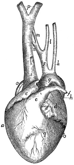
Fig. 115.—Heart, Ventral View.
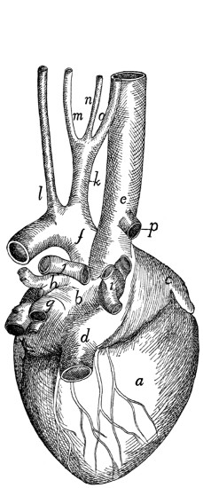
Fig. 116.—Heart, Dorsal View.
Fig. 115.—a, right ventricle; b, left ventricle; c, right auricular appendage; d, left auricular appendage; e, conus arteriosus; f, pulmonary artery; g, aortic arch; h, thoracic aorta; i, vena cava superior; j, innominate artery; k, left subclavian artery; l, left common carotid; m, right common carotid; n, right subclavian; o, azygos vein; p, the two innominate veins; q, coronary artery.
Fig. 116.—a, right ventricle; b, left auricle; c, right auricle; d, vena cava inferior; e, vena cava superior; f, aorta; g, h, i, groups of pulmonary veins (g, dorsal group; h, sinistral group; i, dextral group); j, pulmonary artery (division into two); k, innominate artery; l, left subclavian; m, left common carotid; n, right common carotid; o, right subclavian; p, azygos vein.
The cavity (including that of the auricular appendage) is somewhat egg-shaped, with its long axis transverse. The musculi pectinati, which branch and unite into a network, are most abundant on its dorsal wall at the right. The axes of the two venæ cavæ if produced into the cavity of the auricle would meet one another at its centre and nearly at right angles. Caudad of the opening of the inferior vena cava is seen the slit-like opening of the coronary sinus, guarded craniad by the semilunar valve of the coronary sinus (or valve of Thebesius). The coronary sinus receives blood from coronary veins, which collect it from the walls of the heart.
In the auricular septum ventrad of the opening of the inferior vena cava is seen a faintly marked smooth oval depression, the fossa ovalis. When the auricular septum is examined by transmitted light it is seen to be thinner over the fossa ovalis. There is an opening, foramen ovale, at this point in fœtal life, so that the blood of the inferior vena cava then passes directly from the right auricle to the left auricle. Caudad the cavity of the right auricle communicates with that of the ventricle by the large oval auriculoventricular opening, which is guarded by the tricuspid valves (Fig. 117).
2. The right ventricle (ventriculus dexter) (Figs. 115 and 116, a) does not reach quite to the apex of the heart, so that it makes up less than one-half of its ventricular portion. It makes a half-spiral turn about the left ventricle, from its lateral side at the apex toward its ventral side at the base, where it ends in the pulmonary artery (Fig. 115, e). Its wall is very thin as compared with that of the left ventricle. Its cavity (Fig. 117) is nearly flat on its medial side, convex on its lateral side. It communicates with the auricle craniodorsad. Cranioventrad the cavity is narrower and turns around toward the ventral side of the heart and opens into the pulmonary artery. That portion of the ventricle between the auriculoventricular opening and the pulmonary artery is the conus arteriosus (Fig. 115, e; Fig. 117, f). Internally the wall presents many muscular trabeculæ (Fig. 117, a) of various sizes. These are more numerous over the ventricular wall than on the septum. They do not occur on the conus (f). Surrounding the auriculoventricular opening is the tricuspid valve (Fig. 117, d, d′, d″). It consists of three flaps. One of these (d′) is septal (i.e., it lies against the septum), while of the two others which do not lie against the septum, one is dorsal (d″) and the other ventral. Each flap is thin, semicircular, and membranous, and is attached to the border of the auriculoventricular opening by the diameter of the semicircle. The free border of each is attached to the wall of the heart by numerous delicate tendinous bands, the chordæ tendineæ (c), some of which are attached to the lower face of each valve. The chordæ tendineæ of the septal valve (or most of them) are attached at their opposite ends to the septum directly, while those of the dorsal and ventral flaps are attached to the ends of three or more band-like muscles, columnæ (or trabeculæ) carneæ (b), which are fixed by their opposite ends to the ventricular wall.
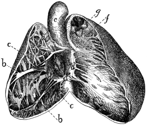
Fig. 117.—Heart, with Right Ventricle laid open to show the Tricuspid Valve.
a, trabeculæ; b, columnæ carneæ; c, chordæ tendineæ; d, d′, d″, the three flaps of the tricuspid valve; e, aorta; f, conus arteriosus, laid open; g, semilunar valves of the pulmonary artery.
Between the conus arteriosus (f) and the pulmonary artery are three pocket-like semilunar valves (Fig. 117, g), one ventral, one dextral, and one sinistral. Between each valve and the wall of the pulmonary artery there is an enlargement of the cavity of the artery, one of the pulmonary sinuses (or sinuses of Valsalva). Beyond the sinuses the pulmonary artery divides into right and left branches (Fig. 116, j).
3. The left auricle (atrium sinistrum) (Fig. 115, d; Fig. 116, b). The auricular appendage (Fig. 115, d) is bent at right angles and its apex turned caudad. The cavity is thus irregular. Muscular trabeculæ occur only in the auricular appendage. When the auricular septum is examined by transmitted light the position of the fossa ovalis is indicated at about the middle of the septum as seen from the left side. Ventrad of it is a fold of the septal wall.
The pulmonary veins enter the dorsal wall of the auricle in three groups (Fig. 116, g, h, i). Each group opens into a more or less pronounced sinus or extension of the auricular cavity. The sinuses may be called, on account of their position, dorsal (g), sinistral (i), and dextral (h).
4. The left ventricle (Fig. 115, b) occupies rather more than the left half of the base of the heart. Its walls are two or three times as thick as those of the right ventricle. Internally its walls present a few muscular bands, comparable to the trabeculæ of the left ventricle. There are two very large columnæ carneæ, one dorsad and one ventrad. At the cranial end it communicates near the lateral wall with the left auricle by the auriculoventricular opening, and near the septum with the aorta. The bicuspid valve (or mitral valve), which guards the auriculoventricular opening, consists of two flaps, one septal and one lateral. Their chordæ tendineæ, which come from their free borders as well as from their outer surfaces, are attached to the columnæ carneæ, chordæ passing from both valves to each columna carnea.
The opening into the aorta is guarded by three aortic semilunar valves, each of which partly conceals an aortic sinus (or sinus of Valsalva). One valve is dorsal, one sinistral, and one dextral. In the dextral sinus is the opening of one of the coronary arteries, which carry blood to the walls of the heart.
Pericardium.
The pericardium is a sac enclosing the heart. It lies in the middle mediastinum, and the two halves of the mediastinal septum with their fat may be dissected away from it. Its wall is composed of two layers, an external firm fibrous layer and an internal layer of flattened epithelial cells similar to the peritoneal epithelium (serous layer). The fibrous layer forms a sac which repeats roughly the form of the heart. This sac is attached to the aorta at the point of origin of the subclavian artery, to the pulmonary artery at its bifurcation, and to the venæ cavæ and pulmonary veins near their entrance into the heart. At these points it is continuous with the fibrous coats of the vessels named, and from them it is reflected over the heart, forming a complete sac enclosing it but not attached to it anywhere. The heart lies within this sac. The serous layer lines the fibrous sac and gives to the surface of the heart and fibrous layer a smooth glistening appearance. It is reflected over the heart. The relation of the heart to it is much the same as the relation of the intestine to the peritoneal sac. The serous layer consists therefore of two portions, parietal, lining the sac, and visceral, covering the heart. The parietal and visceral portions are continuous along a line which runs approximately parallel to the auriculoventricular groove and encloses all the great blood-vessels. Within this line the heart lies against the fibrous layer of pericardium and is not covered by the serous layer. The serous layer is easily dissected free from the heart-wall, but its parietal portion is closely adherent to the fibrous layer.
II. THE ARTERIES. ARTERIÆ.
1. A. pulmonalis, the Pulmonary Artery
(Fig. 115, f).
The pulmonary artery passes craniodorsad and slightly to the left from the cranial end of the conus arteriosus. One to one and a half centimeters from the conus it divides into right and left branches (Fig. 116, j). Just before the division the dorsal surface of the pulmonary artery is connected by the short ligamentum arteriosum, or ligamentum Botalli, with the aorta. This is the remnant of a canal which in fœtal life forms a free communication between the pulmonary artery and the aorta; this canal is known as the ductus Botalli. The ligament is almost or quite obliterated in the adult cat.
The left branch of the pulmonary artery passes to the left lung, crossing ventrad of the thoracic aorta. It then divides into branches which pass to the lobes of the left lung. The point of division of the left branch of the pulmonary artery lies craniad of all the lobes of the lung, so that the lobes of the left lung are said to be all hyparterial, i.e., below (or caudad of) the artery.
The right branch passes under (dorsocaudad of) the aortic arch, and reaches the right lung at about the junction of the cranial lobe with the remainder of the lung. The cranial lobe of the right lung is therefore said to be eparterial, since it is craniad of the pulmonary artery; the other lobes are hyparterial. The right branch divides at its entrance to the lung and is distributed to its lobes.
2. Aorta
(Fig. 115, g; Fig. 118, a).
The aorta is the single great vessel which conveys blood from the left ventricle. It makes a sharp semicircular curve dorsad (Fig. 118, a′) and to the left, passes caudad at the left side of the vertebral column, and passes between the crura of the diaphragm to reach the abdominal cavity. It is divisible into thoracic aorta (Fig. 118) and abdominal aorta (Fig. 126).
A. Thoracic Aorta
(Fig. 118).—The first portion of the thoracic aorta, curved as above described, is the aortic arch (a′). It lies in the thoracic cavity opposite the interval between the third and fourth or fourth and fifth ribs. It is separated from the vertebral column on the right by the superior vena cava (q), and on the left by the œsophagus (4). At its beginning it lies a little to the right of the median plane; but it passes at once to the left side of the vertebral column.
Branches of the Thoracic Aorta.
1. Aa. coronariæ.—The coronary arteries are two. They arise from the aortic sinuses. The left one (Fig. 118, p) leaves the aorta on the left side, passes dorsad of the pulmonary artery, and divides into two branches, one of which follows the auriculoventricular groove (sulcus coronarius) to the dorsal side of the heart and sends branches to the adjacent heart-walls, while the other runs onto the ventricles, following approximately the ventral border of the ventricular septum. The right coronary artery passes in the auriculoventricular groove toward the right and dorsad, and supplies the adjacent walls of the heart.
2. A. anonyma (Fig. 115, j; Fig. 118, b).—The innominate artery passes craniad from the convexity of the aortic arch (a′). It gives rise first to a small mediastinal artery (m) which passes ventrad into the mediastinum, then to the left common carotid (Fig. 115, l), then to the right common carotid (Fig. 115, m). Sometimes the carotids are given off from a common trunk. Beyond the right common carotid the innominate is continued as the right subclavian (Fig. 115, n).
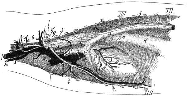
Fig. 118.—Vessels of the Thorax, Viewed from the Left Side.
a, aorta (a′, aortic arch); b, innominate artery; c, left subclavian; d, right subclavian; e, right common carotid; f, left common carotid; g, costocervical axis; g′, vertebral artery; h, internal mammary artery; i, axillary artery; j, thyrocervical axis; k, A. transversa scapulæ; m, mediastinal artery; n, intercostal arteries; o, œsophageal arteries; p, left coronary artery; q, superior vena cava; r, internal mammary vein; s, the two innominate veins; t, sternal artery; u, costocervical vein; v, vertebral vein; w, axillary vein; x, vein to clavotrapezius and lateral ends of the pectoral muscles; y, vein accompanying second branch of A. transversa scapulæ; z, thyrocervical vein. 1, external jugular vein; 2, internal jugular vein; 3, vena cordis magna; 4, œsophagus; 5, thoracic duct; 6, one of the lymphatic ducts from the neck. I, first rib (cut); VIII, eighth rib (cut); XII, twelfth rib (cut).
3. A. subclavia sinistra (Fig. 115, k; Fig. 118, c).—The left subclavian passes craniad from the convexity of the aortic arch. It curves about the first rib (Fig. 118, I) to enter the armpit, where it becomes the axillary artery (i).
4. Aa. intercostales (n).—The intercostal arteries are given off from the dorsal side of the aorta. Each passes to an intercostal space and divides into three branches. One of these passes ventrad along the caudal margin of the cranial one of the two ribs between which it extends. Another goes to the deep muscles of the back, and the third enters the spinal canal through the intervertebral foramen. The arteries for the first and second (and sometimes the third) intercostal space usually arise from the subclavian (costocervical axis, Fig. 118, g).
5. Aa. bronchiales.—The bronchial arteries are two, and arise either from the aorta opposite the fourth intercostal space or from the fourth intercostal arteries. They accompany the bronchi to the lungs.
6. Aa. œsophageæ (Fig. 118, o).—The œsophageal arteries are small branches of varying origin passing to the œsophagus.
7. Aa. lumbales.—The lumbar arteries correspond to the intercostals, but pass off between the lumbar vertebræ. The obliquity of the diaphragm throws the origin of one or two pairs of them within the thorax.
A. carotis communis. The Common Carotid Artery.
(Fig. 115, l and m; Fig. 119, a).
The two common carotid arteries arise from the innominate artery in the manner already described. Each passes craniad along the side of the trachea. In the thorax (Fig. 118, f) the common carotid lies mediad of the subclavian artery (Fig. 118, c) and dorsad of the superior vena cava (Fig. 118, q). In the neck (Fig. 119) the artery lies, accompanied by the vagus and sympathetic nerves and the internal jugular vein (b), in the space between the longus capitis muscle (7) and the trachea (14); it is covered ventrally by the sternomastoid and sternothyroid (2) muscles, lying close to the lateral border of the latter. Near its origin the common carotid may give rise to the small inferior thyroid artery. It then passes to about the level of the larynx without giving off branches; here it gives off the superior thyroid (c) on the ventral side and one or more muscular branches (e) on the dorsal side. One or two centimeters further craniad it gives off on the dorsal side the internal carotid (g) and the occipital artery (f). The main artery now takes the name external carotid (m).
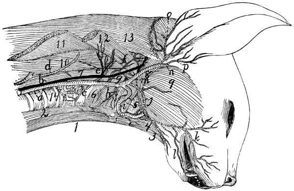
Fig. 119.—Common Carotid Artery and Internal Jugular Vein.
a, common carotid artery; b, internal jugular vein; c, superior thyroid artery; d, cervicalis ascendens artery; e, large muscular branches; f, occipital artery; g, internal carotid; h, branch to larynx; i, lingual artery; j, external maxillary; k, superior labial; l, inferior labial; m, external carotid; n, internal maxillary; o, posterior auricular; p, superficial temporal. 1, M. sternohyoideus; 2, M. sternothyreoideus; 3, M. geniohyoideus; 4. M genioglossus; 5, M. constrictor pharyngis medius; 6, M. constrictor pharyngis inferior; 7, M. longus capitis; 8, M. digastricus; 9, M. masseter; 10, M. scalenus; 11, M. levator scapulæ (cut); 12, M. levator scapulæ ventralis (cut); 13, M. splenius; 14, trachea.
Branches of the common carotid:
1. A. thyreoidea ima.—The inferior thyroid is a small artery which arises either from the common carotid near its origin, or from the innominate before the origin of the carotid. It passes craniad on the trachea as far as the thyroid gland, giving branches to the trachea and œsophagus.
2. A. thyreoidea superior (c).—The superior thyroid leaves the carotid opposite the thyroid cartilage and passes mediad and caudad, sending branches to the thyroid gland, and the sternothyroid (2) and sternohyoid (1) muscles. A small branch, the superior laryngeal, passes to the larynx, and supplies those muscles of the larynx which are not enclosed by the cartilages.
3. Rami musculares (e).—One or two branches, usually of considerable size, leave the common carotid at about the same level as the superior thyroid and pass to the muscles on the dorsal side of the neck, the main trunk of the artery passing between the longus capitis (7) and scalenus muscles (10).
4. A. occipitalis (f).—The occipital artery arises from the common carotid at about the same point as the internal carotid. It immediately sends a large branch dorsad, passing between M. longus capitis (7) and the vertebral column, to the deep muscles of the neck. The occipital then crosses the outer surface of the digastric muscle (8) to the back of the skull, and runs along the lambdoidal crest just beneath the splenius muscle. It sends a number of branches to the muscles of the back of the neck; and one of its branches may unite with the vertebral artery as it lies in the groove on the atlas, or with a branch of the vertebral.
5. A. carotis interna (g).—The internal carotid artery is one of the terminal branches of the common carotid. It is very small. It is given off near or in common with the occipital artery, passes toward the cranial end of the tympanic bulla, enters the bulla with the Eustachian tube, and passes into the skull at the foramen lacerum. Its course is much convoluted before entering the foramen. Within the skull (Fig. 121, g) it joins the posterior cerebral artery (Fig. 121, f) at the side of the hypophysis.
6. A. carotis externa (Fig. 119, m).—After giving off the internal carotid the continuation of the common carotid artery receives the name external carotid (m). It passes craniad and laterad between the digastric (8) and styloglossus muscles, where it gives off cranioventrad the lingual artery (i) and a number of small muscular branches; also sometimes the small laryngeal artery. At the dorsolateral border of the digastric (8) it gives off the external maxillary artery (j), and about one centimeter farther craniad the posterior auricular (o). It now turns mediad, lying against the cartilaginous auditory meatus, on its cranial side, and gives off the superficial temporal (p). The artery then continues mediad, taking the name internal maxillary (n)—so that the internal maxillary artery is to be considered the terminal branch of the external carotid.
Branches of the external carotid artery (Fig. 119):
a. A. lingualis (Fig. 119, i; Fig. 120, d).—The lingual artery leaves the external carotid near its beginning and passes craniomediad along the ventral border of the digastric muscle (Fig. 120, 9), accompanied by the hypoglossal nerve. It gives off numerous small branches to the hyoid and pharyngeal muscles, then passes dorsad of the hyoglossus muscle (Fig. 120, 6), where it gives off a branch which passes transversely across the middle line to communicate with the artery of the other side. Beneath the hyoglossus the artery turns craniad and passes into the tongue. Here it runs along the medial border of the styloglossus to the tip of the tongue, giving off numerous branches into the substance of this organ.
b. Rami musculares.—Muscular branches pass to the digastric and to the hyoid muscles. A small branch (Fig. 119, h), which may arise either from the external carotid or from the common carotid near the beginning of the external carotid, passes to the larynx and supplies the thyroarytenoid and lateral cricoarytenoid muscles of the larynx.
c. A. maxillaris externa (Fig. 119, j).—The external maxillary artery leaves the external carotid (m) opposite the angle of the jaw and at about the dorsal border of the digastric muscle (8). It passes craniad, lying at first beneath the digastric muscle and sending a branch to the submaxillary gland. Opposite the caudal border of the mylohyoid muscle it gives off the submental artery, turns dorsad, emerges from beneath the digastric, and passes along the cranial border of the masseter onto the face. Here it divides into superior (k) and inferior (l) labial branches, which pass along the upper and lower lips, respectively, giving off numerous branches.
The submental artery passes to the symphysis menti between the digastric and mylohyoid muscles, giving off on its course collateral branches to the muscles of this region.
d. A. auricularis posterior (Fig. 119, o).—The posterior auricular leaves the external carotid (m) opposite the middle of the bulla tympani and beneath the submaxillary gland. It passes about the base of the ear on its caudal and dorsal sides outside of the deep muscles of the occipital region, but beneath the auricular muscles, and sends several branches to the muscles of the external ear, passing onto the caudal surface of the concha. It sends also a large branch mediad to the muscles of the occiput, especially to the temporal muscle, within which it ramifies. A large branch (anterior auricular, Fig. 131, u) passes from the caudal side of the concha craniodorsad, and appears on the cranial side of the external ear, running along the cranial margin of the auditory opening.
e. A. temporalis superficialis (Fig. 119, p; Fig. 120, h).—The superficial temporal artery arises from the external carotid as the latter lies between the cartilaginous auditory meatus and the caudal border of the masseter muscle. It passes dorsad and gives off soon after its origin a muscular branch to the masseter, and an auricular branch which passes distad along the concha auris and ramifies over its cranial surface. The superficial temporal itself passes onto the surface of the temporal muscle (Fig. 120, 11), to which it gives numerous branches. It extends to the caudal angle of the eye (Fig. 131, s), where it divides. One branch passes into the lower eyelid; the larger branch passes along the dorsal side of the eye, sending a branch into the orbit and small branches onto the dorsal surface of the nose.
f. A. maxillaris interna (Fig. 119, n; Fig. 120, i).—The internal maxillary artery is the continuation of the external carotid. It turns caudad at the caudal end of the mandible, then passes craniad, lying dorsad of the pterygoid muscles (Fig. 120, 10), and against the medial surface of the mandible. It gives off the inferior alveolar artery (Fig. 120, j), then the middle meningeal (k), and then continuing mediad divides into three or four branches. The branches redivide, and the twigs form a complicated plexus, the carotid plexus (Fig. 120, l), which surrounds the maxillary division of the fifth nerve near its exit from the foramen rotundum. One of the larger branches of the plexus enters the skull through the orbital fissure, lying beside the hypophysis; it divides in the manner described below.
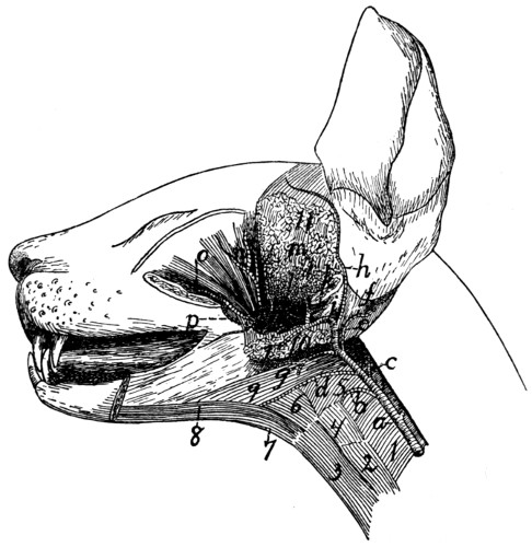
Fig. 120.—Branches of External Carotid Artery.
a, common carotid; b, branch to larynx; c, internal carotid; d, lingual; e, external carotid; f, posterior auricular; g, external maxillary; h, superficial temporal; i, internal maxillary; j, inferior alveolar; k, middle meningeal; l, carotid plexus; m, branch to temporal muscle; n, ophthalmic; o, infraorbital; p, lesser palatine. 1, M. constrictor pharyngis inferior; 2, M. sternothyreoideus; 3, M. sternohyoideus; 4, M. thyreohyoideus; 5, M. constrictor pharyngis medius; 6, M. hyoglossus; 7, M. geniohyoideus; 8, M. genioglossus; 9, M. digastricus; 10, Mm. pterygoidei externus and internus (cut); 11, M. temporalis (cut).
Beyond the carotid plexus a main trunk which may be considered the continuation of the internal maxillary passes craniad, lying on the dorsal surface of the external pterygoid muscle; nearly opposite the molar tooth it divides into the infraorbital (Fig. 120, o) and the sphenopalatine arteries.
Branches of the internal maxillary artery and of the carotid plexus:
1. A. alveolaris inferior (Fig. 120, j).—The inferior alveolar (or inferior dental) artery leaves the inferior maxillary just opposite the condyloid process of the mandible. It enters the mandibular canal by the mandibular foramen, along with the nerve of the same name, and traverses the canal, furnishing branches to the lower teeth. It emerges at the mental foramen, and its terminal branches are distributed to the chin, but a branch is continued in the bone beyond the mental foramen and supplies the incisor and canine teeth of the lower jaw.
2. A. meningea media (Fig. 120, k).—The middle meningeal is a large vessel which leaves the internal maxillary at about the same level as the inferior alveolar. It passes into the foramen ovale and ramifies in the dura mater. Its branches leave distinct impressions on the inner surface of the bones of the skull.
3. A large branch (Fig. 121, h) from the plexus passes into the cranial cavity through the orbital fissure and lies within the skull at the side of the hypophysis. It gives off the following branches:
a. A posterior communicating branch, very short, which extends caudad and joins the internal carotid artery (Fig. 121, g).
b. A. cerebri media (Fig. 121, i).—The middle cerebral artery passes dorsad on the side of the cerebral hemisphere along the fissure of Sylvius and divides into numerous branches which are distributed to the surface of the cerebrum.
c. A. cerebri anterior (Fig. 121, j) Passes dorsad between the cerebral hemispheres. Just craniad of the optic chiasma the two anterior cerebral arteries are united by a small communicating branch, thus completing the circulus arteriosus or circle of Willis (Fig. 121), surrounding the hypophysis (see page 292).
4. From the carotid plexus several branches pass, arising either separately or in common, to the masseter, temporal, and pterygoid muscles.
5. A. ophthalmica (Fig. 120, n).—The ophthalmic artery passes from the carotid plexus to the structures in the orbit. It gives off numerous branches which supply the muscles of the eyeball, and other structures of this region. It sends an ethmoidal branch into the nasal cavity through the ethmoidal foramen in the orbital plate of the frontal bone, then continues distad to emerge from the orbit on the medial side of the eye; here it anastomoses with branches of the superficial temporal.
6. A. palatina minor (Fig. 120, p).—The lesser palatine artery leaves the internal maxillary distad of the carotid plexus, near the caudal border of the maxillary bone. It passes ventrocaudad into the soft palate.
7. A. sphenopalatina.—The sphenopalatine is one of the terminal branches of the internal maxillary; it passes mediad through the sphenopalatine foramen into the nasal cavity, and divides into numerous branches which supply the mucous membrane of the nose. It gives off just before it enters the sphenopalatine foramen the descending palatine (A. palatina descendens), which passes into the posterior palatine canal and emerges on the surface of the hard palate, where it ramifies.
8. A. infraorbitalis (Fig. 120, o).—The infraorbital artery is a direct continuation craniad of the internal maxillary. It sends off numerous small branches to the teeth of the upper jaw, and a rather large branch which passes to the lower eyelid. It then enters the infraorbital foramen, at the same time dividing usually into two or three branches; these emerge from the foramen and supply the parts of the nose and upper lip adjacent to the foramen (Fig. 131, r).
A. subclavia. The Subclavian Artery.
The left subclavian (Fig. 118, c) arises from the convexity of the aortic arch just distad of the origin of the innominate artery, and about two or three centimeters from the heart. It passes craniad and slightly to the left, and turns into the left arm just craniad of the first rib.
The right subclavian (Fig. 115, n) is a direct continuation of the innominate, the artery receiving the name subclavian after the right common carotid is given off, usually at about the level of the second or third intercostal space.
The subclavian has the following branches: the vertebral artery (Fig. 118, g′), the internal mammary (Fig. 118, h), the costocervical axis (Fig. 118, g), the thyrocervical axis (Fig. 118, j). Beyond the last-named branch it continues into the arm as the axillary artery (Fig. 118, i; Fig. 122, g).
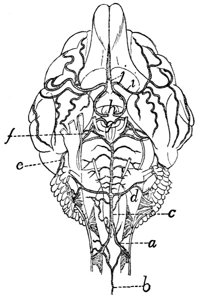
Fig. 121.—Arteries of the Ventral Surface of the Brain.
a, A. vertebralis; b, A. spinalis anterior; c, A. basilaris; d, A. cerebelli inferior posterior; e, A. cerebelli anterior; f, A. cerebri posterior; g, cut ends of the two internal carotid arteries; h, cut ends of branches from the carotid plexus (g and h are placed within the circulus arteriosus or circle of Willis); i, A. cerebri media; j, A. cerebri anterior.
a. A. vertebralis (Fig. 118, g′).—The vertebral artery arises from the dorsal surface of the subclavian opposite the first rib. It passes craniad and dorsad at the side of the thoracic portion of the longus colli muscle, and enters the foramen transversarium of the sixth cervical vertebra. It passes thence craniad through the foramina transversaria (which together form the vertebrarterial canal) and gives off at the intervertebral foramina branches to the muscles of the neck and branches which pass across the ventral surface of the spinal cord to join A. spinalis anterior. Craniad of the foramen transversarium of the atlas the vertebral artery turns dorsad in the groove on the lateral surface of the atlas. Here it gives off a large branch which passes laterodorsad to the muscles of the neck and may anastomose with a branch of the occipital artery. The vertebral artery then passes into the vertebral canal through the atlantal foramen. It passes to the ventral side of the spinal cord and unites at about the level of the foramen magnum with the vertebral artery of the opposite side (Fig. 121, a) to form the basilar artery (A. basilaris) (Fig. 121, c), which passes craniad along the ventral middle line of the brain. Just before their union the two vertebral arteries (a) give off each a branch which passes caudomediad. These two branches soon unite in the middle line, forming the anterior spinal artery (A. spinalis anterior) (b), which passes caudad the entire length of the spinal cord, lying on its ventral middle line and receiving many communicating branches from the vertebral, intercostal, and lumbar arteries.
A. basilaris (c).—This arises by the union of the two vertebral arteries in the manner just described. It passes craniad along the ventral middle line of the medulla and pons, giving numerous small branches to these structures. A large branch, A. cerebelli inferior posterior (d), passes on each side to the caudal surface of the cerebellum, on which it ramifies. At the cranial margin of the pons the basilar artery divides. From each division a very large branch passes on each side across the pedunculi cerebri to the cranial part of the cerebellum; this is A. cerebelli anterior (e). Just craniad of this, arising from nearly the same point, the smaller A. cerebri posterior (f) passes laterad to the caudal part of the cerebrum; it is joined by the internal carotid artery (g). The small continuations of the two halves of A. basilaris then pass craniad at the side of the hypophysis to join the posterior communicating branches from the carotid plexus. As other branches of the carotid plexus unite across the middle line craniad of the optic chiasma, an arterial circle is formed on the base of the brain, surrounding the hypophysis and the optic chiasma. This is known as the circulus arteriosus, or circle of Willis (see page 289).
b. A. mammaria interna (Fig. 118, h).—The internal mammary artery rises from the ventral surface of the subclavian opposite the first rib, and passes in the mediastinum ventrocaudad to reach the sternum opposite the third intercostal space. It sends two or three small branches craniad to the midventral part of the thoracic wall and then extends caudad at the side of the sternum. It sends off lateral branches to the ventral thoracic wall, branches to the mediastinum and pericardium, a branch to the diaphragm which anastomoses with the phrenic; and finally it passes out of the thoracic cavity caudad of the last costal cartilage, extends caudad in a zigzag course at the lateral border of the rectus muscle and anastomoses with the inferior epigastric artery.
c. Truncus costocervicalis (Fig. 118, g).—The costocervical axis arises from the subclavian opposite the first rib, passes craniodorsad, and divides almost at once into two branches. The smaller one of these, the superior intercostal (A. intercostalis suprema), passes caudad and, dividing, supplies the first and second intercostal spaces, and then passes to the deep muscles of the back. The other branch divides almost immediately into two. One of these, A. transversa colli, passes laterad in front of the first rib and enters the serratus anterior muscle. In this it passes dorsad, giving off branches to this muscle and to the levator scapulæ, till it reaches M. rhomboideus, which it likewise supplies. The other branch (A. cervicalis profunda), which seems to form a continuation of the main artery, passes directly dorsad and leaves the thoracic cavity between the heads of the first and second ribs. Here it passes into the deep muscles of the neck; it can be traced in the substance of the complexus muscle as far forward as the atlas.
d. Truncus thyrocervicalis (Fig. 118, j).—The thyrocervical axis (or thyroid axis) arises from the subclavian beneath the first rib, a short distance distad of the origin of the costocervical axis. It passes laterocraniad and dorsad, lying on the mediocranial side of the brachial plexus. A short distance from its origin it gives off a branch, varying much in size, the cervicalis ascendens (Fig. 119, d), which passes craniad on the ventral side of the neck, supplying the sternomastoid, sternohyoid, the cervical portion of the scalenus, and sometimes other muscles of the neck region. Other branches pass from the thyroid axis to the inner surface of the clavotrapezius (Fig. 122, a) and to the ventral end of the pectoral muscles. At the level of the cranial border of the scapula the artery takes the name A. transversa scapulæ, or suprascapularis. This divides into three main branches. The first branch, sometimes large, sometimes small, passes to the lymphatic gland in the hollow of the shoulder, and to the adjacent muscles; it supplies the clavotrapezius, acromiotrapezius, levator scapulæ ventralis, splenius, occipitoscapularis, and rhomboideus. A second branch passes between the subscapularis and supraspinatus muscles, dividing into various branches which supply the muscles named. The third branch pierces the supraspinatus muscle, just craniad of the acromion process, and ramifies in that muscle.
e. A. axillaris (Fig. 122, g).—The axillary artery is the continuation of the subclavian laterad of the first rib. It lies caudad of the brachial plexus and parallel to it. It sends off the following branches:
1. A. thoracica anterior (Fig. 122, h).—The anterior thoracic is a slender artery which leaves the ventral side of the axillary opposite the first rib and passes caudomediad, to supply the medial ends of the pectoral muscles (g).
2. A. thoracica longa (p).—The long thoracic artery is larger than the preceding, leaves the axillary a short distance laterad of it, and passes caudad to the middle portions of the pectoral muscles (8) and continues to the inner surface of the latissimus dorsi.
A short distance beyond the long thoracic the axillary divides into two. The more cranial one of these is the subscapular (l); the other is the brachial (g′).
3. A. subscapularis (l).—The subscapular artery passes laterad and gives off a short distance from its origin the A. thoracicodorsalis (t) and A. circumflexa humeri posterior (see below); it may also give rise to the circumflexa anterior humeri (l′), and to the profunda brachii (u); these two branches, however, rise more frequently from the brachial artery (g′) and are described in connection with it. The subscapular artery then passes through the triangular interval between the scapular end of the long head of the triceps, the latissimus dorsi, and the glenoid border of the scapula. Within this interval it sends muscular branches to the long head of the triceps, the subscapularis, and the latissimus dorsi. That to the latter muscle is very large. At the border of the scapula, opposite the tuberosity of the spine, it turns craniad, passes over the lateral surface of the infraspinatus muscle, supplying it; crosses the spine, and sends branches into the supraspinatus fossa in both directions parallel to the scapular spine. These supply the supraspinatus, acromiotrapezius, and spinotrapezius, and anastomose with the branches of the transversa scapulæ. As the subscapularis turns craniad onto the surface of the infraspinatus it sends dorsad a small branch, the circumflexa scapulæ, which passes in the infraspinatus fossa close to its glenoid border as far as the glenovertebral angle, supplying the infraspinatus and latissimus dorsi by lateral branches.
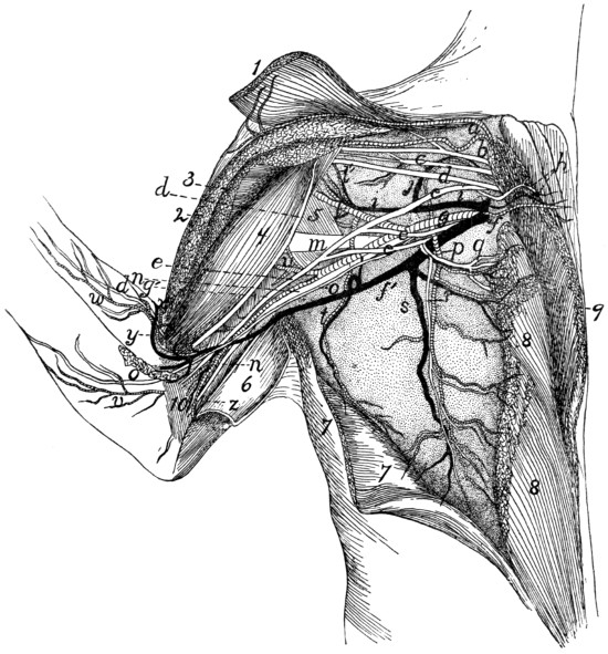
Fig. 122.—Blood-vessels and the more Ventral Nerves of the Axilla, Ventral View.
The pectoral and clavobrachial muscles have been cut and laid aside, their ends being shown; only a part of the nerves are exhibited. 1, M. clavobrachialis; 2, cut end of M. pectoralis major; 3, cut end of M. pectoralis minor; 4, M. biceps; 5, M. teres major; 6, M. epitrochlearis, partly cut and turned back; 7, M. latissimus dorsi, partly cut; 8, M. pectoralis minor; 9, M. pectoralis major; 10, short portion of caput mediale of M. triceps brachii. a, branches of the thyrocervical axis to clavobrachial and clavotrapezius muscles; b, suprascapular nerve; c, first subscapular nerve; d, musculocutaneous nerve; e, median nerve; f, V. axillaris; f′, V. brachialis; g, A. axillaris; g′, A. brachialis; h, first anterior thoracic nerve, accompanied by the anterior thoracic artery and vein; i, V. subscapularis; j, large muscular branch (to subscapular muscle) of the subscapular vein and brachial artery; l, A. subscapularis; l′, A. circumflexa humeri anterior; m, radial nerve; n, ulnar nerve; o, medial cutaneous nerve; p, A. thoracica longa; q, r, second anterior thoracic nerve; s, V. longa thoracica; t, A. and V. thoracicodorsalis; u, A. profunda brachii; v, branch of brachial artery accompanying medial cutaneous nerve; w, branch of A. collateralis radialis superior; x, A. collateralis radialis superior; y, V. mediana cubiti; z, A. collateralis ulnaris.
A. thoracicodorsalis (t).—This arises from the subscapular a short distance from its origin and passes across the teres major to the latissimus dorsi (7), giving branches to both these muscles and to the epitrochlearis.
A. circumflexa humeri posterior.—The posterior circumflex artery arises from the A. subscapularis close to the origin of the latter. It passes between the subscapularis muscle and the teres major, close to the border of the biceps, then between the lateral and long heads of the triceps. It gives off a branch to the inner surface of the spinodeltoid and acromiodeltoid, then passes distad to supply the lateral and long heads of the triceps.
4. A. brachialis (g′).—The brachial artery is the continuation of the axillary (g) into the arm beyond the origin of the subscapular (l). It passes along that side of the biceps which lies next to the humerus. It passes thus, accompanied by the brachial vein (f′) and median and ulnar nerves, through the bicipital arch and afterwards between the biceps (4) and the intermediate division of the medial head of the triceps to the supracondyloid foramen of the humerus, through which it passes with the median nerve to reach the concavity of the elbow. It gives off the following branches:
a. A. circumflexa humeri anterior (l′).—The anterior circumflex artery usually leaves the brachial near its origin (but may arise from the subscapular (l) or one of its branches); it passes to the biceps (4) near the origin of the latter and sends a branch proximad to the head of the humerus.
b. A. profunda brachii or superior profunda (u).—This arises from the first part of the brachial or it may come off from one of the branches of the axillary (e.g., the subscapularis). It passes along with the radial nerve onto the dorsal side of the humerus and supplies the triceps muscle. It also sends branches to the epitrochlearis (6) and latissimus dorsi (7).
c. Rami musculares.—Muscular branches are given off near the supracondyloid foramen to the biceps (4), epitrochlearis (6), and brachialis muscles. A nutrient artery leaves the brachial proximad of the supracondyloid foramen, either separately or in common with the muscular branches, and passes into the nutrient foramen at the junction of the middle and distal thirds of the shaft of the humerus, supplying the bone.
d. A. collateralis ulnaris (superior) (or A. anastomotica magna) (z).—This leaves the brachial artery just proximad of the supracondyloid foramen and passes to the convexity of the elbow, supplying the structures about the olecranon.
e. A. collateralis radialis superior (Fig. 130).—This rises from the axillary artery just proximad of the supracondyloid foramen (Fig. 122, x), in company with the vena mediana cubiti (y), passes across the surface of the biceps (4), beneath the pectoantibrachialis, into the concavity of the elbow. Here it gives branches to the pectoantibrachialis, clavobrachialis, and extensor muscles of the forearm. It then passes onto the ventroradial border of the forearm (Fig. 130) and runs along this border, in company with the vena cephalica (Fig. 130, c) and the superficial radial nerve (g), to the wrist, sending off branches to the integument. At the wrist it turns onto the dorsum of the hand, passing in a gentle curve to the ulnar side and distad, and giving off a branch for the space between each pair of metacarpal bones. These branches (Fig. 130, e) (Aa. digitales dorsales) pass distad and anastomose with branches coming from the palm.
f. A. radialis (Fig. 123).—Distad of the convexity of the elbow the brachial artery takes the name radial artery. It passes from the supracondyloid foramen on the medial side of the biceps tendon (1) and beneath the pronator teres muscle (5), giving off small branches, as far as the middle of the forearm, where it gives off the ulnar artery (k). It then passes from beneath the pronator teres (5) and lies on the surface of the fifth part of the flexor profundus (8), covered only by the fascia and integument. Near the wrist it sends off a branch (m) toward the ulnar side of the arm, which passes into the palm on the surface of the tendon of the flexor profundus, giving twigs to the digits and to the pad in the palm. A branch from this may join the ulnar, and the common trunk thus formed passes into the hand and gives origin to the branches described under the ulnar artery. The presence and size of this branch of the radial varies with the size of the ulnar artery, it being smaller or absent as the ulnar is larger. It is shown at m, Fig. 123. The radial artery (g) now turns gradually dorsad and passes beneath the tendon of the extensor brevis pollicis onto the dorsum of the hand. It passes over the oblique groove on the dorsal surface of the base of the second metacarpal beneath the tendon of the extensor carpi radialis longus and passes between the bases of the second and third metacarpals into the palm of the hand. Here it passes to the ulnar side and anastomoses with the ulnar artery to form the palmar arch.
Branches of the Radial Artery.
1. A. radialis recurrens (i).—The radial recurrent arises in the concavity of the elbow and sends a branch proximad to the structures in the concavity; it then continues to the radial side of the forearm at the elbow, supplying adjacent parts of the brachialis (2), the extensor carpi radialis, and the extensor communis digitorum.
2. A. ulnaris recurrens (j).—The ulnar recurrent arises on the medial side of the tendon of the biceps and supplies structures in the concavity of the elbow on the ulnar side, also the pronator teres (5) and proximal ends of the flexor carpi radialis and flexor profundis digitorum.
3. Rami musculares.—Muscular branches are given off along the course of the artery, to adjacent muscles. A nutrient artery to the radius leaves the radial artery about two centimeters distad of the supracondyloid foramen.
4. Aa interosseæ (anterior (l) and posterior) are given off usually separately between the origin of the ulnar artery and the biceps tendon. Sometimes they arise as a common trunk which soon divides. The posterior interosseous artery passes distad, supplying the flexor muscles of the forearm. It also sends a branch to the pronator teres. The anterior interosseous (l) may arise from the ulnar artery. It passes dorsad to the interosseous membrane, on which it runs distad; sends a nutrient branch to the ulna, and then passes to the wrist in the substance of the pronator quadratus, which it supplies. It sends a branch to the dorsal surface of the wrist; this ramifies on the carpus, forming a network of small arteries.
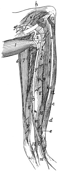
Fig. 123.—Nerves and Arteries of the Forearm, seen from the Flexor Side.
Mm. palmaris longus, flexor carpi radialis, flexor carpi ulnaris, and the third and fourth heads of flexor profundus digitorum have been removed; also part of M. pronator teres. 1, biceps; 2, conjoined tendon of brachialis and clavobrachialis; 3, short portion of caput mediale of triceps brachii; 4, intermediate portion of caput mediale of triceps brachii; 5, cut ends of pronator teres; 6, brachioradialis; 7, extensor carpi radialis longus and brevis; 8, fifth head of flexor profundus digitorum; 9, second head of flexor profundus digitorum; 10, radial part of flexor sublimis digitorum; 11, common tendon of flexor profundus digitorum; 12, first head of flexor profundus digitorum; 13, cut origin of flexor carpi ulnaris. a, ulnar nerve; b, median nerve; c, dorsal cutaneous branch of the ulnar nerve; d, palmar branch of ulnar nerve; e, deep palmar branch; f, superficial palmar branch; g′, A. brachialis; g, A. radialis; h, A. collateralis ulnaris superior; i, A. radialis recurrens; j, A. ulnaris recurrens; k, A. ulnaris; l, A. interossea anterior; m, large branch of A. radialis, joining A. ulnaris.
5. A. ulnaris (k).—The ulnar artery passes beneath the second, third, and fourth parts of the flexor profundus digitorum (but outside of the origin of the fifth part), to the inner surface of the flexor carpi ulnaris. It supplies the flexor carpi ulnaris, the flexor profundus and palmaris longus, and passes on the inner surface of the flexor carpi ulnaris to the wrist. Near the wrist it sends a branch onto the side of the forearm, and another to its midventral part, and ends in a small branch to the wrist on the radial side of the pisiform bone. This branch anastomoses with the radial to form the palmar arch, described below.
6. The palmar arch (Fig. 124) is formed by the termination of the radial artery (a) in the palm and its junction with the end of the ulnar (f). The radial artery reaches the palm between the bases of the second and third metacarpals and passes thence toward the ulnar side and distad, piercing the interosseus muscle of the third digit and lying on the outer surface of the interossei of the third and fourth digits beneath the adductors of the second and fifth digits. A small communicating branch from the radial passes to it between the first and second metacarpals.
Branches of the palmar arch:
A. princeps pollicis et indicis (c) leaves the palmar arch near its radial end and sends a branch onto the ulnar side of the thumb and one onto the radial side of the index.
The palmar interosseæ (d) are three in number. They leave the palmar arch (b) and pass distad and dorsad in the intervals between the four ulnar digits. The radial one passes along the ulnar side of the first digit, the ulnar one along the radial side of the fifth digit. The middle one divides and supplies the contiguous sides of the third and fourth digits. Each of these interosseous arteries sends off muscular branches (e) to the short muscles in the palm, and branches into the fibrous pad which occupies the palm of the hand. The palmar arch also sends branches onto the dorsum of the hand at the sides of the fourth metacarpal. Branches pass from these proximad to the wrist.
B. Aorta abdominalis. The Abdominal Aorta.
—The abdominal aorta emerges into the abdomen from between the crura of the diaphragm, at about the level of the second lumbar vertebra. It passes caudad along the dorsal middle line, lying to the left of the inferior vena cava. It gives off parietal branches to the body wall, and visceral branches to the viscera, and ventrad of the first sacral vertebra it gives off two large branches on each side, the external iliac (Fig. 126, k) and the hypogastric (Fig. 126, l)—a very small median vessel, the sacralis media (Fig. 126, o), continuing the course of the aorta and passing into the tail.
The aorta gives off the following branches: A. cœliaca; A. mesenterica superior; Aa. adrenolumbales; Aa. renales; A. mesenterica inferior; Aa. iliolumbales; Aa. lumbales (seven pairs); Aa. iliacæ externæ; Aa. hypogastricæ.
A. A. cœliaca (Fig. 125).—The cœliac artery is a large branch which is given off from the aorta one centimeter or less caudad of the opening in the diaphragm. It passes directly ventrad about three or four centimeters, then divides, usually at once, into three branches. The most cranial of these is the hepatic (d), the next is the gastrica sinistra (e), while the third and largest, seeming to form a continuation of the cœliac, is the splenic (f) (A. lienalis). The cœliac artery may give rise also, before its division, to the two phrenic arteries (which, however, usually arise from the adrenolumbales), and either before or at the point of division to one or two small Aa. ventriculi dorsales, which, however, frequently arise from the gastrica sinistra.
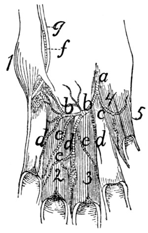
Fig. 124.
The Palmar Arch and its Branches in the Palm of the Hand.
The interosseous muscles have been removed, except those of the third and fourth digits. 1, M. abductor digiti quinti; 2, 3, Mm. interossei of third and fourth digits; 4, M. adductor pollicis; 5, M. flexor brevis pollicis. a, termination of radial artery; b, palmar arch; c, A. princeps pollicis et indicis; d, Aa. interosseæ; e, muscular branches of the same; f, A. ulnaris; g, deep palmar branch of ulnar nerve.
Branches of the cœliac artery:
1. A. hepatica (d).—The hepatic artery passes cranioventrad, pierces the descending limb of the great omentum and passes craniad to the liver, lying, together with the portal vein and common bile-duct, in the ventral boundary of the foramen epiploicum (foramen of Winslow) and contained, together with the last-named vessels, in a fibrous sheath called the capsule of Glisson. Just before entering the sheath it gives off the gastroduodenalis (g). At its termination the hepatic artery divides, sending branches to the lobes of the liver and a cystic artery to the gall-bladder.
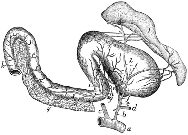
Fig. 125.—The Cœliac Artery and its Branches.
a, abdominal aorta; b, A. cœliaca; c, A. mesenterica superior (cut); d, A. hepatica (cut); e, A. gastrica sinistra; f, A. lienalis; g, A. gastroduodenalis (cut from A. hepatica); h, A. pylorica; i, A. gastroepiploica dextra; j, A. pancreaticoduodenalis superior; k, A. pancreaticoduodenalis inferior (cut from inferior mesenteric); l, A. ventriculi dorsalis. 1, spleen; 2, stomach; 3, duodenum; 4, pancreas.
a. A. gastroduodenalis (g).—This arises from the hepatic near the pylorus and passes caudad, dividing one or two centimeters from its origin into three branches, A. pylorica (h), A. pancreaticoduodenalis superior (j), and A. gastroepiploica dextra (i). The pylorica (h) (which may arise directly from the hepatic) passes to the pylorus, thence along the lesser curvature of the stomach, dividing into numerous branches and anastomosing with the gastrica sinistra. The pancreaticoduodenalis superior (j) passes to the duodenum, supplying it and the duodenal part of the pancreas and anastomosing with the pancreaticoduodenalis inferior (k). The gastroepiploica dextra (i) passes along the greater curvature of the stomach from the pyloric end and supplies the walls of the stomach, sending branches also to the ascending limb of the great omentum. These branches anastomose with the terminal branches of the splenic artery.
2. A. gastrica sinistra (e).—This arises from the cœliac artery and passes to the lesser curvature of the stomach, extending along this to the right. It gives off many branches to the walls of the stomach, and anastomoses with A. pylorica.
A. ventriculi dorsalis (l).—One or two small arteries which arise either from the gastrica sinistra or the cœliac artery, and pass toward the dorsal part of the greater curvature of the stomach near its cardiac end.
3. A. lienalis (f).—The splenic artery is the largest of the branches of the cœliac axis, of which it appears to be the direct continuation. It divides into two large branches, one to the cranial end, the other to the caudal end, of the spleen (1). From the latter a large branch passes to the pancreas and descending limb of the great omentum.
B. A. mesenterica superior (Fig. 126, d).—The superior mesenteric artery is larger than the cœliac. It supplies the blood to the small intestines and caudal portion of the pancreas and to the ascending and transverse colon. It arises from the ventral side of the abdominal aorta about one centimeter caudad of the cœliac axis and passes caudoventrad, forming a curve with the convexity dextrad. It gives off the pancreaticoduodenalis inferior, colica media, colica dextra, ileocolica, and numerous branches to the small intestine.
1. A. pancreaticoduodenalis inferior (Fig. 125, k).—This passes to the caudal end of the pancreas and a part of the duodenum, anastomosing with the pancreaticoduodenalis superior.
2. A. colica media is a large branch to the transverse and descending portions of the large intestine. It divides and sends branches in both directions which anastomose with the colica dextra and with branches from the inferior mesenteric arteries.
3. A. colica dextra.—A small branch which passes to the ascending and transverse colon, anastomosing with the colica media and ileocolica. It is sometimes absent.
4. A. ileocolica.—This passes to the cæcum and supplies that structure, sending branches to the ileocolic valve and the caudal end of the ileum, and anastomosing with the colica dextra and the intestinal branches of the superior mesenteric.
5. The superior mesenteric now divides into about sixteen terminal branches which pass to the small intestine. In many cases these branches unite near the intestine, forming arches, and from these arches numerous short branches pass to the intestine.
3. A. adrenolumbalis (or lumboabdominalis) (Fig. 126, e).—This rises from the aorta, one on each side, about two centimeters caudad of the superior mesenteric. Each passes laterad onto the dorsal body wall, supplying the muscles of this region. A large branch passes caudad along the surface of the muscles dorsad of the kidney and anastomoses with the iliolumbar artery. From the adrenolumbalis rises usually:
A. phrenica (f).—The phrenic artery rises either from the cœliac (c) or the adrenolumbalis (e) and passes to the diaphragm, near its dorsal border. In the diaphragm it passes ventrad, lying at the medial margin of the costal portion of the diaphragm, as far as its sternal portion, where it unites with the artery of the opposite side. The two thus form an arch, from which radiating arteries pass off to supply the diaphragm.
4. A. renalis (g).—The two renal arteries arise from the sides of the aorta, usually at about the same point, so that the left passes caudolaterad and the right craniolaterad (owing to the position of the kidneys). The artery usually divides just before entering the kidney (2). It passes dorsad of the vein (t). The renal artery sometimes sends a branch to the suprarenal body (1). It also occasionally gives origin to the spermatic artery (h).
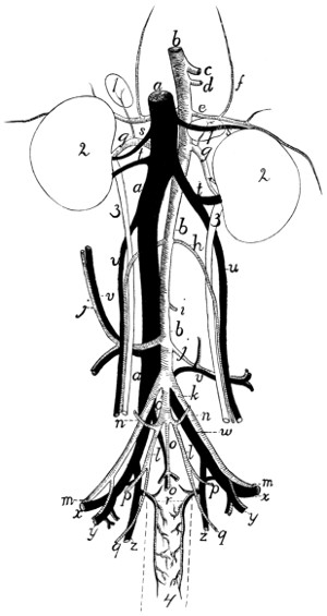
Fig. 126.—Abdominal Blood-vessels, Ventral View.
a, vena cava inferior; b, abdominal aorta; c, A. cœliaca (cut); d, A. mesenterica superior (cut); e, A. adrenolumbalis; f, A. phrenica; g, A. renalis; h, A. spermatica interna; i, A. mesenterica inferior (cut); j, A. iliolumbalis; k, A. iliaca externa; l, A. hypogastrica; m, beginning of A. femoralis; n, A. umbilicalis; o, A. and V. sacralis media (o, the artery; o′, the vein); p, A. glutea superior; q, A. glutea inferior; r, A. and V. hemorrhoidalis media; s, V. adrenolumbalis; t, V. renalis; u, V. spermatica interna; v, V. iliolumbalis; w, V. iliaca communis; x, V. femoralis; y, V. and A. profunda femoris; z, V. hypogastrica. 1, suprarenal body; 2, kidney; 3, ureter; 4, outline (broken) of rectum.
5. A. spermatica interna (h).—This rises from the aorta on each side at about the level of the caudal ends of the kidneys, and passes laterad (or it may arise from the renal). In male specimens the artery turns caudad, along with the corresponding vein (u), and passes to the inguinal canal. Thence it passes along with the vas deferens and the vein (forming all together the spermatic cord) to the testes and the other structures in the scrotum.
In females (Fig. 112, 2, page 265) the artery is larger and passes more nearly directly laterad, being much convoluted. It passes to the ovary, being now called the ovarian artery. It sends branches into the ovary (c) and to the cranial end of the uterus (f); the latter anastomosing with branches of the uterine artery (3).
6. A. mesenterica inferior (Fig. 126, i).—The inferior mesenteric artery has its origin from the aorta at about the level of the last lumbar vertebra. It passes toward the large intestine, and near it divides into two branches, the colica sinistra, which passes craniad along the descending colon, anastomosing with the colica media, and the superior hemorrhoidal, which passes caudad along the descending colon and rectum and anastomoses with the middle hemorrhoidal (page 308).
7. A. iliolumbalis (Fig. 126, j).—The iliolumbar arteries arise from the aorta about two centimeters caudad of the inferior mesenteric and pass laterad over the ventral surface of the psoas minor and iliopsoas muscles. Each divides into two main branches, one passing caudad and the other craniad; these supply the muscles of this region. The cranial branch anastomoses with branches of the adrenolumbalis (e). The caudal branch sends an artery through the abdominal wall to appear at the cranial edge of the thigh; it passes onto the lateral surface of M. sartorius and extends here some distance distad.
8. A. lumbalis.—There are usually seven pairs of lumbar arteries, passing almost directly dorsad from the dorsal surface of the aorta; the first pair arising just craniad of the diaphragm, the last at the same level as the origin of the external iliac. The two arteries of a pair usually arise from the aorta one behind the other, or they may arise by a common trunk. They correspond to the intercostal arteries. They pass between the centra of the vertebræ and the muscles of the dorsal region, and send one main branch laterad, the other dorsad; both supply the dorsal muscles. From the dorsal branch small arteries pass into the vertebral canal to join the anterior spinal artery.
9. A. iliaca externa (Fig. 126, k).—The external iliac artery passes obliquely caudad from the aorta, lying ventrad of the common iliac vein and against the medial surface of the psoas minor muscle. It passes onto the ventral surface of this and the iliopsoas and at the same time reaches the tendon of the abdominal muscles; through a small opening in this tendon lying ventrocaudad of that for the iliopsoas, it leaves the abdominal cavity. On its emergence from the abdominal cavity onto the medial surface of the thigh it receives the name A. femoralis (m). The branches of the external iliac artery are described below (page 309).
10. A. hypogastrica (or iliaca interna) (Fig. 126, l).—The hypogastric or internal iliac arteries are given off from the aorta usually within a centimeter caudad of the external iliac. Each passes caudolaterad, lying on the medial side of the common iliac vein, and divides into branches which supply the structures within the pelvis and the muscles about the pelvic wall. There is much variation in the origin and relations of the branches of this artery. The following seems to be a very common arrangement: A very short distance from the origin the umbilical artery (n) is given off. The hypogastric artery then extends two or three centimeters and gives off A. glutea superior (p), which passes at once to the pelvic wall. A little farther caudad the hypogastric divides into the middle hemorrhoidal (r), to the rectum, and the glutea inferior (q), which passes out of the pelvis.
Branches of the hypogastric artery:
a. A. umbilicalis (Fig. 126, n).—This small artery arises from the hypogastric about one centimeter from the beginning of the latter, and passes ventrad to the bladder. Here it divides into two branches—the superior vesical to the sides of the bladder, the inferior vesical to the neck of the bladder and the urethra.
b. A. glutea superior (Fig. 126, p).—This passes dorsolaterad from the hypogastric and reaches the pelvic wall against the medial surface of the ilium. Here it divides into two branches. One passes between the iliopsoas muscle and the ventral border of the ilium to the medial surface of the gluteus maximus; it gives branches to the gluteus muscles, the pyriformis, and the rectus femoris. The other branch passes dorsad of the ilium, sending a branch to the medial surface of the gluteus medius, and a small branch which joins the lateral sacral artery and enters thus the sacral canal.
c. A. hemorrhoidalis media (Fig. 126, r).—The middle hemorrhoidal passes ventrad from the hypogastric, onto the lateral surface of the rectum. It passes caudad near the ventral side of the rectum as far as the anus. A short distance from the origin it gives off a branch which passes almost directly ventrad toward the beginning of the urethra. In the male this branch is small, sending twigs to the urethra and the prostate. In the female it is much larger, forming A. uterina (Fig. 112, 3, page 265). The uterine artery turns craniad onto the uterus, which it supplies, passing to the cranial end of the uterine cornu and anastomosing with the ovarian artery.
The middle hemorrhoidal gives off in the female branches to the vagina (Fig. 112, m) and neck of the bladder (k), and a large branch to the urogenital sinus (n); it then supplies the anal glands and other structures about the anus, and anastomoses with the terminal branches of the hemorrhoidalis superior. In the male branchlets are given to the urethra, to the bulbourethral (or Cowper’s) gland, and to the penis. The A. dorsalis penis is a small branch which passes along the median dorsal groove of the penis to the glans. The middle hemorrhoidal then supplies the structures about the anus and anastomoses with the superior hemorrhoidal, as in the female.
d. A. glutea inferior (Fig. 126, q).—This is the terminal portion of the hypogastric. It passes along with the great sciatic nerve to the great sciatic notch, and reaches the medial surface of M. pyriformis. It divides into branches which supply the gluteus and pyriformis muscles, while a small branch accompanies the great sciatic nerve. A small branch also passes onto the lateral surface of the tail, extending caudad along its side.
11. A. sacralis media (Fig. 126, o).—This is the continuation of the aorta into the sacral and caudal regions. It extends to near the end of the tail, lying in the ventral middle line against the sacral and caudal vertebræ. In the caudal region it passes through the hæmal arches, covered by the chevron bones. Between the vertebræ it gives off side branches comparable to the intercostal and lumbar arteries of the aorta.
Branches of the sacralis media:
a. A. sacralis lateralis.—Behind the first sacral vertebra a large branch, the lateral sacral, is given off on each side. This enters the first anterior sacral foramen, gives a branch to the structures in the sacral canal, and sends a dorsal branch out through the posterior sacral foramen to the muscles on the dorsal side of the sacrum.
Similar but smaller branches are given off between the succeeding vertebræ, sacral and caudal.
The External Iliac and its Branches.
—The origin of the external iliac (Fig. 126, k) is described above (page 307). It gives off the following branches:
1. A. profunda femoris (Fig. 126, y).—This is given off just before the external iliac leaves the abdominal cavity. It passes caudad, and about one centimeter from its origin it gives off three branches, sometimes all separately, sometimes two in common. One of these passes mediad in the lateral ligament of the bladder and ramifies on the lateral surface of that organ. A second passes through the abdominal wall and into the fat on the medial surface of the thigh; it sends a branch caudad toward the external genital organs, while its main trunk passes distad in the subcutaneous fat almost to the knee. From one of these two a small branchlet passes in the male to the spermatic cord and accompanies this to the testis; it represents thus the external spermatic artery. The third branch, A. epigastrica inferior, passes directly to the ventral surface of the rectus abdominis muscle, on which it passes craniad, sending branches to the muscles of the abdominal walls. It anastomoses with terminal branches of the internal mammary artery.
The profunda femoris itself passes between the iliopsoas and the pectineus muscles, then to the medial surface of the adductor femoris. It divides into branches which supply the pectineus, adductor longus, adductor femoris, caudofemoralis, quadratus femoris, semimembranosus, and semitendinosus.
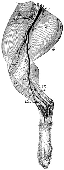
Fig. 127.—Superficial Arteries, Veins, and Nerves on the Medial Side of the Leg.
a, A. and V. femoralis; b, N. femoralis; c, A. profunda femoris; d, A. and V. circumflexa femoris lateralis; e, ramus muscularis; f, A. saphena and V. saphena magna; g, N. saphenus; h, A. and V. articularis genu suprema; i, N. tibialis; j, plantar branch of A. saphena; k, dorsal branch of A. saphena. 1, M. sartorius; 2, M. tensor fasciæ latæ; 3, M. rectus femoris; 4, M. iliopsoas; 5, M. pectineus; 6, M. adductor longus; 7, M. adductor femoris; 8, M. semimembranosus; 9, M. gracilis; 10, M. semitendinosus; 11, medial head of M. gastrocnemius; 12, M. plantaris; 13, M. flexor longus digitorum; 14, M. tibialis anterior; 15, tibia.
2. A. femoralis (Fig. 127).—This is the continuation of the external iliac onto the medial surface of the thigh. The artery lies in a triangular depression between the borders of the sartorius (1) and gracilis (9) muscles. The floor of the depression is formed by the adductor longus (6), pectineus (5), vastus internus, and rectus femoris (3) muscles. This triangular depression is known as the iliopectineal fossa, or Scarpa’s triangle; it contains also the femoral vein (a) and saphenous nerve (g), which pass along with the artery. The artery extends distad, and at about the middle of the length of the thigh it passes, along with the vein and nerve, into a groove between the vastus medialis and the adductor femoris. This groove is converted into a canal by the overlying aponeurosis; it is known as Hunter’s canal or the canalis adductorius. At the distal end of the adductor femoris the femoral artery ceases to be superficial and passes between the vastus medialis and the semimembranosus to the popliteal space; it now receives the name A. poplitea.
Branches of the femoral artery:
a. A. circumflexa femoris lateralis (Fig. 127, d).—The lateral circumflex arises from the femoral about one centimeter from its emergence from the abdominal cavity. It passes craniad and laterad and divides into two main portions. One part passes between the rectus femoris and vastus medialis, gives branches to these muscles, and sends an ascending branch to the structures about the hip-joint, and a descending branch into the vastus medialis muscle. The remainder of the lateral circumflex passes along the inner (lateral) surface of the sartorius (1) to the cranial border of the leg, giving branches to the sartorius (1) and tensor fasciæ latæ (2).
b. A large muscular branch (e) passes caudad (or ventrad) and distad across the distal end of the adductor femoris (7) and between the gracilis (9) and semimembranosus, supplying these muscles. Other small muscular branches may be given off from both sides of the femoral.
c. A. articularis genu suprema (h).—The superior articular artery arises from the femoral, either in common with the saphenous artery or separately, a little proximad of the point where the femoral ceases to be superficial. It passes toward the knee, between the vastus medialis and semimembranosus, and covered by the sartorius (1), and ramifies over the medial surface of the knee-joint and in the structures just proximad of the joint.
d. A. saphena (f).—The saphenous artery frequently takes origin in common with the superior articular (h), but may arise separately at about the same level. It passes distad across the gracilis (9), accompanied by the saphenous nerve (g) and vein, sends several branches dorsad (toward the knee), and at about the middle of the lower leg it divides into two main branches, a dorsal branch (k), passing to the dorsum of the foot, and a plantar branch (j), to the sole of the foot. The dorsal branch is accompanied by the main saphenous nerve (g) and vein. It sends one or two branches to the medial side of the ankle-joint, passes then onto the dorsum of the foot, and divides into four main branches. The medial one passes along the medial side of the medial digit; the other three pass to the intervals between the digits. Each divides into two branches which supply the contiguous sides of the two digits between which the interval lies. The dorsal branch thus supplies arteries to the sides of all the digits except to the lateral side of the lateral digit. This is supplied by A. suralis.
The plantar branch (j) is larger than the dorsal. It passes distad on the medial surface of the flexor longus hallucis, accompanied by the tibial nerve, and across the space between the tendon of Achilles and the flexor. It gives superficial and deep branches to the structures about the ankle-joint, and sends inward a branch from the lateral side (Fig. 128, d) which joins the termination of A. tibialis anterior to form the plantar arch. It then passes distad along the plantar surface of the foot, nearer its medial border. Beneath the pad in the sole of the foot it divides into three branches which supply the interosseous spaces between the three digits.
e. A. poplitea.—The main trunk of the femoral artery passes between the vastus medialis and the semimembranosus and then through the distal portion of the adductor femoris to reach the popliteal space. This is the space ventrad (or caudad) of the knee, between the biceps femoris on one side and the semimembranosus on the other. Here the artery receives the name A. poplitea, or popliteal artery. The popliteal artery gives off a number of large branches as it passes through the popliteal space, passes between the condyles of the femur and underneath the popliteal muscle, and finally turns dorsolaterad between the tibia and fibula, just distad of the head of the fibula. It now receives the name A. tibialis anterior and passes distad along the dorsal (anterior) border of the fibula.
Branches of the popliteal artery:
1. A. suralis.—This is a large branch which passes distad from the caudal side of the popliteal artery. It sends branches to the biceps and to the fat in the popliteal space, and passes onto the ventral border of the lateral head of the gastrocnemius. It sends branches to both heads of the gastrocnemius and to the popliteus, passes distad onto the lateral surface of the tendon of Achilles, and may be traced to the proximal portion of the dorsolateral side of the foot, where it sends many branches to the integument about the ankle-joint. It then passes along the lateral side of the foot and supplies the artery on the lateral side of the fifth digit.
2. Aa. genu posteriores.—Several small arteries which pass to the knee-joint.
3. Rami musculares.—Numerous small branches to the muscles about the popliteal space.
4. A. tibialis posterior.—This is the largest branch of the popliteal artery, which it leaves just before the latter passes beneath the popliteal muscle. The tibialis posterior passes at first mediad, then turns distad, passes across the popliteus muscle onto the surface of the flexor longus hallucis, and ramifies in the substance of this muscle and its tendon. It gives branches also to the gastrocnemius and soleus. (It does not pass into the foot as does the corresponding artery in man.)
5. A. tibialis anterior.—The anterior tibial artery is the continuation of the popliteal after its passage through the interosseous membrane between tibia and fibula to the dorsal side of the lower leg. Here it passes distad, lying against the interosseous membrane, between the extensor longus digitorum and the tibialis anterior muscle, or partly imbedded in the substance of the latter. It passes, lying beneath the tendons of these muscles, through the transverse ligament proximad of the malleoli, thus reaching the dorsum of the foot. It extends distad across the tarsus to the space between the second and third metatarsals, and through this space to the plantar side of the foot (Fig. 128). Here it receives one or two small branches (a and d) from the plantar branch of A. saphena (forming with them the plantar arch (c)), and passing distad and slightly laterad, divides into three main portions (e). These (Aa. digitales plantares) pass to the three intervals between the digits. The medial one passes along the lateral side of the second digit; the next divides, sending a branch to the lateral side of the third digit and the medial side of the fourth; the lateral one passes along the medial border of the fifth digit. The Aa. digitales plantares send numerous branches to the interosseous muscles.
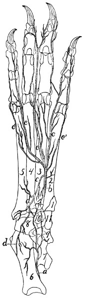
Fig. 128.—Deep Arteries on the Plantar Surface of the Foot.
a, medial twig from plantar branch of A. saphena; b, A. tibialis anterior (or plantaris profunda); c, plantar arch; d, lateral twig from plantar branch of A. saphena; e, terminal branches from A. tibialis anterior.
Branches of the tibialis anterior:
a. A. tibialis recurrens.—Immediately after passing through the interosseous membrane the anterior tibial sends a small artery proximad to the knee-joint.
b. Rami musculares.—Numerous short branches throughout the course of the artery, to the muscles about it.
c. Ramus superficialis.—A larger branch leaves the lateral surface of the artery about one-third the distance from the knee to the ankle and passes along with the superficial peroneal nerve, at first beneath M. peroneus longus, then superficially. It passes onto the dorsal surface of the foot and becomes connected with terminal branches of A. saphena.
d. A. tarsea medialis.—This arises from the tibialis anterior just distad of the transverse ligament through which it passes at the ankle, passes over the medial surface of the astragalus, and is distributed to the ligaments about the ankle-joint. It anastomoses with the plantar branch of A. saphena.
e. A. metatarsea.—The metatarsal artery passes laterad from the tibialis anterior at the distal boundary of the tarsus, on the dorsal surface of the foot. It sends branches distad in the intervals between the third and fourth and the fourth and fifth metatarsals.
The distal branches (Fig. 128) of the tibialis anterior have been described in the general description of the artery.
THE VEINS. VENÆ.
Venæ pulmonales. The Pulmonary Veins.
The pulmonary veins follow in the lungs the course of the bronchi. They enter the left auricle in three groups (Fig. 116, page 276). The first of these (i) comes from the anterior and middle lobes of the right side, the second (h) from the corresponding lobes of the left side, and the third (g) from the terminal lobes of both sides. Each group is composed of two or three veins and opens into a sinus or extension of the auricle. The sinuses are from their position dextral (i), sinistral (h), and dorsal (g).
The Veins of the Body.
The veins of the body may be classified into three main groups: (1) the veins of the heart; (2) the superior vena cava and its branches; (3) the inferior vena cava and its branches. The portal system of veins will be considered with the inferior vena cava.
1. Veins of the Heart.
The veins of the heart consist of one large vein, the vena cordis magna, and a number of small veins.
Vena cordis magna (Fig. 118, 3).—This arises as a number of scattered branches on the surface of both ventricles. These unite to form two main trunks. One of these, on the ventral side of the heart, marks externally the septum between the two ventricles. It passes beneath the left auricular appendage, then turns to the left and passes along the sulcus coronarius to the dorsal side of the heart. Here it unites with the second main trunk which ramifies over the surface of the left ventricle. Arriving at the base of the right auricle it enters this by the coronary sinus, which lies just beneath the opening of the inferior vena cava.
In addition to the vena cordis magna and its branches there are a number of small veins, especially in the wall of the right ventricle. These enter the right auricle by a number of small separate openings along the sulcus coronarius.
2. Vena cava superior (Fig. 129, b).
The superior vena cava is the great vein returning the blood from the head, fore-limb, and cranial part of the trunk. It extends from the level of the first rib on the right side of the vertebral column to the right auricle. Its caudal end lies dorsad of the aortic arch. Opposite the right rib it is formed by the junction of the two innominate veins (m).
Branches of the superior vena cava:
1. V. azygos (d).—The first branch of the superior vena cava is the azygos vein, which enters the vena cava on the right side a centimeter or less craniad of the root of the right lung. It is formed in the abdominal cavity by the confluence of two or three small veins, which collect the blood from the muscles of the dorsal wall of the abdomen. The small median trunk thus formed enters the thoracic cavity between the crura of the diaphragm and lies on the ventral surface of the centra of the thoracic vertebræ, slightly to the right of the middle line. The azygos receives the intercostal veins (n), which correspond to the intercostal arteries and have the same course and distribution. In the caudal part of the thoracic cavity the intercostals enter the azygos separately, but the intercostals of the cranial intercostal spaces usually unite two or three together and enter the azygos by one or more common trunks. The azygos also receives bronchial and œsophageal veins, corresponding to the similarly named arteries.
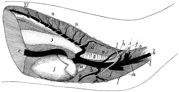
Fig. 129.—Thoracic Blood-vessels, from the Right Side.
a, aorta; b, vena cava superior; c, vena cava inferior; d, V. azygos; e, A. subclavia dextra; f, A. mammaria interna; g, V. mammaria interna; h, costocervical axis; i, united trunk of vertebral and costocervical veins; j, A. and V. axillaris; k, V. jugularis externa; l, V. jugularis interna; m, the two innominate veins; n, intercostal arteries and veins; o, thyrocervical axis. 1, heart; 2, trachea; 3, œsophagus; I-XI, cut ends of the first eleven ribs.
2. V. mammaria interna (g).—The internal mammary veins follow the corresponding arteries. The two veins unite to form a common trunk which, after receiving a branch from the cranial part of the sternum, enters the vena cava opposite the third rib.
3. Vv. anonymæ (Fig. 129, m; Fig. 115, p).—The innominate veins unite opposite the first intercostal space to form the vena cava superior. Each extends from the cranial end of the vena cava superior craniolaterad to a point a short distance craniad of the first rib and is there formed by the union of the external jugular (Fig. 129, k) and subclavian (j) veins. The innominate vein has the following branches:
A and B. Vv. vertebralis and costocervicalis (i).—The vertebral and costocervical veins unite to form a single trunk which enters the innominate about opposite the first rib. These two veins follow the corresponding arteries. (Sometimes the vertebral and costocervical veins enter the innominate separately, and in other cases one or more of the components of the costocervical veins (e.g., the transversa colli) may enter the axillary vein. In other cases the entire common trunk of the vertebralis and costocervicalis may enter the superior vena cava directly.)
C. V. subclavia.—The subclavian vein enters the innominate at about the level of the first rib. Outside of the thorax it is called the axillary vein (j); this comes from the arm.
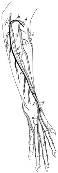
Fig. 130.
Superficial Arteries, Veins, and Nerves on the Extensor Side of the Arm and Dorsum of the Hand.
a, V. cephalica; b, V. mediana cubiti; c, V. cephalica antibrachii; d, A. collateralis radialis superior; e, Aa. and Vv. digitales dorsales; f, A. radialis; g, superficial radial nerve; h, N. musculocutaneus; i, N. cutaneus medialis.
Veins of the Arm.—The blood is returned from the arm by two systems of veins,—a deep and a superficial system.
The deep system is composed of veins which correspond to the branches of the axillary artery, follow in general the same course, and receive the same names (Fig. 122). They unite to form the axillary vein. The brachial vein does not pass with the brachial artery through the supracondyloid foramen.
The superficial system comprises the vena cephalica and its branches.
V. cephalica (Fig. 130).—Several superficial branches lying in the palm of the hand unite to form a common trunk which curves about the radial side of the wrist and here unites with a similarly formed trunk from the dorsum of the hand. The vein formed by this union, vena cephalica (antibrachii) (c), passes to the elbow accompanying the radial nerve (g) and the collateral radial artery (d), and lying on the extensor carpi radialis longus muscle just beneath the integument. It receives small lateral superficial branches. At the elbow it divides into two: these are the vena cephalica (humeri) (a) and the vena mediana cubiti (b). The latter passes beneath the pectoantibrachialis muscle, across the biceps, and unites with the brachial vein in the concavity of the elbow.
The vena cephalica (humeri) passes at the elbow onto the upper arm, following the lateral border of the clavobrachial muscle. It thus reaches the acromiodeltoid muscle, where it divides into two branches. One passes inward beneath the acromiodeltoid, and joins the vena circumflexa posterior (a branch of the axillary). The second continues over the outer surface of the shoulder, and finally joins the vena transversa scapulæ (a branch of the external jugular).
D. V. jugularis externa (Fig. 131; Fig. 65, 5, page 109).—The external jugular vein comes from the head and face. It is formed by the junction of the anterior (Fig. 131, c) and posterior (b) facial veins near the ventral border of the sternomastoid muscle. It passes thence obliquely across the sternomastoid to the triangular interval between it and the clavotrapezius. In the interval it receives a small branch which accompanies the branch of the transversa scapulæ artery to the clavobrachialis muscle, receives next the common trunk of the vena cephalica and transversa scapulæ, and opposite the cranial end of the manubrium receives the internal jugular vein (Fig. 129, l). It then unites with the subclavian to form the innominate vein. Near the point where it receives the internal jugular the external jugular vein receives the thoracic duct (Fig. 118, 5).
Branches of the external jugular:
a. V. transversa scapulæ.—This follows the course of the artery of the same name. One of its branches receives one of the two terminal divisions of the vena cephalica (q. v.). In many cases veins which correspond to branches of the transversa scapulæ artery enter the external jugular separately (as shown in Fig. 118, x and y).
b. V. cervicalis ascendens.—A small branch which follows the corresponding branch of the thyrocervical axis.
c. V. jugularis interna (Fig. 118, 2; Fig. 119, page 284).—The internal jugular vein varies much in size and in the place where it joins the external jugular. The point of junction is usually at about the level of the first rib, but may be much farther craniad. It arises on the ventral side of the basal portion of the occipital by the junction of a number of veins which are described below. The internal jugular passes caudad in the neck region at the side of the trachea, in company with the common carotid artery and the vagus and sympathetic nerves. In this region it receives branches which accompany the branches of the common carotid artery.
The veins which by their union form the internal jugular vein are the following:
(1) One or two veins from the venous sinuses of the brain, leaving the cranial cavity by the jugular foramen. These are described in the account of the veins of the brain.
(2) A large communicating branch from the posterior facial.
(3) The vena occipitalis, a large branch coming from the vertebral column (which it leaves by the atlantal foramen) and from the back of the head.
d. V. facialis anterior (Fig. 131, c).—The anterior facial vein collects the blood from the face, the tongue, and adjacent parts; it joins the posterior facial (b) caudad of the angle of the jaw to form the external jugular vein.
The anterior facial begins over the frontal bone caudad of the orbit, where it is known as the frontal vein (c″). This runs along the dorsal border of the orbit, into which it dips. It sends a communicating branch into the orbit, and receives small veins (superior palpebral) from the upper eyelid. It then turns ventrad, passing along the cranial angle of the eye between the levator labii superioris proprius (5) and the orbicularis oculi (8) muscles, being called in this region the angular vein (c′). This receives branches (external nasal veins, k) from the side of the nose. The vein crosses the malar bone obliquely (now receiving the name anterior facial, c) and follows the cranioventral border of the masseter muscle (9). As it passes the infraorbital foramen it receives through the foramen a small vein from within the orbit. It receives also the inferior palpebral (j) from the lower eyelid, and the superior labial vein (i) from the upper lip. Just caudad of the angle of the mouth it receives from beneath the cranioventral margin of the masseter the V. facialis profunda (h) described below. Still farther caudad it receives the inferior labial vein (g) from the lower lip, and a small branch from the masseter muscle. Caudad of the middle of the cranioventral border of the masseter it receives from beneath the lymphatic gland of this region the large submental vein (f). Next the anterior facial vein receives a large communicating branch (V. transversa, d) which passes transversely across the ventral surface of the throat and connects the two anterior facial veins of the opposite sides. From the middle of this communicating branch an unpaired trunk passes caudad in the middle line and divides into the two laryngeal veins, which pass to the larynx; a small branch is continued from the unpaired trunk craniad into the tongue. The communicating branch receives also near its junction with the anterior facial a rather large superficial branch (e) which comes from the sides of the lower jaw and passes across the lymphatic gland (12) to join the communicating branch.
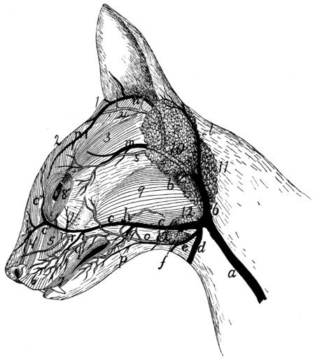
Fig. 131.—Superficial Veins and Arteries of the Face.
a, external jugular vein; b, posterior facial vein; c, anterior facial vein (c′, angular vein; c″, frontal vein); d, communicating branch with opposite anterior facial; e, superficial branch from lower jaw; f, submental vein; g, inferior labial vein; h, deep facial vein; i, superior labial vein; j, inferior palpebral vein; k, external nasal veins; l, posterior auricular vein; m, superficial temporal vein; n, anterior auricular vein; o, external maxillary artery; p, inferior labial artery; q, superior labial artery; r, infraorbital arteries; s, superficial temporal artery; u, anterior auricular branch of posterior auricular artery. 1, M. adductor auris superior; 2, M. corrugator supercilii medialis; 3, M. corrugator supercilii lateralis; 4, M. levator labii superioris alæque nasi; 5, M. levator labii superioris proprius; 6, M. myrtiformis; 7, M. orbicularis oris; 8, M. orbicularis oculi; 9, M. masseter; 10, parotid gland; 11, submaxillary gland; 12, lymphatic glands.
The following larger branches of the anterior facial are not described above:
V. facialis profunda (Fig. 131, h).—The deep facial vein enters the anterior facial from beneath the masseter, just caudad of the angle of the mouth. It collects branches from the soft and hard palates and the teeth, these branches passing along with the corresponding arteries for a distance, then uniting and passing ventrolaterad beneath the zygomatic arch to join the anterior facial. The deep facial vein also receives branches which come from the venous plexus that lies in the same region as the arterial carotid plexus.
V. submentalis (Fig. 131, f).—This enters the anterior facial vein at the ventral border of the masseter muscle and near the cranial end of the two superficial lymph-glands (12) in this region. The submental vein is formed at the dorsal border of the digastric muscle by two branches emerging from between the digastric and the mandible. The more cranial of these, V. lingualis, collects branches from the tongue, from its middle to the tip; it passes through the middle of the lateral border of the mylohyoid muscle to join the other branch. The second branch emerges from between the mandible and the pterygoid muscles. It comes from near the caudal end of the mandible, where it becomes continuous with a communicating branch from the posterior facial vein, and receives a branch which comes from the mandibular canal; also small branches from the adjacent parts.
e. V. facialis posterior (Fig. 131, b).—The posterior facial vein arises from the region of the internal maxillary artery, its terminal branches following the branches of the artery. It collects blood from the pterygoid, masseter (9), and temporal muscles, and forms a plexus which is interwoven with the carotid (arterial) plexus in the region of the orbital fissure. It is connected with the submental vein by a communicating branch. From the posterior facial a branch extends ventrad close against the outer surface of the tympanic bulla and then turns caudad, receives a branch from the pharynx, then continues to join the internal jugular. The posterior facial itself passes laterad along the caudal border of the masseter (9) and the ventral border of the parotid gland (10), then becomes superficial and turns ventrad over the outer surface of the submaxillary gland (11) to join the anterior facial (c) caudad of the angle of the jaw.
As it passes ventrad of the parotid gland (10), just before becoming superficial, the posterior facial receives the superficial temporal (m). This at first follows the superficial temporal artery (s), then enters the substance of the parotid gland, joining the posterior facial near the ventral border of the latter. The superficial temporal receives the anterior auricular (n), a large vein passing along the cranial border of the auricular opening and arising dorsad of the eye.