THE MUSCLES. (continued 2)
Reading edition
Action.—Draws the arm mediad.
M. pectoralis major, or ectopectoralis (Fig. 65, l, l′).—In this it is usually possible to distinguish a superficial and a deep layer.
The superficial (l′) layer is a flat band of fibres one or two centimeters wide.
Origin from a raphe in the midventral line, along the cranial half of the manubrium and for five to ten millimeters craniad of it.
Insertion (Fig. 81, f).—The pectoral ridge of the middle third of the shaft of the humerus, slightly dorsad of the line of insertion of the deep portion.
Relations.—Outer surface with the clavobrachial (Fig. 65, k) and integument, the medial end caudally with the pectoantibrachialis (m), and the distal end with the brachialis (v). Inner surface with the deep portion (l), the fibres of which it crosses obliquely.
The deep portion (l) is a flat band of parallel fibres about three times as broad as the superficial portion.
Origin.—The ventral surface of the manubrium and of the first three divisions of the sternum, and the median raphe for about one centimeter craniad of the manubrium. It passes directly laterad.
Insertion (Fig. 81, e) along a line which begins at the infraspinatus fossa of the great tuberosity, and runs parallel to the deltoid ridge until it reaches the pectoral ridge at the junction of the second and third fourths of the humerus and then continues in the direction of the pectoral ridge as far as the junction of the third and fourth fourths of the humerus. Some of its fibres may pass caudad of the biceps and be inserted with the epitrochlearis. Into the part of the line which is parallel to the deltoid ridge the muscle is inserted by a flat tendon; into the remainder of the line it is usually inserted directly by muscle-fibres, though at its caudal end its insertion may again be tendinous.
Relations.—Outer surface with the cutaneus maximus, the outer layer of the pectoralis major (Fig. 65, l′), the pectoantibrachialis (m), and the clavobrachial (k). Inner surface with the pectoralis minor (o), the proximal end of the humerus, the coracobrachialis (Fig. 77, f), and the supraspinatus (Fig. 77, d) at the insertion of the latter. At the cranial part of its origin it touches the sternomastoid (Fig. 65, g) and sternohyoid (Fig. 65, e).
Action.—Draws the arm mediad and turns the foot forward.
M. pectoralis minor, or entopectoralis (Fig. 65, o).—This is a fan-shaped mass of fibres, flat but thicker than the pectoralis major.
Origin from the lateral half of the first six divisions of the body of the sternum and sometimes from the xiphoid process. The fibres are divisible into several bundles which vary in extent and distinctness and are sometimes described as separate muscles. They pass craniolaterad and converge so that the line of insertion is about one-half as long as that of the origin.
Insertion (Fig. 81, d) into the humerus along a line which forms the ventral border of the bicipital groove at the proximal end of the bone, passes thence onto the pectoral ridge and continues in the direction of the pectoral ridge nearly as far as the middle of the length of the humerus. The cranial and caudal fibres are inserted by means of a thin tendon; the middle fibres directly. The cranial border of the tendon of insertion is continued as a thin tendon to the coracoid process; it is closely united with the insertion of the supraspinatus.
Relations.—By its outer surface with the pectoralis major (Fig. 65, l) over its cranial half; with the cutaneus maximus, latissimus dorsi (Fig. 65, q), and integument over its caudal half. By its inner surface with the xiphihumeralis (Fig. 65, p), rectus abdominis (Fig. 73, k), and a mass of fat in the axilla. The tendon of the xiphihumeralis is also connected with its inner surface near its insertion. Its cranial border is free. Its caudal border is in relation at its medial end with the xiphihumeralis, at its lateral end with the tendon of the latissimus dorsi (Fig. 65, q).
Action.—Draws the arm toward the middle line.
M. xiphihumeralis (Fig. 65, q).—A long, thin, narrow muscle which may be considered as part of the pectoralis minor.
Origin a median raphe along the xiphoid process or at an angle to the median line on the rectus abdominis muscle. It passes craniad, becoming gradually smaller, and about two centimeters from its insertion ends in a thin tendon.
Insertion.—It ends in a flat tendon which in passing is connected by a strong fascia with the tendon of the latissimus (Fig. 65, q). It passes along the inner surface of the pectoralis minor (o) to be inserted with its cranial fibres near the ventral border of the bicipital groove.
Relations.—Outer surface with the cutaneus maximus at its medial end, with the pectoralis minor (o) at its distal end. Inner surface with the external oblique (Fig. 68, p), the rectus abdominis (Fig. 73, k), and a mass of fat in the axilla. Borders free. Some of the fibres of the cutaneus maximus are attached to its outer surface.
Action.—Assists the pectoralis minor.
B. Muscles of the Wall of the Thorax.
—M. serratus posterior superior (Fig. 73, l).—A thin sheet of muscle and tendon beneath the serratus anterior (i) on the dorsal part of the thorax and neck.
Origin by fleshy slips from the outer surfaces of the first nine ribs just ventrad of their angles. The origin may extend as far as the tenth or eleventh ribs. The first slip is usually delicate. The fibres are directed dorsad and craniad and unite to form a continuous sheet. This ends along a longitudinal line opposite the vertebral transverse processes in the aponeurosis which covers the longissimus dorsi and other muscles in this region. Its insertion is into the median dorsal raphe between the axial spinous process and the tenth thoracic spinous process.
Relations.—Outer surface with the serratus anterior (i), the latissimus dorsi (Fig. 68, m), and the rhomboideus. Inner surface with the external intercostals (Fig. 73, m), the iliocostal (Fig. 69, h), the longissimus dorsi (Fig. 69, f), the spinalis dorsi (Fig. 69, g), and the splenius (Fig. 73, b).
Action.—Draws the ribs craniad.
M. serratus posterior inferior (Fig. 73, n).—The serratus posterior inferior is a thin muscle lying caudad of the serratus posterior superior (l) and sometimes overlying the caudal end of the latter.
Origin by four or five heads from the last four or five ribs. In some specimens only three heads may be present. The separate bundles pass dorsocaudad and unite to form a continuous sheet which ends in a continuation of the aponeurosis of the serratus posterior superior (l).
Insertion into the lumbar spinous processes and the intervening interspinous ligaments.
Relations.—Outer surface with the latissimus dorsi (Fig. 68, p), and at the origins with the intercostales externi (Fig. 73, m). Inner surface with the intercostales interni (Fig. 69, k) at the origin and with the intercostales externi (Fig. 69, i), iliocostal (Fig. 69, h), longissimus dorsi (Fig. 69, f), and the caudal end of the spinalis dorsi (Fig. 69, g).
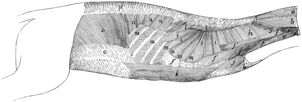
Fig. 73.—Body Muscles exposed after Removal of the Forelimb and the Muscles shown in Fig. 68.
a, M. occipitoscapularis (cut); b, M. splenius; c, M. levator scapulæ ventralis (cut); d, M. cleidomastoideus (cut); e, M. longus capitis; f, M. scalenus (f′, middle division; f″, dorsal division; f‴, ventral division; f, cervical portion); g, M. longissimus capitis; h, M. levator scapulæ (cut); i, M. serratus anterior (cut); j, M. transversus costarum; k, M. rectus abdominis (k′, its tendon); l, M. serratus posterior superior; m, M. intercostales externi; n, M. serratus posterior inferior; o, M. obliquus abdominis internus; p, lumbodorsal fascia.
Action.—Assists the serratus posterior superior.
M. transversus costarum, or sternocostalis externus (Fig. 73, j).—This is a thin muscle applied to the cranial part of the side of the thorax, covering the cranial end of the rectus abdominis (k).
Origin by tendon from the side of the sternum between the attachments of the third and sixth ribs. The muscle passes dorsocraniad to its
Insertion on the first rib and the lateral portion of its costal cartilage.
Relations.—Outer surface with the pectoralis minor (Fig. 65, o). Inner surface with the scalenus (Fig. 73, f‴), the rectus abdominis (Fig. 73, k), and the intercostales externi (Fig. 73, m). At its insertion it is united with a portion of the scalenus (Fig. 73, f‴).
Action.—Draws the sternum forward.
Mm. levatores costarum.—Small muscles having origin on the transverse processes of the thoracic vertebræ, passing caudoventrad, and becoming inserted on the angle of the rib lying immediately caudad of the origin. They are continuous with the external intercostals.
Relations.—Outer surface with the longissimus dorsi (Fig. 69, f), and craniad with the iliocostal (Fig. 69, h). Inner surface with the internal intercostals (Fig. 69, k).
Action.—Pull the ribs dorsocraniad.
Mm. intercostales externi (Fig. 73, m; Fig. 69, i).—The external intercostals are placed in the outer portion of the intercostal spaces. They are composed of bundles of fibres attached by their ends to the adjacent borders of the ribs and having in general the direction of the external oblique muscle, i.e., they pass from their cranial ends caudoventrad. They occupy the intercostal spaces between the true ribs and extend even caudad into the spaces between the false ribs. They are lacking between the ventral ends of the costal cartilages of the first six to eight ribs, so that the internal intercostals (Fig. 69, k) are here exposed. The more caudal external intercostals are more nearly craniocaudal in direction.
Relations.—Outer surface with obliquus abdominis externus (Fig. 68, p), latissimus dorsi (Fig. 68, m), serratus posterior inferior (Fig. 73, n) and superior (Fig. 73, l), serratus anterior (Fig. 73, i), scalenus (Fig. 73, f), and iliocostal (Fig. 69, h). Inner surface with the internal intercostals (Fig. 69, k).
Action.—Protractors of the ribs.
Mm. intercostales interni (Fig. 69, k).—The internal intercostals are similar to the external intercostals, beneath which they lie. Their fibres pass between the ribs at nearly right angles to those of the external intercostals and have nearly the direction of the fibres of the internal oblique. They occupy all the intercostal spaces from the first to the thirteenth ribs.
Relations.—Outer surface with the external intercostals (Fig. 69, i), and ventrad with the scalenus (Fig. 73, f), transversus costarum (Fig. 73, j), and rectus abdominis (Fig. 73, k). Inner surface with the pleura and the transversus thoracis.
Action.—Retractors of the ribs.
M. transversus thoracis (triangularis sterni; sternocostalis internus).—This represents a thoracic portion of the transversus abdominis. It consists of five or six flat muscular bands lying on the inner surface of the thoracic wall.
Origin on the lateral borders of the dorsal face of the sternum, opposite the attachments of the cartilages of the third to the eighth ribs. The six bands thus formed, each about one centimeter wide, pass laterad and are inserted into the cartilages of the ribs near their junction with the ribs, and into the fascia which covers the inner surface of the internal intercostals in this region.
Relations.—Outer surface with the internal intercostals and the cartilages of the ribs. Inner surface with the pleura.
Diaphragma (Fig. 74).—The diaphragm consists of a central so-called semilunar tendon (e) and of muscular fibres which pass radially from the body wall to the tendon. It forms a complete oblique partition between the abdominal and thoracic cavities. The dorsal end is farther caudad than the ventral.
The central tendon (e) is thin and irregularly crescent-shaped, with the convexity ventrad and the horns of the crescent prolonged as two tendinous bands (e′) which end in two triangular membranous portions (d) of the diaphragm, one on each side of the spinal column. It is pierced by an opening for the vena cava (f).
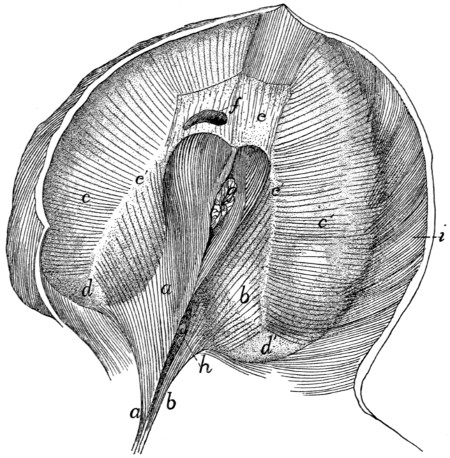
Fig. 74.—Diaphragm, Caudal Surface seen Obliquely from the Right.
a, right crus; b, left crus; c, c′, sternocostal part of diaphragm; d, d′, membranous portions of the diaphragm; e, central tendon; e′, prolongations of central tendon; f, opening for posterior vena cava; g, œsophagus; h, aorta; i, M. transversus abdominis.
The muscular portion is in two parts: (a) The vertebral portion (a, b) arises by a single tendon from the ventral surface of the second, third, and fourth lumbar centra. The tendon diverges into two, the right one (a) of which is much stronger, and from each of the two arise muscle-fibres. Each mass of fibres is one of the two crura (sing. crus) of the diaphragm. The aorta (h) enters the abdomen between the crura dorsally. The fibres of each crus diverge to be inserted into the central tendon and the dorsal continuation of its horn. The two sets of fibres unite ventrad of the opening of the aorta. Between this opening and the central tendon is another opening for the œsophagus (g); this lies entirely in that part of the diaphragm which arises from the right crus (a). The fibres are again united ventrad of the œsophageal opening.
(b) The sternocostal part (c, c′) takes origin from the xiphoid process and the last five ribs, by fleshy bundles which interdigitate with those of the transversus abdominis (i). The fibres converge to the central tendon (e). Between the crus of each side and the most dorsal of the costal fibres is the membranous interval (d) mentioned above.
4. Abdominal Muscles.
M. obliquus abdominis externus (Fig. 68, p).—A large, thin sheet of muscle covering the whole abdomen and part of the thorax ventrally.
Origin.—(a) From the last nine or ten ribs by means of as many tendons, which are interconnected to form arches that span the slips of the serratus anterior. The muscle-fibres arise from these tendons and from their intervening arches. (b) From the lumbodorsal aponeurosis common to it and the internal oblique. The cranial fibres pass nearly ventrad, the caudal fibres caudad, and the intervening fibres take an intermediate course. The fibres end in a thin aponeurosis of insertion along a curved line which passes at first caudad and then laterodorsad. The aponeurosis fibres continue in the direction of the muscle-fibres to the
Insertion into the median raphe ventrad of the sternum from the insertion of the seventh costal cartilage to the xiphoid process, into the linea alba from the sternum to the pubic tubercle, and into the tubercle and the cranial border of the pubis. Caudad of the xiphoid process the aponeurosis is closely united to the superficial layer of the internal oblique, where it forms the outer layer of the sheath of the rectus abdominis. Laterad of the pubic tubercle the tendon is perforated by the inguinal canal. In the cat neither the caudal part of the muscle nor its tendon is attached to the ilium, as it is in man and the dog, so that no Poupart’s ligament, or inguinal ligament, is formed.
Relations.—Outer surface with the cutaneus maximus, the integument, and near the origin with the latissimus dorsi (Fig. 68, m). Inner surface with the obliquus internus (Fig. 73, o), the rectus abdominis (Fig. 73, k), the intercostales externi (Fig. 73, m), a small part of the serratus posterior inferior (Fig. 73, n), and by its dorsal tendon with the longissimus dorsi (Fig. 69, f).
Action.—Constrictor of the abdomen.
M. obliquus abdominis internus (Fig. 73, o).—A thin sheet similar to the preceding but of less extent. Its fibres cross those of the external oblique nearly at right angles and lie beneath them.
Origin.—(1) Between the fourth and seventh lumbar vertebræ from the lumbar aponeurosis which is common to it and the external oblique. The lumbar aponeurosis takes origin from the lumbar spinous processes and the interspinous ligaments, and is continuous craniad with the aponeurosis of the serratus posterior inferior. Laterad it splits into three sheets, two for the above-named muscles and a third which passes to the vertebral transverse processes and forms the fascia covering the supraspinous muscles of the lumbar region. (2) By a similar aponeurosis from the ventral half of the iliac crest. (3) By fleshy fibres from the three crural arches. These are three delicate ligamentous arches which stretch from the crest of the ilium to the pubic spine. The dorsal one gives exit to the iliopsoas muscle, the middle to the femoral vessels, and the ventral to the spermatic cord. In the female the middle and ventral arches may fuse. The pillar between the dorsal and middle arches is attached to the iliopectineal eminence.
Insertion.—The fibres pass cranioventrad and end along a longitudinal line in a thin aponeurosis of insertion, which is united in the linea alba to those of the external oblique and transversus. At the caudal end of the aponeurosis all its fibres pass outside of the rectus abdominis (Fig. 73, k). At its cranial end the fibres divide into two sheets or laminæ, one of which passes outside of the rectus and the other inside. The outer lamina unites with the aponeurosis of the external oblique, while the inner lamina unites with that of the transversus. There is thus formed a sheath for the cranial part of the rectus muscle.
Relations.—Outer surface with the obliquus externus (Fig. 68, p). Inner surface with the transversus abdominis (Fig. 69, l), and by its ventral tendon with the rectus.
Action.—Compressor of the abdomen.
M. transversus abdominis (Fig. 69, l).—A thin sheet covering the whole surface of the abdomen and lying beneath the internal oblique. Its fibres are nearly transverse.
Origin.—(1) By fleshy fibres or by a thin aponeurosis from the cartilages of all the false and floating ribs, by interdigitation with the fibres of the diaphragm. (2) From the tips of all the lumbar transverse processes. (3) From the ventral border of the ilium. (4) From the dorsal and middle of the three crural arches, where it may blend partly with the internal oblique. The muscle is continuous craniad with the transversus thoracis muscle. Near the lateral border of the rectus abdominis the muscle ends in a thin aponeurosis of insertion which is continued (its fibres having the direction of the muscle-fibres) to the
Insertion in the linea alba.
Relations.—Outer surface with the internal oblique (Fig. 73, o) and the rectus abdominis (Fig. 73, k), dorsad also with the longissimus dorsi (Fig. 69, f). Inner surface with a thin fascia covering the peritoneum and ventrocaudad with the rectus (Fig. 73, k). The dorsal edge of the muscle touches the iliopsoas and longissimus dorsi; the ventral edge touches the muscle of the opposite side.
Action.—Constrictor of the abdomen.
M. rectus abdominis (Fig. 73, k).—A rather thick, flat muscle which lies near the median ventral line separated by the linea alba from its fellow of the opposite side and stretching from the pubis to the first costal cartilage. Opposite the first lumbar vertebra it is approximately four centimeters wide. It narrows at both ends.
Origin by a strong tendon from the tubercle of the pubis. The muscle passes craniad at first between the peritoneal fascia and the transversus aponeurosis, then in the sheath formed by the internal and external oblique and the transversus aponeurosis. It emerges from the sheath opposite the xiphoid process and passes ventrad of the costal cartilages. Between the third and fourth costal cartilages it ends in a thin tendon which passes beneath the transversus costarum and is
Inserted into the first costal cartilage near its middle, into the second costal cartilage near its sternal end, and into the sternum between the first and fourth cartilages.
Relations.—Outer surface of the cranial part of the muscle and its tendon with the pectoralis minor (Fig. 68, o) and the transversus costarum (Fig. 73, j); caudad the muscle is covered by the outer layer of the rectus sheath. Lateral edge with the obliquus externus (Fig. 68, p), obliquus internus (Fig. 73, o), and transversus abdominis (Fig. 69, l). Medial edge with the muscle of the opposite side. Inner surface with the internal intercostals (Fig. 69, k) and the rib cartilages; the inner layer of the rectus sheath and the peritoneum.
Action.—Retracts the ribs and sternum and compresses the abdomen.
IV. MUSCLES OF THE THORACIC LIMBS.
The muscles connecting the thoracic limbs with the rest of the body have been described.
1. Muscles of the Shoulder.
A. Muscles on the Lateral Surface of the Shoulder.
M. deltoideus.—The deltoid muscle of the cat is divided into two (or three) portions which are together equivalent to the human deltoid. These are the spinodeltoid, the acromiodeltoid, and possibly the clavobrachial, which is frequently called clavodeltoid.
M. spinodeltoideus (Fig. 75, e; Fig. 68, i).—A rather thick, flat muscle between the scapular spine and the deltoid ridge of the humerus. It forms a chord of the angle between the glenoid border of the scapula and the humerus.
Origin by short tendon-fibres from the glenoid border of somewhat more than the middle third of the spine of the scapula (Fig. 76, g), and from a tendinous raphe between the spinotrapezius (Fig. 68, j), acromiotrapezius (Fig. 68, h), and infraspinatus (Fig. 75, c). The origin may pass toward the vertebral border of the scapula onto the infraspinatus muscle (Fig. 75, c).
Insertion (Fig. 81, h) by a flat tendon upon the deltoid ridge of the humerus, nearly parallel to that of the pectoralis major.
Relations.—Outer surface with the integument and at the insertion with the acromiodeltoideus (Fig. 75, f). Inner surface with the infraspinatus (c), teres minor (Fig. 80, c), caput laterale (Fig. 75, h), and caput longum (Fig. 75, g) of the triceps muscle.
Action.—Flexes the humerus and rotates it outward.
M. acromiodeltoideus (Fig. 75, f; Fig. 68, g).—A flat muscle which overlies the distal end of the spinodeltoideus (Fig. 75, c). It connects the acromion with the humerus.
Origin (Fig. 76, d).—From the glenoid border of the acromion, and sometimes the adjacent metacromion as far as the tip.
Insertion.—Mostly upon the outer surface of the spinodeltoideus (Fig. 75, c). The outer fibres are continued to the bone, especially at the lateral border of the muscle, and are inserted along a line ventrad of the line of insertion of the spinodeltoideus, and extending farther distally (Fig. 81, g). Some of the outer fibres pass into the brachialis (Fig. 80, h).
Relations.—Outer surface with the integument and the clavobrachial (Fig. 68, e). Inner surface with the infraspinatus (Fig. 75, c), teres minor (Fig. 80, c), spinodeltoid (Fig. 75, e), and caput laterale of the triceps (Fig. 75, h).
Action.—Like that of the spinodeltoid.
M. clavobrachialis (Fig. 65, k; Fig. 68, e).—A flat, triangular muscle on the cranial surface of the shoulder, forming a direct continuation of the clavotrapezius (Fig. 68, d). These two are frequently described as constituting a single muscle, the cephalohumeral or cephalobrachial. (The clavobrachial is frequently given the name clavodeltoid; as its homology with the human clavodeltoid appears doubtful, it seems well to use the name clavobrachial, as proposed by Clasen.)
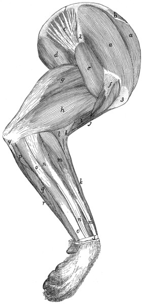
Fig. 75.—Muscles on the Lateral Surface of the Arm.
a, M. supraspinatus; b, part of insertion of M. rhomboideus; c, M. infraspinatus; d, M. teres major; e, M. spinodeltoideus; f, M. acromiodeltoideus; g, caput longum of M. triceps brachii; h, caput laterale of M. triceps brachii; i, M. brachialis; j, M. biceps; k, M. brachioradialis; l, M. extensor carpi radialis longus; m, M. extensor communis digitorum (m′, tendon of same); n, M. extensor lateralis digitorum; (n′, tendon of same); o, M. extensor carpi ulnaris (o′, tendon of same); p, M. extensor indicis proprius; q, fifth head of M. flexor profundus digitorum; r, M. flexor carpi ulnaris. 1, vertebral border of the scapula; 2, spine of the scapula; 3, greater tuberosity of the humerus; 4, olecranon; 5, transverse ligament of the wrist.
Origin.—Its superficial fibres are continuations of the clavotrapezius (Fig. 68, d); other fibres have origin from the clavicle and from a raphe laterad of the clavicle which is common to this muscle and the clavotrapezius. It passes distad along the cranial surface of the arm, growing narrower as it approaches the convexity of the elbow.
Insertion.—Eight to ten millimeters from the ulna it joins the brachialis muscle (Fig. 79, i) to be inserted with it by a flat tendon (Fig. 79, i; Fig. 87, c) upon a rough area on the medial surface of the ulna just distad of the semilunar notch, and about midway between the dorsal and ventral borders.
Relations.—Outer surface with the integument. Inner surface with the pectoralis major (Fig. 65, l), biceps (Fig. 77, g), lateral head of the triceps (Fig. 75, h), acromiodeltoid (Fig. 75, f), and brachialis (Fig. 80, h). Medial border with the pectoantibrachialis (Fig. 65, m). Lateral border free except near the insertion, where it is in relation with the brachialis.
Action.—Flexor of the antibrachium.
M. supraspinatus (Fig. 75, a) occupies the whole of the supraspinatus fossa of the scapula. It is covered by strong fascia which stretches from the free edge of the spine to the coracoid border of the scapula and to the coracoid half of its vertebral border.
Origin (Fig. 76, b) by fleshy fibres from the whole surface of the supraspinatus fossa, from the above-mentioned fascia, and from the subscapularis (Fig. 77, a) craniad of the coracoid border of the scapula.
Insertion (Fig. 83, a).—It passes over the capsule of the shoulder-joint, to which it is closely attached, and is inserted into the free border of the great tuberosity ventrad (or proximad) of the fossa for the infraspinatus (Fig. 83, c).
Relations.—Outer surface with the spinotrapezius (Fig. 68, j), acromiotrapezius (Fig. 68, h), levator scapulæ ventralis (Fig. 68, f), clavotrapezius (Fig. 68, d), and cleidomastoid (Fig. 65, h). Inner surface with the scapula. The distal end of the coracoid border is closely related to the pectoralis minor (Fig. 79, f). The glenoid border is related to the origin of the deltoidei.
Action.—Extends the humerus after it has been flexed on the scapula.
M. infraspinatus (Fig. 75, c).—This fills the infraspinatus fossa, its fibres converging to the insertion on the great tuberosity of the humerus.
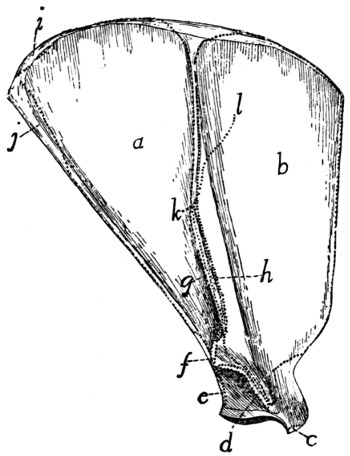
Fig. 76.—Lateral Surface of the Scapula, with the Areas of Attachment of Muscles.
a, M. infraspinatus; b, M. supraspinatus; c, origin of M. biceps; d, M. acromiodeltoideus; e, M. teres minor; f, M. levator scapulæ ventralis; g, M. spinodeltoideus; h, M. acromiotrapezius; i, M. rhomboideus; j, M. teres major; k-l, line of insertion of M. spinotrapezius.
Origin (Fig. 76, a).—By fleshy fibres from the whole infraspinatus fossa, and by a raphe between it and the teres major (Fig. 75, d), and sometimes by fibres from the teres minor (Fig. 80, c), triceps, spinodeltoideus (Fig. 75, e), spinotrapezius (Fig. 68, j), and subscapularis (Fig. 77, a).
Insertion (Fig. 83, c).—By a flat tendon which passes over the capsule of the joint, into the ventral half of the infraspinatus fossa on the great tuberosity of the humerus. (The dorsal half of the fossa is covered by a synovial bursa.)
Relations.—Outer surface with the spinotrapezius (Fig. 68, j), spinodeltoid (Fig. 68, i), acromiodeltoid (Fig. 68, g), teres major (Fig. 75, d), and levator scapulæ ventralis (Fig. 68, f). Inner surface with the scapula. Glenoid border with the teres minor (Fig. 80, c), the teres major (Fig. 75, d), and the long head of the triceps (Fig. 75, g).
Action.—Rotates the humerus outward.
M. teres minor (Fig. 80, c).—A small muscle from the glenoid border of the scapula to the proximal end of the humerus.
Origin (Fig. 76, e) by a sheet of tendinous fibres from the glenoid border of the scapula beginning about one-fifth the length of the border from the glenoid fossa and extending to its middle. It is often attached to the infraspinatus (Fig. 80, b) and the caput longum of the triceps (Fig. 75, g).
Insertion (Fig. 83, d) by a short tendon into the tubercle just distad of the infraspinatus fossa on the great tuberosity of the humerus.
Relations.—Outer surface with the spinodeltoideus (Fig. 75, e), acromiodeltoideus (Fig. 75, f), and the infraspinatus (Fig. 80, b). Inner surface with the lateral (Fig. 75, h) and long (Fig. 75, g) heads of the triceps and the capsule of the joint.
Action.—Assists the infraspinatus to rotate the humerus outward.
B. Muscles on the Medial Surface of the Shoulder.
(Fig. 77.)
M. subscapularis (Fig. 77, a).—A triangular mass from the subscapular fossa to the lesser tuberosity of the humerus.
Origin (Fig. 78, a).—From the whole subscapular fossa except along the fusiform area for the attachment of the levator scapulæ and the serratus anterior near the vertebral border, and except over a quadrangular area about one centimeter long near the glenoid angle. The origin is by fleshy fibres directly from the periosteum except along two or three lines marked by oblique ridges. To these lines are attached tendinous fibres. At the glenoid border the area of origin sometimes occupies part or the whole of the surface of the fossa for the origin of the teres major, so that the teres arises from the fascia on the surface of the subscapularis. At the coracoid border some of the fibres may take origin from the adjacent fascia of the supraspinatus (Fig. 77, d).
Insertion (Fig. 82, b).—The fibres converge to the glenoid border, and the insertion is by a strong, flat tendon into the dorsal border of the lesser tuberosity of the humerus.
Fig. 77.—Muscles on the Medial Side of the Arm.
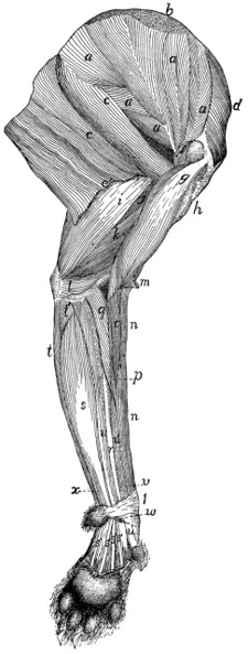
M. epitrochlearis, M. clavobrachialis, and the breast-muscles have been removed. a, M. subscapularis; b, insertion of M. levator scapulæ and of M. serratus anterior; c, M. teres major; d, M. supraspinatus; e, M. latissimus dorsi (e′, cut edge of that part which becomes united with the bicipital arch); f, M. coracobrachialis; g, M. biceps brachii (the capsule of the joint has been laid open to show its tendon); h, cut insertions of pectoralis muscles; i, caput longum of M. triceps brachii; j, long portion of caput mediale of M. triceps brachii; k, intermediate portion of caput mediale of M. triceps brachii; l, short portion of caput mediale of M. triceps brachii; m, cut end of M. clavobrachialis; n, M. brachioradialis; o, M. extensor carpi radialis longus; p, M. extensor carpi radialis brevis; q, M. pronator teres; r, M. flexor carpi radialis; s, M. palmaris longus (s′, its tendons); t, M. flexor carpi ulnaris (t, ulnar head; t′, humeral head); u, third head of M. flexor profundus digitorum; u′, fifth head of M. flexor profundus digitorum; u″, one of the tendons of M. flexor profundus digitorum; v, M. pronator quadratus; w, M. abductor brevis pollicis; x, ulnar part of M. flexor sublimis digitorum. 1, transverse ligament of the wrist.
Relations.—Lateral surface with the scapula and the capsule of the shoulder-joint. Medial surface with the levator scapulæ (Fig. 73, h), serratus anterior (Fig. 73, i), part of the scalenus (Fig. 73, f), the transversus costarum (Fig. 73, j), and the coracobrachialis (Fig. 77, f). Cranial border with the supraspinatus (Fig. 77, d). Caudal border with the teres major (Fig. 77, c) and infraspinatus (Fig. 75, c).
Action.—Pulls the humerus inward (mediad).
M. teres major (Fig. 75, d; Fig. 77, c).—A thick muscle, triangular in cross-section, lying parallel with the glenoid border of the scapula.
Origin (Fig. 76, j; Fig. 78, b) from the vertebral one-third of the glenoid border of the scapula, and from fascia covering the subscapularis (Fig. 77, a) and the infraspinatus (Fig. 75, c) over a small area near the glenovertebral angle of the scapula. It may touch the insertion of the rhomboideus.
Insertion (Fig. 82, f) by a tendon common to it and the latissimus dorsi (Fig. 79, d′), as already described.
Relations.—Outer surface with the caput longum (Fig. 75, g) and the long portion (Fig. 80, f) of the caput mediale of the triceps, the latissimus dorsi (Fig. 77, e), and the cutaneus maximus. Inner surface with the serratus anterior (Fig. 73, i), the scalenus (Fig. 73, f), the transversus costarum (Fig. 73, j), and the biceps (Fig. 77, g). Dorsal border with the subscapularis (Fig. 77, a) and infraspinatus (Fig. 75, c).
Action.—Rotates the humerus inward and flexes it in opposition to the infraspinatus, teres minor, and the deltoidei.
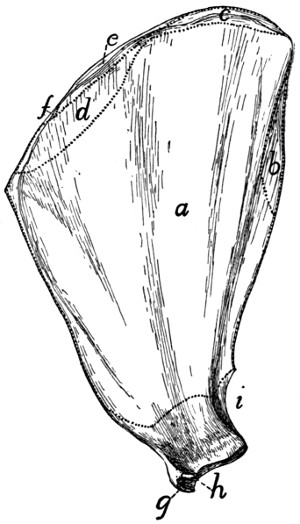
Fig. 78.—Medial Surface of the Scapula with the Areas of Attachment of Muscles.
a, M. subscapularis; b, M. teres major; c, M. serratus anterior; d, M. levator scapulæ; e, M. rhomboideus; f, M. occipitoscapularis; g, M. biceps; h, M. coracobrachialis; i, caput longum of M. triceps.
2. Muscles of the Brachium or Upper Arm.
M. clavobrachialis and the pectoralis group (Fig. 65, page 109) lie partly in this region; they have been described.
M. coracobrachialis (Fig. 77, f; Fig. 79, c).—A very short muscle covering the inner (medial) surface of the capsule of the shoulder-joint. It extends from the coracoid process to the proximal end of the humerus.
Origin (Fig. 78, h).—By a round tendon from the tip of the coracoid process.
Insertion (Fig. 82, e) usually by fleshy fibres on an area five to eight millimeters long and about half as wide, which lies on the medial side of the humerus parallel with the ridge which runs from the dorsal end of the lesser tuberosity to the shaft and close to it. The proximal end of the area is about one centimeter from the proximal end of the head of the humerus.
Relations.—Medial surface with the biceps (Fig. 77, g), pectoralis minor (Fig. 65, o), and teres major (Fig. 77, c). Lateral surface with the capsule of the shoulder-joint. Dorsal border with the subscapularis (Fig. 77, a) and the long portion of the caput mediale of the triceps (Fig. 77, j).
Action.—Adducts the humerus.
The part of the coracobrachialis just described is known as the short head. A long head is sometimes found. It is a conical bundle of fibres of varying size, which arises from the tendon of origin of the short head. It passes distad into a long and extremely slender tendon, the insertion of which varies greatly in different individuals. It is commonly on the humerus in the region of the supracondyloid foramen.
M. epitrochlearis or extensor antibrachii longus (Fig. 65, r).—A thin, flat muscle on the inner or medial side of the brachium, from the lateral surface of the latissimus dorsi (Fig. 65, q) to the olecranon process of the ulna.
Origin from the lateral or outer surface of the ventral border of the latissimus dorsi (Fig. 65, q) near the insertion of the cutaneus maximus. Fibres are often attached to the teres major and the pectoralis minor.
Insertion by a flat tendon which is closely connected with that of the pectoantibrachialis (Fig. 65, m) and is continuous with the general antibrachial fascia, into the caudal border of the cutaneous (dorsal) surface of the olecranon process of the ulna.
Relations.—Outer (medial) surface with the integument, the latissimus dorsi (Fig. 65, q), and the cutaneus maximus. Inner (lateral) surface with the biceps (Fig. 65, t), caput longum (Fig. 65, s), and caput mediale (Fig. 77, j, k) of the triceps.
Action.—Extends the antibrachium and tends to supinate the hand by rotating the ulna.
M. biceps brachii (Fig. 77, g; Fig. 65, t).—A thick, fusiform muscle lying on the front (ventral) surface of the humerus.
Origin (Fig. 76, c) by a strong, round tendon from the bicipital tubercle of the glenoid angle of the scapula, at its coracoid margin. (In man there is a second head—coracoid head—from the coracoid process.) The tendon passes through the capsule of the joint and then along the bicipital groove, which is converted into a canal by a strong ligament.
Insertion by a rounded tendon (Fig. 87, b) on the bicipital tuberosity of the radius (Fig. 86, k).
Relations.—The tendon of origin passes through the capsule of the joint, which is covered by a part of the pectoralis minor. The muscle is then spanned for the middle part of its course by the bicipital arch (Fig. 65, t′), a description of which is given below. Farther distad it is covered by the clavobrachial (Fig. 65, k) and epitrochlearis (Fig. 65, r). At its distal end (Fig. 79, l′) the muscle passes between the pronator teres on the radial side and the conjoined tendon of the brachialis and clavobrachial (Fig. 79, i′) on the other. The biceps lies throughout most of its length on the ventral surface of the humerus, and touches along the medial border of its inner surface the coracobrachialis (Fig. 77, f), the teres major, (Fig. 77, c), the intermediate portion of the caput mediale of the triceps (Fig. 77, k), and the short portion (Fig. 77, l) of the same. Its lateral border touches the pectoralis minor (Fig. 65, o) and the brachialis (Fig. 79, i).
Action.—Flexes the forearm, and tends to supinate the hand.
The bicipital arch (Fig. 65, t′) is a tendinous arch formed over the biceps muscle. Its outer or lateral pillar is formed by the tendon of the pectoralis minor (o), to which may be added part of the latissimus (q) tendon. Its inner or medial pillar is formed by the conjoined tendon of the teres major and latissimus dorsi (Fig. 79, d′), while the xiphihumeralis (Fig. 65, p), cutaneus maximus (Fig. 62, b), and epitrochlearis (Fig. 65, r) are connected with one or the other pillar or with the muscles composing them. The caudal portion of the deep layer of the pectoralis major (Fig. 65, l) may be connected with the inner pillar.
M. brachialis (Fig. 79, i; Fig. 75, i).—From the lateral surface of the humerus to the ulna.
Origin (Fig. 83, g, g′) from a long V-shaped line two to four millimeters wide on the lateral surface of the humerus. The apex of the V is just distad of the teres minor tubercle (d). Its dorsal limb (g′) extends along the lateral supracondyloid ridge to a point opposite the proximal margin of the supracondyloid foramen; its ventral limb (g) extends in the direction of the deltoid ridge to the middle of the bone. No muscle-fibres take origin between the two limbs. The fibres converge and end in a flat tendon which joins the tendon of the clavobrachial (Fig. 79, k).
Insertion (Fig. 87, c).—The dorsal portion of the depressed rough area on the lateral surface of the ulna just distad of the semilunar notch.
Relations.—Outer surface with the acromiodeltoid (Fig. 75, f), the caput laterale of the triceps (Fig. 75, h), the brachioradialis (Fig. 75, k), and the proximal end of the extensor carpi radialis longus (Fig. 75, l). Inner surface with the humerus. Medial edge with the pectoralis major (Fig. 65, l) and the biceps (Fig. 77, g).
Action.—Flexor of the antibrachium.
M. triceps brachii.—The triceps muscle of the cat, like that of man, is divisible (as its name indicates) into three main portions. These are a lateral portion (caput laterale), an intermediate or long portion (caput longum), and a medial portion (caput mediale). The first two correspond to the similarly named heads in man; the medial portion falls into a number of subdivisions whose homologues are uncertain.
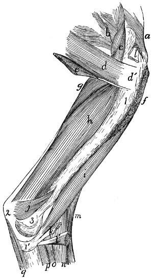
Fig. 79.—Deep Muscles on the Medial Side of the Humerus.
a, M. supraspinatus; b, M. subscapularis; c, M. coracobrachialis; d, M. teres major (d′, its tendon); e, part of M. latissimus dorsi joining the teres major; f, cut end of M. pectoralis minor; g, long part of caput mediale of M. triceps brachii; h, intermediate part of caput mediale of M. triceps; i, M. brachialis (i′, its tendon); j, short part of caput mediale of M. triceps; k, cut end of M. clavobrachialis; l, l′, cut ends of M. biceps; m, M. brachioradialis; n, M. extensor carpi radialis longus; o, M. extensor carpi radialis brevis; p, fifth head of M. flexor profundus digitorum; q, first head of M. flexor profundus digitorum. 1, humerus; 2, olecranon; 3, medial epicondyle of humerus.
Caput laterale (anconeus lateralis) (Fig. 75, h; Fig. 68, l).—The lateral portion is a flat muscle (most of it subcutaneous) on the lateral side of the brachium, connecting the proximal end of the humerus with the olecranon process of the ulna.
Origin (Fig. 81, h′).—By a flat tendon from the proximal portion of the deltoid ridge and the distal border of the teres minor tubercle.
Insertion (Fig. 86, c) by a thin, flat tendon into the lateral border of the dorsal surface of the ulna between a point opposite the distal margin of the semilunar notch and the proximal end of the olecranon.
Relations.—Lateral surface with the integument, and at the proximal end with the spinodeltoid and acromiodeltoid (Fig. 75, e and f). Medial surface with the brachialis (Fig. 75, i), the caput longum (g), the caput mediale, the brachioradialis (k), the anconeus (Fig. 80, l), and the origins of the extensor carpi radialis longus (Fig. 75, l), the extensor communis digitorum (m), and the extensor digitorum lateralis (n).
Caput longum (anconeus longus) (Fig. 75, g; Fig. 68, k; Fig. 77, i).—A prismatic, fusiform mass connecting the scapula with the olecranon.
Origin (Fig. 78, i) by a thick, flat tendon from a triangular area one or two centimeters long at the glenoid end of the scapula on the glenoid border. The base of the area is about one millimeter from the margin of the glenoid fossa.
Insertion (Fig. 86, b).—The muscle ends in a thick tendon which passes over the bifurcated ventral end of the olecranon and is inserted upon the rounded tuberosity which forms the dorsal angle of the olecranon.
Relations.—Lateral surface with the caput laterale (Fig. 75, h), a portion of the caput mediale (Fig. 77, j and k), the spinodeltoid (Fig. 75, e), the infraspinatus (Fig. 75, c), and the integument. Medial surface with the epitrochlearis (Fig. 65, r) and the conjoined portions of the latissimus dorsi and teres major (Fig. 79, d′). Ventral border with the caput mediale (Fig. 77, j and k).
Caput mediale.—The medial head consists of three portions (Fig. 79, g, h, j).
(1) The long portion (anconeus posterior) (Fig. 77, j; Fig. 79, g; Fig. 80, f).
Origin (Fig. 83, e) from a triangular area on the dorsal surface of the humeral shaft. The base of the area is against the articular head, and its apex about one-sixth the length of the humerus from the head. It is between the coracobrachialis and the lateral head of the triceps.
Fig. 80.—Deep Muscles on the Lateral Surface of the Humerus.
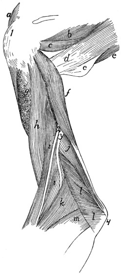
a, part of M. supraspinatus; b, part of M. infraspinatus; c, M. teres minor; d, M. teres major; e, part of M. latissimus dorsi joining M. teres major; f, long part of caput mediale of M. triceps brachii; g, cut insertion of M. acromiodeltoideus; h, M. brachialis; i, M. brachioradialis; j, intermediate part of caput mediale of M. triceps brachii; k, M. extensor carpi radialis longus; l, M. anconeus; m, M. extensor communis digitorum. 1, great tuberosity of humerus; 2, 3, superficial radial and dorsal interosseous branches, respectively, of the radial nerve.
Insertion (Fig. 86, a).—The muscle ends in a long, slender tendon which passes through the furrow on the ventral angle of the olecranon and is inserted into an oblique ridge which forms the dorsal limit of the furrow. The tendon is separated from the floor of the furrow by a synovial bursa.
(2) The intermediate portion (anconeus internus) (Fig. 77, k; Fig. 79, h; Fig. 80, j).
Origin (Fig. 82, g) by fleshy fibres from a triangular area proximad of the middle of the dorsomedial surface of the humerus. The length of the area equals about one-fourth the length of the bone. Its apex points proximad and is almost continuous with the apex of the area of origin of the long portion.
Insertion (Fig. 87, i) by short tendon-fibres into the medial border of the ventral and proximal surfaces of the olecranon. It is connected with the tendon of the long portion.
(3) Short portion (Fig. 79, j; Fig. 77, l).
Origin (Fig. 82, h) from the outer surface of the bony bar which encloses the supracondyloid foramen of the humerus, as far as the medial epicondyle.
Insertion (Fig. 87, e) by fleshy fibres into the medial border of the olecranon distad of the tuberosity for the insertion of the caput longum.
Relations of the caput mediale.—Lateral surface with the caput laterale (Fig. 75, h), the humerus, and the anconeus (Fig. 80, l). Dorsal surface with the caput longum (Fig. 77, i), the epitrochlearis (Fig. 65, r), and the teres major (Fig. 79, d). Ventral surface with the biceps (Fig. 77, g).
Action of the Triceps.—The entire triceps group forms a powerful extensor of the forearm. The short portion of the medial head tends also to rotate the arm outward, so as to supinate the hand.
M. anconeus (Fig. 80, l).—A triangular muscle on the outside of the elbow-joint from the humerus to the ulna.
Origin (Fig. 83, i).—An irregularly triangular area at the distal end of the dorsal surface of the humerus. The area of origin is limited laterally by the sharp lateral supracondyloid ridge. It sometimes extends onto the lateral epicondyle.
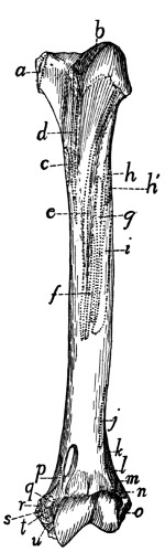
Fig. 81.—Ventral Surface of Humerus, with the Areas of Attachment of Muscles.
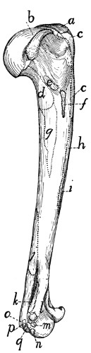
Fig. 82.—Medial Side of Humerus, with the Areas of Attachment of Muscles.
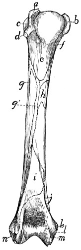
Fig. 83.—Dorsal Surface of the Left Humerus, with the Areas of Attachment of Muscles.
Fig. 81.—a, M. subscapularis; b, M. supraspinatus; c, Mm. teres major and latissimus dorsi; d, M. pectoralis minor; e, deep layer of M. pectoralis major; f, superficial layer of M. pectoralis major; g, M. acromiodeltoideus; h, M. spinodeltoideus; h′, caput laterale of M. triceps; i, M. brachialis; j, second portion of M. brachialis; k, M. extensor carpi radialis longus; l, M. extensor carpi radialis brevis; m. M. extensor communis digitorum; n, M. extensor lateralis digitorum; o, M. extensor carpi ulnaris; p, short portion of caput mediale of M. triceps; q, M. pronator teres; r, M. palmaris longus; s, third and fourth parts of M. flexor profundus digitorum; t, M. flexor carpi radialis; u, second head of M. flexor profundus digitorum.
Fig. 82.—a, M. supraspinatus; b, M. subscapularis; c, M. pectoralis minor; d, long portion of caput mediale of M. triceps; e, M. coracobrachialis; f, Mm. teres major and latissimus dorsi; g, intermediate portion of caput mediale of M. triceps; h, deep layer of M. pectoralis major; i, superficial layer of M. pectoralis major; j, M. anconeus; k, short portion of caput mediale of M. triceps; l, M. pronator teres; m, M. flexor carpi radialis; n, second head of M. flexor profundus digitorum; o, third and fourth heads of M. flexor profundus digitorum; p, M. palmaris longus; q, M. flexor carpi ulnaris.
Fig. 83.—a, M. supraspinatus; b, M. subscapularis; c, M. infraspinatus; d, M. teres minor; e, long portion of caput mediale of M. triceps; f, M. coracobrachialis; g, g′, M. brachialis; h, intermediate portion of caput mediale of M. triceps; i, M. anconeus; j, short portion of medial head of M. triceps; k, M. pronator teres; l, second and third heads of M. flexor profundus digitorum; m, M. palmaris longus; n, M. extensor carpi ulnaris.
Insertion (Fig. 86, d).—The lateral surface of the ulna from the distal margin of the semilunar notch to the proximal end of the olecranon. The muscle covers the capsule of the joint and is closely adherent to it.
Relations.—Outer (dorsal) surface with the caput laterale (Fig. 75, h) and the intermediate portion (Fig. 80, j) of the caput mediale. Inner surface with the bones and the capsule of the joint.
Action.—Keeps the capsule tense and probably rotates the ulna slightly so as to pronate the hand.
3. Muscles of the Antibrachium or Forearm.
Fascia of the Forearm.
—The muscles of the forearm are not in contact with the skin, but are covered by the strong antibrachial fascia. This consists of two layers. The superficial one is a continuation of the general subcutaneous fascia of the arm; it covers the forearm as a continuous sheet which becomes thinner distad, and is finally lost near the wrist. The deep layer forms a dense, tendinous sheet which is closely applied to the muscles. It begins partly as a continuation of the general fascia of the arm, partly from the tendons of the triceps and epitrochlearis muscles. The sheet is attached to the dorsal surface of the ulna, between the extensor carpi ulnaris and the first head of the flexor profundus digitorum. It surrounds the forearm as a continuous sheath. On the lateral surface it dips between the extensor muscles, becoming closely attached to them and forming a partial sheath for each; it is attached with special firmness to their tendons. On the medial side of the arm the fascia passes smoothly over the flexors without dipping between them; it is continuous, however, with the border of the pronator teres, and distad of this muscle it is firmly attached to the radius.
At the wrist the fascia becomes attached to the longitudinal ridges on the dorsal surface of the head of the radius, bridging thus the intervening grooves and holding the tendons in place between the ridges. The fascia then continues to form the dorsal fascia of the hand. On the ventral side of the wrist the fascia becomes thickened to form a strong transverse ligament (Fig. 77, 1), which is attached at its radial and ulnar ends to the carpus, and which holds in place the tendons of the flexor muscles. Distad of the transverse ligament the fascia spreads out in the palm of the hand, and becomes united with the fibrous pad which is situated here. On the fingers the fascia becomes attached to the phalanges, forming tendinous sheaths, the so-called vaginal ligaments, for the flexor muscles. Near the base and the head of the first phalanx strong, narrow, ring-like bands, the annular ligaments (Fig. 88, 2), are formed in these sheaths; these surround and bind down the tendons of the flexor muscles.
A. Muscles on the Ulnar and Dorsal Side of the Forearm (extensors and supinators)
(Fig. 75, p. 158).
M. brachioradialis or supinator longus (Fig. 75, k; Fig. 77, n).—This is a ribbon-like muscle lying on the lateral side of the brachium and the ventrolateral border of the antibrachium just beneath the superficial fascia. It connects the humerus and the radius.
Origin.—By a thin tendon from about the middle fifth of the dorsal border of the humerus, or sometimes proximad of the middle. The muscle curves over the outer surface of the brachialis (Fig. 75, i), to which it is closely applied, and runs along the lateral border of the antibrachium to its insertion.
Insertion by a short tendon into the ridge which forms the ventral limit of the groove on the outer surface of the styloid process of the radius and upon the adjacent ligaments.
Relations.—Outer surface with the integument, and at the proximal end with the caput laterale of the triceps (Fig. 75, h). Inner surface with the brachialis (i), the extensor carpi radialis longus (l) and brevis, and the extensor communis digitorum (m).
Action.—Supinator of the hand.
M. extensor carpi radialis longus (Fig. 75, l; Fig. 77, o).—This lies along the radial side of the forearm, and connects the humerus and the second metacarpal.
Origin (Fig. 81, k) from the middle portion of the lateral supracondyloid ridge between the origin of the brachialis (j) and the anconeus. About the middle of the forearm the muscle ends in a slender tendon (Fig. 84, e) which passes through the groove on the radial side of the dorsal surface of the distal end of the radius.
Insertion (Fig. 84, c).—The tendon passes across the carpus and is inserted onto the dorsal surface of the base of the second metacarpal on the radial side just distad of the groove for the radial artery.
Relations.—Outer surface at the proximal end with the caput laterale of the triceps (Fig. 75, h), farther distad with the integument and the brachioradialis (k); the distal tendon with the extensor pollicis brevis (Fig. 84, d). Radial surface with the brachialis (Fig. 75, i) and the integument. Ulnar surface with the extensor carpi radialis brevis (Fig. 77, p), and near the proximal end with the extensor communis digitorum (Fig. 75, m).
Action.—Extensor of the hand.
M. extensor carpi radialis brevis (Fig. 77, p).—A slender muscle from the humerus to the third metacarpal.
Origin (Fig. 81, l) from the distal part of the lateral supracondyloid ridge distad of the extensor longus. Near the carpus the muscle ends in a slender tendon which passes through the groove with the extensor longus tendon. The tendon then diverges from the extensor longus and has its
Insertion (Fig. 84, f) into the radial side of the dorsal surface of the base of the third metacarpal.
Relations.—Outer surface with the extensor longus (Fig. 77, o), the integument, and distad with the extensor pollicis brevis (Fig. 84, d). Radial surface with the extensor longus. Ulnar surface with the extensor communis digitorum (Fig. 75, m) and the extensor brevis pollicis. Inner surface with the pronator teres (Fig. 77, q), supinator (Fig. 85, b), and extensor brevis pollicis (Fig. 85, a).
Action.—Extensor of the hand.
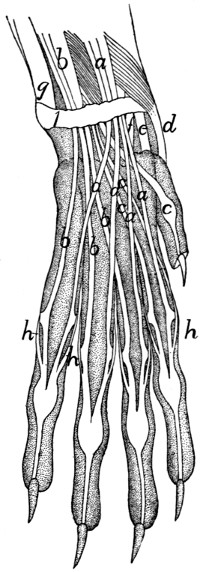
Fig. 84.—Arrangement of the Tendons on the Back of the Hand.
a, M. extensor communis digitorum; b, M. extensor lateralis digitorum; c, M. extensor indicis; d, M. extensor brevis pollicis; e, M. extensor carpi radialis longus; f, M. extensor carpi radialis brevis; g, M. extensor carpi ulnaris; h, tendons of the interossei. 1, transverse ligament of the wrist.
M. extensor digitorum communis (Fig. 75, m).—A slender muscle from the lateral supracondyloid ridge of the humerus to the phalanges of the second, third, fourth, and fifth digits.
Origin (Fig. 81, m).—The distal surface of the lateral supracondyloid ridge dorsad of the origin of the extensor carpi radialis brevis.
At the junction of the middle and distal thirds of the radius the muscle passes into a large, flat tendon (Fig. 75, m′) which passes through the groove on the middle of the dorsal surface of the distal end of the radius. The groove is converted into a canal by a strong transverse ligament. The tendon divides near the groove into four portions (Fig. 84, a) which diverge and pass to the four ulnar digits.
Insertions.—Each division of the tendon as it passes over the dorsal surface of the first phalanx has its border connected by strong fascia to the dorsal surface of the phalanx. It finally passes through the groove at the distal end of the first phalanx and is inserted mostly into the base of the second phalanx; a portion continues distad to be inserted into the third phalanx.
Relations.—Outer surface with the integument and at the origin with the caput laterale (Fig. 75, h) of the triceps. Radial surface with the extensor longus (l) and extensor brevis. Ulnar surface with the extensor digitorum lateralis (n). Inner surface with the supinator (Fig. 85, b) and the extensor pollicis brevis (Fig. 85, a).
Action.—Extensor of the four ulnar digits.
M. extensor digitorum lateralis (Fig. 75, n). (This corresponds in position to the extensor digiti quinti proprius of man.)
Origin (Fig. 81, n) from the lateral supracondyloid ridge of the humerus distad of the origin of the extensor communis.
The muscle passes along the ulnar border and dorsal surface of the forearm between the extensor communis (Fig. 75, m) and the extensor carpi ulnaris (o). Near the carpus it ends in a tendon (n′), which divides immediately into three or four (Fig. 84, b). These pass through the groove on the ulnar side of the dorsal surface of the radius and diverge to the digits lying beneath the extensor communis tendon.
Insertion (Fig. 84, b).—The three tendons on the ulnar side join the ulnar sides of the tendons of the extensor communis (a) at their insertions. The radial of the four tendons (not always present) joins the tendon of the extensor indicis (c) and may also give a branch to the radial side of the base of the first phalanx of the third digit.
Relations.—Outer surface with the integument. Radial border with the extensor communis digitorum (m); ulnar border with the extensor carpi ulnaris (o). Inner surface with the supinator (Fig. 85, b) and the extensor brevis pollicis (Fig. 85, a); distad with the extensor indicis (Fig. 85, c).
Action.—Extensor of the four ulnar digits.
M. extensor carpi ulnaris (Fig. 75, o).
Origin.—(1) By a short, broad tendon from the distal portion of the lateral epicondyle of the humerus (Fig. 81, o) distad of the origin of the extensor lateralis, and (2) by a smaller tendon from the ulna at the dorsal tip of the semilunar notch (Fig. 86, e).
The muscle passes along the ulnar side of the forearm dorsad of the extensor digitorum lateralis (n). It ends near the junction of the middle and distal thirds of the forearm in a large, flat tendon (o′) which is closely adherent to the ligaments of the wrist and which passes over the distal end of the ulna to its
Insertion into the tubercle on the ulnar side of the base of the fifth metacarpal (Fig. 84, g).
Relations.—Outer surface with the integument. Radial (or ventral) border with the extensor digitorum lateralis (Fig. 75, n); ulnar (or dorsal) border with the flexor profundus digitorum (q). Inner surface with the radius, the extensor indicis (Fig. 85, c), and the extensor pollicis brevis (Fig. 85, a).
Action.—Indicated by its name.
M. extensor indicis (proprius) (Fig. 85, c; Fig. 75, p) (includes extensor pollicis longus also).
Origin (Fig. 86, f) by short, fleshy fibres from the lateral surface of the ulna between the semilunar notch and the junction of the third and fourth (distal) fourths.
The muscle-fibres pass obliquely distad and toward the radial side and join the tendon (Fig. 85, c′), which runs nearly the whole length of the radial surface.
Insertion (Fig. 84, c).—The tendon passes through the groove on the ulnar side of the dorsal surface of the base of the radius and divides into two. Both of these usually go to the base of the second phalanx of the second digit, but one may pass to the pollex, or there may be three divisions going to the first three digits.
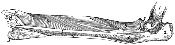
Fig. 85.—Deep Muscles on the Extensor Surface of the Radius and Ulna.
a, M. extensor brevis pollicis (a′, its tendon); b, M. supinator; c, M. extensor indicis (c′, its tendon); d, tendon of M. extensor carpi radialis brevis; e, tendon of M. extensor carpi radialis longus. 1, humerus; 2, olecranon; 3, styloid process of ulna; 4, distal end of radius; 5, head of radius.
Relations.—Outer surface with the integument, the extensor carpi ulnaris (Fig. 75, o), and the extensor digitorum lateralis (Fig. 75, n). Dorsal border with the flexor profundus digitorum (Fig. 75, q). Inner surface with the extensor brevis pollicis (Fig. 85, a).
Action as indicated by the name.
M. supinator (Fig. 85, b).—A flat muscle wrapped spirally about the proximal end of the radius.
Origin by a short, strong tendon from the lateral side of the annular ligament of the radius and by tendinous fibres from the radial collateral ligament passing from the humerus to the radius. From the tendon of origin the fibres diverge, passing toward the radial side and distad.
Insertion (Fig. 86, g).—The dorsal and part of the ventral surface of the proximal two-fifths of the radius to within five millimeters of the articular face of the head.
Relations.—Outer surface with the extensor carpi radialis brevis, extensor communis digitorum (Fig. 75, m), and extensor lateralis digitorum (Fig. 75, n). Radial border with the pronator teres (Fig. 77, q). Inner surface with the radius.
Action.—Supinator of the hand.
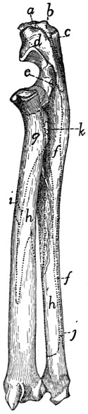
Fig. 86.—Lateral or Extensor Surface of Radius and Ulna, with the Areas of Attachment of Muscles.
a, long portion of caput mediale of M. triceps; b, caput longum of M. triceps; c, caput laterale of M. triceps; d, M. anconeus; e, M. extensor carpi ulnaris; f, M. extensor indicis; g, M. supinator; h, M. extensor brevis pollicis; i, M. pronator teres; j, M. flexor profundus digitorum; k, M. biceps.
M. extensor brevis pollicis or extensor ossis metacarpi pollicis (Fig. 85, a) (includes M. abductor longus pollicis of man).
Origin (Fig. 86, h) by fleshy fibres from the ventral half of the lateral surface of the shaft of the ulna from the semilunar notch to the styloid process; from the ulnar half of the dorsal surface of the radius from the bicipital tubercle to a short distance distad of the middle of the bone; and from the interosseous membrane between these two areas. The fibres converge to form a strong, flat tendon (Fig. 84, d) which passes obliquely over the tendons of the extensor carpi radialis longus (e) and brevis (f) to its
Insertion into the radial side of the base of the first metacarpal. The radial sesamoid bone of the wrist is imbedded in the tendon at its insertion.
Relations.—Outer surface with the extensor carpi radialis brevis, the extensor communis digitorum (Fig. 75, m), extensor lateralis digitorum (Fig. 75, n), and extensor indicis (Fig. 85, c). Inner surface with the radius and ulna, and distad with the tendons of the two extensores carpi radiales (Fig. 84, e and f).
Action.—Extends and abducts the pollex.
B. Muscles on the Radial and Ventral Side of the Forearm (flexors and pronators).
M. pronator teres (Fig. 77, q).
Origin (Fig. 82, l) by a short strong tendon from the extremity of the medial epicondyle of the humerus.
Insertion (Fig. 86, i) by fleshy fibres and short tendinous fibres along the medial border of the radius, at its middle. The radial edge of the muscle is continuous with the strong deep layer of the antibrachial fascia.
Relations.—Outer surface with the superficial fascia. Radial border with the tendon of the biceps (Fig. 79, l′), with the extensor carpi radialis brevis (Fig. 77, p), and the supinator (Fig. 85, b). Ulnar border with the flexor carpi radialis (Fig. 77, r) and part of the flexor profundus digitorum (Fig. 77, u).
Action.—Pronates the hand by rotating the radius.
M. flexor carpi radialis (Fig. 77, r).—A slender fusiform muscle.
Origin (Fig. 82, m) from the tip of the medial epicondyle of the humerus.
Insertion.—The slender tendon passes through a deep groove between the os magnum and the first metacarpal. The groove is converted into a canal by the overlying tendons and muscles. The tendon is finally inserted into the bases of the second and third metacarpals.
Relations.—Outer surface with the pronator teres (Fig. 77, q), the superficial fascia, and the third head of the flexor profundus (u). Radial border with the pronator teres (q). Ulnar border with the third and fourth heads of the flexor profundus. Inner surface with the ulna proximad and the fifth head of the flexor profundus (u′) distad.
Action indicated by the name.
M. palmaris longus (Fig. 77, s).—A flat fusiform muscle beneath the fascia on the medial border of the forearm.
Origin (Fig. 82, p) by a short flat tendon from the distal part of the medial surface of the medial epicondyle of the humerus.
About one centimeter from the wrist the muscle ends in a flat tendon which passes through the transverse ligament and divides on the hand into four or five (or sometimes only three) tendons (Fig. 77, s′) which diverge to the insertions. The ulnar portion of the flexor sublimis digitorum (Fig. 77, x) takes origin from the common tendon before its division.
Insertion.—Each tendon except the first gives off a branch which spreads out in the trilobed pad in the palm. The middle two of these may be traced to the integument covering the pad. The tendon is then inserted (Fig. 88, a) near the base of the first phalanx onto the outer surface of the perforated portion of the flexor sublimis tendon (b, b′) at its side, except that to the thumb, which divides near its distal end into two which are inserted into the sesamoid bones at the base of the first phalanx. The tendons of insertion are closely united to the fibrous pulley-ring at the base of the first phalanx, and each may send a slip to the base of the phalanx at either side of the ring.
Relations.—Outer surface with the superficial fascia and one head of the flexor sublimis (Fig. 77, x). Radial border with the flexor carpi radialis. Ulnar border with the flexor carpi ulnaris and a part of the flexor profundus digitorum. Inner surface with the flexor carpi radialis (r), the flexor carpi ulnaris (t), the flexor profundus digitorum (u), and the radial head of the flexor sublimis digitorum.
Action.—Flexor of the first phalanx of each of the digits.
M. flexor carpi ulnaris (Fig. 77, t, t′).
Origin.—There are two heads. The first or humeral head (t′) takes origin in common with the second part of the flexor profundus from the median surface of the distal end of the humerus just distad of the medial epicondyle (Fig. 82, q). The second or ulnar head (t) takes origin by fleshy fibres from the lateral surface of the olecranon and from the dorsal border of the ulna from the olecranon to a point distad of the semilunar notch (Fig. 87, f). The two heads join proximad of the middle of the forearm. The muscle passes obliquely across the ventral surface of the forearm to its
Insertion by fleshy and tendinous fibres into the proximal surface of the pisiform bone.
Relations.—Outer surface with the integument and the palmaris longus (Fig. 77, s). Inner surface and ulnar border with the flexor profundus digitorum.
Action indicated by the name.
M. flexor sublimis digitorum (or perforatus).—This muscle is in two parts, which are given a common name only because of the similar structure and insertions of their tendons.
The ulnar part (Fig. 77, x) is a conical muscle taking origin from the outer surface of the tendon and muscle of the palmaris longus and from the adjacent ligament. It gives rise to two or three tendons (Fig. 88, b) which arise from distinct slips of the muscle; these pass to the ulnar two or three digits. That passing to the fourth or fifth digit receives an accessory slip from a small mass of fibres attached to the transverse ligament on the radial side of the palmaris tendon.
The radial part is a small flat triangular muscle which takes origin from the outer surface of the tendon formed by the junction of the tendons of the first and second parts of the flexor profundus. It divides into two slips, each giving rise to a tendon. These tendons (Fig. 88, b′) pass to the second and third digits. That to the third digit may divide into two, one of which goes to the fourth digit.
Each of the four tendons (Fig. 88, b and b′) is perforated by a tendon of the flexor profundus (Fig. 88, c) as it passes through the fibrous pulley-ring at the base of the first phalanx. It then continues beneath the flexor profundus tendon through the second pulley-ring (2) and is inserted into the base of the second phalanx.
Relations.—Outer surface of the ulnar part with the integument; inner surface with the palmaris longus (Fig. 77, s). Outer surface of the radial part with the palmaris longus; inner surface with the flexor profundus.
Action.—Flexor of the second phalanx of digits 2-5.
M. flexor profundus digitorum (or perforans).—This arises by five heads, the tendons of which join one another at the wrist to make the strongest and deepest of the flexor tendons.
The first or ulnar head has origin (Fig. 87, g) from the dorsal half of the medial (radial) surface of the ulna from the proximal lip of the semilunar notch to within a centimeter of the styloid process. Its fibres converge to a large flat tendon which forms the lateral (ulnar) and superficial part of the common tendon.
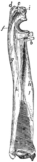
Fig. 87.—Radius and Ulna, Medial or Flexor Side, with M. pronator quadratus and the Areas of Attachment of Other Muscles.
a, M. pronator quadratus; b, tendon of M. biceps; c, conjoined tendon of M. brachialis and M. clavobrachialis; d, insertion of caput longum of M. triceps; e, insertion of short portion of caput mediale of M. triceps; f, origin of M. flexor carpi ulnaris; g, origin of first head of M. flexor profundus digitorum; h, origin of fifth head of M. flexor profundus digitorum; i, intermediate portion of caput mediale of M. triceps.
The second head has origin (Fig. 82, n) from the distal end of the medial epicondyle of the humerus between the flexor ulnaris and the third and fourth heads of the profundus. It may be very closely attached to these muscles at their origins. Its tendon joins the radial border of the tendon of the first head. From this junction the radial part of the flexor sublimis arises.
The third head (Fig. 77, u) has origin (Fig. 82, o) by a strong tendon from the medial epicondyle of the humerus, between the palmaris (p) and flexor radialis (m). Its tendon forms the radial part of the common tendon.
The fourth head has origin by a strong tendon from the medial epicondyle of the humerus just ventrad of the origin of the second head and the flexor ulnaris, to which many of its fibres are attached. Its tendon forms the middle and superficial part of the common tendon.
The fifth or radial head (Fig. 75, q; Fig. 77, u′) has origin (Fig. 87, h) from the middle third of the ventral surface of the shaft of the radius over an area limited by two oblique bony ridges; from the adjacent parts of the interosseous membrane; and from the ventral part of the medial surface of the shaft of the ulna between a point about two centimeters distad of the semilunar notch and the junction of the middle and distal thirds of the shaft. Its very thick tendon forms the middle and deep part of the common tendon.
The common tendon (Fig. 88, c′) covers the carpus and metacarpus ventrally. It divides into five tendons (c) which pass to the five digits and are inserted into the bases of the terminal phalanges. Those of the first four digits perforate the tendon of the flexor sublimis (b) at the base of the first phalanx of each digit. At the same place each passes through a fibrous pulley-ring attached to the base of the phalanx. Each then passes through a second pulley-ring (2) near the head of the phalanx and is finally inserted into the base of the terminal phalanx.
Relations.—Outer surface with the pronator teres (Fig. 77, q), flexor carpi radialis (Fig. 77, r), palmaris longus (s), flexor carpi ulnaris (t), integument, and extensor carpi ulnaris (Fig. 75, o). Inner surface with the radius, ulna, interosseous membrane and pronator quadratus (Fig. 87, a).
Action.—Flexor of all the digits.
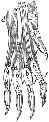
Fig. 88.—Arrangement of the Tendons of M. palmaris longus, M. flexor sublimis digitorum, and M. flexor profundus digitorum, with Mm. lumbricales.
The integument, fibrous pads, and the palmaris longus (except the ends of its tendons) have been removed; the flexor sublimis has been cut. a, cut ends of tendons of M. palmaris longus; b, tendons of M. flexor sublimis digitorum, ulnar division; b′, radial part of M. flexor sublimis digitorum; c, tendons of M. flexor profundus digitorum (c′, the common tendon); d, M. abductor digiti quinti; e, M. flexor brevis digiti quinti; f, Mm. lumbricales (f′, their tendons). 1, transverse ligament of wrist; 2, annular ligaments.
M. pronator quadratus (Fig. 87, a).—A thick quadrangular muscle whose fibres run obliquely between the distal ends of the ulna and radius.
Origin by fleshy fibres from about the distal half of the ventral (flexor) surface or border of the ulna and from the interosseous membrane adjacent to the area. The fibres pass obliquely distad toward the radial side to their
Insertion by fleshy fibres into the ventral (flexor) surface of the radius distad of the area of origin of the fifth head of the profundus.
Relations.—Outer surface with the flexor profundus. Inner surface with the radius, ulna, and interosseous membrane.
Action as indicated by the name.
4. Muscles of the Hand.
A. Between the Tendons.
—Mm. lumbricales (Fig. 88, f, f′).—Four small muscles in the palm of the hand.
Origin by fleshy fibres from the outer (palmar) surface of the tendon common to the ulnar four parts of the profundus (c′).
Insertion (f′).—The four slips are flat at their origin. Each becomes cylindrical and curves about the base of one of the four ulnar digits and is inserted into the radial side of the base of the first phalanx close to its ventral border.
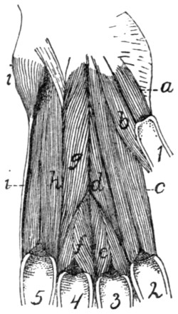
Fig. 89.—Deep Muscles of the Palm of the Hand.
a, M. flexor brevis pollicis; b, M. adductor pollicis; c, M. interosseus of second digit (M. flexor brevis digiti secundi); d, M. adductor digiti secundi; e, M. interosseus of third digit; f, M. interosseus of fourth digit; g, M. opponens digiti quinti; h, M. interosseus of fifth digit (M. flexor brevis digiti quinti); i, M. abductor digiti quinti. 1-5, the digits in order.
Action.—Bend the digits toward the radial side.
B. Muscles of the Thumb.
—M. abductor brevis pollicis (Fig. 77, w) (includes also the opponens pollicis of man).—A very minute, probably rudimentary muscle.
Origin from the transverse ligament (1) which connects the prominent fibrous, hairless projection that lies over the pisiform bone with the radial border of the head of the radius. The muscle passes radiodistad and ends in a very slender tendon, the insertion of which is into the base of the first phalanx of the thumb.
M. flexor brevis pollicis (Fig. 89, a).
Origin by fleshy fibres from the adjacent borders of the os magnum and scapholunar bones and from the fascia which bridges the intervening groove for the flexor carpi radialis.
Insertion by a short tendon into the base of the first phalanx of the pollex.
Action.—Flexor of the thumb.
M. adductor pollicis (Fig. 89, b).
Origin by fleshy fibres from the ventral border of the os magnum. It curves about the base of the first metacarpal on its ulnar side to its insertion.
Insertion by fleshy fibres into the base of the first phalanx of the pollex on its ulnar side.
C. Muscles Lying between the Metacarpals.
—Mm. interossei (Fig. 89).—Small stout muscles lying on the palmar surfaces of the metacarpals of the second, third, fourth, and fifth digits.
Origin by fleshy fibres from the ventral or lateral surfaces of the bases of the metacarpals. Near the distal end of the metacarpal each divides into two masses which pass onto the lateral surfaces of the metacarpal, so as to leave the ventral surface of the distal end exposed.
Insertion partly onto the lateral surfaces of the base of the first phalanx and its sesamoids, and partly by a slender tendon (Fig. 84, h) which is continued dorsad to join the extensor tendon of the digit.
D. Special Muscles of the Second Digit (the Index).
—M. flexor brevis digiti secundi (or indicis).—This name may be applied to M. interosseus of the second digit (Fig. 89, c).
M. abductor digiti secundi.—This name might be applied to a portion of the interosseus of the second digit which is sometimes differentiated from the remainder.
Origin from the radial and ventral surfaces of the base of the second metacarpal, and from the ventral surface of the trapezium.
Insertion into the radial side of the base of the first phalanx of the second digit and into its sesamoid.
M. adductor digiti secundi (Fig. 89, d).
Origin from the ventral surface of the os magnum.
Insertion into the ulnar side of the base of the first phalanx of the second digit.
E. Special Muscles of the Fifth Digit.
—M. abductor digiti quinti (Fig. 89, i).—A thick, small, conical bundle.
Origin from the distal surface of the pisiform bone and from the transverse ligament on the ulnar side of the pisiform. It ends in a slender tendon which runs along the ulnar side of the fifth metacarpal to its
Insertion into the ulnar side of the base of the first phalanx of the fifth digit.
M. flexor brevis digiti quinti (Fig. 89, h; Fig. 88, e).—This covers the ventral surface of the fifth metacarpal, and is really the interosseus of this digit.
Origin by fleshy fibres from the ventral surface of the base of the fifth metacarpal; from the ventral process of the unciform bone, and from the transverse ligament proximad of the fifth metacarpal.
Insertion by fleshy fibres into the ventral border of the proximal end of the first phalanx of the fifth digit.
M. opponens (adductor) digiti quinti (Fig. 89, g).
Origin by a flat tendon from the ventral surface of the os magnum on the ulnar side of the adductor pollicis (b). It passes toward the ulnar side and distad, the fibres diverging to their
Insertion (1) into nearly the whole of the radial surface of the fifth metacarpal, (2) into the base of its first phalanx.
V. MUSCLES OF THE PELVIC LIMBS.
1. Muscles of the Hip.
A. On the Lateral Surface of the Hip.
Fascia of the Thigh.
—After the removal of the superficial fascia with its fat and blood-vessels, there is seen a strong glistening fascia, the fascia lata (Fig. 68, z, page 117), covering the vastus lateralis muscle over the dorsal half of the thigh. Ventrally it dips between the vastus lateralis and the biceps femoris and, becoming gradually thinner, is lost on the surface of the former muscle. Passing over the dorsal border of the thigh, it extends beneath the sartorius and is attached to the dorsal border of the vastus medialis. Toward its distal end the fascia is continuous with the tendon of the biceps femoris and dorsad with the border of the sartorius muscle, while between these points it is united with the tendon of the vastus lateralis; it thus gains insertion into the patella. At its proximal end the fascia receives the insertion of the tensor fasciæ latæ (Fig. 68, r).
M. tensor fasciæ latæ (Fig. 68, r, page 117; Fig. 92, a).—The tensor fasciæ latæ is a thick triangular muscle which may be recognized by its insertion into the proximal end of the fascia lata (Fig. 68, z; Fig. 92, a′).
Origin by fleshy fibres as a thick triangular prism from the outer margin of the ventral border of the ilium craniad of the auricular impression and from the fascia covering the ventral border of the gluteus medius (Fig. 90, b) over its cranial half. The caudal border of the muscle may be continuous with the cranial border of the gluteus maximus. The muscle spreads out in a fan-like manner, covering the proximal third of the dorsal surface of the thigh. The cranial fibres pass farther distad than the others.
Insertion into the fascia lata (Fig. 68, z) along an irregular line which begins caudad at the base of the great trochanter and ends on the cranial side of the thigh at the junction of the first and second thirds.
Relations.—Outer surface with the cutaneus maximus (Fig. 62, b, page 94) and craniad with the sartorius (Fig. 68, q). Cranial border with the sartorius (q); caudodorsal border with the gluteus medius (Fig. 90, b). Inner surface with the gluteus medius, the rectus femoris (Fig. 92, b), and the vastus lateralis (Fig. 90, d).
Action indicated by the name. It also assists the quadriceps femoris in extending the leg.
M. gluteus maximus.—The gluteus maximus is a rather small quadrangular muscle in the angle between the gluteus medius (Fig. 90, b) and the caudal vertebræ.
Origin (Fig. 163, 3, page 401) by fleshy fibres from the tips of the transverse processes of the last sacral and the first caudal vertebræ, from the fascia covering the spinous muscles dorsad of the transverse processes of these vertebræ, and from the fascia covering the gluteus medius. The muscle passes laterad and slightly caudad to its
Insertion by tendon and muscle-fibres into a tubercle on the caudal side of the great trochanter at the middle of its distal end, and for a few millimeters into the ridge which continues distad from this tubercle.
Relations.—Outer surface with the caudofemoralis (Fig. 68, s, page 117), the superficial fascia, and a few fibres of the cutaneus maximus. Inner surface with the abductor caudæ internus, the tenuissimus (Fig. 90, g), the great sciatic nerve (Fig. 163, a), the obturator internus (Fig. 90, e), the gluteus medius (Fig. 90, b), the gemellus superior, and the pyriformis (Fig. 163, 7). Cranial border with the gluteus medius and the tensor fasciæ latæ. Caudal border with the caudofemoralis (Fig. 68, s).
Action.—Abducts the thigh.
M. gluteus medius (Fig. 90, b).—The gluteus medius is a very large triangular muscle which connects the ilium and the sacrum with the great trochanter (1).
Origin by fleshy fibres (1) from the superficial sacral fascia; (2) from the lateral surface of the fascia which covers the surface of the supraspinous extensor muscles of the tail; (3) from the fascia intervening between it and the tensor fasciæ latæ; (4) by tendon fibres from the dorsal half of the crest of the ilium and its dorsal border, and the dorsal half of the lateral surface craniad of the auricular impression; and (5) from the tips of the transverse processes of the last sacral and the first caudal vertebræ. The fibres converge to a strong internal tendon.
Insertion into the proximal end of the great trochanter.
Relations.—Outer surface with the gluteus maximus and tensor fasciæ latæ, and between these with the strong fascia lying beneath the cutaneus maximus muscle. Inner surface with the gluteus minimus, the pyriformis, and the gemellus superior. Cranial border with the tensor fasciæ latæ. Caudal border with the gluteus maximus.
Action.—Abducts the thigh.
M. pyriformis (Fig. 163, 7).—The pyriformis is a triangular muscle covered by the gluteus maximus and the gluteus medius (Fig. 90, b) and overlying the gemellus superior (Fig. 163, 6).
Origin by fleshy fibres from the tips of the transverse processes of the last two sacral and the first caudal vertebræ. The muscle passes laterad through the great sciatic notch to its
Insertion by a flat tendon into an elongated area on the proximal border of the great trochanter just outside (caudad) of the insertion of the gemellus superior.
Relations.—Outer surface with the gluteus medius (Fig. 90, b) and the gluteus maximus. Inner surface with the gemellus superior (Fig. 163, 6), the great sciatic nerve (Fig. 163, a), and the flexor caudæ longus (Fig. 68, x).
Action.—Abductor of the thigh.
M. gemellus superior (Fig. 163, 6).—The gemellus superior is a triangular muscle lying beneath the pyriformis (Fig. 163, 7). It is broader than the pyriformis, so that it projects beyond its borders both caudad and craniad. It is closely united craniad with the gluteus minimus, so that the limits of the two are definable only with difficulty. Its caudal border is closely united to the gemellus inferior.
Origin by fleshy fibres from an elongated area on the dorsal border of the ilium and ischium. The area is narrowed craniad. It does not quite reach the posterior inferior iliac spine in the one direction nor the spine of the ischium in the other direction. The fibres converge to a strong tendon the
Insertion of which is into a triangular area dorsad of the tip of the great trochanter.
Relations.—Outer surface with the gluteus medius (Fig. 90, b), the pyriformis (Fig. 163, 7), the great sciatic nerve (Fig. 163, a), and the gluteus maximus. Inner surface with the ilium, ischium, the capsularis, the caudal edge of the gluteus minimus (Fig. 163, 5), and the cranial edge of the obturator internus (Fig. 90, e).
Action.—Rotates the femur and abducts it so as to carry the foot outward.
M. gluteus minimus (Fig. 163, 5).—The gluteus minimus is a long triangular muscle beneath the middle of the gluteus medius (Fig. 90, b) and with its caudal border against or covering the cranial border of the gemellus superior (Fig. 163, 6), to which it is frequently closely united.
Origin from the ventral half of the ilium, from near its cranial end to a point midway between the posterior iliac spine and the spine of the ischium. The muscle ends in a strong flat tendon.
Insertion into an oval facet at the base of the dorsal surface of the great trochanter on its lateral side.
Relations.—Outer surface with the gluteus medius (Fig. 90, b) and at the caudal border with the gemellus superior (Fig. 163, 6). Inner surface with the capsularis and rectus femoris (Fig. 92, b).
Action.—Rotates the femur so as to carry the foot out.
M. capsularis (gluteus quartus, or epimeralis).—The capsularis is a small flat bundle of muscle-fibres which lies obliquely beneath the gluteus minimus (Fig. 163, 5) and gemellus superior (6) on the lateral or outer surface of the ilium.
Origin by fleshy fibres from the surface of the ilium over a triangular area between the origin of the rectus femoris (Fig. 92, b) ventrad, the gemellus superior (Fig. 163, 6) dorsad, and of the gluteus minimus (Fig. 163, 5) craniad and the acetabulum caudad. The muscle passes over the smooth surface of the ilium and the pubis ventrad of the acetabulum and then over the capsule of the joint.
Insertion by fleshy fibres for about one centimeter in the middle line on the dorsal surface of the femur distad of the great trochanter.
Relations.—Outer surface with the gluteus minimus (Fig. 163, 5), the gemellus superior (6), and the vastus lateralis (Fig. 90, d). Inner surface with the ilium, the rectus femoris (Fig. 92, b), the capsule of the joint, and the vastus medialis (Fig. 92, c).
Action.—Rotates the thigh so as to carry the foot inward, hence antagonizes the iliopsoas.
M. gemellus inferior.—The gemellus inferior is a flat triangular muscle situated just caudad of the gemellus superior and beneath the obturator internus (Fig. 90, e), so that it is seen on reflecting the latter
Origin from the dorsal one-half of the whole lateral surface of the ischium between the ischial spine and the ischial tuberosity. The fibres converge toward its insertion. The inner surface of the muscle is covered by a strong tendon.
Insertion into the inner surface of the tendon of the obturator internus (Fig. 90, e) by tendon- and muscle-fibres. Some of the muscle-fibres are inserted into the capsule of the joint.
Relations.—Outer surface with the obturator internus (Fig. 90, e). Medial surface with the ischium. Caudal border with the quadratus femoris (Fig. 90, f). Cranial border with the gemellus superior.
Action.—Abductor of the thigh. The muscle may be considered as a separate head of the obturator internus (Fig. 90, e).
M. quadratus femoris (Fig. 90, f).—The quadratus femoris is a short thick muscle connecting the ischial tuberosity (2) and the proximal end of the femur. It lies just caudad of the obturator internus (e) beneath the proximal end of the biceps femoris (Fig. 68, t).
The origin is by fleshy fibres from a considerable triangular area on the lateral surface of the ischium near the tuberosity. The area lies between the origin areas of the gemellus inferior, biceps, semimembranosus, and the obturator externus.
Insertion into the distal two-thirds of the ventral border of the great trochanter and about half the adjacent surface of the lesser trochanter.
Relations.—Outer surface with the tenuissimus (Fig. 90, g), the biceps (Fig. 68, t), the semitendinosus (Fig. 90, j), and the great sciatic nerve (Fig. 163, a). Caudal border with the semimembranosus (Fig. 90, i). Ventral border with the adductor femoris (Fig. 90, h); dorsal border with the obturator internus (Fig. 90, e) and gemellus inferior.
Action.—Extensor of the thigh and rotator of the femur so as to carry the foot inward.
M. obturator externus.—The obturator externus is a flat triangular muscle beneath the adductor femoris (Fig. 92, g).
Origin by fleshy fibres from the median lip of the obturator foramen and from both dorsal and ventral surfaces of the rami of the pubis and ischium adjacent to the lip. Also from the outer surface of the ramus of the ischium as far as the area for the quadratus femoris (Fig. 90, f). The fibres converge to a strong flat tendon.
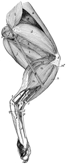
Fig. 90.—Muscles on the Lateral Side of the Leg, after Removal of the Muscles shown in Fig. 68 (Biceps, Tensor fasciæ latæ, Caudofemoralis, and Gluteus maximus).
a, M. sartorius; b, M. gluteus medius; c, M. rectus femoris; d, M. vastus lateralis; e, M. obturator internus; f, M. quadratus femoris; g, M. tenuissimus; h, M. adductor femoris; i, M. semimembranosus; j, M. semitendinosus; k, M. vastus intermedius; l, M. plantaris; m, m′, m″, M. gastrocnemius, outer head (m, part from the external sesamoid bone; m′, from the plantaris; m″, from the superficial fascia); n, M. tibialis anterior; o, M. soleus; p, M. extensor longus digitorum; q, M. peroneus longus; r, M. peroneus tertius (r′, its tendon); s, M. peroneus brevis; t, tendon of Achilles; u, M. extensor brevis digitorum. 1, great trochanter of femur; 2, tuberosity of ischium, with cut origin of M. biceps femoris; 3, patella; 4, calcaneus; 5, transverse ligaments; 6, lateral malleolus.
Insertion into the proximal portion of the bottom of the trochanteric fossa.
Relations.—Ventral surface with the adductor femoris (Fig. 92, g). Dorsal surface with the pubis and ischium. Caudal border with the quadratus femoris (Fig. 90, f).
Action.—Assists the iliopsoas.
B. Muscles on the Medial Surface of the Hip.
—M. obturator internus (Fig. 90, e).—The obturator internus appears as a triangular muscle caudad of the gemellus superior.
Origin by numerous small separate heads from the dorsal surface of the ramus of the ischium along its symphysis and following its medial border from the symphysis nearly to the tuberosity. The fibres form a flat muscle which narrows and passes through the lesser sciatic notch, turns ventrad and ends in a strong flat tendon whose
Insertion is into the bottom of the trochanteric fossa of the femur. The tendon is continued proximad on the inner surface of the muscle and forms a smooth firm surface by which the muscle glides over the dorsal border of the ischium. Into the inner surface of this tendon near its insertion the tendon of the gemellus inferior is inserted.
Relations.—Medial surface within the pelvis with a mass of fat separating it from the pelvic organs. Dorsal or outer surface with the biceps femoris, the tenuissimus, the caudofemoralis, and the great sciatic nerve. Inner surface with the ischium and the great sciatic nerve. Caudal (or ventral) border with the quadratus femoris.
Action.—Abductor of the thigh.
M. iliopsoas (Fig. 162, 8; Fig. 91, c).—The iliopsoas (equivalent to the human psoas and iliacus) is a conical muscle emerging from the abdominal cavity onto the medial surface of the femur.
Origin.—(a) The portion corresponding to the human psoas (Fig. 162, 8) arises by ten vertebral heads. The first five of these come from the five cranial tendons of origin of the psoas minor (Fig. 162, 9); the sixth from a tendinous expansion which passes from the tendon of the first head over the ventral longitudinal muscles to the transverse process of the fifth lumbar vertebra. The seventh, eighth, ninth, and tenth heads arise by fleshy fibres from the ventral surfaces of the centra of the last four lumbar vertebræ.
(b) The portion corresponding to the human iliacus arises by fleshy fibres from the ventral border of the ilium, from opposite the auricular impression to the iliopectineal eminence. The portions of the muscle all converge to form a conical mass which ends in a strong tendon lying on its outer surface.
Insertion by tendon and fleshy fibres into the apex of the lesser trochanter of the femur.
Relations.—Dorsal surface with the quadratus lumborum (with which this muscle is partly united), the rectus femoris (Fig. 91, d), and, by the iliac head, with the gluteus minimus. Ventral and medial surface with the psoas minor (Fig. 162, 9) and the peritoneum. Lateral edge with the transversus abdominis (Fig. 162, 4).
Action.—Rotates the thigh so as to carry the foot out; also flexes the thigh.
2. Muscles of the Thigh.
M. biceps femoris (Fig. 68, t, page 117).—A very large flat muscle covering about two-thirds of the lateral side of the thigh.
Origin (Fig. 90, 2).—From the ventral surface of the tuberosity of the ischium by tendon- and muscle-fibres. The fibres diverge, and near the knee the mass has spread out, ending in a fascia. The dorsal border of the muscle and the common fascia are continuous dorsad with the superficial fascia of the thigh and with the tendon of the caudofemoralis (Fig. 68, s). Ventrad it is continuous with the superficial fascia of the shank.
Insertion into rather more than the proximal one-third of the dorsal border of the tibia along its lateral margin and into the lateral margin of the patella. In passing over the knee-joint it is closely united to the underlying ligaments and tendons.
Relations.—Outer surface with the superficial fascia and with a few of the most caudal fibres of the cutaneus maximus (Fig. 62, b, page 94). Cranial (or dorsal) edge with the caudofemoralis (Fig. 68, s) and the vastus lateralis (Fig. 90, d). Caudal border with the semitendinosus (Fig. 68, u) and a mass of fat. Inner surface with the caudofemoralis (Fig. 68, s), the tenuissimus (Fig. 90, g), the obturator internus (Fig. 90, e), the quadratus femoris (Fig. 90, f), the semitendinosus (Fig. 68, u), the semimembranosus (Fig. 90, i), the adductor femoris (Fig. 90, h), the great sciatic nerve (Fig. 163, a), and distad with the following muscles of the lower leg: the tibialis anterior (Fig. 90, n), the extensor longus digitorum (Fig. 90, p), the peroneus longus (Fig. 90, q), and the lateral head of the gastrocnemius (Fig. 90, m).
Action.—Abductor of the thigh, and flexor of the shank.
M. tenuissimus or M. abductor cruris (Fig. 90, g).—A very slender muscle, only three or four millimeters wide.
Origin from the tip of the transverse process of the second caudal vertebra, in common with the caudofemoralis (Fig. 68, s) or gluteus maximus. It passes obliquely beneath the biceps femoris (Fig. 68, t) distad and ventrad, to the distal end of the ventral border of that muscle. Here it becomes continuous with the ventral border of the biceps, ending in a continuation of the same fascia into which the biceps is inserted.
Relations.—Outer surface with the caudofemoralis (Fig. 68, s) and the biceps femoris (Fig. 68, t); distad with the integument. Inner surface with the obturator internus (Fig. 90, e), quadratus femoris (f), semitendinosus (j), adductor femoris (h), semimembranosus (i), and distad with the muscles of the lower leg.
M. caudofemoralis (parameralis, Strauss-Durckheim) (Fig. 68, s, page 117).
Origin by a flat tendon from the transverse processes of the second and third caudal vertebræ. The muscle forms a flat band which passes distad along the middle of the lateral side of the thigh over the pelvis and caudad of the great trochanter. At the middle of the thigh it ends in a very thin tendon. The tendon passes distad along the medial surface of the biceps femoris (Fig. 68, t), pierces the fascia lata near the knee, and passes to its
Insertion into the middle of the lateral border of the patella.
Relations.—Outer surface with the superficial fascia and a few fibres of the cutaneus maximus; distad with the biceps femoris (Fig. 68, t). Cranial border with the gluteus maximus; caudal border with the biceps femoris. Inner surface with the tenuissimus (Fig. 90, g), the gluteus maximus, the abductor caudæ internus, and with the obturator internus (Fig. 90, e); distad with the vastus lateralis.
Action.—Abducts the thigh and helps to extend the shank.
M. semitendinosus (Fig. 90, j).—A long slender muscle on the ventral (caudal) border of the thigh, between the semimembranosus (i) and the biceps femoris (Fig. 68, t).
Origin from the apex of the tuberosity of the ischium beneath the origin of the biceps femoris. The muscle passes to the medial side of the shank and ends in a thin but strong tendon (Fig. 92, i′) about five millimeters broad. The tendon curves proximad and passes beneath the gracilis tendon (Fig. 91, b) to its