THE CIRCULATORY SYSTEM. (continued 2)
Reading edition
The posterior facial receives also, at the point where it passes from beneath the parotid gland, the posterior auricular or great auricular vein (l). This collects blood from the back of the head and the external ear, its branches following those of the artery of the same name. It passes along the caudal border of the parotid gland to join the posterior facial.
Veins of the Brain and Spinal Cord.
—The veins of the brain form a large number of small vessels which pour their blood into larger veins lying in folds of the dura mater; these are known as the venous sinuses of the dura mater. These sinuses communicate with the venous plexus about the orbital fissure, coming from the posterior facial vein; with the internal jugular through the jugular foramen, and with the vertebral veins, in the vertebral canal. The chief sinuses of the dura mater on the dorsal side of the brain are as follows:
1. Sinus sagittalis superior.—This lies in the dorsal part of the falx cerebri, between the two hemispheres of the cerebrum. It receives veins from the dorsal and middle parts of the cerebrum, and passes caudad to the tentorium. Here it enters the sinus transversus. It receives the vena cerebri magna, a large vein coming from the interior of the brain and passing directly dorsad at the caudal end of the corpus callosum to enter the sinus sagittalis.
2. Sinus transversus.—This lies in a canal in the dorsal border of the tentorium. It receives numerous small veins from the cerebellum, roof of the skull, etc. One or two centimeters on each side of the middle line the sinus transversus passes out of the canal onto the caudal surface of the tentorium, thence proceeds caudoventrad obliquely over the surface of the cerebellum, unites with veins from the ventral side of the brain, leaves the skull by the jugular foramen, forming thus the inferior cerebral vein, and joins the internal jugular vein.
On the ventral side of the brain are the following sinuses of the dura mater:
3. Sinus cavernosus.—A short broad venous sinus, one on each side of the hypophysis, on the body of the sphenoid. It receives veins from the side and ventral surface of the brain. The two sinuses are connected by communicating branches craniad and caudad of the hypophysis. From them branches pass out through the orbital fissure to join the plexus formed by the branches of the posterior facial vein.
4. Sinus petrosus inferior.—This arises from the sinus cavernosus and passes caudolaterad in the groove between the edge of the petrous bone and the basilar portion of the occipital. Reaching the jugular foramen it divides; part joins the termination of the sinus transversus to form the inferior cerebral vein, which passes through the jugular foramen to join the internal jugular vein. The other portion of the sinus petrosus inferior passes through the condyloid canal of the occipital bone, communicates by a strong transverse branch across the surface of the basioccipital with the vein of the opposite side, and enters the spinal canal through the foramen magnum. Here it joins the sinus of the vertebral column.
5. Sinus columnæ vertebralis.—On the ventral surface of the vertebral canal, beneath the periosteum, are two wide venous sinuses, one on each side of the middle line. These sinuses extend the entire length of the spinal cord. At the atlantal foramen each sends a strong branch to the internal jugular vein. Farther caudad they send branches to the vertebral, intercostal, and lumbar veins, and communicate with each other by numerous transverse branches. They receive many small veins from the spinal cord.
3. Vena cava inferior (Fig. 129, c; Fig. 126, a).
The inferior vena cava (Fig. 126, a) is formed at about the level of the last lumbar vertebra by the union of the two common iliac veins (Fig. 126, w). It passes craniad near the dorsal median line, lying at first dorsad of the aorta (b), then to the right, then ventrad. It enters the substance of the liver in the dorsal part of the caudate lobe of the latter, passes through the liver, and then through the diaphragm near the ventrolateral edge of the central tendon. It passes then craniad in the thoracic cavity (Fig. 129, c) ventrad of the caudal lobe of the right lung, and enters the right auricle.
It receives the following branches (Fig. 126): lumbar, iliolumbalis (v), spermatica interna (u), renal (t), adrenolumbalis (s), phrenic, and hepatic veins. All but the last two named accompany the arteries of the same name.
V. phrenica.—The phrenic veins gather the blood from the diaphragm and empty into the vena cava as it penetrates the diaphragm.
V. hepatica.—The hepatic veins vary in number. They gather the blood from the liver (sent in by the portal vein and hepatic arteries) and enter the vena cava just caudad of the diaphragm.
Vena Portæ. The Portal Vein
(Fig. 131).—The portal vein is the large vein carrying the blood from the abdominal viscera to the liver. Within the liver the portal vein breaks up into capillaries; these collect to form the hepatic veins, which enter the vena cava inferior. The portal vein (a) is formed near the pyloric end of the stomach (1) by the union of the superior mesenteric (b) and gastrosplenic (c) veins and passes thence along the ventral border of the foramen epiploicum (foramen of Winslow) to the liver (2), where it divides, going to the lobes of the liver. On its way to the liver it may receive the pancreaticoduodenalis (d), gastroepiploica (e), and coronaria ventriculi (f). These may unite with the portal separately, or any two or all three may unite to form a single trunk before entering the portal vein.
1. V. coronaria ventriculi (f) gathers the blood from the lesser curvature of the stomach and anastomoses with the gastrosplenic veins. It usually empties into the portal vein near the pylorus.
2. V. pancreaticoduodenalis (d) receives the blood from the pancreas and first part of the duodenum, and empties into the vena portæ near to or with the preceding.
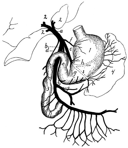
Fig. 132.—Portal Vein.
a, portal vein; b, superior mesenteric; c, gastrosplenic; d, pancreaticoduodenalis; e, gastroepiploica; f, coronaria ventriculi; g, h, i, branches of gastrosplenic; j, inferior mesenteric. 1, stomach; 2, liver; 3, duodenum; 4, spleen.
3. V. gastroepiploica (e) comes from the greater curvature of the stomach and ascending limb of the great omentum. It empties into the vena portæ ventrodextrad of the opening of the coronaria ventriculi, or sometimes in common with it.
4. V. gastrolienalis (c).—The gastrosplenic is one of the veins which unite to make up the portal vein. It is formed by three branches: one (g) from the horizontal or gastric portion of the pancreas, one (h) from the stomach and gastric end of the spleen, the third and largest (i) from the caudal end of the spleen and descending limb of the great omentum. The three branches unite and the common trunk joins the superior mesenteric vein (b) to form the portal vein as above described.
5. V. mesenterica superior (b).—The superior mesenteric vein receives the blood from the small and large intestines. It is formed by numerous branches which follow and have the same name as the corresponding arteries. It receives also the inferior mesenteric (j) from the large intestine. The numerous branches unite, and the main trunk thus formed joins the gastrosplenic (c), and the two united become the portal vein (a).
Vena iliaca communis. Common Iliac Vein (Fig. 126, w; Fig. 127, page 310).
The common iliac veins (Fig. 126, w) are the two large vessels which unite in the sacral region to form the inferior vena cava (a). Each is a large vessel, usually four or five centimeters in length, which is formed by the junction of the large external iliac vein with the smaller hypogastric vein (z). The left common iliac usually receives the V. sacralis media (o); other lateral branches are as a rule not received by the common iliac veins.
There is much variation as to the formation of the common iliac veins and their union to form the vena cava. On this subject the paper by McClure, in the American Naturalist, vol. XXXIV. pp. 135-198 (March, 1900), may be consulted with profit. The more typical variations may be classified as follows: (1) The two common iliac veins may be longer than usual, so that the vena cava is formed farther craniad than usual. (2) There may be in the abdominal region two separate veins (venæ cardinales) representing the inferior vena cava, these uniting in the region of the kidneys. (3) The vena sacralis media may enter into the left common iliac (the usual condition); or the right common iliac (as in Fig. 126); or it may fork, one branch passing to the left, the other to the right common iliac.
Branches of the common iliac veins:
1. V. sacralis media (Fig. 126, o).—This follows the course of the corresponding artery and usually enters the left common iliac.
2. V. hypogastrica (or iliaca interna) (z).—The hypogastric or internal iliac vein joins the medial side of the external iliac to form the common iliac (w). Its branches follow the branches of the corresponding artery except that the vein from the bladder (umbilical vein) joins the V. hemorrhoidalis media. Also the vena glutea inferior receives a large superficial communicating branch (Fig. 163, k′) from the vena saphena parva. This branch leaves the saphena parva (Fig. 163, k) at the popliteal space, and passes over the lateral surface of the biceps muscle to its proximal end. Here it passes between the biceps and caudofemoralis to join the vena glutea inferior.
3. V. iliaca externa (Fig. 127, page 310).—This vein follows the corresponding artery, collecting the blood from the posterior extremity. Its branches are the same and have in general the same distribution as those of the artery. The vein has, however, certain branches in addition to those of the artery, resulting in a somewhat different general arrangement of vessels; it will be necessary therefore to give an account of the system of veins from the foot and lower leg.
The blood is collected from the foot into two sets of veins which may be distinguished as the deep and the superficial sets. The veins of the deep set are those which accompany the branches of the popliteal and anterior tibial arteries. Those of the superficial set are larger and carry most of the blood. On the dorsum of the foot blood is collected from the third, fourth, and fifth digits by two or three superficial metatarsal veins, which unite opposite the proximal end of the metatarsus into a single vein. This receives a similar branch coming from the dorsum of the second digit, and has numerous branches in the ankle region which form here a network, communicating with the vena saphena parva (Fig. 163, k), and with branches from the plantar side of the foot. From this network two or three main veins pass proximad on the medial side of the leg, along with the branches of the arteria saphena, and unite with a similar vein from the plantar side of the foot to form the vena saphena magna (Fig. 127, k, page 310), which accompanies the arteria saphena to join the femoral vein (a).
On the plantar side of the foot small veins pass from the digits to a superficial venous plantar arch, which lies just proximad of the fibrous pad on the sole of the foot. At its medial side this arch extends proximad and dorsad to join that branch of the vena saphena magna which comes from the dorsum of the second digit. On the lateral side it extends proximad along the lateral margin of the foot, sends a communicating branch dorsad to the branches of the V. saphena magna, and continues along the lateral border of the foot to the ankle. It forms the beginning of the V. saphena parva (Fig. 163, k, page 401). This receives branches from the lateral surface of the ankle, which anastomose with those of the saphena magna. The saphena parva then passes proximad along the lateral surface of the leg, close to its ventral border, to the popliteal space. At the distal end of the popliteal space it divides into two branches. One passes inward through the fat of the popliteal space, receiving branches from the lymphatic gland situated here, and joins the popliteal vein. The other (k′) remains superficial and continues proximad over the lateral surface of the biceps to the proximal end of that muscle. Here it passes between the biceps and caudofemoralis to join the inferior gluteal vein.
IV. THE LYMPHATIC SYSTEM. SYSTEMA LYMPHATICUM.
The lymphatic system of the cat has not been worked out in detail, so that only the main features of the system are given in the following account.
The lymphatic system consists of a number of vessels, the lymphatics, containing a colorless fluid called lymph, and of lymphatic glands, connected with the lymphatic vessels. The lymphatic vessels are found throughout the body as slender tubes, frequently united into networks, and containing many valves. They take origin from the spaces in the connective tissue, so that they are at first without definite walls. The fluid in the connective-tissue spaces gradually flows together into tubes with definite walls, and these tubes, the lymphatic vessels, finally join the venous system. In their passage through the lymphatic glands the vessels break up into fine channels which again unite on leaving the gland. In the gland lymph-cells are added to the fluid, and probably other changes are produced.
Most or all of the lymphatic vessels of the body finally unite into two trunks before entering the veins. The largest trunk is the thoracic duct (Fig. 118, 5, page 282), which collects the lymph from the entire caudal half of the body, and from the left forelimb and the left side of the thorax, head, and neck. It passes along the dorsosinistral side of the thoracic aorta, lying against its surface, and enters the left external jugular vein at its junction with the subclavian. The second main trunk is the right lymphatic duct; this collects the lymph from the right side of the thorax, the right forelimb, and the right side of the neck and head; it enters the right external jugular.
1. Lymphatics of the Head.
—A number of lymphatic vessels arise on the sides of the face, especially in the upper and lower lips. They form a superficial network of vessels, lying over the ventral half of the masseter muscle and an area ventrad of it. These lymphatic vessels all enter two large lymphatic glands (Fig. 131, 12, page 322) lying at the ventrocaudal angle of the masseter muscle, covering the union of the anterior and posterior facial veins. Into these glands pass also a number of lymph vessels from the back of the head. From these two lymphatic glands two or three small lymphatic vessels pass caudad, lying on the surface of the external jugular vein. Near the point of the shoulder, some distance craniad of the junction of the external and internal jugular veins, these vessels enter another small lymphatic gland lying on the dorsal surface of the external jugular vein. Thence one or two vessels continue caudad, still on the surface of the external jugular, and finally unite with the deep lymphatics of the head and neck and those of the arm to enter the caudal end of the external jugular vein, usually (on the left side) after junction with the thoracic duct, from the thorax.
The deep lymphatics of the head come from the internal parts of the head,—tongue, pharynx, etc., and enter a large lymphatic gland situated close to the caudoventral surface of the tympanic bulla, at the side of the pharynx. Thence a large lymphatic trunk passes caudad at the side of the trachea, along with the common carotid artery and internal jugular vein, unites with the superficial vessels from the head, above described, joins (on the left side) the thoracic duct, and enters the external jugular vein.
2. Lymphatics of the Neck.
—Two or three lymph-glands are found in the fatty mass in the neck beneath the clavotrapezius and levator scapulæ ventralis muscles. These receive lymphatics from the neck and side of the thorax. The vessels which arise from them join those from the head and arm to enter the external jugular vein.
3. Lymphatics of the Thoracic Limbs.
—The lymphatics of the forelimbs begin as a number of vessels on the dorsum and palm of the hand. These all pass toward the radial side, and at about the base of the first metacarpal the vessels from both sides of the hand unite to form two or three main trunks. These pass towards the elbow, accompanying the vena cephalica (Fig. 130). There are usually two of these main trunks, one lying on each side of the vena cephalica. They accompany this vein over the shoulder, following that portion which finally joins the external jugular. In the hollow of the shoulder they enter the cervical lymph-glands, one or two lymph-glands imbedded in the fat lying beneath the clavotrapezius and levator scapulæ ventralis in this region. From the cervical lymph-glands a single trunk passes mediocaudad, unites with the lymphatics from the head, and usually (on the left side) with the cranial end of the thoracic duct, and enters the external jugular vein at its junction with the subclavian. The common trunk from the head and arm may enter the jugular separately, but close to the termination of the thoracic duct. On the right side the common trunk is joined by one or more small vessels coming from the lymphatic glands of the thorax and forming the right lymphatic duct.
There is said to be also a deep system of lymphatics in the arm, in addition to the superficial system above described; this is said to accompany the branches of the brachial vein. If this system is present in the cat, it is much less easily demonstrated than the superficial system.
4. Lymphatics of the Thorax and Abdomen.
—In the thorax are a number of lymphatic glands which are apparently not constant in number and position. One is commonly found dorsad of the bifurcation of the superior vena cava, one ventrad of the mammary vein, several small ones about the bifurcation of the trachea, one or more in the region of the aortic arch, and a number of small ones scattered in the mediastinum. The vessels from these glands finally join either the right lymphatic duct or the thoracic duct; the details have not been worked out and are probably variable.
In the abdominal cavity a number of mesenteric glands, of considerable size, are found in the mesentery and in the mesocolon. In the mesocolon these are usually separate, forming a chain of glands following the colon about one and a half centimeters from it. In the mesentery the glands are mostly united into a very large one, formerly known as the pancreas aselli; this is the largest lymph-gland in the body. It is a curved structure, four or five centimeters in length, lying in the central region of the mesentery.
Into the mesenteric glands pass lymphatic vessels from the viscera of the abdomen. From the mesenteric glands one or two large lymphatic vessels pass craniodorsad, at first with the portal vein, then dorsad of it. This vessel reaches the dorsal side of the aorta near the cranial end of the kidney, where it enters a large fusiform vessel, the receptaculum chyli, which stretches from the cranial end of the kidney craniad between the crura of the diaphragm into the thorax. The receptaculum receives other lymphatics from the various organs of the abdominal cavity as well as vessels coming from the pelvic region, and from the iliac glands. These lie beside the iliac artery, receive lymphatics from the hind limbs, and send lymphatics craniad to the receptaculum chyli. The latter forms the beginning of the thoracic duct.
The thoracic duct (Fig. 118, 5, page 282) enters the thoracic cavity between the crura of the diaphragm as a continuation of the receptaculum chyli. It lies on the dorsal side of the aorta (a), somewhat to the left. It passes along the aorta, gradually coming to lie more on its left side, and finally leaves that vessel near the aortic arch. The duct continues thence straight craniad along the left side of the œsophagus till it reaches the external jugular vein at its junction with the subclavian. In this region the thoracic duct usually unites with the lymphatics coming from the head and arm, then enters the external jugular vein, or the innominate at the point of union of external jugular and subclavian.
In its passage through the thorax the duct frequently divides into two or three divisions which pass along parallel to each other for a distance, then reunite. Such division into two or three parts is especially frequent just before the union with the jugular vein. The numerous valves in the thoracic duct give it a beaded appearance.
5. Lymphatics of the Pelvic Limbs.
—A number of lymphatic vessels are formed on the dorsum and the sole of the foot. These pass to the lateral side, forming a network on the lateral and dorsal surface of the ankle-joint, and finally unite into two main trunks which accompany the vena saphena parva. In the popliteal space these enter the popliteal gland which lies imbedded in the fat of that region. From the popliteal gland one or two trunks accompany the superficial division of the vena saphena parva across the lateral surface of the biceps muscle, and finally thus reach the pelvic cavity. Here they join the lymphatics of the pelvic region, which all pour their lymph at last into the thoracic duct. Inguinal glands are either very small or not present in the cat.
The pelvic limbs have perhaps a deep system of lymphatics, accompanying the deep veins; if so, they are not easily demonstrable.
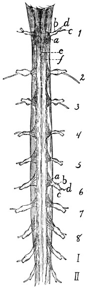
Fig. 133.—Cranial Portion of the Spinal Cord, with the Cervical Enlargement. Dorsal View.
1-8, the eight cervical nerves. I-II, first two thoracic nerves. a, dorsal roots (ventral roots not shown); b, spinal ganglia; c, dorsal rami; d, ventral rami; e, posterior median sulcus; f, posterior lateral sulcus.