THE SKELETON OF THE CAT.
Reading edition
The skeleton of the cat consists of 230 to 247 bones exclusive of the sesamoid bones (44) and the chevron bones (8). These are divided as follows: head 35-40, vertebral column 52-53, ribs 26, sternum 1-8, pelvis 2-8, upper extremities 62, lower extremities 54-56. The number of bones varies with the age of the individual, being fewer in the old than in the young animal, owing to the fact that in an old animal some bones that were originally separate have united.
I. THE VERTEBRAL COLUMN. COLUMNA VERTEBRALIS.
The vertebral column, spinal column, or back-bone, consists of a varying number of separate bones, the vertebræ. At its cranial end are seven vertebræ (cervical, Fig. 1, c) which are without ribs and support the head; caudad of these are thirteen rib-bearing vertebræ (thoracic, Fig. 1, m); caudad of these are seven that are again without ribs (lumbar, Fig. 1, o); these are followed by three vertebræ (sacral, Fig. 1, x) which are united into a single bone, the sacrum, which supports the pelvic arch. Following the sacral vertebræ are twenty-two or twenty-three small ribless vertebræ which support the tail (caudal, Fig. 1, y).
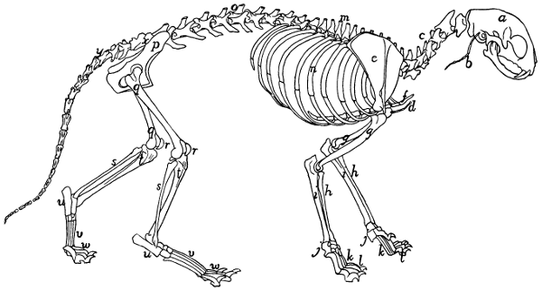
Fig. 1.—Skeleton of Cat.
a, skull; b, hyoid; c, cervical vertebræ; d, clavicle; e, scapula; f, sternum; g, humerus; h, radius; i, ulna; j, carpus; k, metacarpus; l, phalanges; m, thoracic vertebræ; n, ribs; o, lumbar vertebræ; p, innominate bones; q, femur; r, patella; s, fibula; t, tibia; u, tarsus; v, metatarsus; w, phalanges; x, sacrum; y, caudal vertebræ.
Thoracic Vertebræ. Vertebræ thoracales
(Fig. 4).—The thoracic vertebræ are most typical, and the fourth one of these may therefore be first described (Figs. 2 and 3). It forms an oval ring which has numerous processes and surrounds an opening which is the vertebral foramen (a). The ventral one-third of this ring is much thickened and forms the centrum or body (corpus) (b) of the vertebra. The centrum is a semicylinder, the plane face of which bounds the vertebral canal, while the curved surface is concave longitudinally and is directed ventrad. The dorsal plane surface of the centrum is marked by a median longitudinal ridge on either side of which is an opening (nutrient foramen) for a blood-vessel. The ends are nearly plane, the caudal being slightly concave; they are harder and smoother than the other surfaces. They may be easily separated in a young specimen as thin plates of bone known as epiphyses.
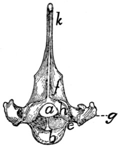
Fig. 2.—Fourth Thoracic Vertebra, Cranial End.
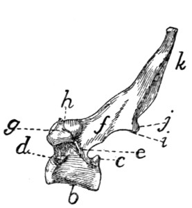
Fig. 3.—Fourth Thoracic Vertebra, Side View.
a, vertebral foramen; b, centrum; c, caudal, and d, cranial, costal demifacets; e, radix or pedicle; f, lamina; g, transverse process; h, cranial articular facet; i, caudal articular facet; j, caudal articular process; k, spinous process.
At the caudal end of the centrum, at its dorsolateral angle, is a smooth area on each side continuous with the surface of the epiphysis and bounded dorsolaterally by a sharp ridge of bone (c). It is a costal demifacet. In corresponding positions at the cranial end of the centrum are two demifacets not limited by bony ridges (d). When the centra of two contiguous thoracic vertebræ are placed together in the natural position the cranial costal demifacets of one together with the caudal demifacets of the other form two costal facets (Fig. 4, e), one on each side, and each receives the head of a rib.
The dorsal two-thirds of the vertebral ring forms the vertebral arch which is continued dorsally into the long, bluntly pointed spinous process (Figs. 2 and 3, k) for attachment of muscles.
The vertebral arch (each half of which is sometimes called a neurapophysis) rises on each side from the cranial two-thirds of the dorsolateral angle of the centrum, as a thickened portion, the radix or pedicle (Figs. 2 and 3, e), which forms the ventral half of the lateral boundary of the vertebral canal. From the dorsal end of each radix a flat plate of bone, the lamina (f), extends caudomediad to join its fellow of the opposite side and form the vertebral arch. Owing to the fact that the radix rises from only the cranial two-thirds of the centrum there is left in the caudal border of the vertebral arch a notch bounded by the radix, the lamina, and the centrum. There is also a slight excavation of the cranial border of the radix. When the vertebræ are articulated in the natural position, these notches form the intervertebral foramina (Fig. 4, d), for the exit of the spinal nerves.
At the junction of radix and lamina the arch is produced craniolaterad into a short process, the transverse process (g), knobbed at the end. On the ventral face of its free end the transverse process bears a smooth facet, the transverse costal facet or tubercular facet (Fig. 4, c), for articulation with the tubercle of a rib.
On the dorsal face of each lamina at its cranial border is a smooth oval area, the cranial articular facet (superior articular facet of human anatomy) (Figs. 2 and 3, h). Its long axis is oblique and it looks dorsolaterad. The slight projections of the cranial edge of the laminæ on which the facets are situated are the inconspicuous cranial articular processes (prezygapophyses).
On the ventral surface of each lamina at the caudal border, near the middle line is a similar area, the caudal articular facet (inferior articular facet of human anatomy) (i); these occupy the ventral surfaces of two projections which form the caudal (inferior) articular processes (postzygapophyses) (j). These are separated by a median notch. When the vertebræ are in their natural position the caudal articular facets lie dorsad of the cranial facets and fit against them. They thus strengthen the joint between contiguous vertebræ, while permitting slight rotary motion.
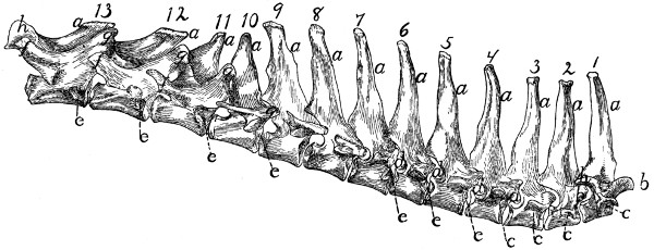
Fig. 4.—Thoracic Vertebræ, Side View.
a, spinous processes; b, cranial articular processes; c, transverse costal facets; d, intervertebral foramina; e, costal facets; f, accessory processes; g, mammillary processes; h, caudal articular processes.
Differential Characters of the Thoracic Vertebræ (Fig. 4).—Following the thoracic vertebræ caudad there is to be seen a gradual increase in the size of the centra brought about by an increase in their craniocaudal and transverse measurements. The dorsoventral measurements remain nearly the same. The costal facets (Fig. 4, e) shift caudad so that on the eleventh, twelfth, and thirteenth thoracic vertebræ each lies entirely on the cranial end of its centrum, while the caudal end of the centrum immediately preceding is not marked by any part of it. In the eleventh thoracic vertebra each costal facet is usually still confluent with the smooth cranial end of the centrum. In the twelfth vertebra the facets are separated by smooth ridges from the cranial end of the vertebra, while in the thirteenth vertebra they are separated by rough ridges.
The spinous processes (a) of the first four are of about the same length. They then decrease in length to the twelfth, while the twelfth and thirteenth are slightly longer than the eleventh. The first ten slope more or less caudad, while the spinous process of the tenth (anticlinal) vertebra is vertical and those of the eleventh, twelfth, and thirteenth point craniad.
Each of the transverse processes of the seventh thoracic vertebra shows a tendency to divide into three tubercles; one of these is directed craniad, the mammillary process (or metapophysis), one caudad, the accessory process (or anapophysis), while the third (transverse process proper) looks ventrad and bears the transverse costal facet. This division becomes more prominent in the succeeding vertebræ, being most marked in the ninth and tenth. In the eleventh, twelfth, and thirteenth vertebræ the mammillary (g) and accessory (f) processes are very pronounced, while the transverse costal facet and that part of the transverse process which bears it have disappeared. The ribs of the eleventh, twelfth, and thirteenth vertebræ are thus attached to their respective centra by their heads alone.
The cranial articular processes (b) are prominent on the first two thoracic vertebræ; back of these they are very small as far as the eleventh, so that the articular facets seem to be borne merely upon the dorsal surface of the cranial edge of the laminæ. In the eleventh, twelfth, and thirteenth the cranial articular processes are large, bearing the articular facets on their medial surfaces, while the mammillary processes appear as tubercles on the lateral surfaces of the articular processes. The caudal articular processes (h) are prominent in the first thoracic, then smaller until the tenth is reached; in the tenth, eleventh, twelfth, and thirteenth they are large and their facets are borne laterally, so as to face the corresponding cranial facets. Thus from the tenth to the thirteenth thoracic vertebra rotary motion is very limited, owing to the interlocking of the articular processes.
The Lumbar Vertebræ. Vertebræ lumbales
(Fig. 5).—The last thoracic vertebræ form the transition to the typical lumbar vertebræ. These are larger than the thoracic vertebræ. The centra are of the form of the centra of the thoracic vertebræ, and increase in length to the sixth, but the seventh is about the length of the first. They increase in breadth to the last.
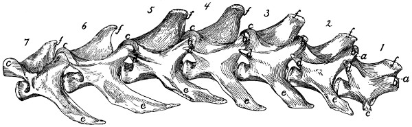
Fig. 5.—Lumbar Vertebræ.
a, cranial articular processes; b, mammillary processes; c, caudal articular processes; d, accessory processes; e, transverse processes; f, spinous processes.
The cranial articular processes (Fig. 5, a) are prominent and directed craniodorsad; they have the facets on their medial surfaces, while their dorsolateral surfaces bear the mammillary processes (b) as prominent tubercles. The caudal articular processes (c) are likewise large; their facets look laterad. When the vertebræ are articulated they are received between the medially directed cranial processes.
The accessory processes (d) are well developed on the first vertebra, diminish in size to the fifth or sixth, and are absent on the seventh and sometimes on the sixth.
The transverse processes (more properly pseudo-transverse processes) (e) arise from the lateral surface of the centra; are flat and are directed ventrocraniolaterad. The first is small, and they increase in length and breadth from the first to the sixth, those of the last being slightly smaller than in the sixth. The free ends of the last four are curved craniad.
The spinous processes (f) are flat and directed craniodorsad. They increase in length to the fifth and then decrease. The first five are knobbed at the end. In a dorsal view the spinous process and cranial articular processes of each vertebra are seen to interlock with the caudal articular processes and accessory processes of the preceding vertebra in such a way as to prevent rotary motion, and this arrangement may be traced craniad as far as the eleventh thoracic vertebra.
Sacral Vertebræ. Vertebræ sacrales
(Figs. 6 and 7).—The three sacral vertebræ are united in the adult into a single bone, the os sacrum, or sacrum. In a kitten the three vertebræ are separate, while in an animal almost mature the first two are united and the third is still separate. The sacrum lies between the last lumbar and the first caudal vertebræ and articulates laterally with the two innominate bones. It is pyramidal, with the base of the pyramid directed craniad, and is perforated by a depressed longitudinal canal, the sacral canal, which is a continuation of the vertebral canal, and by four large foramina dorsally and four ventrally. It may be described as having a cranial end or base and a caudal end or apex, a dorsal, a ventral, and two lateral surfaces.
The base is slightly oblique and presents a smooth transversely oval articular facet (the cranial end of the centrum of the first sacral vertebra), for articulation with the centrum of the last lumbar vertebra. Dorsad of this is the sacral canal, more depressed than the vertebral arch craniad of it. It supports a spinous process (Fig. 6, a) which is directed dorsad. At the junction of its lamina and radix is seen the prominent cranial articular process (b) with sometimes slight indications of a mammillary process on its lateral surface. Laterad of the articular facet is seen the cranial face of the expanded “pseudo-transverse process” (c) of the first sacral vertebra. The ventral border of the base is concave ventrad, forming an arc of about 120 degrees. The apex shows the caudal end of the last sacral centrum. Dorsad of this are the vertebral arch with a very short spinous process (a′), and the caudal articular processes (d). Laterad of the centrum appears the laterally directed thin transverse process (e).
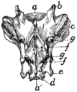
Fig. 6.—Sacrum, Dorsal Surface.
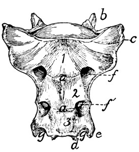
Fig. 7.—Sacrum, Ventral Surface.
Fig. 6.—1, 2, 3, the three sacral vertebræ. a, a′, spinous processes; b, cranial articular process of first sacral vertebra; c, expanded transverse process of first sacral vertebra; d, caudal articular processes of third sacral vertebra; e, transverse processes of third sacral vertebra; f, tubercles formed by fused articular processes of the vertebræ; g, dorsal (or posterior) sacral foramina.
Fig. 7.—1, 2, 3, the three sacral vertebræ. a, the transverse ridges formed by the union of the centra; b, cranial articular processes of first vertebra; c, transverse process of first vertebra; d, caudal articular processes of third vertebra; e, transverse processes of third sacral vertebra; f, f′, ventral (or anterior) sacral foramina; g, notch which helps to form third ventral sacral foramen.
The ventral or pelvic surface (Fig. 7) is smooth, concave craniad, convex caudad, and crossed by two transverse ridges (a) along which are seen the ossified remains of the intervertebral fibro-cartilages. At the ends of the first ridge is a pair of nearly circular ventral (or anterior) sacral foramina (f) for the passage of sacral nerves. At the end of the second ridge is a pair of ventral sacral foramina (f′), smaller than the first pair and continued laterocaudad into shallow grooves for the ventral rami of the sacral nerves. That portion of the bone lying laterad of a line joining the medial borders of these two pairs of foramina is known as the lateral mass of the sacrum and is composed of the fused transverse processes of the sacral vertebræ. At the caudal margin of the ventral surface there is a notch between the lateral mass and the centrum (g). When the caudal vertebræ are articulated, this notch helps to form a foramen for the third sacral nerve.
The dorsal surface (Fig. 6) is narrower at its cranial end than is the ventral surface. Its cranial border bears laterally a pair of cranial articular processes (b) with their medially directed facets and between them it is concave, so that a large dorsal opening is left into the vertebral canal between the last lumbar vertebra and the sacrum. Caudad of the articular processes are two pairs of tubercles (f). These are the fused cranial and caudal articular processes of the sacral vertebræ. Caudad of them are the caudal articular processes of the last sacral vertebra (d). Craniolaterad of the middle and cranial tubercles are dorsal (posterior) sacral foramina (g) for the transmission of the dorsal rami of the sacral nerves. Three spinous processes (a) appear between these rows of tubercles. They decrease in height caudad. That part of the surface included between the spinous process and the tubercles is made up of the fused laminæ of the sacral vertebræ. That part between the tubercles and a line joining the lateral margins of the dorsal (posterior) sacral foramina is formed by the fused radices of the sacral vertebræ.
The lateral surface may be divided into two parts. Craniad is a large rough triangular area with equal sides and with one of its angles directed ventrocraniad. It is the lateral face of the pseudo-transverse process of the first sacral vertebra (Fig. 6, c). A smooth curved surface (the auricular facet) along its ventral edge articulates with the ilium, while the dorsal portion is rough for attachment of ligaments. Caudad is the narrow longitudinal triangular area of the lateral faces of the fused transverse processes of the second and third sacral vertebræ.
Caudal Vertebræ. Vertebræ caudales
(Fig. 1, y, and Figs. 8 and 9).—The caudal vertebræ (21-23 in number) decrease gradually in size to the last one. Caudad they become longer and more slender and lose the character of vertebræ. They become finally reduced to mere centra,—slender rods of bone knobbed or enlarged at their two ends (Fig. 8). The last one is more pointed than the others and bears at its caudal end a small separate conical piece, the rudiment of an additional vertebra.
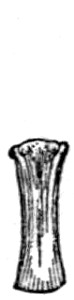
Fig. 8.
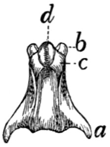
Fig. 9.
Fig. 8.—Caudal Vertebra, from near the caudal end of the tail.
Fig. 9.—Fourth Caudal Vertebra, ventral view. a, transverse processes; b, cranial articular processes; c, hæmal processes; d, chevron bone.
The parts of a typical vertebra—vertebral arch, transverse processes, cranial and caudal articular processes—may be recognized in the vertebræ as far back as the eighth or ninth. The transverse processes (Fig. 9, a) are directed caudad and decrease rapidly in length. They are very small on the ninth vertebra, but may be recognized for a considerable distance back of this. The spinous process disappears at about the fourth caudal vertebra, and the vertebral canal becomes gradually smaller caudad, until on the eighth or ninth vertebra it becomes merely a groove open dorsad.
Caudad of the third vertebra for a considerable distance, each centrum bears on each lateral face at its cranial end a short anterior transverse process, and on its ventral face at its cranial end a pair of rounded tubercles, hæmal processes (c), which articulate with a small pyramidal chevron bone (d) so as to enclose a canal. These structures disappear caudad.
Cervical Vertebræ. Vertebræ cervicales
(Fig. 10).—The cervical vertebræ number seven. The first two of these are so peculiar as to require a separate description, so that the last five may be first considered.
Passing craniad from the fourth thoracic vertebra to the third cervical there is a gradual transition. The centra of the cervical vertebræ are broader and thinner than those of the thoracic vertebræ, while the vertebral arches and vertebral canal are larger (Fig. 11). The caudal end of each centrum is concave and looks dorsocaudad when the centrum is held with its long axis horizontal. The cranial end of the centrum is convex and looks ventrocraniad when the centrum is horizontal. These peculiarities are more marked in the third vertebra than in the seventh. The spinous processes grow rapidly shorter as we pass craniad; the fifth, sixth, and seventh are directed dorsocraniad, the third and fourth dorsad.
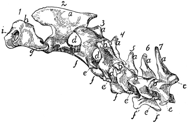
Fig. 10.—Cervical Vertebræ, Side View.
a, spinous processes; b, cranial articular processes; c, caudal articular facet; d, intervertebral foramina; e, transverse process proper; f, processus costarius; g, wing of the atlas; h, dorsal arch of the atlas; i, atlantal foramen.
The caudal articular processes are situated at the junction of the radices and laminæ; their facets (Fig. 10, c) look ventrocaudolaterad. The cranial articular processes also become more prominent than is the rule in the thoracic vertebræ; they are borne at the junction of radix and lamina and have their facets (Fig. 11, b) directed dorsomediad. The cranial and caudal articular processes of each side are joined by a prominent ridge which is most pronounced in the third, fourth, and fifth vertebræ.
The characteristic feature of the cervical vertebræ is their transverse process, so called. In each of them it arises by two roots, one from the centrum and one from the arch. These two roots, which are broad and thin, converge and unite so as to enclose a canal or foramen, the foramen transversarium (Fig. 11, g), for the vertebral artery. Laterad of the foramen the two parts of the process are, in the third cervical, almost completely united, the dorsal part being, however, distinguishable as a tubercle at the caudolateral angle of the thin plate formed by the process as a whole. This dorsal component is the transverse process proper (Figs. 10 and 11, e), while the ventral portion represents a rib, and is hence known as the processus costarius (f). The expanded plate formed by the union of these two processes is directed nearly ventrad and somewhat craniad in the third, fourth, and fifth vertebræ. The two components of the process gradually separate as we pass caudad; in the fourth and fifth vertebræ the part which represents the transverse process proper forms a very prominent tubercle at the caudolateral angle of the plate formed by the processus costarius. In the sixth (Fig. 11) the two parts are almost completely separated; the dorsal part forms (e) a slender knobbed process, while the processus costarius is divided into two portions (f and f′) by a broad lateral notch. In the seventh the ventral part (processus costarius) is usually quite lacking, though sometimes represented by a slender spicule of bone. In the former case the foramen transversarium is of course likewise lacking.
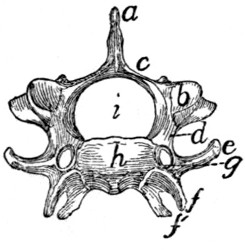
Fig. 11.—Sixth Cervical Vertebra, Cranial End.
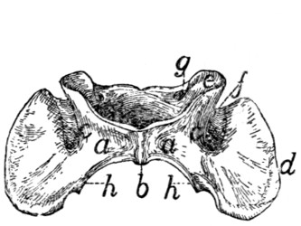
Fig. 12.—Atlas, Ventral View.
Fig. 11.—a, spinous process; b, cranial articular facet; c, lamina; d, radix or pedicle; e, transverse process proper; f, f′, processus costarius; g, foramen transversarium; h, centrum; i, vertebral canal.
Fig. 12.—a, ventral arch; b, tuberculum anterius; c, lateral masses; d, transverse processes; e, cranial articular facets; f, groove connecting the foramen transversarium with the atlantal foramen; g, atlantal foramen; h, caudal articular facets.
The Atlas (Fig. 10, 1; Fig. 12).—The first cervical vertebra or atlas has somewhat the form of a seal ring. The centrum is absent; it has united with the second vertebra to form the odontoid process or dens. Its place is taken in the atlas by a narrow flat arch of bone, narrower at the ends than in the middle, the ventral arch (Fig. 12, a) of the atlas. This connects the lateral, thicker portions of the ring ventrally and bears on its caudal margin a blunt tubercle (tuberculum anterius, Fig. 12, b). Laterally the ring is thickened, forming thus the lateral masses (c) which are continued into the broad thin transverse processes (Fig. 10, g; Fig. 12, d). Each lateral mass bears at its cranial end on its medial surface a concave, pear-shaped facet, cranial (or superior) articular facet, (Fig. 12, e) for articulation with the condyles of the skull. These facets look craniomediad. Dorsad of each is a foramen, the atlantal foramen (Fig. 10, i; Fig. 12, g), which pierces the dorsal arch at its junction with the lateral mass. Caudal to the facet, on the medial face of each lateral mass, within the vertebral canal, is a tubercle. To the two tubercles are attached the transverse ligament (Fig. 14, b) which holds in place the odontoid process (dens) of the axis.
That part of the lateral mass which bears the articular facet projects craniad of the dorsal arch and is separated by a deep triangular notch from the transverse process. Along the bottom of this notch runs a groove (Fig. 12, f), convex craniad, which connects the cranial end of the foramen transversarium and the atlantal foramen. The vertebral artery passes along it. The foramen transversarium is circular. It is bounded laterally by the lateral masses, and dorsally by the dorsal arch.
The dorsal arch (Fig. 10, h) is two to three times as broad as the ventral, has a thick convex cranial border with a median notch, and a thin concave caudal border.
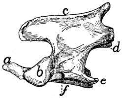
Fig. 13.—Axis or Epistropheus, Side View.
a, odontoid process or dens; b, cranial articular facets; c, spinous process; d, caudal articular facet; e, transverse process; f, foramen transversarium.
The caudal articular facets (Fig. 12, h) are borne by the caudal ends of the lateral masses. They are slightly concave, triangular, and look caudomediad, so that their dorsal borders form with the caudal border of the dorsal arch nearly a semicircle. The transverse processes are flat and directed laterad. The attached margin of each is about two-thirds the length of the thinner free margin. The somewhat thicker caudal end of the transverse process projects further caudad than any other part of the vertebra and is separated by a slight notch from the caudal articular facet. From the bottom of this notch the foramen transversarium extends craniad and opens at the middle of the ventral face of the transverse process.
Epistropheus or Axis (Fig. 10, 2; Fig. 13).—The second cervical vertebra (epistropheus or axis) is not so wide as the atlas but is much longer. Craniad the centrum is continued into a slender conical, toothlike projection, the dens or odontoid process (Fig. 13, a) which represents the centrum of the atlas. The dens is smooth below for articulation with the ventral arch of the atlas. It is rougher above. Laterad of the dens the centrum bears a pair of large cranial articular facets (b) which look craniolaterad. These have each the form of a right-angled triangle with rounded angles, one side of the triangle being nearly horizontal. Each is separated from the articular face of the dens by a roughened groove. The spinous process (c) runs the length of the vertebral arch. It extends craniad of the vertebral arch nearly as far as the dens, as a flat rounded projection. Caudad of the vertebral arch it projects for a short distance as a stout triangular spine. The caudal articular facets (d) are borne on thickenings of the caudolateral portions of the arch; they face almost directly ventrad. The transverse process (e) is slender and triangular and directed nearly caudad. Its apex reaches no farther than the caudal or articular face of the centrum. Its base is traversed by the foramen transversarium (f).
Differential Characters of the Cervical Vertebræ.—It is possible to identify each of the cervical vertebræ:
The first by the absence of the centrum.
The second by the dens or odontoid process.
The third by the small spinous process and slightly marked tubercle of the transverse process, and by a median tubercle on the cranial border of the vertebral arch.
The fourth by the spinous process directed dorsad, and the short thick tubercle of the transverse process not trifid.
The fifth by the spinous process directed craniad, and the more slender spine-like tubercle of the transverse process not trifid.
The sixth by the trifid transverse process.
The seventh by the long spinous process and the slender simple transverse process, and by the usual absence of the foramen transversarium.
LIGAMENTS OF THE VERTEBRAL COLUMN.
Fibro-cartilagines intervertebrales.—The separate vertebræ (except the atlas and axis) are united by the disk-shaped intervertebral fibro-cartilages, which are situated between the centra of the vertebræ. Each consists of a central pulpy portion and a fibrous outer portion, covered by strong intercrossing tendinous fibers which unite with the periosteum of the vertebræ.
Ligamentum longitudinale anterius.—On the ventral face of the centra of the vertebræ, from the atlas to the sacrum, lies a longitudinal ligament, the anterior longitudinal ligament. It is very small, almost rudimentary, in the cervical region: large and strong in the thoracic and lumbar regions.
Ligamentum longitudinale posterius (Fig. 14, a).—A corresponding ligament (posterior longitudinal ligament) lies on the dorsal surface of the centra (therefore within the vertebral canal). It is enlarged between each pair of vertebræ and closely united to the intervertebral fibro-cartilages.
Ligamentum supraspinale.—Between the tips of the spinous processes of the thoracic and lumbar vertebræ extend ligamentous fibers. They are not united to form a distinct band, and can hardly be distinguished from the numerous tendinous fibers of the supraspinous muscles. Together they represent the supraspinous ligament. From the tip of the spinous process of the first thoracic vertebra to the caudal end of the spine of the axis extends a slender strand representing the ligamentum nuchæ or cervical supraspinous ligament. It is imbedded in the superficial muscles of this region, some of which take origin from it.
Ligamentous fibers are also present between the spinous processes of the vertebræ (ligamenta interspinalia): between the transverse processes (ligamenta intertransversaria), and between the vertebral arches (ligamenta flava).
Capsulæ articulares.—The joints between the articular processes are furnished with articular capsules attached about the edges of the articular surfaces. These are larger and looser in the cervical region.
Atlanto-occipital Articulation.—The joint between the atlas and the occipital condyles has a single articular capsule, which is attached about the borders of the articular surfaces of the two bones. This capsule is of course widest laterally, forming indeed two partially separated sacs, which are, however, continuous by a narrow portion across the ventral middle line. This capsule communicates with that which covers the articular surface of the dens, and through this with the capsule between the atlas and axis. That portion of the capsule which covers the space between the ventral arch of the atlas and the occipital bone represents the anterior atlanto-occipital membrane; it is strengthened by a slender median ligamentous strand. The posterior atlanto-occipital membrane covers in the same way the space between the dorsal arch of the atlas and the dorsal edge of the foramen magnum. In it a number of different sets of fibers, with regard to direction and to degree of development, may be distinguished; these have sometimes been considered separate ligaments.
The lateral ligaments of the atlas begin at the lateral angle of the cranial margin of the atlas, at about the junction of its dorsal and ventral arches, and pass cranioventrad to the jugular processes.
Articulation between the Axis and Atlas.—The articular capsule is large and loose, being attached to dorsal and ventral borders of the atlas, about the articular surfaces of the axis, and to the cranial projection of the spine of the atlas. It also passes craniad along the ventral side of the dens and communicates here with the capsule of the atlanto-occipital articulation. In the dorsal part of the capsule a short strong ligamentous strand is developed, connecting the caudal border of the dorsal arch of the atlas with the tip of the cranial projection of the spinous process of the axis.
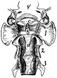
Fig. 14.—Ligaments of the Odontoid Process or Dens.
First three cervical vertebræ and base of the skull, with dorsal surface removed. a, ligamentum longitudinale posterius; b, transverse ligament of the atlas; c, ligamenta alaria; d, odontoid process; e, occipital condyles; 1, 2, 3, the first three cervical vertebræ; 4, basal portion of the occipital bone.
The dens or odontoid process is held in place by the transverse ligament (Fig. 14, b) of the atlas, which passes across the process as it lies within the vertebral canal of the atlas. The transverse ligament is attached at its two ends to the medial surface of the sides of the atlas at about the region where the dorsal and ventral arches of the atlas unite.
From the cranial end of the odontoid process the two ligamenta alaria (Fig. 14, c) diverge craniolaterad to the rough ventromedial angle of the condyles of the occipital bone.
II. RIBS. COSTÆ (Figs. 1 and 15.)
The cat has thirteen pairs of ribs. One of the fifth pair (Fig. 15) may be taken as typical. It is a curved flattened rod of bone attached at its dorsal end to the vertebral column, and at its ventral end to a cartilage (costal cartilage, Fig. 15, f) which serves to unite it to the sternum.
The most convex portion of the bone is known as the angle (e). Each rib presents a convex lateral and a concave medial surface, a cranial and a caudal border. The borders are broad dorsad and narrow ventrad, while the surfaces are narrow dorsad and broad ventrad. The rib has thus the appearance of having been twisted.
The rib ends dorsad in a globular head or capitulum (a), by which it articulates with the costal demifacets of two contiguous thoracic vertebræ. Between the capitulum and angle on the lateral surface is an elevated area, the tubercle, marked by the smooth tubercular facet (c) for articulation with the transverse process of a vertebra. The constricted portion between the head and tubercle is known as the neck (collum) (d). The angle is marked by a projecting process (e) (angular process) on its lateral border, for attachment of a ligament.
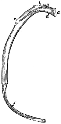
Fig. 15.—Fifth Rib of Left Side, Cranial View.
a, head; b, tubercle; c, tubercular facet; d, neck; e, angle, with angular process; f, cartilage.
The ribs increase in length to the ninth (the ninth and tenth are of the same length) and then decrease to the last. They decrease in breadth behind the fifth. The first is nearly in a dorsoventral plane, while the others have their dorsal ends inclined slightly craniad.

Fig. 16.—Sternum, Ventral View.
a, manubrium; b, the separate pieces forming the body; c, bony part of the xiphoid process (the expanded cartilaginous portion not being shown); d, facet for attachment of first rib.
The tubercles become less prominent as we pass caudad and are absent on the last two or three ribs, which do not articulate with the transverse process.
The first nine ribs (true ribs or costæ veræ) are attached separately to the sternum by their costal cartilages. The last four (false ribs or costæ spuriæ) are not attached separately to the sternum. The costal cartilages of the tenth, eleventh, and twelfth are united to one another at their sternal ends. They may be united also to the ninth costal cartilage or to the sternum by a common cartilage of insertion, or they may be quite free from the sternum. The thirteenth costal cartilages are free (floating ribs).
Ligaments of the Ribs.—The articular surfaces between the head of the rib and the centra, and between the tubercle and the transverse process of the vertebra, have each an articular capsule. There are also a number of small ligamentous bands from the tuberosity and the neck of the rib to the transverse process of the vertebra.
III. STERNUM. (Fig. 16.)
The sternum consists of three portions, a cranial piece or manubrium (a), a caudal piece or xiphoid process (c), and a middle portion or body (corpus), which is divided into a number of segments (b).
To the sternum are united the ventral ends of the first nine ribs. It thus forms the median ventral boundary of the thorax. Since the thorax decreases in dorsoventral measurement craniad, the long axis of the sternum is inclined from its caudal end dorsocraniad, and if continued would strike the vertebral column in the region of the first cervical vertebra.
The manubrium (a) makes up about one-fifth the whole length of the sternum and projects craniad of the first rib. It has the form of a dagger and presents a dorsal surface and two lateral surfaces, the latter uniting ventrad to form a sharp angle. In the middle of the lateral surface near the dorsal margin is an oval articular surface (d) borne on a triangular projection. It looks caudodorsad and is for the first costal cartilage.
The caudal end articulates with the body by a synchondrosis and presents a slightly marked oval facet on each side for the second costal cartilage.
The body consists of six cylindrical pieces (b) enlarged at their ends and movably united by synchondroses. They increase in breadth from the first, and decrease slightly in length and thickness. At the caudal end of each near its ventral border there is a pair of facets looking caudolaterad. They are for the costal cartilages.
The xiphoid process (c) is a broad thin plate of cartilage at its caudal end; bony and cylindrical at its cranial end. It is attached by its base to the last segment of the body by a considerable cartilaginous interval, while the opposite end is free and directed caudoventrad.
The cartilage of the ninth rib is attached to the lateral face of the cartilage between the xiphoid and the body, and just caudad of this the common cartilage of insertion of the tenth, eleventh, and twelfth costal cartilages is attached, if present.
IV. THE SKULL.
The bones of the head consist of the skull proper together with a number of separate bones forming part of the visceral skeleton; these are the lower jaw, the hyoid, and the ear-bones.
The skull proper is considered as divided into cranial and facial portions. The former includes all the bones which take part in bounding the cranial cavity or cavity of the brain; the latter includes the bones which support the face.
The cranial portion of the skull includes all that part enclosing the large cavity which contains the brain. For convenience this portion may be considered as made up of three segments, each of which forms a ring surrounding a part of the cranial cavity. The first or caudal segment or ring consists of the occipital bone (with the interparietal) surrounding the foramen magnum. The second segment consists of the sphenoid ventrad, the parietals laterad and dorsad. Between the first and second segments are intercalated laterally the temporal bones containing the auditory organ. The third segment or ring consists of the presphenoid ventrad, of the frontals laterad and dorsad. The cranial opening of this ring is closed by the lamina cribrosa of the ethmoid.
The cranial portion of the skull therefore contains eleven separate bones: one occipital, one interparietal, two temporals, one sphenoid, two parietals, one presphenoid, two frontals, and one ethmoid.
The facial portion of the skull is much smaller than the cranial, and lies craniad of the latter; it encloses the nasal cavity. It contains the following thirteen bones: two palatines, one vomer, two maxillaries, two lachrymals, two premaxillaries, two nasals, two malar or zygomatic bones. The two halves of the mandible or lower jaw are frequently included in the facial portion of the skull, making in all fifteen separate bones in this part of the skull.
In the following the bones of the skull are first described separately, then an account is given of the skull as a whole.
Occipital Bone. Os occipitale
(Figs. 17 and 18).—The occipital bone forms the most caudal portion of the cranium, entering into the formation of its caudal wall and of its base. It connects the cranium with the vertebral column and surrounds a large opening, the foramen magnum (d), by means of which the cranial cavity communicates with the vertebral canal.
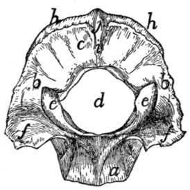
Fig. 17.—Occipital Bone, Caudal or Outer Surface.
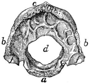
Fig. 18.—Occipital Bone, Inner Surface.
a, basilar portion; b, lateral portions; c, squamous portion; d, foramen magnum; e, occipital condyles; f, jugular processes; g, jugular notch; h, lambdoidal ridge; i, external occipital crest; j, external occipital protuberance.
In young kittens four portions may be distinguished in this bone. These are, a basal portion (the basioccipital bone), two lateral portions (the exoccipital bones), and a dorsal portion (the supraoccipital bone). These four bones remain separate through life in many lower vertebrates. In the adult cat they are completely united into a single bone, but it is convenient to describe this bone as made up of four parts: a basal portion (a) (pars basilare), corresponding to the basioccipital, two lateral portions (b) (partes laterales), corresponding to the exoccipitals, and a squamous portion (c) (squama occipitalis) corresponding to the supraoccipital.
The basilar portion (a) as seen from the dorsal or ventral surface is oblong and flattened. It is broadest at the junction of its middle and last thirds, and tapers toward both ends. It presents a cranial end and a caudal end, a dorsal, a ventral, and two lateral surfaces. Its caudal end, which helps to form the ventral boundary of the foramen magnum, is concave from side to side. The cranial end is transversely elongate, about five times as broad as high, pointed laterally and roughened for attachment to the caudal end of the body of the sphenoid, which has a corresponding form. The joint is a synchondrosis.
The dorsal surface is concave from side to side, forming a longitudinal groove in which rest the pons and medulla. The concavity is more pronounced caudad, where the bone is thinner at its middle.
The ventral surface is marked by three parallel longitudinal ridges. One of them is median and expands caudad into a smooth triangular elevated area which extends to the foramen magnum. The other two ridges run near the lateral edges of the bone. Between them and the median ridge the surface is smooth and is depressed caudad. Laterad of each lateral ridge is a rough triangular surface overlaid in the natural state by the medial edge of the tympanic bulla.
The lateral surfaces are smooth and sharp and abut against the petrous portion of the temporal bone. They pass into the lateral portions of the occipital caudad.
The lateral portions (b) of the occipital arise from the caudal margin of the basilar portion in the transverse plane; a short distance laterad of the line of junction they turn dorsad at an angle of nearly ninety degrees. They form the lateral boundaries of the foramen magnum and pass dorsally into the squamous portion.
The external surface of each presents an elongated elevated spirally curved surface, the occipital condyle (c), for articulation with the atlas. A small part of each condyle is formed from the basal portion. The two condyles are separated from one another ventrally by a narrow notch, and each extends (laterad) along the border of the foramen magnum to a point slightly dorsad of the transverse diameter of the latter.
Laterad of each condyle the bone is elevated into a blunt triangular projection, the jugular process (f), which covers the caudal end of the tympanic bulla. Between the jugular process and the condyle is a deep depression.
The internal surface (Fig. 18) is concave dorsoventrally, following the outline of the foramen magnum. It is convex from side to side. It is smooth except at its outer margin, which is rough for articulation with the mastoid portion of the temporal bone. The cranial face of the jugular process (f) shows a rough concavity for the reception of the bulla tympani. Mediad of the jugular process is a notch (jugular notch) (g) which when the bones are articulated forms part of the boundary of the jugular foramen. Mediad of this notch is a foramen which forms one end of the hypoglossal canal. It passes dorsocaudad into the cranial cavity and transmits the hypoglossal nerve. Dorsad of the hypoglossal canal is the cranial opening of the condyloid canal, which passes caudad and opens just craniad of the dorsal end of the condyle. It transmits a vein. The outer border of this portion is rough for articulation with the petrous and mastoid portions of the temporal.
The squamous portion (c) has the form of a sector of a circle whose arc is a little more than ninety degrees. The central angle of the sector is truncated and bounds the foramen magnum dorsally. The arc of the sector forms the dorsal margin of the bone, while along the radii it passes into the lateral portions. Its dorsal portion is thick and porous; its ventral portion near the foramen magnum is thin and compact.
The external surface (Fig. 17) is marked by a prominent ridge, the lambdoidal ridge (h), parallel with the dorsal border and near to it. The narrow portion of the outer surface that lies dorsad of the ridge forms an angle of about ninety degrees with the remainder of the surface.
A median crest (i) extends ventrad from the middle of the lambdoidal ridge toward the foramen magnum; this is the external occipital crest. At its junction with the lambdoidal ridge it is elevated into a tubercle, the external occipital protuberance (j).
The inner surface (Fig. 18) presents depressions for the convolutions of the cerebellum.
The dorsal border is thick and rough for articulation with the parietals and interparietal. The ventral border abuts on the foramen magnum and is thin and smooth.
Interparietal Bone. Os interparietale
(Fig. 19).—This is a small triangular bone lying between the parietals, with its apex directed craniad, and its base in contact with the squamous portion of the occipital.
Its dorsal surface is arrow-shaped and has its posterior border notched. It is marked by a median crest (part of the sagittal crest) which is continued craniad from the middle of the lambdoidal crest.
The ventral surface is irregularly triangular, smooth, and concave. The three borders are rough for articulation with the parietals and occipital.
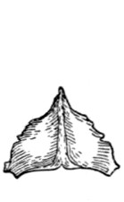
Fig. 19.—Interparietal Bone, Outer Surface.
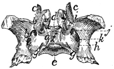
Fig. 20.—Sphenoid Bone, Dorsal or Internal Surface.
Fig. 19, showing the sagittal crest running craniocaudad across its middle.
Fig. 20.—a, body; b, wings; c, pterygoid process; d, tuberculum sellæ; e, dorsum sellæ; f, sella turcica; g, notch which aids in forming the foramen lacerum; h, longitudinal groove of alisphenoid; i, notch which aids in forming the orbital fissure; j, foramen rotundum; k, foramen ovale.
Sphenoid Bone. Os sphenoidale
(Fig. 20).—The sphenoid bone of man is represented in the cat by two entirely distinct bones,—one cranial, the other caudal. The cranial portion may be designated as the presphenoid (Fig. 21); the caudal part will be described as the sphenoid (proper) (Fig. 20).
The sphenoid bone in the kitten is in three parts: a central portion, the basisphenoid, and two lateral portions, the alisphenoids. In many lower vertebrates these three bones are permanently distinct, but in the adult cat they are united to form the sphenoid bone. To these there is added a fourth element, separate in many vertebrates as the pterygoid bone. The sphenoid may thus be described as composed of a central portion, the body (a) (basisphenoid), and of two thin expanded wings (b) (alisphenoids, alæ magnæ of the human sphenoid); each of which has arising from it a thin curved process, the pterygoid process (c), directed craniad and largely made up of the pterygoid bone.
The body of the sphenoid (a) lies in the middle line of the base of the skull. It is wedge-shaped, with the converging sides of the wedge directed laterad and its apex pointed craniad.
It has six surfaces, of which the dorsal and a part of the laterals look into the cranial cavity. The cranial end articulates with the body of the presphenoid, and the caudal with the body of the occipital.
The dorsal surface is triangular, with one apex of the triangle truncated, elevated, and directed craniad. This elevation is the tuberculum sellæ (d). Just caudad of the middle the surface presents a rectangular elevation with rounded angles, the dorsum sellæ (e). The cranial end of the dorsum sellæ presents at each dorsolateral angle a very small smooth tubercle which represents one of the posterior clinoid processes of man. Between this elevation and the elevated cranial end of this surface there is a deep excavation, the sella turcica (f), in which in the natural state is lodged the hypophysis. Near the cranial end of the sella is a small foramen, probably nutrient. At the caudal end of the body a slight notch (g) separates it from the wing: this notch forms a part of the foramen lacerum. Against this notch fits the apex of the petrous bone, and from it a groove (carotid groove) is continued mediocraniad to the sella turcica.
The ventral surface (Fig. 41, 3) is triangular, smooth, and nearly flat; it is marked by a median ridge which is the continuation craniad of the ridge on the ventral face of the basilar portion of the occipital.
Its caudal angles are separated from the rest of the bone by sharp triangular elevations, laterad of which are rough triangular areas, overlaid when the bones are articulated by a triangular spine from the tympanic bulla.
Its lateral surfaces are mostly covered by the wings. They appear at the sides of the elevated cranial end of the dorsal surface as triangular areas.
The caudal end is concave, rough, and has the form of the cranial end of the basilar part of the occipital.
The cranial end is nearly square and rough for articulation with the body of the presphenoid.
The Wing (alisphenoid; ala magna of the human sphenoid) (Fig. 20, b).—This is a thin quadrilateral plate of bone attached by its medial border to nearly the whole of the lateral surface of the body. Its middle portion lies nearly in the same plane as the body, but its ends are curved dorsad so that its internal surface is concave and its external surface is convex. The curvature is most pronounced near the long lateral border, so that this border forms nearly a semicircle.
The internal surface supports the occipital lobe of the cerebrum. It is marked by a rounded groove (h) which is parallel with the lateral surface of the body. The dorsal margin of the groove projects mediad in the form of a sharp ridge which is broadest caudad, where it often reaches nearly to the posterior clinoid process. The groove passes craniad into three foramina. The first (cranial) of these, the orbital fissure (i), is large and lies between the wing, the body, and the pterygoid process. It is incomplete, but is completed by the presphenoid. The second foramen is small and rounded; it is the foramen rotundum (j). The third, foramen ovale (k), is larger and oval and penetrates the wing through about the middle of its longitudinal axis. Another minute foramen penetrates the sphenoid between the wing and the body of the bone, just laterad of the tuberculum sellæ. This foramen is continuous craniad with a groove on the dorsal surface of the pterygoid process; the groove and foramen constitute the pterygoid canal. It transmits a nerve.
The external surface shows the orbital fissure, the foramen rotundum and the foramen ovale, bounded ventrally by a sharp ridge, which is continued onto the pterygoid process. Between this ridge and the body the surface is longitudinally grooved for the tuba auditiva or Eustachian tube.
The semicircular margin of the bone articulates with the squamous portion of the temporal. At the junction of its caudal and middle third there is sometimes a toothlike projection which underlies the root of the zygoma.
The whole of the cranial margin, except the lateral end, articulates with the wing of the presphenoid. At this end the angle formed by the junction of lateral and cranial borders is produced into a flat process, which passes dorsocaudad between the squamous portion of the temporal and the frontal, and articulates by the roughened internal surface of its free end with a similar process from the parietal.
The caudal margin laterad of the groove is bevelled and roughened at the expense of the dorsal surface and is overlaid by the ventral end of the tentorium. Mediad of the groove it projects caudad as a slender point, the lingula of the sphenoid. This is received into a narrow cleft between the apex of the petrous bone and the bulla tympani.
The pterygoid process (c) is a nearly square, thin plate of bone. The medial surface is smooth and concave, the lateral face is convex and marked by two parallel ridges. The medial one of these is continued craniad from the bony septum which separates the orbital fissure from the foramen rotundum, and the lateral one from the septum which separates the foramen rotundum from the foramen ovale. A sharp triangular spine projects laterad from near the caudal end of the lateral ridge.
The two ridges and that part of the lateral surface of the bone included between them form a part of the sphenoid bone known as the pterygoid process of the sphenoid bone, in those cases where the pterygoid is a separate bone.
The remainder of the process is equivalent to the pterygoid bone of other vertebrates.
Between the caudal margin of this bone and the lateral of the two ridges, i.e., between the pterygoid bone and the pterygoid process of the sphenoid, is a long deep fossa, the internal pterygoid fossa (Fig. 40, s). The laterocaudal margin of the pterygoid process projects caudad, as a curved triangular spine, the hamulus or hamular process (Fig. 40, t; Fig. 43, i).
The Presphenoid Bone. Os presphenoidale
(Fig. 21).—In a young cat this bone is in three pieces, a basal portion (presphenoid) and two wings (orbitosphenoid bones). These bones remain distinct throughout life in many lower vertebrates, but in the adult cat they fuse to form a single bone. We may nevertheless conveniently describe this bone as made up of a body (a) (the basisphenoid), and two wings (b), the orbitosphenoids (the alæ parvæ of the human sphenoid).
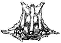
Fig. 21.—Presphenoid Bone, Ventral View.
a, body; b, wings; c, optic foramina.
The body (a) lies in the base of the skull in the median line, craniad of the basisphenoid. It has the form of a rectangular prism about twice as long as broad. It is hollow, and the cavity is divided by a median longitudinal partition into two cavities (sphenoidal sinuses, Fig. 43, l). The sphenoidal sinuses are continued craniad into the cavities of the ethmoid. The body has six surfaces:
The dorsal or internal surface (Fig. 42, n) looks into the cranial cavity and is continuous with the dorsal surface of the wings. The caudal end of the body is depressed, and when united to the basisphenoid aids in forming the cranial wall of the sella turcica. At each caudolateral angle is a short spine, the anterior clinoid process. At about one-third the length of the bone from the caudal end is a transverse groove (chiasmatic groove, Fig. 42, m) for the optic chiasma. Its ends lead into two round foramina (the optic foramina, Fig. 42, l; Fig. 21, c) which pass craniolaterad between the body and the wings of the presphenoid and transmit the optic nerve and the ophthalmic artery.
The ventral surface (Fig. 21) is hour-glass-shaped and marked by a smooth median ridge, continuous with the ridge on the basisphenoid and overlaid at its cranial end by the vomer. The caudal end presents a rough triangular area on each side, for articulation with the pterygoid process of the sphenoid bone, while the cranial end has similar areas overlaid by the nasal portion of the palatine bones.
The lateral surface looks towards the orbitotemporal fossa. It is notched near the caudal end by the ventral border of the optic foramen (c). Caudad of this foramen the surface presents an oblique groove which forms in the natural condition the medial boundary of the orbital fissure.
Craniad of the optic foramen the surface is smooth and marked near its ventral border by a longitudinal ridge which forms part of the dorsal boundary of a fossa, the external pterygoid fossa (Fig. 40, p).
The caudal end presents ventrally a quadrangular rough surface for articulation with the body of the sphenoid. The cranial end presents the two sphenoidal sinuses separated by a median partition.
The median partition articulates by its free border with the lamina perpendicularis of the ethmoid. At its ventral end is the abruptly truncate end of the median ridge of the ventral surface, which is continuous with the ventral cartilaginous portion of the lamina perpendicularis. The lateral walls of the sphenoidal sinuses are continued craniad of the dorsal and ventral walls and of the median partition, and articulate ventrally with the nasal portion of the palatine bones, and dorsally with the orbital plate of the frontal. Between them is received the caudal ends of the labyrinths of the ethmoid in the middle, while between their dorsal edges is received the caudal end of the cribriform plate, and between their ventral edges the expanded end of the vomer.
The wings (b) arise each from nearly the whole of the dorsolateral angle of the body. They form prominent nearly horizontal triangular projections over the optic foramina.
The dorsal and ventral surfaces are smooth and continuous respectively with the dorsal and lateral surfaces of the body. The dorsal surface looks into the cranial cavity, while the ventral looks into the orbitotemporal fossa.
Craniad of the apex of the wing its border articulates with the ventral edge of the orbital portion of the frontal bone. Caudad of the apex the border articulates with the cranial border of the wing of the sphenoid.
Temporal Bone. Os temporale
(Figs. 22 and 23).—This forms a part of the lateral wall of the cranium, filling the gap between the occipital and the sphenoidal segments. It is made up of three portions which are distinct in kittens but somewhat firmly united in adult cats. In lower vertebrates these portions are distinct bones called the Squamous, the Petrous, and the Tympanic bones. In the cat they may be described as the squamous (a), petrous (b), and tympanic (c) portions of the temporal bone.
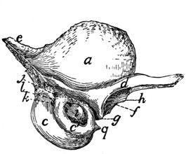
Fig. 22.—Temporal Bone, External Surface.
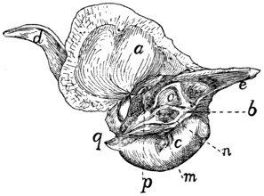
Fig. 23.—Temporal Bone, Internal Surface.
a, squamous portion; b, petrous portion; c, c′, tympanic portion (c, entotympanic; c′, ectotympanic); d, zygomatic process; e, mastoid portion of the petrous; f, mandibular fossa; g, postmandibular process; h, tuberculum articulare; i, external auditory meatus; j, stylomastoid foramen; k, pit for tympanohyal bone; l, mastoid process; m, grooves bounding the jugular foramen; n, internal auditory meatus; o, appendicular fossa; p, hiatus facialis; q, styliform process of tympanic bulla.
The squamous portion (a) (squama temporalis) is thin and oval or has the form of an equilateral triangle with rounded angles, with a curved process, the zygomatic process (d), arising from its ventral border. Its outer surface (Fig. 22) is convex and smooth and gives origin to part of the temporal muscle. Its inner surface (Fig. 23) is concave and smooth except near the margins, where it is bevelled and rough. The roughened border is broader dorsad and caudad. The ventral margin of the bone is turned mediad at its cranial end so that the lateral face of the inverted portion looks ventrad. By this portion of its lateral face the squamous rests upon the tympanic bulla, and its edge articulates with the tentorium and the wing of the sphenoid. The remaining (caudal) portion of the ventral border overlies the mastoid portion (e) of the petrous. By the remainder of its border the bone articulates with the parietal dorsad and with the wing of the sphenoid craniad. The roughened portion of its inner surface overlies the margins of both these bones.
The zygomatic process (d) is formed by the confluence of two roots. One of these starts from the ventral end of the lambdoidal ridge and passes along the ventral margin of the squama dorsad of the external auditory meatus. The other arises abruptly from the cranioventral angle of the bone. The process thus formed is at first broad and passes horizontally laterad and slightly craniad. It soon grows more slender and turns gradually craniad, while at the same time it twists so that the surface which is dorsal at the base becomes medial at the tip; the posterior root which is continuous with the caudal border at the base is continuous with the dorsal border at the apex. On the ventral surface of the base is a transversely elongated concave articular surface, the mandibular fossa (f), for the condyloid process of the lower jaw. Caudad of this is a sharp transverse ridge, the postmandibular or postglenoid process (g), and craniad of the lateral end of the fossa a slight tubercle, the tuberculum articulare (h). Near its apex the zygomatic process is more slender and its ventral border is bevelled for articulation with the malar or zygomatic bone.
The tympanic (Figs. 22 and 23, c; Fig. 24) is expanded into a large hollow olive-shaped bone which is known as the auditory bulla and encloses the tympanic cavity. Its substance is very compact. Unlike the tympanic of most other mammals it is developed from two bones, known as the ectotympanic (Fig. 22, c′) and entotympanic (c). These are strongly marked in young kittens, and can usually be easily distinguished in adult cats. The entotympanic (Figs. 22 and 23, c) forms the larger part of the bulla, constituting its ventral and medial surfaces; it is thin, smooth, and transparent. The ectotympanic (Fig. 22, c′) surrounds the external auditory meatus: it is thicker and more opaque than the entotympanic. The bulla lies ventrad of the squamous, and in an external view conceals a large part of the petrous.
On its lateral surface it presents near the dorsal border an irregular oval opening, that of the external auditory meatus (Fig. 22, i), which leads into the tympanic cavity. Caudad of the external auditory meatus is a nearly dorsoventral groove, which, when the bone is articulated, forms a part of the boundary of the stylomastoid foramen (Fig. 22, j); just ventrad of this groove is a pit (k) which lodges the tip of the tympanohyal bone.
Craniad the bone is produced into a short spine, the styliform process (q), which lies in a horizontal groove in the ventral surface of the basisphenoid. Laterad of this spine is a groove for the tuba auditiva or Eustachian tube.
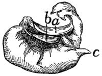
Fig. 24.—Tympanic Bulla, Isolated, Medial Surface.
a, inner end of auditory meatus; b, partition dividing tympanic cavity; c, styliform process.
The medial surface (Fig. 24) presents in the middle near its ventral margin a short triangular spine which lies in the natural state against the ventral surface of the basilar portion of the occipital.
Caudad of this spine the surface is marked by two or three vertical parallel grooves (Fig. 23, m). They indicate the portion of the bone which bounds the jugular foramen, and are possibly impressions of the ninth, tenth, and eleventh nerves.
The dorsal two-thirds of the medial surface is lacking in the disarticulated bulla (Fig. 24), so that the cavity of the bone is exposed. This opening is in the natural state closed by the petrous bone. The caudal end is rough where it is overlaid by the jugular process.
On the inner surface of the tympanic bulla is seen the thickened margin of the inner end of the auditory meatus (Fig. 24, a). To it is attached the membrana tympana. In the median dorsal line this margin is notched for the reception of the incus and head of the malleus. From the lateral wall of the cavity at the line of junction of the ectotympanic and entotympanic a thin bony partition (b) rises. It runs almost directly mediad; is concave dorsally and divides the tympanic cavity into two chambers.
The Petrous Portion (Fig. 23, b, and Fig. 25).—This consists of two parts, a very dense part (the petrous portion proper, Fig. 25), which has the form of a triangular pyramid and encloses the auditory labyrinth, and a less dense part, the mastoid portion (Figs. 22 and 23, e), which is flattened and triangular and is attached by its base to the base of the pyramid.
The petrous portion may be described as having a base and three sides, lateral, dorsal, and medial. It completes the medial wall of the tympanic bulla, so that it is not possible to see it from the exterior of a skull except through the auditory meatus (Fig. 22, i). When the bones of the skull are articulated its dorsal surface is covered by the tentorium and alisphenoid. Its lateral face looks into the tympanic cavity, while the medial face looks into the cranial cavity.
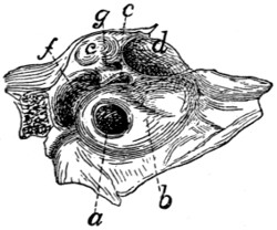
Fig. 25.—Petrous Bone of Right Side, Lateral Surface, Enlarged.
a, fenestra cochleæ; b, promontory; c, fenestra vestibuli; d, fossa for the tensor tympani muscle; e, fossa for incus and malleus; f, fossa continuous with stylomastoid foramen; g, foramen leading to facial canal.
Its lateral face (Fig. 25) (medial wall of the tympanic cavity) presents just ventrad of the middle of its base a large circular foramen, the fenestra cochleæ (a) (or fenestra rotunda), which looks caudolaterad; it leads into the cochlea. The fenestra cochleæ lies at the summit of a nipple-like elevation, the promontory (b), which is continued toward the apex of the bone as a gradually diminishing semicylindrical ridge, due to the presence within it of the bony cochlea. Dorsad of the fenestra cochleæ is the much smaller fenestra vestibuli (c) (or ovalis) which leads into the vestibule. It is occupied in the natural condition by the base of the stapes.
Dorsocraniad of the fenestra vestibuli is a large fossa (d) which contains the tensor tympani muscle. Dorsocaudad of this, partly bounded by the squamous portion of the temporal, is another large fossa (e), the cranial end of which is occupied by the incus, while its caudal end is occupied by the head of the malleus.
Nearly caudad of this fossa and separated from it by an oblique bony septum is a third fossa (f) which is narrow and curved. It is continuous with a notch in the mastoid portion of the bone. When the tympanic is articulated the notch is converted into a foramen (stylomastoid foramen, Fig. 22, j) for the exit of the seventh nerve. The fossa gives passage to the seventh nerve and also lodges the stapedius muscle. A groove may be traced from the stylomastoid foramen to the caudal border of the fossa for the tensor tympani muscle, where it passes into a canal (g). The groove and canal are parts of the canalis facialis or facial canal (aqueductus Fallopii) for the passage of the seventh nerve through the petrous bone.
The medial surface (Fig. 23, b) of the petrous portion shows near its middle a fossa, the internal auditory meatus (n). This is divided by a partition of bone into a dorsal and ventral part. The dorsal portion is the beginning of the facial canal (aqueductus Fallopii) by which the seventh nerve passes through the petrous bone to emerge at the stylomastoid foramen. The ventral portion shows at its bottom several small foramina for the auditory nerve.
Dorsocaudad of the internal auditory meatus is a deep fossa (o) for a small lobe, the so-called appendicular lobe, of the cerebellum. This may be called the appendicular fossa.
The dorsal surface is triangular and presents near its apex a foramen—the hiatus facialis (p), the opening of a canal which joins the canalis facialis and transmits the superficial petrosal branch of the nerve of the pterygoid canal (Vidian nerve). That part of the dorsal surface which lies caudad of the hiatus facialis is known as the tegmen tympani.
The base of the petrous is attached to the mastoid portion (Fig. 23, e).
(For an account of the structures within the petrous bone and the tympanic cavity, see the description of the internal and middle ear.)
The mastoid portion (Figs. 22 and 23, c) is attached by its base to the pyramidal petrous portion, with which it forms an angle of about 120 degrees. It appears in the lateral wall of the skull between the parietal bone and the occipital (Fig. 40, d). The lambdoidal ridge is continued on its outer surface to the caudal border of the external auditory meatus. Caudad of the stylomastoid foramen it forms a slight nipple-like eminence, the mastoid process (Fig. 22, l). Its inner face looks into the cranial cavity.
Parietal Bone. Os parietale
(Figs. 39, 40, and 43, 3).—The parietal bones form the larger part of the lateral and dorsal boundary of the cranial cavity. Each is a thin rectangular bone, compact and curved and with a deeply notched shelf of bone, the tentorium (Fig. 42, e, and Fig. 43, f), projecting inward from near the caudal margin.
The outer surface is smooth and convex. The highest part of the convexity, a little caudad of the middle of the bone, is known as the parietal tubercle or eminence (Fig. 39, d); it marks the point of beginning ossification. An obscure curved ridge (Fig. 39, e), running from the caudodorsal angle or a point craniad of it craniolaterad, indicates the boundary of the origin of the temporal muscle. Near the ventral border the surface is roughened and is covered in the natural state by a part of the squamous portion of the temporal bone.
The inner surface (Fig. 43, 3 and 3′) is smooth and marked by ridges and grooves for the convolutions of the cerebrum. Near the medial border is a ridge which, when the bone is articulated with that of the opposite side, forms a shallow groove for the superior sagittal sinus. Beginning near the middle of the ventral margin and passing dorsad is a groove for the middle meningeal artery. The tentorium (Fig. 43, f) arises from the inner surface near its caudal margin and projects mediad as a thin curved or notched shelf of bone which separates the cerebellar fossa (Fig. 43, I) of the cranium from the cerebral fossa (Fig. 43, II). When the parietals are articulated there is left between the tentoria a large foramen by means of which the two fossæ communicate. The foramen is bounded laterally and dorsally by the free margins of the tentoria, while the ventral end of each tentorium articulates with the alisphenoid, and its dorsal end with the opposite tentorium.
The medial border is straight and is united by suture to the opposite bone.
The cranial border is bevelled at the expense of the inner surface and articulates with the frontal. Just ventrad of the middle of the border projects a sharp spine which fits into a corresponding notch in the caudal border of the frontal.
The ventral border is concave, sharp, and bevelled at the expense of the outer surface, for articulation with the squamous portion of the temporal, except near the cranial end, where it articulates with the wing of the sphenoid.
The caudal border is thick and porous medially, but thin laterally, and bevelled at the expense of the inner surface for articulation with the interparietal and mastoid portion of the temporal.
Frontal Bone. Os frontale
(Figs. 39, 40, and 41, 5; Fig. 43, 8; Fig. 26).—The frontal bones meet one another in the median dorsal line so as to form the roof of the skull between the parietal and nasal bones. A part extends also ventrad, forming a large part of the medial wall of the orbit and a part of the temporal fossa.
The bone may be divided into two portions, a plate forming the cranial portion of the roof of the skull and a part of the roof of the nasal cavity, the frontal plate (Fig. 40, 5), and a part descending into the orbit, the orbital plate (Fig. 40, 5′).
The frontal plate (Fig. 40, 5) is a right-angled triangle with the hypothenuse lateral. Its dorsal surface is convex and smooth. The cranial two-thirds of its lateral border is separated from the orbital fossa by a ridge, the supraorbital arch or margin (Fig. 39, i; Fig. 40, o); the caudal third passes gradually into the temporal fossa. At its cranial angle is a triangular projection, the frontal spine or nasal spine (Fig. 26, a), which fits into a space between the nasal and maxillary bones.
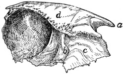
Fig. 26.—Frontal Bone, Medial Surface.
a, frontal spine; b, transverse ridge; c, surface applied to the ethmoid; d, vertical plate of medial border.
The ventral surface is concave and smooth over its caudal one-half and helps to form the cranial part of the brain-case. It presents slight ridges and depressions for convolutions of the cerebrum. At its narrowed middle region the ventral surface is marked by a thick transverse ridge (Fig. 26, b). Caudally the ridge descends by a gentle slope to the level of the ventral surface of the bone. The cranial end of the ridge is pierced by an oval foramen through which the frontal sinus (Fig. 43, m, m′), which lies within the ridge, communicates with the spaces in the ethmoid bone (nasal cavity). Craniad of the ridge the surface (Fig. 26, c) is rough and, together with the raised medial border of the bone and the orbital plate, encloses a rectangular space which in the natural state receives a portion of the labyrinth of the ethmoid. The ventral surface is marked at its medial edge by a thin longitudinal ridge which, when the bones are articulated, is continuous with one of the vertical lamellæ of the ethmoid.
The medial border forms a vertical plate (d), broadest craniad and roughened for articulation with its fellow of the opposite side except at its cranial end, where it articulates with the border of the nasal bone.
The caudal border is roughened, bevelled at the expense of the outer surface, and articulated with the parietal bone except at its ventral end, where it articulates with the alisphenoid.
The lateral border is smooth, and it is here that the orbital plate is joined to the frontal plate at right angles. Along its cranial two-thirds this union is marked by a sharp ridge, the supraorbital margin (Fig. 40, o) or arch. This ridge extends caudolaterad as a triangular projection, the zygomatic (or postorbital) process (Fig. 40, n), which is flattened on its cranioventral face near its extremity and forms part of the boundary of the orbital fossa. At its cranial end the lateral border articulates with the nasal and maxillary bones.
The orbital plate (Fig. 40, 5′) arises from the ventral surface of the lateral border of the frontal plate. It is directed ventrad, is smooth and concave on its outer surface, and forms the dorsal portion of the medial wall of the orbital fossa. Near its ventral border it bears the small ethmoidal foramen, for the artery of the same name.
On the caudal one-half of its inner surface (Fig. 26) it assists the caudal part of the dorsal plate in forming the brain-case. The cranial one-half of its inner surface is marked off from the remainder of the surface by a sharp irregular ridge which is for articulation with the cribriform plate of the ethmoid. Craniad of this the surface is marked by ridges and looks into the nasal cavity.
The cranial margin is produced dorsally in the form of a blunt triangular spine. Mediad of this spine the bone articulates with the lachrymal bone.
The ventral border articulates by its cranial one-third with the orbital plate of the palatine, and by its caudal two-thirds with the body and wing of the presphenoid.
Maxillary Bone. Maxilla
(Figs. 27 and 28).—The maxillary bone forms the cranial and lateral portions of the roof of the mouth. The bones of opposite sides meet craniad, but diverge caudad to enclose the palatal plates of the palatine bones. Each consists of a thick prismatic ventral portion or body (a) and a thin flat plate, the frontal process (b), extending dorsad from the cranial part of the bone.
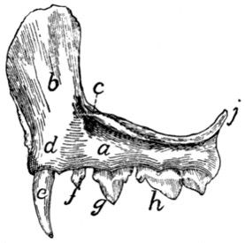
Fig. 27.—Maxillary Bone, Lateral Surface.
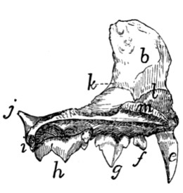
Fig. 28.—Maxillary Bone, Medial Surface.
a, body; b, frontal process; c, infraorbital foramen; d, elevation for root of canine tooth; e, canine tooth; f, first premolar; g, second premolar; h, third premolar; i, molar tooth; j, zygomatic process; k, beginning of lachrymal canal; l, ridge to which the ventral nasal concha is attached; m, nasal crest of palatine process.
The body (a) has the form of a triangular prism whose broader dorsal face looks into the nasal cavity and orbit, while the ventral face looks into the mouth, and the lateral face toward the cheek. From the junction of the dorsal and lateral surfaces at the cranial end the large flat curved frontal process (b) passes dorsad, while the teeth are implanted along the border, alveolar border or process, formed by the junction of the ventral and lateral surfaces.
The lateral surface is continuous with the lateral surface of the frontal process and shows at the base of the frontal process on its caudal border the large infraorbital foramen (Fig. 27, c), for the vessels and nerves of the same name. Near the medial end of the surface is a cylindrical elevation (d) for the root of the canine tooth (e).
The ventral surface is smooth and looks into the roof of the mouth.
On the dorsal surface caudal and cranial halves may be distinguished. The caudal one-half enters into the floor of the orbit. The lateral edge of this portion is divided into two laminæ, between which is received the end of the malar bone. Caudad this edge is prolonged into the short dorsally directed zygomatic process (j). The cranial half of the dorsal surface looks into the nasal cavity and is separated from the caudal half by a sharp vertical lamina of bone which runs caudomediad from the base of the nasal process. To the dorsal edge of this lamina are articulated the lachrymal bone and a part of the palatine. At the point where the lamina joins the base of the nasal process a foramen is seen leading into a canal, the nasolachrymal canal (k). Craniad of the lamina the surface is concave. Where it becomes continuous with the inner edge of the frontal process there is attached to it a thin bone, the ventral nasal concha (or maxilloturbinal), which is rolled into an irregular spiral. The nasolachrymal canal opens ventrad of its cranial end.
The cranial third of this part of the bone projects further mediad than does the rest of the medial border, forming thus the broad palatine process. This is rough on its medial edge for articulation with the premaxillary and the palatine process of the opposite bone. This medial edge rises also dorsally into a low ridge, the nasal crest (m), which is roughened for articulation with the vomer. The caudal two-thirds of the medial edge articulates with the palatine bone.
The cranial end of the bone articulates with the premaxilla.
The caudal end is smooth.
The frontal process (b) presents on its inner surface, which looks into the nasal cavity, certain transverse ridges which are in relation with the ethmoid bone. Its outer surface is smooth. By its cranial border it articulates with the nasal bone dorsally and with the premaxillary bone ventrally.
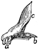
Fig. 29.—Premaxillary Bone, Obliquely Craniolateral Aspect.
a, the three incisor teeth; b, palatal portion of the bone; c, nasal process.
Its dorsal end articulates medially with the nasal spine of the frontal bone, and caudally with the orbital plate of the same bone.
Premaxillary Bone. (Os incisivum BNA.) Premaxilla
(Fig. 29).—The premaxillary bones bear the incisor teeth and form the cranial portion of the roof of the mouth.
Each consists of an irregular, horizontal palatal portion (b) and of a perpendicular nasal process (c) which forms part of the lateral boundary of the nares and enters into the formation of the lateral wall of the nasal cavity.
The palatal portion has in its caudal border a deep notch for the foramen incisivum or anterior palatine canal, which lies between it and the maxillary and transmits blood-vessels and nerves. It articulates with the maxillary bone by this border.
The medial border is raised into a thin crest of bone which, besides forming the medial wall of the foramen incisivum or anterior palatine canal, articulates by its medial border with the bone of the opposite side, forming a sort of median trough (sulcus palatinus) which projects dorsad into the nasal cavity and receives the ventral border of the nasal septum. The caudal end of this border articulates laterad with the maxilla, dorsad with the vomer.
Its craniolateral border bears the incisor teeth (a).
The nasal process (c) presents three surfaces, all elongated and triangular; one, the medial surface, is smooth and concave and looks into the nasal cavity. Its dorsal border is rough for articulation with the nasal bone dorsad, and smooth ventrad where it aids in forming the nares.
The lateral surface is smooth.
The caudal surface is rough for articulation with the maxillary bone.
Nasal Bone. Os nasale
(Fig. 30).—The nasal bones fill the space between the nasal process of the premaxillary, the frontal process of the maxillary, and the nasal spine of the frontal bone (Fig. 39, 7). They thus form part of the dorsal wall of the nasal cavity near the middle line.
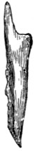
Fig. 30.—Nasal Bone, Dorsal View.
Each may be described as consisting of two elongated triangular lamellæ, one vertical, the other horizontal. The vertical lamella is curved slightly ventrad and has its apex directed craniad. It is applied by its medial surface against the vertical lamella of the opposite bone, the two thus forming a median vertical partition, the nasal crest (Fig. 43, 12), which extends ventrad into the nasal cavity and, by joining the dorsal edge of the lamina perpendicularis, helps to form the internasal septum.
The horizontal lamella is attached to the dorsal margin of the vertical lamella in such a way that its apex lies opposite the base of the vertical lamella. It helps to roof in the nasal cavity, and by its base forms a part of the dorsal boundary of the narial opening. By its lateral margin it articulates with the nasal spine of the frontal at its caudal end, with the frontal process of the maxillary at its middle, and with the nasal process of the premaxilla at its cranial end. The lateral angle of its base projects in a curved line which forms the dorsal part of the lateral boundary of the narial opening.
From the lateral border of the horizontal lamella a bony plate curves ventrad and mediad, enclosing a narrow fossa which receives a part of the ethmoid. This is the concha nasalis superior (nasoturbinal bone).
Ethmoid Bone. Os ethmoidale
(Figs. 31 and 32).—The ethmoid bone closes in the cranial cavity at its cranial end and extends forward into the nasal cavity, which it largely fills.
It consists of a median vertical portion, the lamina perpendicularis (Fig. 43, n; Fig. 42, p), forming a part of the nasal septum, of two lateral portions made of thin sheets of bone variously folded and united—the labyrinths (or ethmoturbinals), which fill the greater part of the nasal cavity; and of a transverse perforated plate, the cribriform plate (lamina cribrosa), attached to the caudal end of the lamina perpendicularis and the labyrinths.
The lamina perpendicularis (Fig. 43, n; Fig. 42, p) is a flat four-sided bone. By its caudal margin it is continuous with the cribriform plate; by its ventral margin it is enclosed by the halves of the vomer; by its dorsal margin it unites with the crest formed by the vertical portion of the nasal bone craniad and with the vertical lamina of the medial margin of the frontal caudad, while its cranial margin is continued into the septal cartilage of the nose. Its lateral faces are smooth and free.
The lamina cribrosa or cribriform plate (Fig. 42, o) is elongated heart-shaped, with the apex of the heart ventrad. Its caudal face is concave and looks into the cranial cavity. It presents three irregular longitudinal rows of holes, one median and two lateral, for the passage of the olfactory fibres from the cranial cavity into the nasal cavity. Its cranial face is continuous along the medial line with the lamina perpendicularis, and at the sides with the labyrinths.
The notch in the heart is directed dorsad and receives the vertical lamina of the medial border of the frontal bone. The apex of the heart articulates with the cranial end of the dorsal surface of the presphenoid. Its lateral margins are articulated with the ethmoidal ridges on the medial surface of the frontal bone.
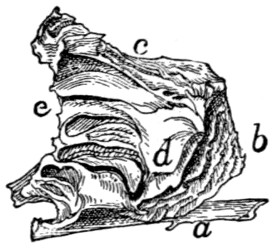
Fig. 31.—Ethmoid and Vomer, Side View.
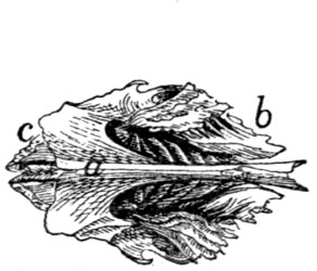
Fig. 32.—Ethmoid and Vomer, Ventral View.
a, vomer; b, vertical cells of the labyrinth of the ethmoid; c, horizontal cell of the same; d, part of the ethmoid that forms the lamina papyracea; e, edge of cribriform plate.
The labyrinths (Figs. 31 and 32) are attached to the cranial face of the lamina cribrosa, one on each side of the lamina perpendicularis. Each is made of thin bony plates irregularly folded so as to enclose spaces, the ethmoid cells. In each may be distinguished a cranial portion (b), in which the cells are nearly vertical, and a caudal portion (c), in which the cells are nearly horizontal.
The medial surfaces are separated by a space from the lamina perpendicularis. This space is broadest along the junction of the horizontal and vertical portions of the labyrinth. There are thus formed two passageways which correspond to the superior meati of human anatomy.
The lateral surfaces come into contact with the frontal process of the maxillary and the orbital plate of the frontal bone. On the lateral surface of each labyrinth there is a thin irregular lamina of bone lying in a dorsoventral longitudinal plane and closing in some of the ethmoid cells laterally (d). A small part of this lamina, situated near the caudoventral angle of the bone, appears in the orbital fossa on the external surface of the skull between the presphenoid, palatine, and frontal bones or between the lachrymal, palatine, and frontal bones. Sometimes in the entire skull two such pieces may be seen, one in each of these positions. This corresponds to the lamina papyracea of human anatomy.
The dorsocaudal angle of each bone is received into the space between the orbital plate of the frontal and the vertical lamina of the medial border of the frontal. Its ventrocaudal angle is received between the cranial extensions of the lateral walls of the presphenoid, while its ventral surface is overlaid caudally by the expanded portion of the vomer, to which it is attached at its caudolateral angles.
Vomer
(Figs. 31 and 32, a).—The vomer consists of two thin laminæ of bone which ensheath the ventral margin of the lamina perpendicularis (or the cartilaginous plate which continues ventrad from this margin) and unite ventrad of it; the two thus form a trough open dorsad.
Each becomes horizontal near its caudal end and at the same time expands. The expanded portion lies ventrad of the labyrinth of the ethmoid, closing in some of its cells: its lateral angles are united with the labyrinths.
At its caudal end the bone articulates with the body of the presphenoid, and each half of it is produced caudad near the middle line into a triangular spine which lies ventrad of the body of the presphenoid. The horizontal portion of the bone helps to separate the olfactory and respiratory passages of the nasal chamber, while its vertical portion contributes to the formation of the nasal septum.
The ventral margin formed by the junction of the two halves of the bone is smooth and free caudad, but at its cranial end is broad and rough for articulation with the palatal processes of the maxillæ.
Palatine Bone. Os palatinum
(Fig. 33).—The palate bone or palatine bone consists of two portions, a horizontal or palatal portion (a) and a perpendicular or nasal portion (b), uniting at an angle of about forty-five degrees.
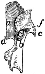
Fig. 33.—Palatine Bone, Dorsal View.
a, horizontal portion; b, perpendicular portion; c, maxillary spine; d, posterior nasal spine; e, sphenopalatine foramen; f, caudal opening of posterior palatine canal.
The horizontal portions (a) of the two bones are received between the maxillary bones and form the caudal and medial part of the roof of the mouth. Each is irregularly quadrilateral in form, with the caudolateral angle produced caudad into a long process which is continuous with the perpendicular portion of the bone. The lateral margin of the horizontal portion articulates over its cranial half with the maxillary bone. At about its middle a short thick maxillary spine (c) projects caudolaterad. The remainder of the lateral margin is directly continuous with the perpendicular plate of the bone. The medial margin is rough for articulation with the corresponding margin of the opposite palatine; the caudal angle of this margin projects caudad as the short posterior nasal spine (d). The caudal margin forms a free edge which bounds the choanæ; it passes laterally into the perpendicular portion.
The ventral surface (Fig. 41, 8) looks into the mouth. Near the middle of its craniolateral margin are two or more small foramina (Fig. 41, q) which form the cranial termination of the posterior palatine canal. The dorsal surface is smooth and looks into the nasal cavity.
The perpendicular or nasal portion (Fig. 33, b) of the palatine is thin and irregularly quadrilateral in form. It is attached by its cranial two-thirds to the dorsal surface of the horizontal portion. The outer surface is concave and looks into the orbital fossa. The inner surface is convex and looks into the nasal cavity.
The perpendicular portion is marked by two foramina just craniad of the middle. The larger dorsal oval foramen is the sphenopalatine foramen (e). The smaller ventral foramen is the caudal opening of the posterior palatine canal (f). From this opening the canal passes craniomediad, lying in the substance of the palatine bone; it opens on the ventral surface of the horizontal portion at the small openings previously described (Fig. 41, q).
By its cranial margin it articulates with the lachrymal bone. By its dorsal margin it articulates craniad with the orbital plate of the frontal: with the lamina papyracea at its middle, and with the body of the presphenoid caudad. The caudal half of the dorsal margin is partially divided into two lamellæ with a rough surface between them: this rough surface lies against the ventral surface of the presphenoid. The caudal margin articulates with the pterygoid portion of the sphenoid.
Lachrymal Bone. Os lachrymale
(Fig. 34; Fig. 39, 10).—The lachrymal bone is a thin pentagonal scale of bone filling the interval between the horizontal plate of the palatine, the maxillary, and the orbital plate of the frontal. Its outer surface looks into the orbit, its inner surface into the nasal cavity.
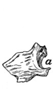
Fig. 34.—Lachrymal Bone of Left Side, External Surface.
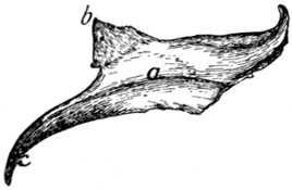
Fig. 35.—Malar Bone of Right Side, Lateral Surface.
Fig. 34.—a, notch forming the beginning of the lachrymal canal.
Fig. 35.—a, ridge for origin of the masseter muscle; b, frontal process; c, zygomatic process.
Near the middle of its cranial border it is notched obliquely by a foramen (a), the beginning of the nasolachrymal canal.
Malar Bone. Jugal Bone. Os zygomaticum
(Fig. 35).—The malar or zygomatic bone is a flat curved plate of bone which forms the lateral wall of the orbit and together with the zygomatic process of the temporal forms the zygomatic arch. Its outer surface is smooth and marked by a longitudinal ridge (a) for attachment of the masseter muscle.
At its caudal end the bone is continued into two processes: one, the frontal process or orbital process (b), is a triangular spine of bone directed caudomediad; when the bones are articulated it lies opposite the zygomatic process of the frontal to which it is joined by a ligament (orbital ligament). The other, zygomatic process (c) of the malar bone, extends ventrocaudad and articulates with a similar process from the temporal to form the zygomatic arch above mentioned.
Its inner surface is smooth and looks into the orbit, except that of the zygomatic process, which looks into the temporal fossa.
Its cranial border is roughened at the expense of both surfaces and articulates with the maxillary bone. Its other borders are smooth except the dorsal border of the zygomatic process, which is roughened for attachment to the zygomatic process of the temporal.
The Mandible. Mandibula
(Figs. 36 and 37).—The mandible (or inferior maxillary bone) is composed of two halves which come together at the cranial end and form the lower jaw. At its caudal end each half articulates with the temporal bone at the mandibular fossa, and at its cranial end it joins the opposite bone, the suture being known as the symphysis of the jaw (symphysis menti) (Fig. 37, a).
Each half consists of a horizontal portion, the body (b), bearing teeth on one of its borders (the alveolar border), and of a vertical portion, the ramus (c).
The body (b) has the form of a flattened cylinder and has two surfaces and two borders. The lateral surface (Fig. 36) is smooth and presents near its cranial end a foramen (or sometimes two), the mental foramen (d), forming the cranial termination of the mandibular canal. At its caudal end is a deep fossa continuing on to the ramus, the coronoid fossa, or masseteric fossa (e).
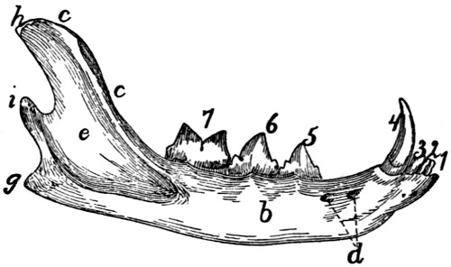
Fig. 36.—Mandible, Lateral Surface.
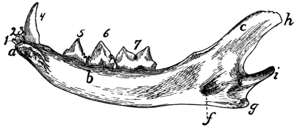
Fig. 37.—Mandible, Medial Surface.
a, symphysis; b, body; c, ramus; d, mental foramina; e, coronoid fossa; f, mandibular foramen; g, angular process; h, coronoid process; i, condyloid process; 1, 2, 3, the three incisor teeth; 4, the canine tooth; 5, 6, the premolars; 7, the molar tooth.
The medial surface (Fig. 37) is smooth and has near its caudal end a foramen, the mandibular foramen (f), which communicates with the mandibular canal leading lengthwise through the bone to the mental foramen. The cranial end is roughened for attachment to the bone of the opposite side.
The ventral border is smooth and rounded; it ends caudally in a blunt point, the angular process (g). The dorsal (alveolar) border is slightly curved and bears the sockets (alveoli) for the teeth. It is continuous with the cranial margin of the coronoid process.
The ramus is divided into two portions, the coronoid process (h) and the condyloid process (i). The coronoid process (h) extends dorsocaudad as a thin plate of bone with smooth surfaces and borders. Its outer surface is partly occupied by the coronoid fossa (e). The condyloid process (i) has the form of a semicylindrical transverse piece of bone attached to the caudal margin of the coronoid process. It articulates with the mandibular fossa of the temporal bone.
Hyoid Bone. Os hyoideum
(Fig. 38 and Fig. 104).—The hyoid bone forms the support for the tongue and gives origin to muscles passing to the tongue and larynx. It also supports the thyroid cartilage (Fig. 104, 1).
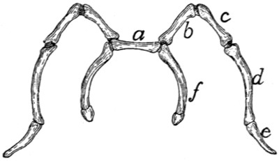
Fig. 38.—Hyoid Bone, Dorsal View.
a, body; b, c, d, e, cranial cornu; f, caudal cornu; b, ceratohyal; c, epihyal; d, stylohyal; e, tympanohyal; f, thyrohyal.
It consists of a transverse bony bar, the body (Fig. 38, a) and of two cornua or horns attached to each end of the body.
The cranial cornu (lesser cornu of human anatomy) is the longer (Fig. 38, b-e). Each arises from the cranial face of the body at its lateral end, curves laterad, and then caudodorsad. It consists of four bony pieces movably united by cartilage.
The terminal piece is the tympanohyal (e); it is imbedded in the tympanic bulla just ventrad of the stylomastoid foramen. It is not therefore seen attached to the cornu after the latter has been separated from the skull. The other pieces become successively shorter toward the body, and are called stylohyal (d), epihyal (c), and ceratohyal (b).
The caudal cornua (f) (greater cornua of human anatomy) arise from the ends of the body. Each consists of a single piece of bone, the thyrohyal (f), which passes caudolaterad; its free end is united to a process of the thyroid cartilage (Fig. 104, 1).
The Skull as a Whole.
—In the following description of the skull as a whole the mandible, hyoid, and ear-bones are not included.
The skull forms a bony box which contains the brain and is produced craniad into the facial portion which encloses the nasal cavity and forms the framework of the face.
In dorsal view (Fig. 39) the skull presents a smooth convex surface, broadest caudad, with the two zygomatic arches (g) curving out some distance laterally. The following bones are visible in dorsal view: the occipital (1), interparietal (2), parietals (3), temporals (4), frontals (5), malar or zygomatic bones (6), nasals (7), maxillaries (8), premaxillaries (9), and lachrymals (10).
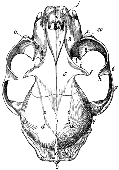
Fig. 39.—Skull, Dorsal Surface.
1, occipital bone; 2, interparietal bone; 3, parietal bones; 4, temporal; 5, frontal; 6, malar; 7, nasal; 8, maxillary; 9, premaxillary; 10, lachrymal, a, lambdoidal ridge; b, external occipital tubercle; c, sagittal crest; d, parietal eminence; e, line which forms the dorsal boundary of the temporal fossa; f, zygomatic process of the frontal; g, zygomatic arch; h, frontal process of the malar; i, supraorbital arch; j, nares; k, foramen incisivum or anterior palatine foramen; l, sphenopalatine foramen; m, zygomatic process of the temporal; n, infraorbital foramen; o, opening of lachrymal duct.
The caudal boundary of the dorsal surface is marked by the prominent lambdoidal ridge (a) which passes from the middle cranioventrad along each side to the root of the zygomatic arch: it is borne by the occipital and temporal bones. From the middle of the lambdoidal ridge a second ridge, the sagittal crest (c), passes craniad in the middle line across the interparietal bone: it varies greatly in extent, reaching in a very old and muscular cat to the cranial border of the parietals, while in kittens it does not exist. The most prominent portions of the skull in this region, just craniad of the middle of the parietal bones, are known as the parietal tubercles or eminences (d). A faint curved line (e) runs from the cranial end of the sagittal crest craniolaterad to the base of the zygomatic process of the frontal: it marks the dorsal boundary of the origin of the temporal muscle, and may therefore be considered the dorsal boundary of the temporal fossa. This fossa extends from its dorsal boundary as far laterad and caudad as the lambdoidal ridge (a), and as far craniad as a line connecting the tip of the zygomatic process of the frontal (f) with the frontal process of the malar (h). The temporal muscle takes origin from its surface.