THE MUSCLES.
Reading edition
I. THE MUSCLES OF THE SKIN. (Fig. 62.)
M. cutaneus maximus (Fig. 62, b).—This is a very large, thin muscle which covers almost the whole side of the body. It arises from the outer surface of the latissimus dorsi (Fig. 68, m) near its ventral end and from the bicipital arch (Fig. 65, t′) in the axilla; from the linea alba for a considerable distance (two or three inches) caudad of the base of the xiphoid process, and from the thorax over a line joining the axilla and the base of the xiphoid. Sometimes a few fibres take origin from the fascia which covers the pectoantibrachialis on the ventral side of the arm.
From their origin the fibres diverge. The cranial ones curve about the base of the forelimb and are inserted into the skin at or near the middle line caudad of the first thoracic vertebra. The most cranial fibres of all are inserted about one to one and a half centimeters from the middorsal line; thence the line of insertion approaches the middorsal line to reach it at about the eighth or ninth thoracic vertebra. The middle fibres run parallel to the middorsal line in the lumbar and sacral regions as far as the root of the tail, a small bundle passing onto the dorsal side of the tail, another onto the ventral side. The fibres of the caudal portion pass onto the thigh, the ventral ones running in the fold of skin which stretches from thigh to abdomen, and are finally lost in the fascia along a line connecting the knee and the root of the tail. A strong fascia connects the adjacent borders of the cutaneus and platysma and lies over the scapular region.
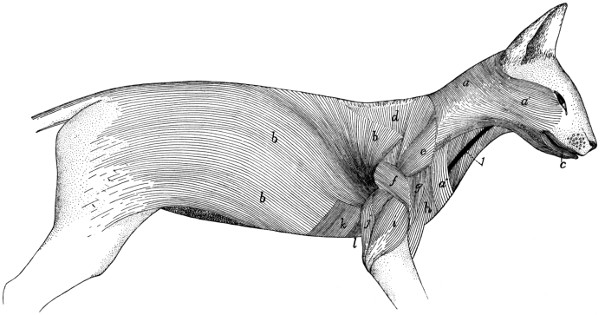
Fig. 62.—Muscles of the Skin.
a, a′, a″, M. platysma; b, M. cutaneus maximus; c, M. orbicularis oris. Between the platysma and the cutaneus maximus are seen the following deeper muscles: d, M. acromio-trapezius; e, M. levator scapulæ ventralis; f, M. spino-deltoideus; g, M. acromio-deltoideus; h, M. clavobrachialis; i, caput laterale of M. triceps brachii; j, caput longum of M. triceps brachii; k, M. xiphi-humeralis; l, M. pectoralis minor. 1, external jugular vein.]
This muscle lies immediately beneath the integument. It covers the first layer of body muscles.
Action.—Moves the skin.
M. platysma (Figs. 62 and 64, a, a′, a″).—This muscle forms a thin layer of fibres covering the sides of the neck and face, in close relation with the integument. Several more or less distinct portions may be distinguished.
(1) Most of the fibres (a) arise from the middorsal line, from the occiput to the first thoracic vertebra, in a narrow fascia common to the muscles of the two sides. The most cranial fibres of this region arise as a small bundle from the external occipital crest, beneath the levator auris longus (Fig. 63, g, g′).
From this origin in the middle line the fibres pass craniolaterad. The most cranial fibres curve about the ventral side of the ear and pass toward the caudal angle of the eye, where they unite with fibres of the zygomaticus (Fig. 64, d) or corrugator supercilii lateralis (Fig. 64, k), or pass to the lower eyelid. Caudad of these the fibres cover the side of the face and become lost among the facial muscles, some passing to the lower eyelid, some to the fibrous pad which supports the vibrissæ, some to the angle of the mouth, some to the lower lip. The most ventral fibres meet the fibres of the opposite muscle just ventrad of the symphysis of the mandible.
The ventral free border of the platysma is separated on the ventral side of the neck from the border of the opposite muscle by a wedge-shaped area having its point at the symphysis menti.
The fibres of this portion of the muscle are interrupted by an attachment to the skin, along a line passing from the base of the ear to about the middle of the coracoid border of the scapula, The dorsal (a) and ventral (a′) portions of the muscle, separated by this line of attachment, are sometimes described as separate muscles (the supercervicocutaneus and cervicofacial, respectively, of Strauss-Durckheim).
(2) A band of fibres one or two centimeters across (a″) arises in the fascia of the side of the neck just craniad of the middle of the coracoid border of the scapula, and passes caudoventrad toward the manubrium, its fibres crossing the fibres of the first part of the platysma at right angles. These fibres become lost in the fascia ventrad of the manubrium, or pass across the middle line to intermingle with the corresponding fibres of the opposite side. This portion of the platysma is sometimes absent.
The platysma is everywhere subcutaneous, except at its dorsocranial angle, where a small bundle of fibres is covered by the levator longus auris. It covers the deeper muscles of the neck and head. Closely attached to its inner surface are the submentalis and depressor conchæ, whose fibres bridge over the ventral interval between the borders of the platysmas of opposite sides.
Action.—Moves the skin of the face and neck.
II. THE MUSCLES OF THE HEAD.
A. Superficial Muscles.
—The most superficial layer of muscles on the face and head is formed by differentiation of the fibres of the platysma. The muscles thus formed are not clearly distinct from each other; in this region sets of fibres differing in direction and in origin or insertion receive separate names even though the different sets of fibres are closely interwoven.
In the quadrangle on the dorsal surface of the head enclosed between the two eyes and the two ears, a thin superficial sheet of fibres is found, in which a number of different sets may be distinguished (Fig. 63). These have received the following names.
M. intermedius scutulorum (Fig. 63, a).—This consists of a broad thin sheet of transverse fibres between the two external ears. The fibres are attached at either end to the scutiform cartilage (1) of the two ears, and pass without interruption across the middle line. At its cranial edge this muscle is continuous with the corrugator supercilii medialis (b); at its lateral edge with the frontoscutularis; at its caudal edge with the levator auris longus (g).
Relations.—Outer surface with the integument. Inner surface with the galea aponeurotica (to which the muscle is closely united), the epicranius muscle (h) and the temporal muscle (n).
Action.—Draws the two ears dorsad, toward the middle line.
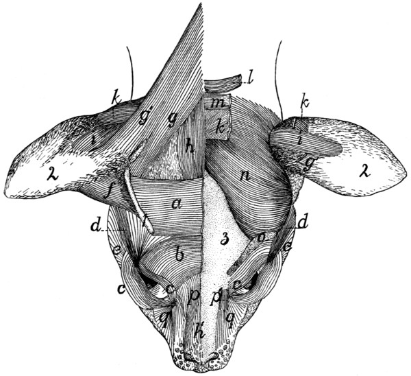
Fig. 63.—Muscles on the Dorsal Surface of the Head.
On the right side are shown the superficial muscles; on the left side the superficial muscles have mostly been removed, exposing the deeper muscles and the bone. a, M. intermedius scutulorum; b, M. corrugator supercilii medialis; c, M. orbicularis oculi; d, M. corrugator supercilii lateralis; e, cranial end fibres of M. platysma; f, M. adductor auris superior; g, g′, M. levator auris longus (g, cranial portion; g′, caudal portion); h, h′, M. epicranius (h, M. occipitalis; h′ M. frontalis); i, M. transversus auriculæ; k, M. auricularis superior (cut on the left side); l, M. abductor auris brevis; m, M. abductor auris longus; n, M. temporalis; o, cut origin of M. frontoscutularis; p, p′, M. levator labii superioris alæque nasi (p′, the origin from the maxillary bone); q, angular head of M. quadratus labii superioris. 1, scutiform cartilage; 2, external ear; 3, bones of the skull.
M. corrugator supercilii medialis (b).—This consists of a thin sheet of scattered fibres lying craniad of the last and intermingling with it. The fibres take origin near the middle line, pass laterad, then curve craniad, converging, to be inserted into the whole extent of the upper eyelid, especially near the caudal angle. Here the fibres unite with those of the orbicularis oculi (c).
This muscle is continuous caudad with the intermedius scutulorum (a), craniad with the orbicularis oculi (c); laterad with the corrugator supercilii lateralis (d). Toward the medial side the fibres lose themselves in a tendinous sheet that joins the galea aponeurotica.
Relations.—Outer surface with the integument. Inner surface with the frontoscutularis and the skull.
Action.—Raises the upper eyelid.
M. orbicularis oculi (Fig. 63, c; Fig. 64, s).—This consists of two thin bands of muscle-fibres which lie one in either eyelid parallel to its border and unite at the angle of the eye.
Origin by short tendon-fibres from a tubercle on the surface of the frontal process of the maxillary bone just dorsad of the orbital end of the lachrymal canal and between the two parts of the quadratus labii superioris (Fig. 63, p and q).
The muscle splits into two parts which pass into the two eyelids. At the outer angle of the eye the two bands unite by the intervention of tendon-fibres between the muscle-fibre bundles of the two.
Relations.—Outer surface with the integument. Inner surface with the inner membrane of the eyelid.
Action.—Closes the eye.
M. corrugator supercilii lateralis (Fig. 63, d; Fig. 64, k).—This consists of a number of scattered fibres which arise from among the fibres of the frontoscutularis, and from the tendon lying just craniad of the external opening of the ear, to which are united also parts of the zygomaticus (Fig. 64, d) and submentalis (Fig. 64, c). The fibres pass craniad, converging so as to form a narrow band which is inserted at the caudolateral angle of the eye, where it unites with the orbicularis oculi (Fig. 64, s). This muscle is continuous on the medial side with the corrugator supercilii medialis (Fig. 64, j) and the frontoauricularis, on the lateral side with the platysma.
Relations.—Outer surface with the integument. Inner surface with the frontal bone and the frontoscutularis.
Action.—Pulls the angle of the eye caudad; at the same time pulls the external ear craniad.
M. frontoauricularis.—A few of the fibres which are attached along the upper eyelid sometimes pass dorsocaudad, mingled with fibres of the corrugatores supercilii medialis and lateralis and the frontoscutularis, to the craniomedial angle of the auricular cartilage, where they unite with the fibres of the adductor auris superior. These fibres are sometimes distinguished as the frontoauricularis muscle.
M. levator auris longus (Fig. 63, g and g′). (Part of the auricularis posterior of man.)—This lies on the caudal half of the dorsal surface of the head, forming a laterocaudal continuation of the intermedius scutulorum (a).
Origin from the middle line of the neck dorsad of the atlas, and from the sagittal crest for about one centimeter craniad of the external occipital tubercle. The fibres form a broad thin sheet which passes craniolaterad as far as the caudal end of the scutiform cartilage (1). Here the muscle divides; the major portion is attached to the scutiform cartilage (1), its fibres intermingling with those of the intermedius scutulorum (a). The caudal portion of the muscle (g′) passes onto the surface of the auricle, extending one or two centimeters distad; here it is inserted on an oblique line which lies directly craniad of the insertion of the transversus auriculæ (i).
The caudal portion of this muscle (g′) having origin above the atlas and insertion on the auricle, is sometimes considered a separate muscle, the supercervicoauricular or cervicoauricular. The cranial portion (g) with origin on the sagittal crest and insertion on the scutiform cartilage might be distinguished as the occipitoscutularis.
The levator auris longus is continuous at its cranial end medially with M. epicranius (h); laterally with M. intermedius scutulorum (a).
Relations.—Outer surface with the integument. Inner surface with the temporal muscle (n), the auricularis superior (k), the abductor longus auris (m), a narrow strand of the platysma, and the clavotrapezius.
Action.—Pulls the external ear dorsocaudad.
The four following muscles lie partly or entirely beneath those already described; they are doubtless formed as differentiations of the inner layers of the platysma.
M. auricularis superior (or attollens auris) (Fig. 63, k).—This muscle forms a band about one centimeter broad lying just beneath the levator longus (g).
Origin on the sagittal crest for about one centimeter craniad of the interparietal bone. The muscle passes laterad onto the surface of the auricle and is inserted into the auricular cartilage a little caudad of the middle of its convex surface. At its lateral end the cranial margin is closely united to the under surface of the levator auris longus (g).
Relations.—Outer surface at the medial end with the epicranius (h), the abductor auris longus (m), and levator auris longus (g); at the lateral end with the skin. Inner surface with the temporal muscle (n) and the auricular cartilage.
Action.—Pulls the external ear dorsad.
M. abductor auris longus (Fig. 63, m; Fig. 64, q). (Part of the auricularis posterior of man.)
Origin on the sagittal crest dorsad of the interparietal bone, caudad of that of the auricularis superior (k), which it partly covers.
The muscle passes laterad as a flat band 8 to 10 millimeters wide over the caudal surface of the concha of the ear, and is inserted (Fig. 64, q) on the lateral surface of the eminentia conchæ, caudad of the antitragus.
Relations.—Outer surface with the levator auris longus (g) and the integument. Inner surface with the auricularis superior (k), the abductor auris brevis (l), and the concha.
Action.—Pulls the external ear caudad.
M. abductor auris brevis (Fig. 63, l).
Origin by a tendon from the lambdoidal crest for one or two centimeters laterad of the middle.
The muscle passes lateroventrad lying beneath the abductor longus (m) as a flat band 6 to 8 millimeters wide which is inserted into the medial surface of the most proximal portion of the concha, just distad of its junction with the cartilaginous auditory meatus.
Relations.—Outer surface with the abductor longus (m), a small strand of the platysma and the concha. Inner surface with the temporal muscle (n).
Action.—Pulls the concha caudad.
M. epicranius (or occipitofrontalis) (Fig. 63, h and h′).
Origin on the sagittal crest or suture, just craniad of the origin of the levator longus auris, to the inner surface of which this muscle is closely united. From the origin the fibres pass craniad forming a band (h) about 8 or 10 millimeters in width, the two muscles lying close to one another and partly united in the middle line. About two centimeters craniad of their origin the fibres of both muscles end in a tendinous sheet, the galea aponeurotica, which covers the surface of the skull in the region between the ears and eyes, and is formed by the inner surface of the intermedius scutulorum (a) and other muscles of this region. The galea aponeurotica passes craniad onto the surface of the nose, where it gives origin again to a thin sheet of muscle-fibres (h′) which are inserted into the integument near the cranial ends of the nasal bones.
M. epicranius is thus formed of two muscular portions (h and h′), connected by a long tendinous sheet. The caudal portion is frequently distinguished as the occipital muscle (M. occipitalis, h), the cranial portion, on the nose, as the frontal muscle (M. frontalis, h′).
Relations.—Outer surface of the occipitalis (h) with the intermedius scutulorum (a), the levator auris longus (g), and the integument; inner surface with the auricularis superior (h), the abductor auris longus, and the bone. Outer surface of the frontalis (h′) with the integument; inner surface with the bones of the skull.
Action.—Moves the integument of the dorsal surface of the head and of the nose.
Ventrad of the external ear the following three differentiated portions of the platysma may be distinguished.
M. zygomaticus (major) (Fig. 64, d).—A slender band connecting the angle of the mouth with the scutiform cartilage of the ear.
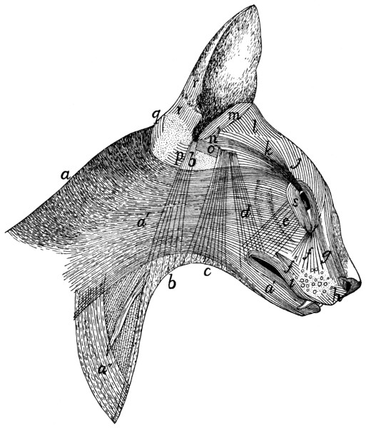
Fig. 64.—Superficial Muscles on the Lateral Surface of the Head and Neck.
a, a′, a″, M. platysma; b, M. depressor conchæ; c, M. submentalis; d, M. zygomaticus (b, c, and d lie beneath the platysma and are seen through it); e, M. zygomaticus minor; f′, M. caninus; f, g, parts of M. quadratus labii superioris (f, angular head; g, M. levator labii superioris alæque nasi); h, M. myrtiformis; i, M. orbicularis oris; j, M. corrugator supercilii medialis; k, M. corrugator supercilii lateralis; l, M. adductor auris superior; m, M. helicis; n, M. antitragicus; o, M. adductor auris inferior; p, M. conchæus externus; q, M. abductor auris longus; r, parts of M. auricularis externus; s, M. orbicularis oculi.
At the angle of the mouth the fibres arise from among those of the orbicularis oris (i), from both the upper and the lower lip. They pass thence dorsocaudad across the zygomatic arch as a band 8 or 10 millimeters wide which is attached to a tendinous aponeurosis lying just craniad of the external opening of the ear. To this aponeurosis are attached also, wholly or partly, the corrugator supercilii lateralis (k) and the submentalis (c). The aponeurosis is united with the caudoventral angle of the scutiform cartilage and with the ventral edge of the cranial margin of the auricular cartilage.
This muscle is united at its cranial end with the orbicularis oris (i); throughout its length more or less with the platysma (a, a′); at its caudodorsal end with the corrugator supercilii lateralis (k) on its medial side, and with the submentalis (c) on its lateral side.
Relations.—Outer surface with the integument and the platysma (a′). Inner surface with the masseter, temporal, frontoscutularis, and adductor auris inferior (o).
Action.—Draws the angle of the mouth dorsocaudad; the external ear ventrocraniad.
M. submentalis (Fig. 64, c).—A flat band, similar to the last, and closely connected for some distance to its caudal border.
It arises as scattered fibres near the ventral middle line at about the level of the larynx, the fibres of the opposite muscles crossing the middle line and intermingling. The fibres pass dorsad, converging so as to form a narrower band, which unites dorsad with the zygomaticus (d), to be inserted with it into the tendinous aponeurosis above described.
Relations.—Outer surface with the integument and platysma (a′). Inner surface with the muscles of the hyoid, the digastricus, the parotid gland, the masseter, the zygomatic arch and the temporal muscle.
Action.—Draws the external ear ventrad.
M. depressor conchæ (Fig. 64, b).—A thin band of fibres, caudad of the last and parallel with it. The muscle arises as scattered fibres on the ventral surface of the neck, one or two centimeters craniad of the manubrium, the fibres of the muscles of opposite sides crossing the middle line and interdigitating. They pass dorsad, gathering together to form a narrow band which is inserted into the summit of the antitragus.
Relations.—Outer surface with the skin at the ventral end, and with the platysma (a′) further dorsad. Inner surface with the deep muscles of the neck and with the parotid gland.
Action.—Draws the external ear ventrad.
Lying deeper than the muscles thus far described, but closely connected with a number of them, are the following:
M. frontoscutularis. Origin (Fig. 63, o) on the frontal bone, along the supraorbital margin, from the craniomedial angle of the eye caudad to the zygomatic process of the frontal.
The fibres form a large muscle which passes dorsocaudad to the scutiform cartilage and is mostly attached along its ventrolateral border. Some of the outer fibres, however, pass distad of the scutiform cartilage toward the cartilage of the ear, thus joining the adductor auris superior (f).
The outer surface of this muscle is closely connected with fibres of the intermedius scutulorum (a), the corrugatores supercilii medialis (b) and lateralis (d), and the frontoauricularis, when this exists. The outer surface is covered near the origin by the orbicularis oculi (c), farther dorsad by the muscles just mentioned and the integument. The inner surface is in relation with the frontal bone and the adductor auris inferior (Fig. 64, o).
Action.—Pulls the ear craniad.
M. adductor auris inferior. (Fig. 64, o.) (Part of the auricularis anterior of man.)—A small muscle lying beneath the frontoscutularis.
Origin on the ligament which connects the zygomatic process of the frontal with the frontal process of the malar. The fibres form a thin band about 7 millimeters wide which passes craniad, closely united to the inner surface of the frontoscutularis. At the caudal edge of the zygomaticus (d) the muscle is interrupted by a short tendinous interval; the fibres then continue to their insertion on the tip of the antitragus.
Relations.—Outer surface with the frontoscutularis, the zygomaticus (d), and the integument. Inner surface with the temporal muscle and M. antitragicus (n).
Action.—Pulls the ear craniodorsad.
The remainder of the muscles of the ear, which, with the exception of the tragicus lateralis, merely interconnect the cartilages of the external ear or parts of these cartilages, are described in connection with the account of the Auditory Organ.
Owing to the presence of the fibrous pad to which the whiskers are attached, the facial muscles between the eye and the mouth differ in the cat in some respects from those of related animals. This fibrous pad interrupts the muscles, frequently breaking muscles which are elsewhere single into two.
M. zygomaticus minor, or malaris (Fig. 64, e).—This muscle is not always present; when it exists it forms a very thin flat band of fibres passing from the ventral side of the eye toward the angle of the mouth.
Origin among the fibres of the orbicularis oculi (s) in the lower eyelid. The fibres pass ventrad; the insertion varies. In some specimens the insertion is among the fibres of the orbicularis oris (i), at the angle of the mouth, beneath the zygomaticus (d). In other cases this muscle is said to be inserted into the pad on which the whiskers rest.
Relations.—Outer surface with the integument and near its insertion with the zygomaticus major (d). Inner surface with the malar bone and the masseter muscle.
Action.—Pulls the angle of the mouth (or the whiskers) dorsad.
M. orbicularis oris (Fig. 64, i).—This forms a very thin layer of subcutaneous concentric fibres which surround the mouth, that of the upper lip being thicker than that of the lower lip. The part of the muscle in the lower lip has its fibres intermingled with those of the platysma (a′). In the median line the fibres of the upper lip are interrupted by a raphe, and caudad of this are intermingled with those of the caninus.
M. quadratus labii superioris (Fig. 63, p and q; Fig. 64, f and g).—This is a complex of muscle-fibres attached chiefly to the fibrous pad on which the whiskers rest. In it two parts can be distinguished more or less completely.
(1) M. levator labii superioris alæque nasi (Fig. 63, p; Fig. 64, g) on the sides of the nose. This arises (a) as a continuation of the frontal portion of the epicranius, and (b) in a small bundle (Fig. 63, p) from near the dorsal end of the frontal process of the maxillary bone, in common with the next. The two slips thus formed unite craniad and are inserted partly into the integument on the outer side of the wing of the nose, partly into the skin of the upper lip, lateroventrad of the nose, and partly into the pad on which the whiskers are located, in connection with the next.
(2) The angular head (caput angulare), or levator labii superioris proprius (Fig. 63, q; Fig. 64, f) is much larger than the last. It takes origin from a small tubercle at the cranial border of the orbit, close to the origin of the orbicularis oculi. The fibres diverge from their origin to their insertion among the whiskers and in the fibrous pad on which the whiskers rest.
Action.—Erects the whiskers and raises the upper lip.
M. caninus or levator anguli oris (Fig. 64, f′).
Origin in a depression on the lateral surface of the maxillary bone, just craniad of the last premolar tooth. The fibres pass craniad, diverging, toward the whiskers, and have their insertion into the pad of tissue on which the whiskers rest.
Action.—Retracts the whiskers and raises the upper lip.
M. buccinator.—A thin muscle against the mucous membrane of the upper lip, partly united with the orbicularis oris and lying beneath M. caninus.
Origin on the outer surface of the superior maxillary bone in the depression between the infraorbital foramen and the first premolar tooth. The fibres pass ventrad, diverging, and mingle with those of the deep part of the orbicularis oris.
Relations.—Outer surface with the caninus and with the orbicularis oris. Inner surface with the mucous membrane of the upper lip.
Action.—Raises the upper lip.
M. myrtiformis (probably corresponds to a part of the human M. nasalis) (Fig. 64, h).—A thin sheet of muscle-fibres lying craniad of the levator labii superioris alæque nasi and passing from the whiskers to the nose and upper lip. It is distinguishable from the alæque nasi by the direction of the fibres.
Origin from the pad upon which the whiskers rest. The fibres diverge and have their
Insertion upon the wing of the nose and into the skin of the upper lip near the median line.
Action.—Dilator of the nares and elevator of the upper lip.
M. “moustachier” (Str.-D.) (probably corresponds to one of the Mm. incisivi).—It lies on the upper lip near the median line.
Origin from the outer surface of the premaxilla near the suture along the ventral border of the narial opening. The fibres diverge and pass caudad into the upper lip within the superficial fibres of the orbicularis oris.
Insertion.—The skin of the upper lip just craniad of the whiskers. Some of the fibres pass into the pad on which the whiskers rest.
Action.—It carries the lip craniad.
M. quadratus labii inferioris.—A thin flat band extending almost the length of the lower lip.
Origin from the alveolar border of the mandible, between the molar tooth and the canine. The fibres pass dorsad into the lower lip, where they intermingle with those of the orbicularis oris. The muscle is perhaps not constant.
Action.—Depressor of the lower lip.
Sometimes a few fibres arising from the cranial portion of the alveolar border of the mandibula pass mediad, seeming to join corresponding fibres from the opposite side. Such fibres would constitute a M. transversus menti.
B. Deep Muscles of the Head.
(a) Muscles of Mastication.
M. digastricus (Fig. 65, b).—The digastric is a thick prismatic muscle lying mediad of the angle of the lower jaw, connecting it with the base of the skull.
Origin by fleshy fibres from the outer surface of the jugular process of the occipital bone, and by a thin tendon from the tip of the mastoid process and from the ridge between the mastoid and the jugular processes. The muscle passes craniad, becoming at the same time broader and more voluminous.
Insertion.—The ventral border of the mandible craniad of the caudal border of the molar tooth. The insertion area extends slightly onto both surfaces of the bone and is triangular, with its apex craniad.
Relations.—Outer (ventral) surface with the following, beginning at the caudal end: the rectus capitis lateralis, the cleidomastoid (h), the submaxillary gland (2), the stylohyoid (d), a large lymph-gland (3), and the integument. Inner (dorsal) surface with the jugulohyoid, the tympanic bulla, the masseter (a), the hyoglossus, and the mylohyoid (c).
Action.—Depressor of the lower jaw.
M. masseter (Figs. 65 and 66, a).—The masseter forms the projecting mass so prominent in the cat behind and below the eye. Its outer surface is covered by a strong aponeurosis. The muscle is divided into three layers which are distinct as to origin, insertion, and direction of fibres, but which are otherwise not distinct.
The superficial layer takes
Origin by the superficial aponeurosis from the ridge which runs lengthwise of the outer surface of the malar bone, and by fleshy fibres from the surface of the malar bone ventrad of this ridge. The fibres pass obliquely caudad to their insertion.
Insertion.—The caudal half of the lateral margin of the ventral border of the mandible, by a sheet of fascia which lies on the inner surface of the muscle. The superficial fibres curve ventrad of the mandible and are inserted into a tendinous intersection between this muscle and the internal pterygoid (Fig. 66, c).
The middle layer takes
Origin from the ventral border of the malar bone, from opposite the molar tooth to within three millimeters of the mandibular fossa. The fibres pass ventrad and have their
Insertion into the mandible at the ventral border of the external coronoid fossa by means of the aponeurosis which covers the inner surface of the muscle.
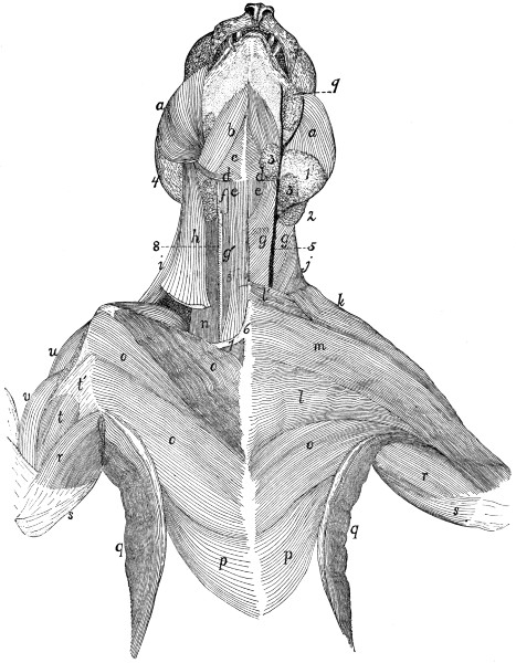
Fig. 65.—Muscles on the Ventral Surface of the Thorax, Neck and Head.
The left side shows the first layer of muscles beneath the skin and skin-muscles; the right side shows the second layer, except in the head region. a, M. masseter; b, M. digastricus; c, M. mylohyoideus; d, M. stylohyoideus; e, M. sternohyoideus; f, M. thyreohyoideus; g, M. sternomastoideus; g′, M. sternothyreoideus; h, M. cleidomastoideus; i, M. levator scapulæ ventralis; j, M. clavotrapezius; k, M. clavobrachialis; l, M. pectoralis major; m, M. pectoantibrachialis; n, part of M. scalenus; o, M. pectoralis minor; p, M. xiphihumeralis; q, M. latissimus dorsi; r, M. epitrochlearis; s, caput longum of M. triceps brachii; t, M. biceps brachii; t′, bicipital arch; u, M. acromiodeltoideus; v, M. brachialis. 1, parotid gland; 2, submaxillary gland; 3, lymphatic glands; 4, external ear; 5, external jugular vein; 6, manubrium; 7, first rib; 8, common carotid artery; 9, molar gland.
The deep layer takes
Origin by a strong tendon from the ventral border of the zygoma just craniad of the mandibular fossa. The fibres pass cranioventrad, diverge and have their
Insertion into the external coronoid fossa.
Relations.—Outer surface with the following, beginning at the caudal border: the external ear, the parotid gland (Fig. 65, 1), the platysma, the submentalis, the zygomaticus (major), the zygomaticus minor. Ventral border with a lymph gland caudad of the angle of the mouth (Fig. 65, 3), and the digastric muscle (b). Inner surface with the bones.
Action.—A very powerful elevator of the lower jaw.
M. temporalis (Fig. 63, n).—The temporal muscle is the great mass taking origin from the temporal fossa and having its insertion on both surfaces and both borders of the coronoid process of the mandible. It may be divided into two layers, deep and superficial.
The superficial layer. The temporal fascia stretches over the temporal fossa, being attached to its borders; that is, to the sagittal and lambdoidal crests, to the curved ridge which connects the sagittal crest with the zygomatic process of the frontal bone, to the caudal border of this process, to the caudal border of the malar bone, to the caudal border of the ligament connecting the malar bone and the zygomatic process of the frontal, and to the dorsal border of the zygomatic process of the temporal bone and its dorsal root. The craniolateral part of this fascia is much stronger than the remainder.
The muscle-fibres take origin from the strong craniolateral part of the inner surface of the temporal fascia, from the groove on the temporal bone dorsad of the dorsal root of its zygomatic process, from the whole inner surface of this process and of the zygomatic process of the malar bone, and sometimes also from that part of the frontal bone which lies within the temporal fossa.
Insertion.--The outer surface of the coronoid process of the mandible dorsad of the coronoid fossa, and both borders of the coronoid process. The cranial fibres may be inserted craniad of the coronoid process onto the aponeurosis covering the deep portion of the muscle. The caudal part of this portion is more or less distinct and is sometimes described as a separate head.
The deep portion.
Origin by fleshy fibres from the whole surface of the temporal fossa, except in cases where the cranial part of the fossa is occupied by the superficial portion. The fibres converge toward the coronoid process, and the ventral third of the outer surface is covered by strong fascia.
Insertion.—The whole inner surface of the coronoid process of the mandible.
Relations.—Outer surface with the external ear, the ear-muscles (Fig. 63), the epicranius (Fig. 63, h), the corrugatores supercilii lateralis (d) and medialis (b). Inner surface with the skull, the pterygoideus externus (Fig. 66, b), and the structures within the orbit.
Action.—Elevator of the lower jaw.
M. pterygoideus externus (Fig. 66, b).—This lies on the medial side of the mandible, ventrad of the ventral portion of the temporal.
Origin.—The external pterygoid fossa (Fig. 40, h), which lies on the lateral surface of the perpendicular plate of the palatine bone, and on the surface of the pterygoid process of the sphenoid bone dorsad of the internal pterygoid fossa. The fossa extends from the foramen rotundum to the sphenopalatine foramen. The muscle passes with parallel fibres laterad, ventrad, and caudad and has its
Insertion by a strong flat tendon into the medial surface of the mandible near its ventral border between the opening of the mandibular canal and the base of the angular process.
Relations.—Ventral surface with the masseter (a), the digastric, and the soft palate with its muscles. Dorsal surface with the temporal, and near its origin with the muscles of the eye, and other structures in the orbit.
Action.—Elevator of the lower jaw.
M. pterygoideus internus (Fig. 66, c).—This lies caudad of the pterygoideus externus, nearly parallel to it, and is connected with it at its lateral end.
Origin from the whole surface of the internal pterygoid fossa (Fig. 40, s).
Insertion by fleshy fibres into the ventral surface of the external pterygoid and its tendon, into the medial surface of the angular process of the mandible and caudad of the angular process for about one centimeter into a fascia common to it and the masseter muscle, and into the stylomandibular ligament.
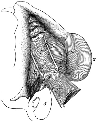
Fig. 66.—Obliquely Ventral View of the Deep Muscles of the Lower Jaw and Palate.
The tongue and other structures forming the floor of the mouth have been removed, showing the roof of the mouth. On the right side the masseter and pterygoideus internus have been removed, a, a′, M. masseter; b, M. pterygoideus externus; c, M. pterygoideus internus; d, d′, M. tensor veli palatini; e, e′, M. levator veli palatini; f, M. constrictor pharyngis superior. 1, aponeurosis of the velum palatinum; 2, ridges formed by the palatines and pterygoid portions of the sphenoid; 3, hamular processes; 4, free caudal border of the velum palatinum; 5, bulla tympani.
Relations.—Ventral surface with the styloglossus and the soft palate. Dorsal surface with the bulla tympani (5) and base of the skull. Cranial surface with the pterygoideus externus (b), with which it is partly united.
Action.—Assists the pterygoideus externus.
(b) Muscles of the Hyoid Bone.
M. stylohyoideus (Fig. 65, d).—The stylohyoid is a ribbon-like muscle about three millimeters wide. It lies on the superficial surface of the digastric, caudad and ventrad of the angle of the jaw.
Origin by fleshy fibres from the outer surface of the stylohyal bone near its middle. The muscle curves over the outer surface of the digastric (b), and ends about one centimeter from the middle line in a slender flat tendon.
Insertion into the middle of the ventral surface of the body of the hyoid bone. Some of the fibres of the mylohyoid (c) are inserted into its tendon.
Relations.—Outer surface with the submaxillary gland (2) and the submentalis muscle. Inner surface with the digastric (b), the hyoglossus (Fig. 67, h), and the geniohyoid (Fig. 67, g).
Action.—Raises the hyoid.
M. geniohyoideus (Fig. 67, g).—The geniohyoid is a long slender muscle which lies next to the median line between the symphysis menti and the hyoid bone, closely applied to its fellow of the opposite side and within (dorsad of) the mylohyoid (Fig. 65, c).
Origin from the ventral half of the inner surface of the mandibula for about one millimeter from the symphysis.
Insertion by muscle-fibres into the lateral half of the ventral surface of the body of the hyoid bone.
Relations.—Outer (ventral) surface with the stylohyoid (Fig. 65, d) and the mylohyoid (Fig. 65, c); medial border with the muscle of the opposite side. Lateral border with the hyoglossus (Fig. 67, h) and genioglossus (Fig. 67, f). Inner (dorsal) surface with the genioglossus.
Action.—Draws the hyoid forward.
M. jugulohyoideus (Fig. 67, b).—The jugulohyoid is a small flat quadrangular muscle covering the outer part of the ventral surface of the tympanic bulla.
Origin from the ventral border of the jugular process.
Insertion into the caudal side of the cartilaginous part of the stylohyoid opposite the origin of the styloglossus.
Relations.—Ventral surface with the digastric. Dorsal surface with the tympanic bulla (3).
Action.—Draws the hyoid backward and thus gives a firmer surface of origin for the styloglossus.
M. mylohyoideus (Fig. 65, c).—The mylohyoid is a large thin muscle seen after the reflection of the digastric, filling with its fellow of the opposite side the whole angle between the rami of the lower jaw. Its fibres are transverse.
Origin from the middle of the medial surface of the body of the mandible between the caudal opening of the mandibular canal and the symphysis of the jaw.
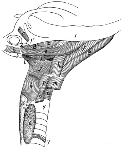
Fig. 67.—Muscles of Tongue, Hyoid Bone, and Pharynx.
a, M. tragicus lateralis; b, M. jugulohyoideus; c, M. pterygoideus externus; d, partially cut surface of M. pterygoideus internus; e, M. styloglossus; f, M. genioglossus; g, M. geniohyoideus; h, M. hyoglossus; i, M. glossopharyngeus; j, M. constrictor pharyngis medius; k, M. constrictor pharyngis inferior; l, M. stylopharyngeus; m, M. sternohyoideus (cut); n, M. cricothyreoideus; o, M. sternothyreoideus (cut); p, M. thyreohyoideus. 1, mandible; 1′, angular process of mandible; 2, stylomandibular ligament; 3, bulla tympani; 4, trachea; 5, œsophagus; 6, thyroid gland; 7, isthmus of the thyroid gland.
Insertion with the opposite muscle into a median raphe which extends from the symphysis of the jaw to the hyoid bone and is closely united to the external surface of the geniohyoid. Some of the fibres gain insertion into the body of the hyoid bone through their insertion into the tendon of the stylohyoid (d).
Relations.—Outer surface with the submentalis (Fig. 64, c) and the digastric (Fig. 65, b). Lateral edge with the masseter (a), the digastric (b), and the mandibula. Caudal border with the stylohyoid (d). Inner (dorsal) surface with the geniohyoid (Fig. 67, g), the hyoglossus (Fig. 67, h), and the genioglossus (Fig. 67, f).
Action.—Raises the floor of the mouth and thus brings the hyoid forward.
M. ceratohyoideus.
Origin, the two proximal pieces of the cranial horn of the hyoid. The fibres pass caudad parallel to one another and within the middle constrictor of the pharynx.
Insertion into the whole length of the caudal cornu of the hyoid bone.
Relations.—Outer surface with the middle constrictor of the pharynx. Inner surface with the pharynx.
Action.—Draws craniad the body of the hyoid by bringing its cornua together and bending the joint between the two pieces of the cranial cornua.
The muscles connecting the hyoid and the tongue are described in connection with the description of the tongue.
III. MUSCLES OF THE BODY.
1. Muscles of the Back.
A. Muscles of the Shoulder
(connecting the forelimb with the back) (Fig. 68).
M. trapezius.—The trapezius muscle is divided in the cat into three portions, the spinotrapezius, the acromiotrapezius, and the clavotrapezius.
M. spinotrapezius, or trapezius inferior (Fig. 68, j).—The spinotrapezius (caudal part of the human trapezius) is a flat triangular muscle.
Origin from the tips of the spinous processes of all or nearly all the thoracic vertebræ and from the intervening supraspinous ligament. The origin may extend from a point anywhere between the first and fourth spines to any point between the eleventh and thirteenth.
Insertion along an S-shaped line (Fig. 76, k-l) which crosses the tuberosity of the scapular spine and forms an angle with the spine of about forty-five degrees. The line extends on one side onto the fascia covering the supraspinatus muscle, and on the other side onto the fascia covering the infraspinatus, but does not reach the borders of the scapula. The cranial two-thirds of the insertion is by a flat tendon which is broader craniad.
Relations.—Outer surface with the cutaneus maximus, and craniad with the acromiotrapezius (h). Inner surface with the latissimus dorsi (m) caudad and with the rhomboideus, supraspinatus, and infraspinatus craniad. Caudal border free; cranial border in contact with the acromiotrapezius (h).
Action.—Draws the scapula dorsocaudad.
M. acromiotrapezius, or trapezius superior (Fig. 68, h).—The acromiotrapezius (middle part of the human trapezius) is a flat four-sided muscle just craniad of the spinotrapezius, from the median dorsal line to the scapular spine.
Origin along the median dorsal line from the spinous process of the axis to a point anywhere between the spinous processes of the first and fourth thoracic vertebræ. Its caudal two-thirds is from a flat tendon which is directly continuous with the muscle of the opposite side and thus bridges the depression between the vertebral borders of the scapulæ. This tendon narrows craniad, and its cranial fourth is attached by a fascia to the spinous processes of the cervical vertebræ. At the cranial border of the muscle the fibres sometimes reach the middle line. The fibres of the muscle run nearly transversely.
Insertion (Fig. 76, h).—Into the outer surface of the metacromion, the glenoid border of the scapular spine from the metacromion to the tuberosity, and along a line continued for about one centimeter from the tuberosity onto the surface of the spinotrapezius (j) at the junction of its muscular and tendinous portions.
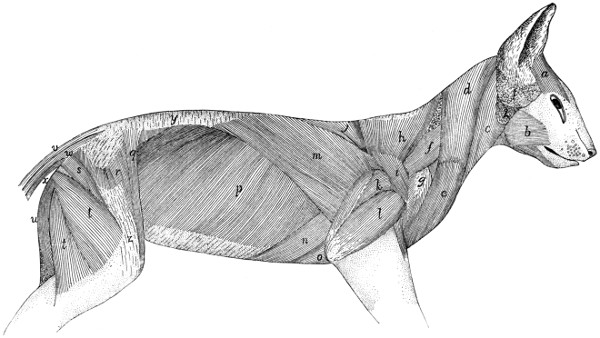
Fig. 68.—First Layer of Body-muscles, beneath the Skin and Skin-muscles.
a, M. frontoscutularis; b, M. masseter; c, M. sternomastoideus; d, M. clavotrapezius; e, M. clavobrachialis; f, M. levator scapulæ ventralis; g, M. acromiodeltoideus; h, M. acromiotrapezius; i, M. spinodeltoideus; j, M. spinotrapezius; k, caput longum of M. triceps brachii; l, caput laterale of M. triceps brachii; m, M. latissimus dorsi; n, M. xiphihumeralis; o, M. pectoralis minor; p, M. obliquus abdominis externus; q, M. sartorius; r, M. tensor fasciæ latæ; s, M. caudofemoralis; t, M. biceps femoris; u, M. semitendinosus; v, M. extensor caudæ lateralis; w, M. abductor caudæ externus; x, M. flexor caudæ longus; y, lumbodorsal fascia, superficial layer; z, fascia lata. 1, parotid gland; 2, submaxillary gland.
Relations.—Outer surface with the cutaneus maximus, the integument and platysma. Inner surface with a mass of fat partially covering the rhomboideus; with the occipitoscapularis, spinotrapezius, supraspinatus, levator scapulæ, and splenius. Near its origin its caudal border touches the outer surface of the spinotrapezius (j). Its cranial border is overlaid near its insertion by the levator scapulæ ventralis (f) and is closely related to the caudal border of the clavotrapezius (d) near its origin.
Action.—The two muscles hold the scapulæ together.
M. clavotrapezius (Fig. 68, d).—The clavotrapezius (cranial part of the human trapezius) is a flat muscle passing from the lambdoidal crest and middorsal line to the clavicle and covering the side of the neck.
Origin.—The medial half of the lambdoidal crest and the middorsal line between the crest and the caudal end of the spine of the axis. The fibres are parallel and pass caudoventrad so that the muscle covers the ventral surface of the neck caudally and fills the concavity at the front of the shoulder.
Insertion into the clavicle and into a raphe between the clavotrapezius and the clavobrachial (e) muscles. This raphe extends along the clavicle and for some distance laterad of it. The muscle is partly continuous with the clavobrachial (e).
Relations.—Outer surface with the platysma, and at its craniomedial angle with the levator auris longus. Inner surface with the rhomboideus, splenius, occipitoscapularis, levator scapulæ, cleidomastoid, and supraspinatus. The cranial edge touches the sternomastoid. The caudal border touches the acromiotrapezius near the origin.
Action.—Draws the scapula craniodorsad.
This muscle and the clavobrachial (e) are sometimes considered as forming a single muscle, the cephalohumeral, or cephalobrachial.
M. occipitoscapularis (or levator scapulæ dorsalis or rhomboideus capitis) (Fig. 73, a).—The occipitoscapularis (equivalent to a part of the human rhomboideus) is a slender flat muscle from the lambdoidal ridge to the coracovertebral angle of the scapula.
Origin, the medial half of the lambdoidal ridge beneath the clavotrapezius (Fig. 68, d). The origin does not extend quite to the middle line. The muscle passes almost directly caudad. Its caudal end is thicker and narrower than the cranial end and is wedged between the rhomboideus dorsally and the levator scapulæ ventrally.
Insertion either into the inner surface of the levator scapulæ near its dorsocranial border at its insertion or into the scapula near the coracovertebral angle (Fig. 78, f) or into both. Its fibres are very closely related to those of the rhomboideus.
Relations.—By its outer surface with the clavotrapezius (Fig. 68, d) and acromiotrapezius (h). At its insertion it is often overlaid by either the rhomboideus or the levator scapulæ or both. By its inner surface with the splenius (Fig. 73, b). Its caudal third is wedged between the rhomboideus and the levator scapulæ.
Action.—Draws forward and rotates the scapula.
M. rhomboideus (probably equivalent to the human rhomboideus major and rhomboideus minor).—A rather thick trapezoid muscle connecting the vertebral border of the scapula with the spinous processes of the adjacent vertebræ.
Origin from the caudal two or three fifths of the cervical supraspinous ligament and from the sides and tips of the first four thoracic vertebral spines and the interspinous ligaments caudad of these four spines. It passes lateroventrad, and the fibres converge somewhat so that the line of insertion is shorter than the line of origin.
Insertion by a short tendon (1 millimeter long) into the vertebral border of the scapula, the line of insertion passing gradually from the inner (Fig. 78, e) to the outer (Fig. 76, i) surface; and by fleshy fibres into the outer surface of the glenovertebral angle of the scapula (Fig. 76, i) in close connection with the origin of the teres major (j). A bundle of fibres at the cranial border may be inserted into the cranial border of the levator scapulæ at its insertion.
Relations.—By its outer surface with the spinotrapezius (Fig. 68, j), acromiotrapezius (h), latissimus dorsi (m), and clavotrapezius (d); it is usually partially separated from these muscles by a mass of fat. By its inner surface near its insertion with the serratus anterior (Fig. 73, i) and levator scapulæ (Fig. 73, h), and craniad with the splenius (Fig. 73, b), and the tendon of the serratus posterior superior (Fig. 73, l). At its lateral end it touches the occipitoscapularis (Fig. 73, a), serratus anterior (i), levator scapulæ (h), supraspinatus (Fig. 75, a), and infraspinatus (Fig. 75, c), and the teres major (Fig. 75, d).
Action.—Draws the scapula toward the vertebral column.
M. cleidomastoideus (clavicular portion of the human sternocleidomastoid) (Fig. 65, h).—A narrow flat muscle between the mastoid process and the clavicle.
Origin from the apex and caudal margin of the mastoid process of the temporal bone, by tendon and muscle fibres. The muscle passes caudad, becoming broader and thinner.
Insertion into the lateral four-fifths of the clavicle within the insertion of the clavotrapezius, and into the raphe which continues laterad of the clavicle.
Relations.—Outer or lateral surface at the caudal end with the clavotrapezius (Fig. 65, j); further craniad with the depressor conchæ (Fig. 64, b), the platysma, and the sternomastoid (Fig. 65, g). Inner or medial surface with the supraspinatus (Fig. 75, a) caudad, with the mass of fat in the axilla at its middle, with the levator claviculæ ventralis (Fig. 65, i) and obliquus capitis superior (Fig. 71, e) craniad.
Action.—Pulls the clavicle craniad when the head is fixed. Turns the head and depresses the snout when the clavicle is fixed.
M. levator scapulæ ventralis, or levator claviculæ (Fig. 68, f; Fig. 65, i; Fig. 72, c, c′, c″) (not usually found in man).
Origin (Fig. 72, c′, c″) in two parts. (1) By fleshy fibres from the ventral surface of the atlantal transverse process near its caudal border (c′). (2) By a small tendon from the ventral surface of the basioccipital opposite the middle of the length of the bulla tympani (c″) in common with the longus capitis (a). The two heads unite to form a flat band which passes caudoventrad and is inserted (Fig. 76, f) by a flat short tendon, about eight millimeters broad, into the outer surface of the metacromion along its ventral border, and into the infraspinatus fossa for one or two millimeters distad of that border.
Relations.—Near the insertion the dorsal border overlaps the border of the acromiotrapezius (Fig. 68, h), and the ventral border is attached by strong fascia to the border of the clavotrapezius (Fig. 68, d). The outer surface is in relation with the longissimus capitis (Fig. 73, g), clavotrapezius (Fig. 68, d), cleidomastoid (Fig. 73, d), platysma, and the skin. The inner surface is in relation caudad with the supraspinatus and with a mass of fat and a lymphatic gland which occupy the hollow of the shoulder. Craniad it is in relation with the cleidomastoid (Fig. 73, d), the scalenus (Fig. 73, f), the longus capitis (Fig. 73, e), and the obliquus superior (Fig. 71, e).
Action.—Pulls the scapula craniad.
M. latissimus dorsi (Fig. 68, m; Fig. 65, q; Fig. 77, e).—A large triangular sheet covering rather more than the dorsocranial half of the abdomen and thorax. It arises from the middorsal line, from the pelvis to the fifth thoracic spine, and is inserted into the humerus.
Origin from the tips of the neural spines of the vertebræ from the fourth or fifth thoracic to about the sixth lumbar. As far as the tenth or eleventh thoracic vertebra the origin is directly by muscle-fibres. Caudad of this is a broad triangular tendon which may be so closely united to the tendons of the underlying muscles that it cannot be separated. The muscle passes cranioventrad, the fibres converging to the axilla. In the axilla a part of the cutaneus maximus and the epitrochlearis (Fig. 65, r) take origin from the outer surface.
Insertion.—The muscle then ends in a flat tendon, to the cranial surface of which the fibres of the teres major (Fig. 77, c) are attached. Five to ten millimeters from the humerus the muscle-fibres of the teres give place to the tendon-fibres, and the conjoined tendon of the teres and latissimus which is thus formed (Fig. 79, d′) is inserted into a rough elongated area (Fig. 82, f) which lies parallel with the pectoral ridge on the medial surface of the shaft of the humerus (Fig. 48, g). The teres tendon forms the proximal part of the conjoined tendon. A part of the tendon of the latissimus may join the pectoralis minor at its distal or caudal border (Fig. 65), and thus it may contribute to the formation of both pillars of the bicipital arch (Fig. 65, t′).
Relations.—Its outer surface is covered caudad by the cutaneus maximus, and craniad by the spinotrapezius (Fig. 68, j) and the long head of the triceps (Fig. 68, k). The cranial border comes into relation with the spinotrapezius (j) and teres major (Fig. 77, c). The caudal border is in contact at the ventral end with the xiphihumeralis (Fig. 65, p) and pectoralis minor (Fig. 65, o). The inner surface of the latissimus covers near its cranioventral end parts of the xiphihumeralis (Fig. 68, n), pectoralis minor (Fig. 68, o), teres major (Fig. 77, c), and serratus anterior (Fig. 73, i). The caudal border covers a portion of the obliquus externus abdominis (Fig. 68, p); the dorsal and middle portions cover parts of the longissimus dorsi (Fig. 69, f″), the spinalis dorsi (Fig. 69, g), the serratus posterior superior (Fig. 73, l), and serratus posterior inferior (Fig. 73, n).
Action.—Pulls the arm caudodorsad.
M. serratus anterior and levator scapulæ (Fig. 73, i and h) in the cat are continuous at both origin and insertion, and it is barely possible to separate their contiguous borders without cutting the fibres. The muscle extends from the transverse processes of the last five cervical vertebræ and from the first nine or ten ribs to the medial surface of the scapula near its vertebral margin (Fig. 78, c and d).
M. serratus anterior (or serratus magnus) (Fig. 73, i).
Origin.—From the first nine or ten ribs in as many subdivisions. The first subdivision arises from the whole or nearly the whole of the caudal border of the first rib. The next four or five arise either from the ribs or from the costal cartilages near or at their junction with the ribs. The last four subdivisions arise from the ribs at increasing distances from the cartilages. The fibres converge and the subdivisions join one another.
Insertion (Fig. 78, c) into a narrow area on the medial surface of the scapula near the vertebral border. The glenoid half of the insertion is by a short tendon, the remainder by muscle-fibres.
Relations.—Outer (lateral) surface with the latissimus dorsi (Fig. 68, m), teres major (Fig. 77, c), subscapularis (Fig. 77, a), and near the origin with the rectus abdominis (Fig. 73, k), the scalenus (Fig. 73, f-f‴), and the obliquus externus (Fig. 68, p). Inner (medial) surface with the serratus posterior superior (Fig. 73, l) and its tendon, and with the intercostales externi (Fig. 73, m). At its insertion the muscle touches the rhomboideus. The cranial edge is united to the caudal edge of the levator scapulæ (Fig. 73, h).
Action.—Depressor of the scapula.
M. levator scapulæ (Fig. 73, h).
Origin.—From the dorsal tubercles of the transverse processes of the last five cervical vertebræ and from the ligaments between the tubercles. A slip sometimes arises from the atlas.
Insertion (Fig. 78, d) into a triangular area on the medial surface of the scapula near its vertebral border. It is continuous with the insertion of the serratus anterior.
Relations.—Outer (lateral) surface with a mass of fat separating it from the clavotrapezius (Fig. 68, d) and cleidomastoid (Fig. 65, h), and with the subscapularis (Fig. 77, a). Inner (medial) surface with the splenius (Fig. 73, b), the tendon of the serratus posterior superior (Fig. 73, l), and the cervical portion of the longissimus dorsi (Fig. 69, f″). Near the insertion the muscle touches the occipitoscapularis (Fig. 73, a) and rhomboideus. Caudal border united with the serratus anterior (Fig. 73, i).
Action.—Draws the scapula cranioventrad.
B. Muscles of the Back
(interconnecting parts of the vertebral column) (Fig. 69).—The muscles connected with the vertebral column form a mass which is less markedly differentiated into distinct muscles than is the case in the limbs. A great longitudinal mass of fibres begins in the sacral region and extends along the vertebral column to the head. This mass contains fibres running in various directions and attached at one or both ends to the sacrum, the innominate bones, the spinous, transverse, and articular processes, and to the arches, of the vertebræ; to the ribs and to the head, and having in general the function of moving the vertebræ in various ways, or of moving parts connected with the vertebræ. In some regions, especially the cervical, it is possible to distinguish clearly distinct muscles which have been differentiated from this mass. In the greater part of its extent, however, it is possible to distinguish only more or less closely interconnected bundles,—the more clearly marked of which have received special names.
The great dorsal mass running along each side of the vertebral column, occupying the space between the spinous processes and the transverse processes, receives the general name of M. extensor dorsi communis; the separate muscles are to be considered as parts or differentiations of this. The largest part of the mass is in the lumbar region (first portion of the longissimus dorsi, Fig. 69, f). At the caudal end of the thorax a lateral mass, parts of which become connected with the ribs, is separated off from the main part of the muscle; this extends craniad into the cervical region as M. iliocostalis (h). The main portion of the longissimus dorsi (f′) continues craniad on the medial side of the iliocostal into the neck region. At about the level of the eighth or ninth thoracic vertebra a strip begins to become separated off on the medial side of the longissimus; further forward it becomes clearly distinct, forming the spinalis dorsi (g). The separate muscles connecting the cervical vertebræ with each other and with the head are differentiations of the extensor dorsi communis.
A still deeper set of fibres, interconnecting the vertebræ, forms the multifidus spinæ and a number of other small muscles.
A description of the different bundles of fibres, under the names usually given, follows. Certain general principles are observed in some of the names used. Muscles which interconnect the spinous processes of the vertebræ receive the name spinalis. Those interconnecting contiguous spinous processes are called interspinalis. Muscles attached at one end to transverse processes, at the other to the spinous processes, receive the name transversospinalis. Semispinalis has the same signification as transversospinalis, but is a name usually applied to subdivisions of the transversospinalis group. The intertransversarii are muscles interconnecting the transverse processes.
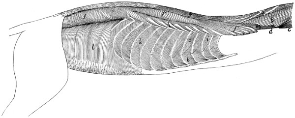
Fig. 69.—Deep Muscles of Body, Exposed after Removal of the Muscles shown in Fig. 73.
a, M. biventer cervicis; b, M. complexus; c, M. obliquus capitis superior; d, M. longus atlantis; e, cut end of M. longissimus capitis; f, f′, f″, M. longissimus dorsi; g, M. spinalis dorsi; h, M. iliocostalis; i, Mm. intercostales externi; k, Mm. intercostales interni; l, M. transversus abdominis.
(a) Muscles of the Lumbar and Thoracic Regions.
—The muscles of the lumbar and thoracic regions are mostly covered by a strong fascia, known as the lumbodorsal fascia (Fig. 68, y). This consists of two sheets, the superficial sheet being applied directly to the outer surface of the inner sheet, or separated from it by a mass of fat.
The superficial sheet (Fig. 68, y) overlies the lumbar region and the caudal half of the thoracic region. On the medial side it is attached to the spinous processes of the vertebræ and is united closely to the deeper layer. Laterally this fascia is continuous with the latissimus dorsi (m) and obliquus abdominis externus (p). Caudad it is attached to the spine of the ilium and becomes continuous with the fascia covering the gluteus muscles.
The deeper sheet (Fig. 70, c) is of a tendinous character, forming the external tendinous layer of the longissimus dorsi, many of whose fibres take origin from its under surface. It is described more fully in the account of this muscle.
M. longissimus dorsi (Fig. 69, f, f′, f″; Fig. 70, a and b).—A very large muscle, filling most of the region between the spinous processes and transverse processes of the lumbar and thoracic vertebræ, and extending into the cervical region.
The muscle is largest in the lumbar region (Fig. 69, f; Fig. 70), where it is divided into a narrow medial (Fig. 70, a) and a thick lateral portion (Fig. 70, b), the latter being again partially subdivided by the fascia. The two parts unite farther craniad.
The medial division (Fig. 70, a) consists of muscular bundles connecting the spinous processes of the vertebræ with the accessory and mammillary processes of other vertebræ; it is continuous caudad with the extensor caudæ lateralis (Fig. 70, f). The muscle-fibres take origin in the sacral region by small round tendons from the spinous processes of the last two sacral and the caudal vertebræ. They curve cranioventrad, forming a large belly, and are inserted into the accessory and mammillary processes of the lumbar vertebræ. In the thoracic region this division unites with that portion of the lateral division in which the fibres coming from the fascia of origin are inserted into the laminæ and transverse processes of the thoracic vertebræ.
The lateral division (Fig. 70, b) is much larger than the medial one, forming in the lumbar region a nearly cylindrical mass.
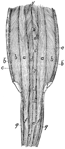
Fig. 70.—Muscles on the Dorsal Side of the Vertebral Column in the Lumbar, Sacral, and Caudal Regions.
Both sheets of the lumbodorsal fascia have been removed, the deep layer (c) being cut where it passes into the longissimus dorsi. 1, crest of ilium; 4-7, tips of spinous processes of the fourth to seventh lumbar vertebræ. I, II, tips of spinous processes of first two sacral vertebræ. a, b, M. longissimi dorsi (a, medial portion; b, lateral portion; b′, portion taking origin from the lumbodorsal fascia); c, cut edge of deep layer of lumbodorsal fascia; d, M. multifidus spinæ; e, M. extensor caudæ medialis; f, M. extensor caudæ lateralis, g, M. abductor caudæ externus.
Origin from the crest of the ilium (Fig. 70, 1) and the medial surface of the ilium as far caudad as the auricular impression; also from the deep layer of the lumbodorsal fascia (c). This fascia is connected with the crest of the ilium and with the tips of the spinous processes of the vertebræ in the lumbar and thoracic regions, and from it a large proportion of the fibres of the longissimus take origin. In the lumbar region it dips into the muscle as an intermediate longitudinal sheet (c), partially dividing it lengthwise into two parts. Fibres taking origin from the lateral surface of this intermediate sheet curve cranioventrad, and are inserted on the transverse processes of the lumbar vertebræ. Fibres taking origin from the medial surface of the sheet pass mediocraniad and are inserted into the accessory processes and the surfaces of the vertebral arches. In the thoracic region (Fig. 69, f′) most of the fibres are inserted in separate bundles by small tendons on the transverse processes of the vertebræ, while some of the medial fibres unite with tendinous strands which become attached to the laminæ and articular processes of the vertebræ. At the region of the eighth or ninth thoracic vertebra the spinalis dorsi (Fig. 69, g) begins to be separated off on the medial side, the separation becoming complete only some distance farther craniad; the longissimus dorsi then continues into the cervical region (f″). Bundles of fibres become attached in the manner above described to transverse processes of all the thoracic vertebræ. In the cervical region (f″) the muscle spreads out and becomes thinner, and bundles of fibres become attached to the transverse processes of the cervical vertebræ as far forward as the second. In the more cranial portion of its extent the muscle receives fibres having origin on the articular processes and laminæ of the cervical and a few of the more cranial thoracic vertebræ.
The portion of the muscle which is inserted on the cervical transverse processes (f″) is sometimes distinguished as the longissimus cervicis; it is not well separated from the rest of the muscle in the cat. The longissimus capitis (Fig. 69, c; Fig. 73, g) is to be considered a differentiated cranial portion of this muscle.
Relations.—Outer surface with the following, beginning with the caudal end: the sartorius (Fig. 68, q), the latissimus dorsi (Fig. 68, m), the lumbodorsal fascia (Fig. 68, y), the serratus posterior inferior (Fig. 73, n) and superior (Fig. 73, l), and the levator scapulæ (Fig. 73, h). Lateral margin with the abdominal muscles, the iliocostal (Fig. 69, h), and the levator scapulæ (Fig. 73, h). Medial side with the multifidus spinæ (Fig. 70, d), the spinous processes of the thoracic vertebræ, the spinalis dorsi (Fig. 69, g), the complexus (Fig. 69, b), the biventer cervicis (Fig. 69, a), and the longissimus capitis (Fig. 69, e).
Action.—Extends the vertebral column.
M. iliocostalis (Fig. 69, h).—This is a muscle about 1½ to 2 centimeters wide, lying on the dorsal portion of the ribs, laterad of the longissimus dorsi (f′). It is composed of many partly separated bundles, each with an oblique tendon. The muscle begins at the last or next to the last rib, where it is connected with the longissimus by a rather small bundle of fibres. The rest of the muscle-fibres take origin in bundles from the lateral surface of the ribs, at about the angles, or from thin tendons connecting the angles, of the ribs; they pass obliquely craniad and are inserted, usually by tendons, on the lateral surface of the third or fourth rib craniad of the one on which the given bundle has origin. The insertions on any given rib lie ventrad of the origins on the same rib. In the cat this muscle is confined to the thoracic region.
Relations.—Outer surface with the serratus posterior inferior (Fig. 73, n) and superior (Fig. 73, l), and the levator scapulæ (Fig. 73, h). Inner surface with the dorsal surface of the ribs, the external intercostals (Fig. 69, i), and craniad with the levatores costarum. Medial edge in contact with the longissimus dorsi (Fig. 69, f′).
Action.—Draws the ribs together.
M. spinalis dorsi (Fig. 69, g).—A muscle interconnecting the spinous processes of vertebræ some distance apart, in the thoracic and cervical regions. It lies on the medial side of the longissimus dorsi (f′).
Origin by strong tendons from the tips of the spinous processes of the tenth to the thirteenth thoracic vertebræ. These tendons represent the cranial portion of the deep layer of the lumbodorsal fascia, and give origin also to many fibres of the longissimus dorsi, so that the two muscles are completely united at the origin of the spinalis. They become separated at about the level of the sixth thoracic vertebra, the spinalis forming a strong separate bundle passing into the neck region close against the sides of the spinous processes. The insertion is by fleshy bundles into the sides of the spinous processes of the first nine or ten thoracic vertebræ and of the cervical vertebræ as far forward as the second. Some of the fibres of this muscle pass craniad into the biventer cervicis (Fig. 69, a).
Relations.—Outer surface with the tendons of the serratus posterior inferior (Fig. 73, n) and serratus posterior superior (Fig. 73, l), and with the splenius (Fig. 73, b), biventer cervicis (Fig. 69, a), and complexus (Fig. 69, b). Lateral and ventral surface with the longissimus dorsi (f′); medial surface with the spinous processes and the multifidus spinæ.
Action.—Extensor of the vertebral column.
M. multifidus spinæ.—This consists of bundles of fibres which have origin on the transverse processes or neighboring parts, pass craniodorsad across one or more vertebræ, and are inserted into the spinous processes of vertebræ lying some distance craniad of the origin. They lie deeper than the muscles previously described. The muscle is most strongly developed in the lumbar region (Fig. 70, d), where it forms a thick interwoven mass in which it is difficult to distinguish separate bundles. The fibres in this region have origin on the accessory or mammillary processes and usually pass over more than one vertebra between origin and insertion; their insertions reach the dorsal ends of the spinous processes, so that part of the muscle lies immediately beneath the lumbar fascia. In other regions the multifidus is covered by other muscles. In the thoracic region the separate bands are more distinct, and usually pass in their course over but one intervening vertebra. In the cervical region the bands are interconnected, forming a fairly distinct single muscle, which is described separately below as the semispinalis cervicis (Fig. 71, c). The portion of this muscle attached to the head (semispinalis capitis) forms the biventer cervicis (Fig. 69, a) and complexus (b). Caudad this muscle passes onto the tail as the extensor caudæ medialis (Fig. 70, c).
Relations.—Outer and lateral surface in the lumbar region with the longissimus dorsi (Fig. 69, f) and the lumbodorsal fascia (Fig. 68, y); in the thoracic region with the longissimus dorsi (Fig. 69, f′) and spinalis dorsi (g). Inner surface with the arches, articular processes, and spinous processes of the vertebræ.
Action.—Extends the back when the muscles of both sides work together. Turns the vertebral column obliquely sideways when one set acts alone.
The deepest layer of the multifidus forms what is sometimes distinguished as the Mm. rotatores; no separate layer of this sort is to be made out in the cat.
Mm. interspinales.—Muscle-bundles passing from the spinous process of one vertebra to that of the vertebra immediately craniad or caudad of it. They are best developed in the lumbar region.
Mm. intertransversarii.—Muscle-fibres interconnecting the transverse processes. In the lumbar region the intertransversarii mediales connect the accessory and mammillary processes; the intertransversarii laterales lying between the transverse processes. In other regions only one set of the intertransversarii is to be distinguished.
(b) Dorsal Muscles of the Cervical Region.
—The clavotrapezius (Fig. 68, d) and occipitoscapularis (Fig. 73, a) have been described in connection with the muscles of the shoulder. The remaining muscles of this region may be considered as differentiations of the general vertebral musculature (M. extensor dorsi communis).
M. splenius (Fig. 73, b.)—A large sheet of muscle covering the dorsal part of the side of the neck, beneath the trapezii.
Origin from the whole cervical ligament and from the fascia covering the deeper muscles along a line which extends from the first thoracic spinous process caudolaterad for about two centimeters.
Insertion by a thin tendon into the whole lambdoidal ridge. Laterad the tendon may be fused with that of the longissimus capitis (Fig. 73, g).
Relations.—Outer surface with the sternomastoid (Fig. 68, c), occipitoscapularis (Fig. 73, a), clavotrapezius (Fig. 68, d), rhomboideus, tendon of the serratus posterior superior (Fig. 73, l), and the levator scapulæ (Fig. 73, h). Lateral edge closely united with the longissimus capitis (Fig. 73, g). Inner surface with the longissimus capitis, complexus (Fig. 69, b), and biventer cervicis (Fig. 69, a).
Action.—Lateral flexor of the head. The two together elevate the head.
M. longissimus capitis (trachelomastoideus) (Fig. 73, g; Fig. 69, e).—This is a slender muscle lying close against the lateral border of the splenius (Fig. 73, b), and sometimes fused with the splenius at its craniad end. It is a cranial continuation of the longissimus dorsi (Fig. 69, f″).
Origin by five slips which are attached by strong tendons to the anterior articular processes of the last four cervical vertebræ. The tendons are common to this muscle and the complexus. The five slips unite to form a flat belly which has its
Insertion by a strong rounded tendon into the mastoid process of the temporal bone. The tendon may be closely united to that of the splenius.
Relations.—Lateral surface with the sternomastoid (Fig. 68, c), the levator scapulæ (Fig. 73, h), and longissimus dorsi (Fig. 69, f″). Medial surface with the splenius (Fig. 73, b) (to which it is partly united), the complexus (Fig. 69, b), and the longus atlantis (Fig. 69, d).
Action.—Lateral flexor of the head.
M. biventer cervicis (Fig. 69, a) (medial portion of M. semispinalis cervicis et capitis).—The biventer cervicis is a flat muscle which lies beneath the splenius (Fig. 73, b), next the median line of the neck, with its fibres longitudinal.
Origin in three or four slips from the surface of the three or four aponeurotic arches which take origin from the tips of the spinous processes of the last cervical and the first three thoracic vertebræ and pass laterad and caudad to the anterior processes of the second, third, fourth, and fifth thoracic vertebræ. These arches are deviated from their straight course by four aponeurotic bands which pass from them obliquely caudad and dorsad to the sides of the spinous processes of the vertebræ into which the arches are inserted. The arches form the beginning of the sheet of fascia which passes outside of the longitudinal supraspinous muscles, and the anchoring bands form the beginning of a similar sheet which passes within the longitudinal supraspinous muscles. An additional slip may take origin from the cervical ligament between the last cervical and the first thoracic vertebræ. The fibres form a flat band divided by two or three oblique tendinous intersections. It adheres closely to the cervical supraspinous ligament.
Insertion by a strong tendon into the medial part of the lambdoidal crest.
Relations.—Outer surface with the splenius (Fig. 73, b) and at the caudal end with the longissimus dorsi (Fig. 69, f″). Inner surface with the spinalis dorsi (Fig. 69, g), the semispinalis cervicis (Fig. 71, c), and the rectus capitis posterior major (Fig. 71, a). Medial edge with the muscle of the opposite side; lateral edge with the complexus (Fig. 69, b).
Action.—Raises the snout.
M. complexus (Fig. 69, b) (lateral portion of M. semispinalis cervicis et capitis).—The complexus is closely associated with the biventer cervicis (a) and lies on its lateral side beneath the splenius (Fig. 73, b).
Origin by tendinous bands from the anterior articular processes of the last five or six cervical and the first one, two, or three thoracic vertebræ. The same bands give origin externally to the fibres of the longissimus capitis (Fig. 69, e), and internally to those of deeper muscles of the neck. The six or more muscular slips continued from these tendons unite to form a flat band, the
Insertion of which is by a flat tendon into the medial third of the lambdoidal crest some distance ventrad of the free edge of the crest. Near the insertion the lateral border of the muscle is connected by an aponeurotic band to the lateral border of the transverse process of the atlas.
Relations.—Outer surface at the dorsal border with the biventer cervicis (Fig. 69, a); at the middle with the splenius (Fig. 73, b); at the ventral border with the longissimus capitis (Fig. 69, e) and the longissimus dorsi (Fig. 69, f″). Inner surface with the cervical portion of the spinalis dorsi, with the semispinalis cervicis (Fig. 71, c), the obliquus capitis inferior (Fig. 71, b), the rectus capitis posterior major (Fig. 71, a), and the obliquus superior (Fig. 71, e).
Action.—Raises the head.
M. spinalis dorsi extends into the cervical region; it has been described.
M. semispinalis cervicis (Fig. 71, c).—This represents that portion of the multifidus spinæ which extends into the cervical region. It is not composed of distinct bundles, like the multifidus of the thoracic region, but all the fibres are united into a fairly well-defined muscle.
Origin from the articular processes of the last five cervical vertebræ.
Insertion into the spinous processes of the cervical vertebræ up to the second, the largest part of the muscle being inserted into the caudal end of the spine of the atlas.
Relations.—Outer surface with the spinalis dorsi (Fig. 69, g) and the complexus (Fig. 69, b). Inner surface with the vertebræ.
Action.—Extends the neck.
M. longissimus dorsi extends into the cervical region, where it is often distinguished as the longissimus cervicis (Fig. 69, f″). It has been described.
M. longus atlantis (Fig. 71, f; Fig. 69, d).—This represents a differentiated portion of the longissimus dorsi.
Origin from the transverse process and the side of the vertebral arch of the third cervical vertebra.
Insertion into the caudolateral angle of the wing of the atlas.
Relations.—Dorsal surface with the complexus (Fig. 69, b), the longissimus capitis (Fig. 69, e), and the obliquus inferior (Fig. 71, b). Ventral surface with the scalenus (Fig. 71, g), longissimus dorsi (Fig. 69, f″), and levator scapulæ (Fig. 71, h).
Action.—Extends the neck and turns the head sideways.
M. rectus capitis posterior major (Fig. 71, a).
Origin.—The whole length of the spinous process (or crest) of the axis, or its caudal part only. It is united by a raphe to the opposite muscle. The muscle passes craniad and laterad to its
Insertion into an area ventrad of the medial part of the lambdoidal crest beneath the insertion of the complexus (Fig. 69, b) and the biventer cervicis (Fig. 69, a).
Relations.—Dorsal surface with the biventer cervicis (Fig. 69, a). Ventral surface with the rectus capitis posterior medius (Fig. 71, d) and the obliquus capitis inferior.
Action.—Raises the snout.
M. rectus capitis posterior medius (Fig. 71, d). (This is in man a part of the rectus capitis posterior major.)—It lies beneath the rectus capitis posterior major (Fig. 71, a).
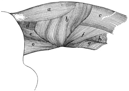
Fig. 71.—Deep Muscles of the Neck, after the Removal of Mm. Biventer Cervicis and Complexus.
a, M. rectus capitis posterior major; b, M. obliquus capitis inferior; c, M. semispinalis cervicis; d, M. rectus capitis posterior medius; e, M. obliquus capitis superior; f, M. longus atlantis; g, M. scalenus (part of cervical portion); h, cut end of M. levator scapulæ.
Origin from the cranial end of the axial spine. It forms a triangular prismatic mass which passes craniad and has its
Insertion into an elongated area on the occipital bone, ventrad of the median half of the lambdoidal crest.
Relations.—Dorsal surface with the rectus capitis posterior major (Fig. 71, a). Ventral surface with the atlas and the rectus capitis posterior minor.
Action.—Assists the rectus capitis posterior major.
M. rectus capitis posterior minor.—A small triangular muscle beneath the rectus capitis posterior medius (Fig. 71, d).
Origin by the apex of the triangle from the cranial border of the dorsal arch of the atlas for about two millimeters laterad of the median line. The muscle passes over the ligament which connects the atlas to the occiput and adheres to it.
Insertion into an elongated area on the occipital bone, ventrad of the insertion of the rectus capitis posterior medius for about five millimeters next to the median line.
Relations.—Dorsal surface with the rectus capitis posterior medius. Ventral surface with the posterior atlanto-occipital membrane.
Action.—Assists the rectus capitis posterior major.
M. obliquus inferior (Fig. 71, b).
Origin from the whole lateral surface of the spine of the axis. The parallel fibres form a thick mass which passes laterad and craniad.
Insertion into the dorsal surface of the transverse process of the atlas along a narrow area near its lateral margin.
Relations.—Outer surface with the rectus capitis posterior major (Fig. 71, a), the complexus (Fig. 69, b), and the obliquus superior (Fig. 71, e). Inner surface with the axis.
Action.—Rotates the head.
M. obliquus capitis superior (Fig. 71, e; Fig. 72, e).—This is a triangular muscle passing from the outer border of the atlas laterad of the recti capitis posteriores to the occiput.
Origin from the lateral border of the transverse process of the atlas on its ventral margin. The fibres pass craniad and diverge.
Insertion into the caudal side of the mastoid process of the temporal bone and into a line parallel with the lambdoidal ridge and ventrad of it, from the mastoid process to within one centimeter of the median line.
Relations.—Outer surface with the splenius (Fig. 73, b), complexus (Fig. 69, b), and longissimus capitis (Fig. 73, g). Inner surface with the obliquus inferior (Fig. 71, b), the wing of the atlas, and the rectus lateralis (Fig. 72, d). Ventral border with the cleidomastoid (Fig. 73, d) and one head of the levator scapulæ ventralis (Fig. 72, c′).
Action.—Flexes the head laterally.
Mm. interspinales and intertransversarii are found in the neck region also; they are described with the muscles of the back.
C. Muscles of the Tail.
—M. extensor caudæ medialis (Fig. 70, e).—This is a continuation caudad of the multifidus spinæ (Fig. 70, d); it lies next to the dorsal median line, the muscles of right and left side touching one another in the middle line.
Origin by numerous fleshy bundles from the spinous processes of the sacral and first caudal vertebræ. The fibres pass caudad and are inserted by tendons into the articular processes and the dorsal surface of the caudal vertebræ.
Action.—Extends (raises) the tail.
M. extensor caudæ lateralis (Fig. 70, f).—This is a continuation caudad of the medial portion of the longissimus dorsi (Fig. 70, a); it lies just laterad of the extensor caudæ medialis (Fig. 70, e).
Origin in many fleshy bundles from the articular processes of the sacral vertebræ, and the transverse processes of the caudal vertebræ. The fibres curve dorsocaudad and are inserted by many long slender tendons on the dorsal surfaces of the caudal vertebræ. The muscle grows continually smaller as it passes caudad.
Action.—Raises the tail.
M. abductor caudæ (coccygis) externus (Fig. 68, w; Fig. 70, g).—A rounded muscle on the lateral surface of the cranial part of the tail.
Origin from the medial side of the dorsal border of the ilium, and from the dorsal surface of the sacrum. The muscle passes caudad, lying just ventrad of the extensor lateralis, and is inserted into the transverse processes and lateral surfaces of the caudal vertebræ, as far back as the eighth or ninth.
Action.—Bends the tail sideways.
M. abductor caudæ internus (or M. coccygeus).—A large flat muscle, having origin on the spine of the ischium. The muscle passes dorsomediad, spreading out, and is inserted into the transverse processes of the second to the fourth caudal vertebræ.
Relations.—Lateral surface with the caudofemoralis (Fig. 68, s), gluteus maximus, and pyriformis. Medial surface with the iliocaudalis and the flexor caudæ longus (Fig. 68, x).
M. iliocaudalis (Str.-D.) (Fig. 162, 11′, page 398).—This represents a portion of the levator ani of man, and in some specimens is united with that muscle (Fig. 162, 11) in the cat.
Origin along the ventral half of the medial surface of the ilium, caudad of the sacrum. The fibres pass caudad and are inserted by a flat tendon into the ventral surface of the caudal vertebræ, from the second or third to about the seventh.
Relations.—Lateral surface with the gluteus maximus and abductor caudæ internus (Fig. 68, w). Medial surface with the levator ani (Fig. 162, 11), with which this muscle may be partly united.
Action.—The two muscles together flex the tail.
M. flexor caudæ longus (Fig. 162, 12; Fig. 113, c, page 270).
Origin on the ventral surface of the last lumbar vertebra, of the sacrum, and of the transverse processes of the caudal vertebræ. Caudad the muscle forms long, strong tendons which are inserted on the ventral surface of the tail.
Relations.—Lateral surface with the ilium, the gluteus maximus, the abductor caudæ internus, the iliocaudalis, and the skin of the tail. Dorsally it touches in the caudal region the abductor caudæ externus (Fig. 68, w), medially the flexor caudæ brevis (Fig. 162, 13).
Action.—Flexes the tail.
M. flexor caudæ brevis (Fig. 162, 13).
Origin on the ventral surface of the caudal vertebræ, from the first to the seventh or eighth. The principal head comes from the first vertebra. The bundles thus formed pass caudad and are inserted each into the ventral surface of a vertebra some distance caudad of the origin. The muscle extends to about the tenth caudal vertebra, and its most caudal parts are closely interconnected with the flexor caudæ longus.
Relations.—Lateral edge with the flexor caudæ longus. Medial edge in the sacral region with the levator ani (Fig. 162, 11) and the iliocaudalis (Fig. 162, 11′); in the caudal region with the muscle of the opposite side.
Action.—Flexes the tail.
2. Muscles on the Ventral Side of the Vertebral Column.
A. Lumbar and Thoracic Regions.
—The iliopsoas (Fig. 162, 8) belongs in this group, but since it moves the leg it is described with the muscles of the pelvic limbs.
M. psoas minor (Fig. 162, 9, page 398).—This muscle lies along the ventral surface of the vertebral column, next to the peritoneum, and extends from the thoracic vertebræ to the ilium.
Origin usually by five heads, from the caudal border of the centra of the last two (or one) thoracic and first three (or four) lumbar vertebræ. These heads also serve as origin for a part of the iliopsoas, and are closely united at their origin with the quadratus lumborum. They unite to form an, at first, rather large flat muscle, which rapidly becomes smaller caudad, finally forming a slender tendon which is inserted on the iliopectineal line, just craniad of the acetabulum.
Relations.—Dorsolateral surface with the quadratus lumborum and iliopsoas (Fig. 162, 8). Ventral surface with the pleura and diaphragm near the origin; with the peritoneum caudad.
Action.—Flexes the back in the lumbar region.
M. quadratus lumborum.—A flat muscle lying on the ventral surface of the transverse processes of the lumbar vertebræ. Craniad it has origin on the ventral surface of the last two thoracic vertebræ, and by a few fibres from the last rib. The muscle passes caudad, lying on the lateral side of the psoas minor (Fig. 162, 9), and becomes attached to each of the transverse processes of the lumbar vertebræ. Caudad the muscle passes into a strong flat tendon which is inserted into the anterior inferior spine of the ilium.
Relations.—Dorsal surface with the transverse processes and the intertransverse muscles. Ventral and medial surfaces with the psoas minor (Fig. 162, 9) and iliopsoas (Fig. 162, 8).
Action.—Bends the vertebral column sideways.
B. Muscles on the Ventral Side of the Neck
(Fig. 65, page 109).—M. sternomastoideus (Fig. 65, g; Fig. 68, c).—The sternomastoid (sternal portion of the human sternocleidomastoid) is a flat band one to three centimeters wide extending from the cranial end of the manubrium and the midventral line craniad of it to the lambdoidal ridge.
The origin extends from the cranial end of the manubrium along the midventral line as far as the caudal border of the cricoid cartilage, and is in two parts. The caudal portion arises from the lateral surface of the manubrium along the dorsal half of its cranial end, and from the median raphe for about one centimeter craniad of the manubrium. Its fibres are parallel and tend to diverge into two layers. The cranial portion arises by means of fibres which cover the median line and interdigitate with the fibres of the opposite muscle between the cricoid cartilage and a point a few millimeters craniad of the manubrium. Its caudal border is thus overlaid by the caudal portion. Its fibres converge and join those of the caudal portion. The muscle passes dorsocraniad and is
Inserted by means of a flat tendon into the lateral half of the lambdoidal ridge and into a continuation of the ridge onto the mastoid portion of the temporal bone as far as the mastoid process. The thickest part of the tendon is inserted into the mastoid process.
Relations.—Outer surface at the caudal end with the pectoralis major (Fig. 65, l); in the middle part with the platysma, the depressor conchæ (Fig. 64, b), and with the external jugular vein (Fig. 65, 5), which crosses it obliquely; at the cranial end with the submaxillary (Fig. 65, 2) and parotid (1) glands. Inner surface with the sternohyoid (Fig. 65, e), sternothyroid (g′), internal jugular vein, longus capitis muscle, the cleidomastoid (h), levator scapulæ ventralis (i), a large lymphatic gland ventrad of the ear, and the splenius (Fig. 73, b).
Action.—One muscle turns the head and depresses the snout. Both together depress the snout.
M. sternohyoideus (Fig. 65, e).—A slender muscle on the midventral line of the neck close to the opposite muscle.
Origin from the cranial border of the first costal cartilage. The muscle passes craniad closely united near its caudal end to the sternothyroid (g′).
Insertion (Fig. 67, m) into the outer half of the ventral surface of the body of the hyoid bone caudad of the origin of the geniohyoid (Fig. 67, g).
Relations.—Outer surface with the sternomastoid (Fig. 65, g). Inner surface with the thyrohyoid (Fig. 65, f; Fig. 67, p), the trachea (Fig. 67, 4), and the larynx.
Action.—Draws the hyoid caudad. Raises the ribs and sternum when the hyoid is fixed.
M. sternothyreoideus (Fig. 65, g′).—The sternothyroid lies beneath the sternohyoid and is connected with it at its caudal end.
Origin from the first costal cartilage beneath the sternohyoid.
Insertion (Fig. 67, o) into the lateral part of the caudal border of the thyroid cartilage of the larynx.
Relations.—Outer surface with the sternohyoid (Fig. 65, e) and laterally with the sternomastoid (Fig. 65, g). Inner surface with the trachea (Fig. 67, 4), and at the lateral edge with the thyroid gland (Fig. 67, 6).
Action.—Pulls the larynx caudad.
M. scalenus (Fig. 73, f-f‴).—This is a large and complex muscle lying on the ventral side of the neck and the lateral surface of the thorax. It might equally well be classified with the muscles of the thorax. It is divisible into a considerable number of interconnected bundles which are sometimes described as separate muscles.
The largest, middle portion (scalenus medius) (f′) takes origin by thin tendons from the sixth, seventh, eighth, and ninth ribs, just dorsad of their junction with the cartilages; these tendons unite near their origins to form a flat band which becomes fleshy at about the fifth rib, passes craniad, and unites just craniad of the first rib with the other parts of the muscle. The dorsal portion (scalenus posterior) (f″) has origin by a very slender tendon from about the middle of the outer surface of the third or fourth rib; it extends craniad as a narrow band which passes laterad between the heads of the serratus anterior (i) that are attached to the second and third (or third and fourth) ribs, and unites with the other heads. The ventral head (part of scalenus anterior) (f‴) arises by one or two minute tendons from the cartilages of the second and third ribs; they are partly united with the transversus costarum (j). This ventral head passes craniad and unites with the other heads.
These three divisions unite just craniad of the first rib with each other and with the cervical portion of the muscle (f) (part of scalenus anterior). This consists of a number of small bundles of fibres which arise from the first rib and the transverse processes of the first thoracic and the last six cervical vertebræ.
The insertion is onto the transverse processes of all the cervical vertebræ, including the axis and atlas. According to Strauss-Durckheim the fibres from any given transverse process are inserted into the transverse processes of all the vertebræ craniad of it.
The muscle is partly continuous craniad with the longus capitis (e). The cervical nerves pass out between the bundles of the cervical portion.
Relations.—Outer (lateral) surface in the thoracic region with the external oblique (Fig. 68, p), the pectoralis minor (Fig. 65, o), and in part with the serratus anterior (Fig. 73, i); in the cervical region with the sternomastoid (Fig. 65, g). Inner (medial) surface with the serratus anterior (Fig. 73, i), the levator scapulæ (Fig. 73, h), the longus atlantis, the transverse processes of the cervical vertebræ, and the longus capitis (Fig. 73, e).
Action.—Flexes the neck and draws the ribs craniad.
M. longus capitis (or rectus capitis anterior major) (Fig. 72, a; Fig. 73, e).—This is a long muscle lying on the ventral aspect of the cervical vertebræ. With the muscle of the opposite side and the underlying longus colli (Fig. 72, g′) it forms a trough in which lie the œsophagus, pharynx, and trachea.
Origin by five (or six) heads from the ventral margins of the transverse processes of the cervical vertebræ from the second to the sixth inclusive. The heads unite into a common belly, the lateral border of which is united with the levator scapulæ ventralis (Fig. 72, c) near its insertion.
Insertion into the body of the sphenoid bone between the bulla and the middle line. The insertion extends craniad onto the basisphenoid.
Relations.—Ventral surface with the sternomastoid (Fig. 65, g) and the large lymphatic gland ventrad of the external ear. Lateral surface with the scalenus (Fig. 72, h) and craniad with the levator scapulæ ventralis (Fig. 72, c-c″). Medial surface with the carotid artery, the trachea, œsophagus, and pharynx. Dorsal edge with the vertebræ, the longus colli (Fig. 72, g′), and the rectus capitis anterior minor (Fig. 72, b).
Action.—Lowers the snout.
M. rectus capitis anterior minor (Fig. 72, b).—This is a flat band beneath (dorsad of) the longus capitis (a).
Origin from the ventral surface of the inferior arch (body) of the atlas (3) for about five millimeters laterad of the middle line.
Insertion into a deep depression on the basioccipital caudad of the insertion of the longus capitis (a) and between the caudal end of the bulla (1) and the median line.
Relations.—Ventral surface with the longus capitis (a). Dorsal surface with the anterior (ventral) atlanto-occipital membrane (2).
Action.—Depresses the snout.
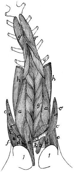
Fig. 72.—Muscles on the Ventral Surface of the Cervical Vertebræ.
On the left side the longus capitis and one head of the levator scapulæ ventralis have been cut, in order to show the rectus capitis anterior minor. a, M. longus capitis; b, M. rectus capitis anterior minor; c, M. levator scapulæ ventralis (c′, its atlantal head; c″, its occipital head); d, M. rectus capitis lateralis; e, M. obliquus capitis superior; f, M. longissimus capitis; g, M. longus colli (g′, its cervical portion; g″, its thoracic portion); h, part of M. scalenus. 1, bulla tympani; 2, ventral atlanto-occipital membrane; 3, ventral arch of atlas; 4, first rib.
M. rectus capitis lateralis (Fig. 72, d).—This lies ventrad of the obliquus superior (e) on the ventral face of the transverse process of the atlas and at first appears to be a part of the obliquus superior.
Origin from the median half of the ventral surface of the transverse process of the atlas. The fibres form a cylindrical mass which passes craniad and slightly laterad.
Insertion into the fossa laterad of the condyle of the occipital bone.
Relations.—Ventral surface with the digastric (Fig. 65, b) and the lymphatic gland ventrad of the ear. Dorsal surface with the wing of the atlas.
Action.—Flexes the head laterally.
M. longus colli (Fig. 65, g′, g″).—A long, rather slender muscle lying on the ventral surface of the cervical and first six thoracic vertebræ. It consists of many separate bundles, and is divisible into a thoracic and a cervical portion.
The thoracic portion (g″) has origin by six heads from the ventral surface of the first six thoracic vertebræ. The separate heads join to form a band which passes craniad and is inserted for the most part into the processus costarius of the sixth cervical vertebra; a portion of it joins the cervical division of the muscle.
The cervical portion (g′) is a continuation craniad of the thoracic portion. It arises in small bundles from the transverse processes and sides of the ventral surfaces of the centra of the cervical vertebræ. These bundles pass craniomediad, so that those from the muscles of opposite sides meet and are inserted on the centra of the vertebræ in the middle line, each pair of bundles forming a V opening caudad. The most cranial insertion is into the tubercle on the middle of the ventral surface of the ventral arch of the atlas (3).
Relations.—Dorsal surface with the centra of the vertebræ. Ventral surface in the thorax with the trachea; in the neck with the œsophagus. Medial edge with the muscle of the opposite side. Lateral margin in the cervical region with the longus capitis (a).
Action.—Bends the neck.
3. Muscles of the Thorax.
A. Breast-muscles (connecting the arm and thorax)
(Fig. 65, page 109).
The muscles connecting the arm with the sternum, corresponding to the pectoralis muscles of man, fall into a considerable number of not always clearly separated bundles in the cat. We shall distinguish by name four such bundles; these might be, and frequently are, further subdivided.
M. pectoantibrachialis (Fig. 65, m).—This is a small flat bundle about one or two centimeters wide, forming the most superficial portion of the pectoralis group.
Origin on the lateral surface of the manubrium. The muscle passes laterad onto the arm and is inserted by a flat tendon into the superficial fascia of the dorsal border of the forearm, near the elbow-joint. The tendon is continuous craniad with the border of the clavobrachial (k), so that a bundle of fibres may pass from this muscle to be inserted with the clavobrachial, or a bundle from the clavobrachial to be inserted with this. The tendon is continuous caudad with that of the epitrochlearis (r). The fascia of insertion may be traced to an attachment into the dorsal border of the ulna. This muscle usually receives near its insertion a slip, sometimes called its caudal division, from the deep layer of the pectoralis major.
Relations.—Outer surface with the integument. Inner surface with both divisions of the pectoralis major (l) on its medial two-thirds, and with the biceps (t) and bicipital arch (t′) in its lateral one-third. The cranial border is closely applied to that of the clavobrachial (k), except at the two ends.